Cloning and characterization of newly isolated lipase from ... · PDF fileCloning and...
11
Cloning and characterization of newly isolated lipase from Enterobacter sp. Bn12 Parisa Farrokh 1 , Bagher Yakhchali 2 , Ali Asghar Karkhane 2 1 Department of Genetics, School of Biological Science, Tarbiat Modares University, Tehran, Iran. 2 Industrial and Environmental Biotechnology Department, National Institute of Genetic Engineering and Biotechnology, Tehran, Iran. Submitted: February 11, 2013; Approved: September 9, 2013. Abstract A mesophilic Enterobacter sp. Bn12 producing an alkaline thermostable lipase was isolated from soil in Tehran, Iran. The lipase gene (ELBn12) was identified from a genomic library. Sequence anal- ysis of the DNA fragment revealed an open reading frame of 879 bp encoding a lipase with a molecu- lar mass of 31.3 kDa. The deduced amino acid sequence showed 96% identity with a lipase of Enterobacter sp. Ag1 and the identity of their DNA sequences was 88.9%. ELBn12 belongs to the lipase subfamily I.1 and its catalytic triad consists of Ser82, Asp237 and His259. The lipase was ex- pressed in Escherichia coli (BL21) pLysS and partially purified by anion exchange chromatography. The maximum activity of ELBn12 was obtained at temperature of 60 °C and pH 8.0 towards tricaprylin (C 8 ) and its specific activity was around 2900 U/mg. ELBn12 was stable within a broad pH range from 6.0 to 11.0. The enzyme showed high stability in both polar and nonpolar or- ganic solvents at 50% (v/v). The lipase activity was enhanced in the presence of 10 mM of Ca 2+ , Mg 2+ and K + , while heavy metals (Fe 3+ and Zn 2+ ) had strong inhibitory effect. ELBn12 showed high activ- ity in the presence of 1% (w/v) nonionic surfactants, however ionic surfactants inhibited the lipolytic activity. ELBn12 characteristics show that it has a potential to be used in various industrial processes. Key words: Enterobacter, thermostable lipase, alkaline, organic solvent. Introduction Lipases (EC 3.1.1.3) belong to the family of carbo- xylic hydrolases, which catalyze both the hydrolysis and synthesis of esters (Treichel et al., 2010). They have com- mon a/b fold and their active site usually consist of serin, histidin and aspartic acid (Arpigny and Jaeger, 1999). Li- pases are an important group of biocatalysts for a wide vari- ety of industrial applications (food, detergent and pharma- ceutical industries) and account for about 5 to 10% of the biggest worldwide enzyme market (Salihu and Alam, 2012). Lipases have been isolated from plants, animals and microorganisms (Treichel et al., 2010). Microbial en- zymes, because of their high production ability, ease of ge- netic manipulation and versatile catalytic activities in vari- ous environmental conditions, have greater advantages in industrial usage than the other sources (Andualema and Gessesse, 2012). Some of the desired characteristics for in- dustrial application of lipases are stability in high tempera- tures, alkaline conditions and in the presence of organic solvents. As the lipid substrates have high melting points, ther- mal stability is so crucial for lipases as biocatalysts in many industrial processes. Moreover thermophilic lipases show higher resistance to chemical denaturation (Lee et al., 1999). Stability of lipases in an organic solvent environ- ment is beneficial since these solvents are able to increase the solubility of substrate, facilitate the recovery of nonpolar products, reduce the degree of undesirable sub- strate and/or product inhibition in organic solvent-water two-phase system (Rahman et al., 2003). Thermostable lipases have potential applications in detergent, biodiesel, pharmaceutical and leather industries. Usage of organic solvent stable lipases has advantages in synthesis of biopolymers, biodiesel and pharmaceutical in- dustry. Alkaline lipases are also being used for detergent, leather and pulp industries (Hasan et al., 2006). Brazilian Journal of Microbiology 45, 2, 677-687 (2014) Copyright © 2014, Sociedade Brasileira de Microbiologia ISSN 1678-4405 www.sbmicrobiologia.org.br Send correspondence to B. Yakhchali. Industrial and Environmental Biotechnology Department, National Institute of Genetic Engineering and Biotech- nology, P.O. Box 14965/161, Tehran, Iran. E-mail: [email protected]. Research Paper
Transcript of Cloning and characterization of newly isolated lipase from ... · PDF fileCloning and...

Cloning and characterization of newly isolated lipase from Enterobacter sp. Bn12
Parisa Farrokh1, Bagher Yakhchali2, Ali Asghar Karkhane2
1Department of Genetics, School of Biological Science, Tarbiat Modares University, Tehran, Iran.2Industrial and Environmental Biotechnology Department,
National Institute of Genetic Engineering and Biotechnology, Tehran, Iran.
Submitted: February 11, 2013; Approved: September 9, 2013.
Abstract
A mesophilic Enterobacter sp. Bn12 producing an alkaline thermostable lipase was isolated from
soil in Tehran, Iran. The lipase gene (ELBn12) was identified from a genomic library. Sequence anal-
ysis of the DNA fragment revealed an open reading frame of 879 bp encoding a lipase with a molecu-
lar mass of 31.3 kDa. The deduced amino acid sequence showed 96% identity with a lipase of
Enterobacter sp. Ag1 and the identity of their DNA sequences was 88.9%. ELBn12 belongs to the
lipase subfamily I.1 and its catalytic triad consists of Ser82, Asp237 and His259. The lipase was ex-
pressed in Escherichia coli (BL21) pLysS and partially purified by anion exchange chromatography.
The maximum activity of ELBn12 was obtained at temperature of 60 °C and pH 8.0 towards
tricaprylin (C8) and its specific activity was around 2900 U/mg. ELBn12 was stable within a
broad pH range from 6.0 to 11.0. The enzyme showed high stability in both polar and nonpolar or-
ganic solvents at 50% (v/v). The lipase activity was enhanced in the presence of 10 mM of Ca2+, Mg2+
and K+, while heavy metals (Fe3+ and Zn2+) had strong inhibitory effect. ELBn12 showed high activ-
ity in the presence of 1% (w/v) nonionic surfactants, however ionic surfactants inhibited the lipolytic
activity. ELBn12 characteristics show that it has a potential to be used in various industrial processes.
Key words: Enterobacter, thermostable lipase, alkaline, organic solvent.
Introduction
Lipases (EC 3.1.1.3) belong to the family of carbo-
xylic hydrolases, which catalyze both the hydrolysis and
synthesis of esters (Treichel et al., 2010). They have com-
mon �/� fold and their active site usually consist of serin,
histidin and aspartic acid (Arpigny and Jaeger, 1999). Li-
pases are an important group of biocatalysts for a wide vari-
ety of industrial applications (food, detergent and pharma-
ceutical industries) and account for about 5 to 10% of the
biggest worldwide enzyme market (Salihu and Alam,
2012). Lipases have been isolated from plants, animals and
microorganisms (Treichel et al., 2010). Microbial en-
zymes, because of their high production ability, ease of ge-
netic manipulation and versatile catalytic activities in vari-
ous environmental conditions, have greater advantages in
industrial usage than the other sources (Andualema and
Gessesse, 2012). Some of the desired characteristics for in-
dustrial application of lipases are stability in high tempera-
tures, alkaline conditions and in the presence of organic
solvents.
As the lipid substrates have high melting points, ther-
mal stability is so crucial for lipases as biocatalysts in many
industrial processes. Moreover thermophilic lipases show
higher resistance to chemical denaturation (Lee et al.,
1999). Stability of lipases in an organic solvent environ-
ment is beneficial since these solvents are able to increase
the solubility of substrate, facilitate the recovery of
nonpolar products, reduce the degree of undesirable sub-
strate and/or product inhibition in organic solvent-water
two-phase system (Rahman et al., 2003).
Thermostable lipases have potential applications in
detergent, biodiesel, pharmaceutical and leather industries.
Usage of organic solvent stable lipases has advantages in
synthesis of biopolymers, biodiesel and pharmaceutical in-
dustry. Alkaline lipases are also being used for detergent,
leather and pulp industries (Hasan et al., 2006).
Brazilian Journal of Microbiology 45, 2, 677-687 (2014) Copyright © 2014, Sociedade Brasileira de Microbiologia
ISSN 1678-4405 www.sbmicrobiologia.org.br
Send correspondence to B. Yakhchali. Industrial and Environmental Biotechnology Department, National Institute of Genetic Engineering and Biotech-
nology, P.O. Box 14965/161, Tehran, Iran. E-mail: [email protected].
Research Paper
With an increasing interest in biotechnological appli-
cations of lipolytic enzymes, searching on lipase-producing
bacteria having satisfactory properties is an ongoing pro-
cess. Although there are some characterized alkaline
thermostable lipases from different microorganisms or
metagenomic libraries (Ahmed et al., 2010; Guanasekaran
et al., 2006; Khoramnia et al., 2010; Kim et al., 1996;
Nawani et al., 1998; Sarkar et al., 2012), a few of them are
stable in the presence of organic solvents (Ahmed et al.,
2010; Guanasekaran et al., 2006; Nawani et al., 1998). In
this study, we report functional screening, sequencing and
characterization of a novel organic solvent- and thermo-
stable alkaline lipase from a mesophilic Enterobacter spe-
cies.
Materials and Methods
Bacterial strains and plasmids
The pTZ57R/T (Fermentas, Vilnius, Lithuania) and
pBC KS+ (Agilent Technologies, Santa Clara, California,
United States) were used for the PCR product cloning and
genomic library construction respectively. Escherichia coli
Top10 (Invitrogen, Grand Island, New York, United
States) was used for cloning and plasmid preparation. E.
coli (BL21) pLysS and pET-26(+) (Novagen, Madison,
Wisconsin, United States) were used for expression of
lipase gene.
Screening of thermostable alkaline lipase-producingbacteria
Two hundred fifty bacterial isolates, used in this
study, were isolated from different soils (Zarenejad et al.,
2012) and wastewaters of leather and edible oil industries
(in this study) of Tehran, Iran. Lipase-positive colonies
were detected by formation of clear halo on Luria-Bertani
(LB) agar containing 0.5% (v/v) olive oil after 48 h of incu-
bation at 37 °C. Among all isolates exhibiting lipolytic ac-
tivity, isolates showing greater clear zones were subjected
to hydrolysis of p-nitrophenyl palmitate (pNPP) (Sigma-
Aldrich, Germany) at high temperature (50 °C) and alkaline
conditions (pH range 9.0-10.0). Out of all isolates, one
(Bn12) was selected for further studies because it showed
the largest clear halo and superior hydrolysis activity at
mentioned conditions.
Identification of the isolated bacterium
Genomic DNA was extracted from the isolate using
phenol-chloroform method, as described by Sambrook et
al. (2001). The 16S rRNA gene was amplified with two
universal eubacterial primers: fD1 and rD1 (Weisburg et
al., 1991). The PCR product was purified using an Agarose
Gel DNA Purification Kit (Roche, Switzerland) and cloned
into pTZ57R/T according to manufacturer’s structure.
DNA sequencing was carried out by dideoxy chain termi-
nation method (Macrogen, Seoul, Korea), using pUC/M13
primers.
The 16S rRNA gene sequence was analyzed through
NCBI BLAST (http://www.ncbi.nlm.nih.gov/blast/) and
EzTaxon servers (http://www.eztaxon.org/). The most sim-
ilar sequences were aligned in Molecular Evolutionary Ge-
netics Analysis (MEGA) 5.01 software and a phylogenetic
tree was made by the neighbor-joining (NJ) method with
1,000 bootstrap replicates, in the MEGA (Tamura et al.,
2011).
Morphological and biochemical identification of the
isolate was made according to Bergey’s manual of system-
atic bacteriology (Bernner et al., 2005).
Genomic DNA library construction
The chromosomal DNA was separately digested with
Sau3AI, HindIII and pstI and then partial digest products
with size of 1.5-10 kb were isolated by Agarose Gel DNA
Purification Kit. These fragments were cloned into dephos-
phorylated BamHI-, HindIII- and pstI-digested pBC KS+
and transformed to E. coli TOP 10 by thermal shock trans-
formation according to Sambrook et al. (2001). Screening
of the transformants was carried out on 0.5% (v/v) olive oil
LB agar plates supplemented with Chloramphenicol
(30 �g/mL). Lipase-positive colonies were detected by the
formation of clear haloes on olive oil LB agar plates after
48 h incubation at 37 °C.
Lipase sequence analysis
From a lipase-producing clone, the recombinant
plasmid was extracted and sequenced with pUC/M13 prim-
ers. The upstream sequence of the lipase gene from
genomic DNA of Bn12 strain was amplified using thermal
asymmetric interlaced (TAIL)-PCR (Liu et al., 1995) and
then cloned and sequenced.
Homology searches against DNA and protein data-
bases were performed using NCBI BLAST server. Multiple
sequence alignment was done with Clustal Omega
(http://www.ebi.ac.uk/Tools/msa/clustalo/). Theoretical
molecular mass and isoelectric point value of the lipase
were obtained using ExPASy bioinformatic resource portal
(http://www.expasy.org/tools/). Prediction of N-terminal
signal peptide sequence was done by SignalP 4.0 (Petersen
et al., 2011) and SecretomeP 2.0 (Bendtsen et al., 2005)
servers. Transmembrane regions analysis was performed
using TMHMM 2.0 server (Krogh et al., 2001).
Cloning of the lipase gene
The lipase gene was amplified with the following
primers; YKF1 (5’-
ATCATATGTCTACATCCCTGAAGTAC-3’) and YKF2
(5’- TAGAGCTCTTACAGTCCCTTCGCTTGC-3’). The
underlined nucleotides indicate NdeI and SacI restriction
sites respectively. The obtained PCR product was first
cloned into the pTZ57R/T and then subcloned into the ex-
678 Farrokh et al.
pression vector pET26(b+) using NdeI and SacI restriction
sites. The nucleotide sequence of the insert in the resulting
recombinant plasmid was confirmed by DNA sequencing.
E. coli BL21 (DE3) pLysS was used for expression of re-
combinant lipase.
Expression and purification of the recombinantlipase
The E. coli BL21 (DE3) pLysS cells harboring the
plasmid encoding lipase were grown in 200 mL LB me-
dium supplemented with the required antibiotics at 37 °C
until the absorbance at 600 nm of 0.6. The culture was then
induced with a final concentration of 0.5 mM isopropyl-�-
D-thiogalactopyranoside (IPTG) and incubated for another
18 h at 20 °C. After centrifugation, the bacterial pellet was
resuspended in 20 mL of lysis buffer (50 mM Tris-HCl
buffer (pH 8.5), 100 mM NaCl, 10 M EDTA, 1 mM PMSF,
1 mM DTT, 0.5% (v/v) Triton X-100) and disrupted by ul-
trasonic treatment (Hielscher GmbH, Teltow, Germany) at
4 °C.
The lysis extract was centrifuged at 15000 g for
15 min at 4 °C to pellet the cell debris and insoluble frac-
tions. Supernatant was dialyzed overnight against 50 mM
Tris-HCl buffer (pH 5.5). The dialyzed solution containing
lipase was purified by anion exchange chromatography on
diethylaminoethyl (DEAE)-cellulose column (1.5 x
7.0 cm) which had been equilibrated with the dialysis
buffer. Under the experimental conditions used, most of the
contaminating proteins bind to DEAE, while the lipase re-
mains in unbounded protein fraction. Purity of the lipase
was analyzed by sodium dodecyl sulfate polyacrylamide
gel electrophoresis (SDS-PAGE) (Laemmli, 1970) and
Coomassie Blue (G-250) staining and then dialyzed over-
night against 20 mM Tris-HCl buffer (pH 8.0).
Zymographic analysis of lipase activity
Lipase activity was detected by zymography follow-
ing standard SDS-PAGE. The SDS-PAGE gel was washed
by agitating in 50 mM Tris-HCl buffer (pH 8.0) containing
2% (v/v) Triton X-100 for 1 h, and then rinsed 20 min with
distilled water. The gel was placed on an agar plate contain-
ing 50 mM Tris-HCl buffer (pH 8.0), 0.5% (v/v) trigly-
ceride and 20 mg/ml gum arabic (Sigma). Tributyrin and
olive oil were used as substrates and appearance of hydro-
lysis halos exhibits lipase activity.
Protein estimation
The protein concentration of purified lipase was de-
termined by Bradford’s method using bovine serum albu-
min as the standard protein (Bradford, 1976).
Lipase assay
The lipase activity was measured titrimetrically using
pH-Stat assay system (Metrohm Ltd., Herisau, Switzer-
land). Substrate solution comprised of 2% (w/v) gum arabic
and 2 mg/ml triglycerides in deionized water. Emulsion of
the solution was prepared by ultrasonic treatment. For
lipase activity assay, 10 �L of 0.2 mg/ml enzyme solution
was added to the substrate solution and 0.1 M NaOH used
as the titrant. One unit of lipase was determined as the
amount of enzyme releasing 1 �mol of fatty acid per min.
All the experiments were done in triplicates.
Effect of temperature and pH on lipase activity andstability
Optimum temperature of the lipase activity was deter-
mined at different temperatures from 30 to 70 °C. The ac-
tivity assay was carried out with tricaprylin (C8) at pH 8.0.
Thermostability was assayed after incubation of lipase at
50, 60 and 70 °C for 1 h and the residual activity was ana-
lyzed every 20 min. Optimum pH for the lipase activity was
measured over a pH range from 6.0 to 10.0 at 60 °C using
tricaprylin as substrate. pH stability was determined at opti-
mal conditions after incubating the lipase at 30 °C for 1 h
over a pH value from 5.0 to 11.0. 50 mM sodium acetate
was used for pH 5.0, 50 mM sodium phosphate buffer
for pH 6.0-8.0 and 50 mM glycine-NaOH for pH 9.0-11.0.
Effect of substrate on lipase activity
Substrate specificity was studied by using triglyce-
ride substrates; tributyrin (C4), tricaproin (C6), tricaprylin
(C8), tricaprin (C10), trilaurin (C12), trimyristin (C14),
tripalmitin (C16) and olive oil (C18) (Sigma) at 60 °C
and pH 8.0.
Effect of various effectors on lipase activity
To determine the effect of various chemical effectors,
the lipase activity was studied at optimal conditions after
1 h pre-incubating at 30 °C with 1% (w/v) of surfactants,
50% (v/v) of organic solvents, 10 mM of metal ions or
10 mM of EDTA.
Nucleotide sequence accession number
The nucleotide sequences of the 16S rDNA and
ELBn12 lipase gene of Enterobacter sp. Bn12 were depos-
ited in the EMBL/DDBJ/GenBank databases under the ac-
cession numbers JX456175 and JX456176 respectively.
Results
Isolation and identification of the lipase-producingbacterium
From 250 different isolates used in this study, 10 iso-
lates with extracellular lipase activity were detected on LB
agar containing 0.5% (v/v) olive oil through formation of
clear haloes. Among these isolates, the Bn12 isolated from
soil, with the largest clear zone and notable hydrolysis ac-
tivity at alkaline pH and high temperature (data not shown),
was selected for further studies.
A new lipase from Enterobacter sp. 679
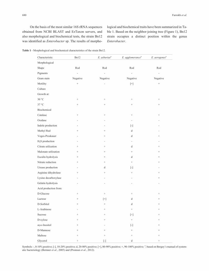
On the basis of the most similar 16S rRNA sequences
obtained from NCBI BLAST and EzTaxon servers, and
also morphological and biochemical tests, the strain Bn12
was identified as Enterobacter sp. The results of morpho-
logical and biochemical traits have been summarized in Ta-
ble 1. Based on the neighbor-joining tree (Figure 1), Bn12
strain occupies a distinct position within the genus
Enterobacter.
680 Farrokh et al.
Table 1 - Morphological and biochemical characteristics of the strain Bn12.
Characteristic Bn12 E. asburiaea E. agglomerancea E. aerogenesa
Morphological
Shape Rod Rod Rod Rod
Pigments - - - -
Gram stain Negative Negative Negative Negative
Motility + - [+] +
Culture
Growth at:
30 °C + + + +
37 °C + + + +
Biochemical
Catalase + + + +
Oxidase - - - -
Indole production - - [-] -
Methyl Red + + d -
Voges-Proskauer - + d +
H2S production - - - -
Citrate utilization + + d +
Malonate utilization + + + +
Esculin hydrolysis + + d +
Nitrate reduction + + + +
Urease production - d [-] -
Arginine dihydrolase + + + +
Lysine decarboxylase - - - +
Gelatin hydrolysis - - - -
Acid production from:
D-Glucose + + + +
Lactose + [+] d +
D-Sorbitol + + d +
L-Arabinose - + + +
Sucrose + + [+] +
D-xylose + + + +
myo-Inositol + - [-] +
D-Mannose + + + +
Maltose + + + +
Glycerol + [-] d +
Symbols: -, 0-10% positive; [-], 10-20% positive; d, 20-80% positive; [+], 80-90% positive; +, 90-100% positive. a, based on Bergey’s manual of system-
atic bacteriology (Bernner et al., 2005) and (Promsai et al., 2012).

Functional assay and analysis of the lipase geneand the flanking regions
From nearly 3500 E. coli transformants, one halo-
forming colony was identified from pstI genomic library of
Enterobacter sp. Bn12. Nucleotide sequencing of the re-
combinant plasmid, pLIP, from the halo-forming colony
was shown a 1847 bp fragment. BLAST analysis of the
fragment revealed that there was an ORF from position 89
to 967 related to lipases (Figure 2) which was named as
ELBn12. Since the upstream region of the lipase gene was
short, it was obtained through TAIL-PCR. Sequencing of
the plasmid containing upstream region (pUPLIP) of the
ELBn12 gene, revealed 881 bp fragment with 77% identity
to a hypothetical protein (data not shown).
ELBn12 encodes a protein of 292 amino acids and
BLASTx search of GenBank showed that the deduced
ELBn12 had the most similarity to the putative lipases:
96% identity with lipase from Enterobacrter sp. Ag1
(EJF30243), and 70% identity with lipase from Yersinia
enterocolitica (YP_004298016) over the entire length of
the protein and 92% identity with LipC12 from
metagenomic library (AEK97793) over 99% of the length
of the protein. Identity of ELBn12 at the nucleotide level
with the lipases of Enterobacrter sp. Ag1, LipC12, and Y.
enterocolitica was 88.9% , 85.1% and 65.5% respectively.
There was a potential ribosome-binding site, GAGG,
10 bp upstream from the start codon. The putative -10 and
-35 regions were predicted according to the E. coli pro-
moter consensus sequences (McClure, 1985) (Figure 2).
Catalytic triad of the ELBn12 consists of serine
(Ser82), aspartic acid (Asp237) and histidin (His259). The
catalytic nucleophile Ser82 being present in Gly-His-Ser-
Gln-Gly sequence of ELBn12 (Figure 3) matches with the
conserved motif, Gly-X-Ser-X-Gly (where X is any amino
acid), found in the family of lipases. The predicted molecu-
lar mass and pI of the ELBn12 were 31307.68 Da and 6.13
respectively. There were not any classical or non-classical
predictable signal peptide and transmembrane domain in
ELBn12 sequence.
Cloning, expression, and purification of ELBn12
The amplified ELBn12 (892 bp) by YKF primers was
cloned into pET26(b+) and the resulting recombinant
plasmid was named pYKF. The pYKF was transformed
into E. coli BL21 (DE3) pLysS to express ELBn12 lipase.
IPTG induction of pYKF at 20 °C for 18 h produced the
maximum amount of ELBn12 lipase. The recombinant
A new lipase from Enterobacter sp. 681
Figure 1 - Neighbour-Joining phylogenetic tree based on 16S rRNA gene sequences showing the position of isolate Bn12 with the closest members
within the Enterobacter genus. Numbers at nodes show bootstrap values (1000 resampling).

lipase was detected in the soluble fraction with molecular
weight of approximately 30 kDa on SDS-PAGE which was
close to the predicted size. The ELBn12 was purified in one
step chromatography using DEAE-cellulose column. The
purified lipase showed a single band on SDS-PAGE corre-
sponding to molecular mass of ~30 kDa (Figure 4).
Zymogram analysis
Zymographic analysis showed that the purified lipase
had lipolytic activity. The hydrolysis halos were detected in
the proximity of the 30 kDa molecular weight marker,
while clear zone for tributyrin substrate was larger than ol-
ive oil substrate (Figure 4).
Effect of temperature and pH on the lipase activityand stability
Purified ELBn12 exhibited maximum activity at tem-
perature range of 45 to 65 °C with an optimum activity at
60 °C (Figure 5). ELBn12 retained more than 51% of its
initial activity after 1 h incubation at 50 and 60 °C. How-
ever, the relative activity was reduced to 43% when incu-
bated for 1 h at 70 °C (Figure 6). The ELBn12 showed the
highest activity at pH 8.0. The enzyme retained more than
80% of its original activity after 1 h incubation at pH range
from 6.0 to 11.0 (Figure 7).
Substrate specificity
The relative activity of the purified lipase against var-
ious triglycerides (C4 to C18) was determined at 60 °C; pH
8.0. ELBn12 did not show any specific activity for long or
short chain triglycerides. It had a maximum activity to-
wards C8 (100%), C12 (86.87%) and C4 (73.36%). The
lipase activity was negligible against C16 and C10 (Figu-
re 8). The specific activity of ELBn12 lipase was 2900
U/mg when measured at optimum conditions.
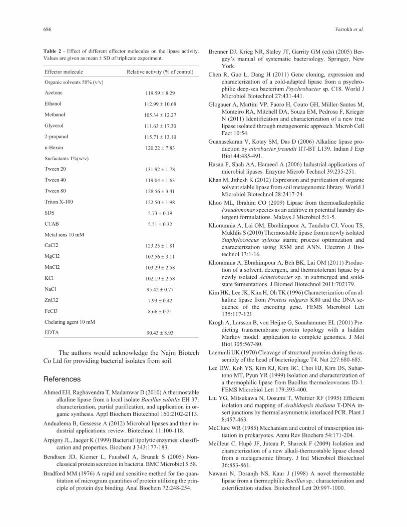
Effect of various effectors on the lipase activity
The lipase activity was partially activated by Ca2+,while K+, Mg2+ and Mn2+ increased enzyme activityslightly. Sodium ion had little inhibitory effect on the
682 Farrokh et al.
Figure 2 - Nucleotide sequence of the ElBn12 from Enterobacter sp. and its deduced amino acid sequence. The putative -10 and -35 promoter sequences
and Shine-Dalgarno ribosome binding site (RBS) of the ELBn12 are underlined.

ELBn12 activity. In contrast, the enzyme activity wasstrongly inhibited by Zn2+ and Fe3+. The non-ionic deter-gents Tween 40, Triton X-100, Tween 80 and Tween 20were found to increase (19 to 31%) the lipase activity.However, the ELBn12 activity was negligible in the pres-ence of ionic detergents (SDS and cetyltrimethyl-ammonium bromide (CTAB)). Various organic solvents at50% (v/v) partly enhanced the relative activity of theELBn12 lipase. In the presence of 10 mM EDTA, the lipasewas significantly stable (Table 2).
Discussion
At present study a novel organic solvent- and ther-
mo-stable alkaline lipase was isolated from a mesophilic
Enterobacter sp. Bn12 through genomic library. Although
there are several studies describing cloning and character-
ization of microbial lipases (Chen et al., 2011; Kim et al.,
1996; Rahman et al., 2003), a few of them describe charac-
teristics of lipase from Enterobacter genus (Zhen-qian and
Chun-yan, 2009).
Most of the thermostable enzymes are produced by
thermophilic microorganisms, but there are some reports
describing thermostable lipases from mesophilic ones.
Some examples of these include thermostable lipases from
Bacillus subtilis EH 37 (Ahmed et al., 2010), citrobacter
freundii (Guanasekaran et al., 2006), Staphylococcus
xylosus (Khoramnia et al., 2010), Acinetobacter sp. (Kho-
ramnia et al., 2011), and Proteus vulgaris K80 (Kim et al.,
1996).
A new lipase from Enterobacter sp. 683
Figure 3 - Multiple sequence alignment of the predicted amino acid sequence of ELBn12 (AFU92748) with some of the closest lipases using the Clustal
Omega. *, Identical residues; :, very similar residues and ., similar residues. The catalytic triad is boxed.

According to the classification of Arpigny and Jaeger
(1999), the primary structure of ELBn12 lipase and multi-
ple sequence alignment of the most similar lipases showed
that it was belonging to the subfamily I.1 of bacterial
lipolytic enzymes. Molecular mass of ELBn12 was also in
the range of the other members of subfamily I.1 (30-
32 kDa) (Arpigny and Jaeger, 1999).
Similar to some lipases belong to the subfamily I.1
(Glogauer et al., 2011; Kim et al., 1996), there were no pre-
dictable signal sequences in the N- or C-terminal regions of
ELBn12. Neither chaperon-encoding gene nor regulatory
sequences were detected in the flanking regions of ELBn12.
This observation, in addition to the functional expression of
the lipase gene in E. coli BL21 (DE3) pLysS, indicates that
its expression and folding do not depend on any helper pro-
teins.
The maximum activity of ELBn12 was observed at
60 °C and pH 8.0 towards tricaprylin with specific activity
of about 2900 U/mg. In comparison with the other thermo-
stable bacterial lipases (Kim et al., 1996; Meilleur et al.,
2009; Sarkar et al., 2012), ELB12 had notable specific ac-
tivity. Unlike many of them (Khoramnia et al., 2010, 2011;
Sarkar et al., 2012), the lipase showed less thermostability
at high temperatures. Nevertheless, the specific activity of
ELBn12 even after 1 h incubation at 70 °C (1000 U/mg)
was remarkable and more than original activity of the many
reported thermostable bacterial lipases (Khoramnia et al.,
2011; Meilleur et al., 2009; Sarkar et al., 2012). ELBn12
also showed acceptable stability within a broad pH range
from 6.0 to 11.0.
Stimulatory effects of some metal ions, such as Ca2+
were reported for several bacterial lipases (Andualema and
684 Farrokh et al.
Figure 4 - SDS-PAGE and zymogram of purified ELBn12. Lane 1: crude
extract from induced recombinant E. coli; Lane 2: purified ELBn12 lipase;
M: protein molecular weight markers. Lipolytic activity of the lipase on
tributyrin (a) and olive oil (b) substrates.
Figure 5 - Effect of temperature on ELBn12 lipase activity. The experiments were performed in triplicate.
Figure 6 - Effect of temperature on enzyme stability. The lipase stability
was measured every 20 min after incubation at various temperatures for 1
h. The experiments were carried out in triplicate.

Gessesse, 2012; Khan and Jithesh, 2012; Lee et al., 1999).
Sharma et al. explained that this was due to the formation of
insoluble Ca-salts of fatty acids released in the hydrolysis,
thus avoiding product inhibition (2009). The result of this
study showed that Ca2+ (10 mM) increased ELBn12 activ-
ity. However, its activity did not depend on the metal ions
(Ca2+ and Mg2+) because in the present of chelating agent,
EDTA (10 mM), the lipase maintained more that 90% of its
original activity. Inhibitory effects of heavy metals (Fe3+
and Zn2+) on the lipase activity, as seen here, may be due to
the alteration of enzyme conformation (Sharon et al.,
1998).
Stability of lipases in different organic solvents was
variable, but generally most of the lipases were more stable
in the presence of nonpolar solvents than polar solvents
(Ahmed et al., 2010; Rahman et al., 2003). In spite of
destabilizing effect of polar solvents on the enzyme’s con-
formation, the results of this study showed that both polar
and nonpolar organic solvents at 50% (v/v) increased
ELBn12 activity.
Catalytic activity of lipases is affected in the presence
of surfactants differently. Many of the lipases were inhib-
ited by ionic surfactants, while nonionic surfactants had
stimulatory effects (Chen et al., 2011; Khoo and Ibrahim,
2009). The same result was observed for ELBn12.
Among the most similar lipases to the ELBn12, only
LipC12 has been well characterized (Glogauer et al., 2011).
In spite of their high amino acid sequence similarity (92%),
the characteristics of ELBn12 were noticeably different
from LipC12, especially in optimal temperature (maximum
activity of LipC12 was at 30 °C). By characterization of
more bacterial lipases, interpretation of these differences
might be accessible in future. Furthermore, identification
of new lipases with multiple appropriate characteristics has
advantages to be applied in more industrial processes.
ELBn12, which was described here, could be a useful can-
didate for application in various industries such as bio-
diesel, pharmaceutical and leather industries.
Acknowledgments
A new lipase from Enterobacter sp. 685
Figure 7 - Effect of pH values on activity and stability of ELBn12. The stability of lipase was assayed after 1 h incubation at various pH values at 30 °C.
The experiments were carried out in triplicate.
Figure 8 - Substrate specificity of ELBn12 against various triacylglycerols. The experiments were performed in triplicate.
The authors would acknowledge the Najm Biotech
Co Ltd for providing bacterial isolates from soil.
References
Ahmed EH, Raghavendra T, Madamwar D (2010) A thermostable
alkaline lipase from a local isolate Bacillus subtilis EH 37:
characterization, partial purification, and application in or-
ganic synthesis. Appl Biochem Biotechnol 160:2102-2113.
Andualema B, Gessesse A (2012) Microbial lipases and their in-
dustrial applications: review. Biotechnol 11:100-118.
Arpigny JL, Jaeger K (1999) Bacterial lipolytic enzymes: classifi-
cation and properties. Biochem J 343:177-183.
Bendtsen JD, Kiemer L, Fausbøll A, Brunak S (2005) Non-
classical protein secretion in bacteria. BMC Microbiol 5:58.
Bradford MM (1976) A rapid and sensitive method for the quan-
titation of microgram quantities of protein utilizing the prin-
ciple of protein dye binding. Anal Biochem 72:248-254.
Brenner DJ, Krieg NR, Staley JT, Garrity GM (eds) (2005) Ber-
gey’s manual of systematic bacteriology. Springer, New
York.
Chen R, Guo L, Dang H (2011) Gene cloning, expression and
characterization of a cold-adapted lipase from a psychro-
philic deep-sea bacterium Psychrobacter sp. C18. World J
Microbiol Biotechnol 27:431-441.
Glogauer A, Martini VP, Faoro H, Couto GH, Müller-Santos M,
Monteiro RA, Mitchell DA, Souza EM, Pedrosa F, Krieger
N (2011) Identification and characterization of a new true
lipase isolated through metagenomic approach. Microb Cell
Fact 10:54.
Guanasekaran V, Kotay SM, Das D (2006) Alkaline lipase pro-
duction by citrobacter freundii IIT-BT L139. Indian J Exp
Biol 44:485-491.
Hasan F, Shah AA, Hameed A (2006) Industrial applications of
microbial lipases. Enzyme Microb Technol 39:235-251.
Khan M, Jithesh K (2012) Expression and purification of organic
solvent stable lipase from soil metagenomic library. World J
Microbiol Biotechnol 28:2417-24.
Khoo ML, Ibrahim CO (2009) Lipase from thermoalkalophilic
Pseudomonas species as an additive in potential laundry de-
tergent formulations. Malays J Microbiol 5:1-5.
Khoramnia A, Lai OM, Ebrahimpour A, Tanduba CJ, Voon TS,
Mukhlis S (2010) Thermostable lipase from a newly isolated
Staphylococcus xylosus starin; process optimization and
characterization using RSM and ANN. Electron J Bio-
technol 13:1-16.
Khoramnia A, Ebrahimpour A, Beh BK, Lai OM (2011) Produc-
tion of a solvent, detergent, and thermotolerant lipase by a
newly isolated Acinetobacter sp. in submerged and soild-
state fermentations. J Biomed Biotechnol 2011:702179.
Kim HK, Lee JK, Kim H, Oh TK (1996) Characterization of an al-
kaline lipase from Proteus vulgaris K80 and the DNA se-
quence of the encoding gene. FEMS Microbiol Lett
135:117-121.
Krogh A, Larsson B, von Heijne G, Sonnhammer EL (2001) Pre-
dicting transmembrane protein topology with a hidden
Markov model: application to complete genomes. J Mol
Biol 305:567-80.
Laemmli UK (1970) Cleavage of structural proteins during the as-
sembly of the head of bacteriophage T4. Nat 227:680-685.
Lee DW, Koh YS, Kim KJ, Kim BC, Choi HJ, Kim DS, Suhar-
tono MT, Pyun YR (1999) Isolation and characterization of
a thermophilic lipase from Bacillus thermoleovorans ID-1.
FEMS Microbiol Lett 179:393-400.
Liu YG, Mitsukawa N, Oosumi T, Whittier RF (1995) Efficient
isolation and mapping of Arabidopsis thaliana T-DNA in-
sert junctions by thermal asymmetric interlaced PCR. Plant J
8:457-463.
McClure WR (1985) Mechanism and control of transcription ini-
tiation in prokaryotes. Annu Rev Biochem 54:171-204.
Meilleur C, Hupé JF, Juteau P, Shareck F (2009) Isolation and
characterization of a new alkali-thermostable lipase cloned
from a metagenomic library. J Ind Microbiol Biotechnol
36:853-861.
Nawani N, Dosanjh NS, Kaur J (1998) A novel thermostable
lipase from a thermophilic Bacillus sp.: characterization and
esterification studies. Biotechnol Lett 20:997-1000.
686 Farrokh et al.
Table 2 - Effect of different effector molecules on the lipase activity.
Values are given as mean � SD of triplicate experiment.
Effector molecule Relative activity (% of control)
Organic solvents 50% (v/v)
Acetone 119.59 � 8.29
Ethanol 112.99 � 10.68
Methanol 105.34 � 12.27
Glycerol 111.63 � 17.30
2-propanol 115.71 � 13.10
n-Hexan 120.22 � 7.83
Surfactants 1%(w/v)
Tween 20 131.92 � 1.78
Tween 40 119.04 � 1.63
Tween 80 128.56 � 3.41
Triton X-100 122.50 � 1.98
SDS 5.73 � 0.19
CTAB 5.51 � 0.32
Metal ions 10 mM
CaCl2 123.25 � 1.81
MgCl2 102.56 � 3.11
MnCl2 103.29 � 2.58
KCl 102.19 � 2.58
NaCl 95.42 � 0.77
ZnCl2 7.93 � 0.42
FeCl3 8.66 � 0.21
Chelating agent 10 mM
EDTA 90.43 � 8.93

Petersen TN, Brunak S, Heijne GV, Nielsen H (2011) SignalP 4.0:
discriminating signal peptides from transmembrane regions.
Nat Meth 8:785-786.
Promsai S, Tragoolpua Y, Jatisatienr A, Thongwai N (2012) Ad-
hesion of wilt causing bacteria in Curcuma alismatifolia tis-
sue. Int J Agric Biol 14:377-382.
Rahman RNZA, Chin JH, Salleh AB (2003) Cloning and expres-
sion of a novel lipase gene from Bacillus sphaericus 205y.
Mol Gen Genomics 269:252-260.
Salihu A, Alam MZ (2012) Production and applications of micro-
bial lipases: a review. Sci Res Essays 7:2667-2677.
Sambrook J, Russell DW, (2001) Molecular cloning: a laboratory
manual. Cold Spring Harbor Laboratory Press, New York,
USA, 6.9-6.11 and 1.112-1.115.
Sarkar P, Yamasaki S, Basak S, Bera A, Bag PK (2012) Purifica-
tion and characterization of a new alkali-thermostable lipase
from Staphylococcus aureus isolated from Arachis
hypogaea rhizosphere. Process Biochem 47:858-866.
Sharma A, Bardhan D, Patel R (2009) Optimization of physical
parameters for lipase production from Arthrobacter sp.
BGCC#490. Indian J Biochem Biophys 46:178-183.
Sharon C, Furugoh S, Yamakido T, Ogawa H, Kato Y (1998) Pu-
rification and characterization of a lipase from Pseudomo-
nas aeruginosa KKA-5 and its role in castor oil hydrolysis. J
Ind Microbiol Biotechnol 20:304-307.
Tamura K, Peterson D, Peterson N, Stecher G, Nei M, Kumar S
(2011) MEGA5: molecular evolutionary genetics analysis
using maximum likelihood, evolutionary distance, and max-
imum parsimony methods. Mol Biol Evol 28:2731-2739.
Treichel H, Oliveira DD, Mazutti MA, Luccio MD, Oliveira JV
(2010) A review on microbial lipases production. Food
Bioprocess Technol 3:182-196.
Weisburg W, Barns SM, Pelletier DA, Lane DJ (1991) 16S ribo-
somal DNA amplification for phylogenetic study. J
Bacteriol 173:697-703.
Zarenejad F, Yakhchali B, Rasooli I (2012) Evaluation of indige-
nous potent mushroom growth promoting bacteria (MGPB)
on Agaricus bisporus production. World J Microbiol
Biotechnol 28:99-104.
Zhen-qian Z, Chun-yan G (2009) Screening for lipase-producing
Enterobacter agglomerans for biodiesel catalyzation. Afr J
Biotechnol 8:1273-1297.
All the content of the journal, except where otherwise noted, is licensed under a
Creative Commons License CC BY-NC.
A new lipase from Enterobacter sp. 687