Clinicopathological and genetic study in cerebral ... · aspergillosis and leukemic infitration in...
5
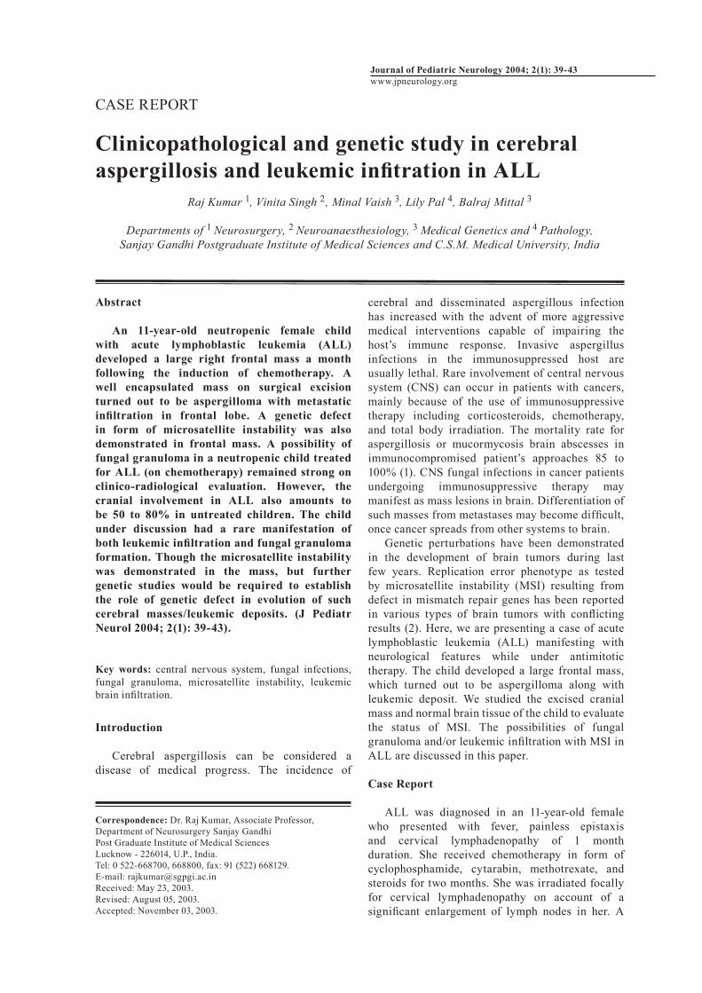
Journal of Pediatric Neurology 2004; 2(1): 39-43 www.jpneurology.org CASE REPORT Clinicopathological and genetic study in cerebral aspergillosis and leukemic infitration in ALL Raj Kumar 1 , Vinita Singh 2 , Minal Vaish 3 , Lily Pal 4 , Balraj Mittal 3 Departments of 1 Neurosurgery, 2 Neuroanaesthesiology, 3 Medical Genetics and 4 Pathology, Sanjay Gandhi Postgraduate Institute of Medical Sciences and C.S.M. Medical University, India Abstract An 11-year-old neutropenic female child with acute lymphoblastic leukemia (ALL) developed a large right frontal mass a month following the induction of chemotherapy. A well encapsulated mass on surgical excision turned out to be aspergilloma with metastatic infiltration in frontal lobe. A genetic defect in form of microsatellite instability was also demonstrated in frontal mass. A possibility of fungal granuloma in a neutropenic child treated for ALL (on chemotherapy) remained strong on clinico-radiological evaluation. However, the cranial involvement in ALL also amounts to be 50 to 80% in untreated children. The child under discussion had a rare manifestation of both leukemic infiltration and fungal granuloma formation. Though the microsatellite instability was demonstrated in the mass, but further genetic studies would be required to establish the role of genetic defect in evolution of such cerebral masses/leukemic deposits. (J Pediatr Neurol 2004; 2(1): 39-43). Key words: central nervous system, fungal infections, fungal granuloma, microsatellite instability, leukemic brain infiltration. Introduction Cerebral aspergillosis can be considered a disease of medical progress. The incidence of cerebral and disseminated aspergillous infection has increased with the advent of more aggressive medical interventions capable of impairing the host’s immune response. Invasive aspergillus infections in the immunosuppressed host are usually lethal. Rare involvement of central nervous system (CNS) can occur in patients with cancers, mainly because of the use of immunosuppressive therapy including corticosteroids, chemotherapy, and total body irradiation. The mortality rate for aspergillosis or mucormycosis brain abscesses in immunocompromised patient’s approaches 85 to 100% (1). CNS fungal infections in cancer patients undergoing immunosuppressive therapy may manifest as mass lesions in brain. Differentiation of such masses from metastases may become difficult, once cancer spreads from other systems to brain. Genetic perturbations have been demonstrated in the development of brain tumors during last few years. Replication error phenotype as tested by microsatellite instability (MSI) resulting from defect in mismatch repair genes has been reported in various types of brain tumors with conflicting results (2). Here, we are presenting a case of acute lymphoblastic leukemia (ALL) manifesting with neurological features while under antimitotic therapy. The child developed a large frontal mass, which turned out to be aspergilloma along with leukemic deposit. We studied the excised cranial mass and normal brain tissue of the child to evaluate the status of MSI. The possibilities of fungal granuloma and/or leukemic infiltration with MSI in ALL are discussed in this paper. Case Report ALL was diagnosed in an 11-year-old female who presented with fever, painless epistaxis and cervical lymphadenopathy of 1 month duration. She received chemotherapy in form of cyclophosphamide, cytarabin, methotrexate, and steroids for two months. She was irradiated focally for cervical lymphadenopathy on account of a significant enlargement of lymph nodes in her. A Correspondence: Dr. Raj Kumar, Associate Professor, Department of Neurosurgery Sanjay Gandhi Post Graduate Institute of Medical Sciences Lucknow - 226014, U.P., India. Tel: 0 522-668700, 668800, fax: 91 (522) 668129. E-mail: [email protected] Received: May 23, 2003. Revised: August 05, 2003. Accepted: November 03, 2003.
Transcript of Clinicopathological and genetic study in cerebral ... · aspergillosis and leukemic infitration in...
Journal of Pediatric Neurology 2004; 2(1): 39-43 www.jpneurology.org CASE REPORT
Clinicopathological and genetic study in cerebral aspergillosis and leukemic infitration in ALL
Raj Kumar 1, Vinita Singh 2, Minal Vaish 3, Lily Pal 4, Balraj Mittal 3
Departments of 1 Neurosurgery, 2 Neuroanaesthesiology, 3 Medical Genetics and 4 Pathology,Sanjay Gandhi Postgraduate Institute of Medical Sciences and C.S.M. Medical University, India
Abstract
An 11-year-old neutropenic female child with acute lymphoblastic leukemia (ALL) developed a large right frontal mass a month following the induction of chemotherapy. A well encapsulated mass on surgical excision turned out to be aspergilloma with metastatic infiltration in frontal lobe. A genetic defect in form of microsatellite instability was also demonstrated in frontal mass. A possibility of fungal granuloma in a neutropenic child treated for ALL (on chemotherapy) remained strong on clinico-radiological evaluation. However, the cranial involvement in ALL also amounts to be 50 to 80% in untreated children. The child under discussion had a rare manifestation of both leukemic infiltration and fungal granuloma formation. Though the microsatellite instability was demonstrated in the mass, but further genetic studies would be required to establish the role of genetic defect in evolution of such cerebral masses/leukemic deposits. (J Pediatr Neurol 2004; 2(1): 39-43).
Key words: central nervous system, fungal infections, fungal granuloma, microsatellite instability, leukemic brain infiltration.
Introduction
Cerebral aspergillosis can be considered a disease of medical progress. The incidence of
cerebral and disseminated aspergillous infection has increased with the advent of more aggressive medical interventions capable of impairing the host’s immune response. Invasive aspergillus infections in the immunosuppressed host are usually lethal. Rare involvement of central nervous system (CNS) can occur in patients with cancers, mainly because of the use of immunosuppressive therapy including corticosteroids, chemotherapy, and total body irradiation. The mortality rate for aspergillosis or mucormycosis brain abscesses in immunocompromised patient’s approaches 85 to 100% (1). CNS fungal infections in cancer patients undergoing immunosuppressive therapy may manifest as mass lesions in brain. Differentiation of such masses from metastases may become difficult, once cancer spreads from other systems to brain. Genetic perturbations have been demonstrated in the development of brain tumors during last few years. Replication error phenotype as tested by microsatellite instability (MSI) resulting from defect in mismatch repair genes has been reported in various types of brain tumors with conflicting results (2). Here, we are presenting a case of acute lymphoblastic leukemia (ALL) manifesting with neurological features while under antimitotic therapy. The child developed a large frontal mass, which turned out to be aspergilloma along with leukemic deposit. We studied the excised cranial mass and normal brain tissue of the child to evaluate the status of MSI. The possibilities of fungal granuloma and/or leukemic infiltration with MSI in ALL are discussed in this paper.
Case Report ALL was diagnosed in an 11-year-old female who presented with fever, painless epistaxis and cervical lymphadenopathy of 1 month duration. She received chemotherapy in form of cyclophosphamide, cytarabin, methotrexate, and steroids for two months. She was irradiated focally for cervical lymphadenopathy on account of a significant enlargement of lymph nodes in her. A
Correspondence: Dr. Raj Kumar, Associate Professor, Department of Neurosurgery Sanjay Gandhi Post Graduate Institute of Medical SciencesLucknow - 226014, U.P., India.Tel: 0 522-668700, 668800, fax: 91 (522) 668129.E-mail: [email protected]: May 23, 2003.Revised: August 05, 2003.Accepted: November 03, 2003.

Aspergilloma in ALL R Kumar et al40
month following commencement of chemotherapy, she had an episode of generalized tonic clonic seizures for which she was put on antiepileptic treatment and investigated further. Neurological examination revealed left sided hemiparesis with bilateral papilloedema. On investigation, the hemogram showed typical picture of ALL. X-ray chest and radiological screening of air sinuses remained unremarkable. Magnetic resonance imaging (MRI) brain scan showed a large mass in right posterior frontal
region, isointense on T1 weighted with intralesional hypointensities and surrounding large hypointense perifocal region. It was hyperintense on T2 weighted as a whole with only a rim of hypointense area surrounded by perifocal hyperintensity, suggestive of grade II edema (Figure 1). Considering the diagnosis of leukemic deposits or fungal granuloma, excision of mass was done via right frontal craniotomy on account of progressive symptoms (Figure 2). On operation table it was a well capsulated, well defined large whitish mass with capsule stripping off easily from the surrounding brain. It had dark yellowish soft solid material inside, which could be evacuated easily. Total enucleation of encapsulated mass was achieved. Histology showed large areas of necrosis of brain parenchyma along with patchy infiltration by atypical immature lymphoid cells (Figure 3). At places septate fungal hyphae (aspergillus) were invading the brain parenchyma with necrosis (Figure 4). Inflammatory reaction at the site of fungal invasion was minimal probably due to immunosuppression. Genetic defect as exemplified by microsatellite instability was assessed using a panel of ten markers (BAT-26, BAT-40, hMSH3, BAX, IGFIIR, TGFβRIII, D2S123, D9S283, D9S1851 and D18S58) and polymerase chain reaction (PCR), was performed to examine the pattern of microsatellite alleles in the DNA of normal neural tissue and
Figure 1. Proton density weighted MRI showing right frontal hypointense rim lesion with intralesional heterointensities and hyperintense areas and surrounded by hypointense area: mass effect.
Figure 2. Contrast enhanced CT of the same child following surgery depicting hypodense area at operation site. Note persisting posterior edema.
Figure 3. Photomicrograph of H and E stained section showing patchy leukemic deposits composed of small, round immature cells on a necrotic background (H and E x 200).

Aspergilloma in ALL R Kumar et al 41
the excised mass. The sequences of primers were searched from human genomic data base. PCR amplified products were then analyzed on 8% denaturing polyacrylamide gel followed by autoradiography. Out of the total markers evaluated all were found to be stable except a change was observed at D9S283 locus. This locus exhibited loss of heterozygosity (LOH) in study tissue as compared to the heterozygous pattern observed in normal tissue (Figure 5). The child had smooth postoperative recovery without added deficit. She was transferred to the department of immunology on 7th postoperative day for further treatment of ALL. Post-operatively she also received amphotericin B in the dose of 1 mg/kg/day over the period of 3 months. The drug was started following sensitivity testing with minimal dose. The child could not tolerate the total dose of more than 1.5 g on account of her already compromised blood counts and immunological status. This dose was considered sufficient for her weight and nutritional status by immunologist. At 9 months follow-up she had minimal residual left hemiparesis.
Discussion
Aspergillus fumigatus is the most frequent cause of aspergillosis of the CNS. The following conditions tend to favour the development of the disease: environmental or professional factors that expose the organism to prolonged contact with the infective agent, depressed immune response, acquired by corticosteroids therapy, antibiotics, cytotoxic drugs, immunosuppressive agents or a state of general debilitation pulmonary tuberculosis (3), hepatitis, alcoholism (4), and other drug addictions (5), diabetes (6) leukemia (7) and other neoplastic diseases (8). It seems that leukemia,
radiation and cytotoxic drugs were predisposing factors for developing aspergilloma in our child as described in the literature (7). There has been no report till now on intracerebral aspergillosis aspergilloma associated with leukemia deposits/metastases. Cerebral infection is usually secondary to a primary focus elsewhere in the body. The infection reaches into brain either directly from the nasal sinuses (9) or via vascular channels as blood born from the lungs and gastrointestinal trac (3,10). It may be airborne, contamination of the tissue during neurosurgical procedures (11). Nosocomial transmission has also been documented (12). Aspergillosis remains as one of the most difficult infection’s to treat, especially in immunocompromised patients, where it is mostly hematogenous dissemination from lung (13). Cases of intracranial aspergillosis occur worldwide with no demonstrable predisposition for sex or race, but mostly occur in adults. Most cases of invasive aspergillosis have occurred in neutropenic patients, who have an underlying hematological neoplasias (7) as it occurred in our neutropenic child of ALL with a history of radiation and multidrug antimitotic therapy. The brain is the second or third most commonly involved organ in patients with aspergillosis (14). Concurrent intracranial involvement is seen in 13 to 16% of patients with pulmonary aspergillosis. The brain involvement is found in 40 to 60% of cases in one series (14). The presenting symptoms of metastases and aspergillomas are usually nonspecific in the form of stroke like symptoms, seizure or intracranial
Figure 4. Photomicrograph of haematoxylene and eosin stained section showing areas of necrosis with clusters of septate fungal hyphae (aspergillus) (H and E x 200).
Figure 5. D9S283 allele showing heterozygous pattern in normal tissue (N) as compared to tumors (T) which exhibits LOH.
Aspergilloma in ALL R Kumar et al42
mass effects, while fever may not be present (15) and signs of meningeal irritation are also rare (16). Cerebrospinal fluid is usually sterile with slightly elevated protein levels and organism can rarely be cultured (13,15). Serologic tests to detect aspergillous antigen in the serum and/or cerebrospinal fluid remain experimental (17). For these reasons the diagnosis is usually difficult, only special staining and histopathological findings can confirm the diagnosis. Imaging studies provide a useful adjunct to diagnosis in CNS aspergillosis. Three different neuroimaging pattern of cerebral aspergillosis have been identified in immunosuppressed patients: (a) It may manifest with multiple areas of hypodensities on computerized tomography scan or hyperinstensities on T2 weighted MRI involving the cortex/subcortical white matter with multiple areas of embolic infarction. (b) Multiple intracerebral ring enhancing lesions, often hypointense on T1 weighted images and isointense on proton density and T2 weighted images with surrounding edema (18) with a low signal peripheral rim in cases of hemorrhagic infarction are demonstrated (19). (c) Dural enhancement associated with enhancing lesion in the adjacent tissues may be another imaging manifestation. Recognition of these three patterns of aspergillosis in immunosuppressed patients may lead to more effective diagnosis and treatment. Our child however presented with large well defined ring lesion, isointense on T1 weighted with intralesional hypointensities and large area of perifocal hypointensity. It was hyperintense on T2 weighted with hypointense rim, surrounding hyperintense edema and mass effect. In the literature, few descriptions of patients with ring lesion or nodular enhancement lesions exist as in our case (8,18). Despite intensive treatment with amphotericin B in many cases the mortality rate ranges from 75 to 100% and diagnosis is often made after death (20). The literature contains description of only a few patients who survived after surgical intervention and antifungal therapy (8,18). A reasonably good host defense mechanism may have role in good outcome as seen in our child who is fairing well despite having undergone surgery for fungal granuloma, while ALL has been taken care by further chemotherapy. Genetic changes like activation of proto-oncogenes, loss of tumor suppressor genes, stimulation of growth factors and aberrant immune surveillance play an important role in the induction of many neoplasms. Many genetic changes including loss of 12p detected by comparative genome hybridization, preferential loss of maternal 9p alleles, whole chromosome 6 loss and duplication of the remaining chromosome, mono and bi-allelic 9p 21 deletions are frequently seen is childhood
ALL (21-24). However, recently the disruption of mismatch repair genes indicated by MSI is observed as one of the key feature present in ALL. Cytogenetic and FISH (fluorescent in situ hybridiation) analysis has demonstrated chromosomal abnormalities in 68% of patients with ALL (25). LOH of 6q, 9p, 11q and 12p in 20%, 40%, 12%, and 25% of individuals, respectively, has been observed as a very frequent alteration in children with ALL (26). In our present case of aspergillosis along with leukemic deposits/ metastases, which is behaviorally different from brain tumors, we report LOH at long arm of chromosome 9 at D9S283 encompassing CA repetitions. To the best of our knowledge the genetic instability has not been reported till now either in fungal granuloma or in patients with fungal granuloma associated with ALL. It is hypothesized that infection caused due to antimitotic therapy and/or radiation accumulates genetic alterations in the locally infected and metastatic tissue as compared to normal tissue of brain which manifests as loss of one allele and makes the locus at 9q chromosome susceptible to genomic instability as seen in our child.
Conclusion
The CNS aspergillosis in immunocompromised neutropenic child may manifest with large cerebral mass and mass effect. Metastases from ALL may coexist with aspergilloma. Usually such cases have fatal course in short duration of time even after surgical intervention and appropriate antifungal therapy. But our child remained intact even after 9 months, most probably because of localized disease and duly offered treatment for fungal granuloma and ALL. The contrast enhanced MRI and computerized tomography characteristics have an important role in establishing the early diagnosis and treatment in high risk, immunocompromised patients. Genetic defect demonstrated as genomic instability has also been found to be present, which may signify the correlation between cerebral masses/ALL and microsatellite instability. Further studies would be required to establish, the susceptibility of infection/tumors in patients harboring genetic defect.
Acknowledgement
The authors are extremely grateful to Mr. A.P. Dhar Dwivedi for preparation of this manuscript. One of the authors Minal Vaish is thankful to CSIR, goverment of India, for providing financial support.
References
1. Goodman ML, Coffey RJ. Streotactic drainage of aspergillus brain abscess with long-term survival:

Aspergilloma in ALL R Kumar et al 43
case report and review. Neurosurgery 1989; 24: 96-99.
2. Alvino E, Fernandez E, Pallini R. Microsatellite instability in primary brain tumors. Neurol Res 2000; 22: 571-575.
3. Spens N, Tattersall WH. Fungal infection of the central nervous system supervening during routine chemotherapy for pulmonary tuberculosis. Br Med J 1965; 5466: 862.
4. Klein HJ, Richter HP, Schachenmayr W. Intracerebral aspergillus abscess: case report. Neurosurgery 1983; 13: 306-309.
5. Kasantikul V, Shuangshoti S, Taecholarn C. Primary phycomycosis of the brain in heroin addicts. Surg Neurol 1987; 28: 468-472.
6. Kim DG. Hong SC, Kim HJ, et al. Cerebral aspergillosis in immunologically competent adults. Surg Neurol 1993; 40: 326-331.
7. Yamamoto N, Miyara T, Kawakami K, et al. A case of disseminated aspergillosis with smoldering adult T-cell leukemia. Kansenshogaku Zasshi 2002; 76: 460-465 (in Japanese).
8. Lin Zy, Sheng RY, Li XL, Li TS, Wang AX. Nosocomial fungal infections, analysis of 149 cases. Zhonghua Yi Xne Za Zhi 2003; 83: 399-402 (in Chinese).
9. Erdoğan E, Beyzadeoğlu M, Arpacı F, Celasun B. Cerebellar aspergillosis: case report and literature review. Neurosurgery 2002; 50: 874-876.
10. Hay RJ. Fungal infections. Med Int (Middle Eastern Edition) 1998; 53: 2203-2209.
11. Sharma RR, Gurusinghe NT, Lynch PG. Cerebral infarction due to Aspergillus arteritis following glioma surgery. Br J Neurosurg 1992; 6: 485-490.
12. Darras-Joly C, Veber B, Bedos JP, Gachot B, Regnier B, Wolff M. Nosocomial cerebral aspergillosis: a report of 3 cases. Scand J Infect Dis 1996; 28: 317-319.
13. Cohen J. Clinical manifestations and management of aspergillosis in the compromised patients. In: Warnock DW, Richardson MD (eds). Fungal Infection in the Compromised Patients. Chichester: John Wiley Sons, 1991, pp 117-151.
14. Denning DW, Stevens DA. Antifungal and surgical treatment of invasive aspergillosis: review of 2,121 published cases. Rev Infect Dis 1990; 12: 1147-1201.
15. Meyer D, Young LS, Armstrong D, Yu B.
Aspergillosis complicating neoplastic disease. Am J Med 1973; 54: 6-15.
16. Lyons RW, Andriole VT. Fungal infections of the CNS. Neurol Clin 1986; 4: 159-170.
17. Armstrong D, Wong B. Central nervous system infection in immunocompromised hosts. Annu Rev Med 1982; 33: 293-308.
18. Fisher BD, Asmstrong D, Yu B, Gold JW. Invasive aspergillosis: progress in early diagnosis and treatment. Am J Med 1981; 71: 571-577.
19. Miaux Y, Ribaund P, Williams M, et al. MR of cerebral aspergillosis in patients who have had bone marrow transplantation. AJNR Am J Neuroradiol 1995; 16: 555-562.
20. Cox J, Murtagh FR, Wilfong A, Brenner J. Cerebral aspergillosis: MR imaging and histopathologic correlation. AJNR Am J Neuroradiol 1992; 13: 1489-1492.
21. Kanerva J, Niini T, Vettenranta K, et al. Loss at 12p detected by comparative genomic hybridization (CGH): association with TEL-AML1 fusion and favorable prognostic features in childhood acute lymphoblastic leukemia (ALL). A multi-institutional study. Med Pediatr Oncol 2001; 37: 419-425.
22. Morison IM, Ellis LM, Teague LR, Reeve AE. Preferential loss of maternal 9p alleles in childhood acute lymphoblastic leukemia. Blood 2002; 99: 375-377.
23. McEvoy CR, Morley AA, Firgaira FA. Evidence for whole chromosome 6 loss and duplication of the remaining chromosome in acute lymphoblastic leukemia. Genes Chromosomes Cancer 2003; 37: 321-325.
24. Bertin R, Acquaviva C, Mirebeau D, Guidal-Giroux C, Vilmer E, Cave H. CDKN2A, CDKN2B, and MTAP gene dosage permits precise characterization of mono-and bi-allelic 9p21 deletions in childhood acute lymphoblastic leukemia. Genes Chromosomes Cancer 2003; 37: 44-57.
25. Andreasson P, Hoglund M, Bekassy AN, et al. Cytogenetic and FISH studies of a single center consecutive series of 152 childhood acute lymphoblastic leukemias. Eur J Haematol 2000; 65: 40-51.
26. Takeuchi S, Tsukasaki K, Bartram CR, et al. Long-term study of the clinical significance of loss of heterozygosity in childhood acute lymphoblastic leukemia. Leukemia 2003; 17: 149-154.