Dr Don Ajith Karawita MBBS, PgD Ven, MD Venereology National STD/AIDS Control Programme.
Volume 23, No.2, October 2016The Gulf Journal of Dermatology and Venereology
8
ORIGINAL ARTICLE
Clinicomycological study of coexisting toenail onychomycosis and tinea pedisHamed Mohamed Abdo,1 MD, Sabah Ibrahim Abd Elrahem,2 MD
1Dermatology and Venereology Department, Faculty of Medicine - Al-Azhar University, Cairo, Egypt2Clinical Pathology Department, Faculty of Medicine - Al-Azhar University for girls, Cairo, Egypt
Correspondence: Dr. Hamed Mohamed Abdo, Department of Dermatology and Venereology, Faculty of Medicine, Al Azhar University, Cairo, Egypt Phone: +201066339011 - E-mail: [email protected]
ABSTRACTBackground: Tinea pedis is frequently associating with toenail onychomycosis. The causative fungi are usually the same but may also be different.Aim: To correlate and find out the etiology of coexisted toenail onychomycosis and tinea pedis in patients presenting with both of them. Patients and methods: The study population included 44 patients with clinically suspected, combination of toenail onychomycosis and tinea pedis. The nails and feet were evaluated clinically and mycologically. Nail bed/plate specimens were obtained with sterilized instruments and scales were collected from clinically suggestive tinea pedis lesions. All samples were examined by direct microscopy and fungal culture.Results: Of the 44 patients, 33 had toenail onychomycosis (75%) while 40 had tinea pedis (90.9%). Distal/lateral subungual onychomycosis and dry hyperkeratotic tinea pedis were the most common types of nail and foot diseases (n=22-66.66%; n=19- 47.5% respectively). Middle aged males were the commonest infected population. From nails, direct microscopy and culture were positive in 31 (70.45%) and 33 (75%) patients respectively. Dermatophytes were the most prevalent isolated species (n=24/33; 72.73%) followed by non-dermatophytic molds (n=5/33; 15.15%) and yeasts (n=4/33; 12.12%). From tinea pedis lesions, direct microscopy and culture were positive in 36 (81.81%) and 35 (79.54%) respectively. Dermatophytes were the most prevalent species (n=25/35; 71.43%) followed by yeasts (n=7/35; 20%) and non-dermatophytic molds (n=3/35; 8.57%). Trichophyton rubrum was the most common isolate from nail and foot lesions (45.83% and 56% respectively)Conclusion: Majority of the coexisting toenail onychomycosis and tinea pedis were caused by single and the same pathogen. The presence of the same fungi in toenail and foot lesions may explain auto-infectivity, chronicity and recurrence of these infections. When both conditions are concurrent, a nail-targeted treatment plan should be considered.
KEY WORDS: Tinea pedis, toenail onychomycosis, KOH test, culture.
INTRODUCTIONThe prevalence of superficial mycotic infections worldwide is 20-25%, of which dermatophytes are the most common agents.1 These infections are widespread in the developing communities, especially in the tropical and subtropical countries, where the environmental temperature and relative humidity are high. Other factors such as increased urbanization including the use of occlusive footwear and tight fashioned clothes,
has been linked to higher prevalence.2 Over the last few years, studies on epidemiology of dermatophytic infection from tropical countries have shown a rising trend in the prevalence of cutaneous dermatophytosis, with change in spectrum of infection and isolation of some uncommon species.3 Tinea pedis is a frequently encountered dermatophytosis affecting primarily the adults. Its prevalence has increased over the last several
Volume 23, No.2, October 2016The Gulf Journal of Dermatology and Venereology
9
decades due to an increase in multiple risk factors. Infection from dermatophytes is most common, but infection from other fungi can also result in tinea pedis. Four distinct clinical presentations occur: interdigital, moccasin, vesicular, and acute ulcerative types.4 The frequency of onychomycosis in developed countries ranges between 3 and 8, depending on the examined population, and increases with patient age. Toenail onychomycosis is about 4 to 7 times more frequent than fingernail disease. In about a third of patients with toenail onychomycosis, concomitant tinea pedis can be observed. The coexistence of toenail onychomycosis with other types of fungal infections is also frequent.5,6 Relapse or re-infection in onychomycosis is common, occurring in 10 to 53% of patients. Although a number of factors have been suggested to play a role in recurrence, the co-existence of diabetes has been shown to have a significant impact.7 The dermatophytes Trichophyton (T) rubrum and T. interdigitale are the common dermatophytes known to cause nail infection. Non-dermatophytes such as yeasts from Candida species, as well as molds from Fusarium and Acremonium species account for the remainder of infections.8 Changes over time within a region in the prevalence of particular dermatophyte species are common. A change that has occurred in last decades is the growing prevalence of dermatophytoses of the foot and nails.9 Studies regarding coexistence of toenail onychomycosis with tinea pedis are lacking; therefore, we aimed to study both diseases in patients presenting with clinically highly suggestive toenail and foot skin fungal infection.
PATIENTS AND METHODSThis prospective study was carried out for a period of 3 years (October 2012 to December 2015). In this study, 44 patients with clinically highly suggestive, coexisting toenail onychomycosis and tinea pedis were enrolled after strict inclusion and exclusion criteria. The study was approved by the local committee of medical ethics, and written prior informed consent was obtained from every participant. The lesions of the nails and feet were evaluated clinically and mycologically. For every patient, duplicate samples of scrapings were collected from the nail and foot lesions. One sample was examined by direct microscopy and the other by fungal culture. After cleaning the lesions with 70% alcohol, nail/skin samples were collected from the affected site(s). According to the clinical type, nail specimens were obtained from the nail bed or nail plate with sterilized instruments. Scales were collected from clinically suggestive tinea pedis lesions.
1. Direct microscopy with potassium hydroxide (KOH): Specimen was placed on a clean glass slide, and a drop of 20% KOH/40% Dimethyl Sulfoxide (DMSO) mixture was added (DMSO increases sensitivity of the preparation and softens keratin more quickly than KOH alone in the absence of heat).10 A coverslip was applied with gentle pressure to drain away excess solution. The sample was then examined thoroughly for the presence of fungal elements. Dermatophytes were identified by the presence of hyaline, filamentous, septate, branching hyphae with or without arthrospores. Non-dermatophytic molds (NDMs) were identified by the presence of hyaline or non-
Hamed Mohamed Abdo et, al.
Volume 23, No.2, October 2016The Gulf Journal of Dermatology and Venereology
Clinicomycological study of coexisting toenail onychomycosis and tinea pedis
10
hyaline filamentous, usually aseptate, branching broad hyphae. Yeasts were identified by presence of multiple oval and/or round cells with some budding with or without hyphae/pseudohyphae.
2. Fungal culture: The second sample of scraping was inoculated onto two Sabouraud Dextrose Agar (SDA) media: One with cycloheximide (to suppress the growth of contaminant fungi) and the other without cycloheximide. Chloramphenicol was added to both media to prevent bacterial overgrowth. The media were then incubated in a warm, moist environment at 28°C and examined regularly to detect growth of any fungus. Observation for growth was done periodically for at least 4 weeks after which the culture was reported as positive or negative. For fungal identification, we relied upon macro and micro-morphological characters of the isolates. The fungi were identified by noting their growth rate, colonial macro-morphology, and microscopic structures. Macroscopic examination included color, size, texture, and topography of the colony. The microscopic structures of fungi usually provide definitive identification.11 Using the tease mount technique, the following microscopic features were looked for: the type, size, shape and arrangement of spores and the size, color, septation and special shapes of hyphae.
STATISTICAL ANALYSISThe Statistical Package for Social Studies (SPSS version 18.0) was used for data analysis. Numerical parameters were expressed as mean ± SD. Data were analyzed statistically using the chi-square test. Results were assumed to be statistically significant when P value was < 0.05.
RESULTS1. Demographic results: Out of the 44 patients, there were 27 (61.36%) males and 17 (38.64%) females. Their ages ranged from 14-72 years (mean ± SD 43.13± 3.28). Middle aged males were the commonest infected population (n=15; 34.1%) while older patients (over 60 yrs) represented the next common group (n=11; 25%). Most cases were office employees (n=12; 27.27%), followed by manual workers (n=9; 20.45%) and farmers (n=8; 18.18%). Other miscellaneous groups (n=15; 34.1%) included teachers, house wives, sportsmen and students. Low education level was noticed in a considerable number of patients (n=17; 38.64%). Small fraction (n=9; 20.45%) had history of repeated exposure to animals including farm animals, cats and rabbits.
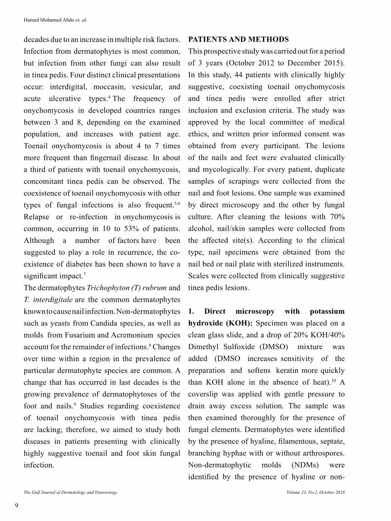
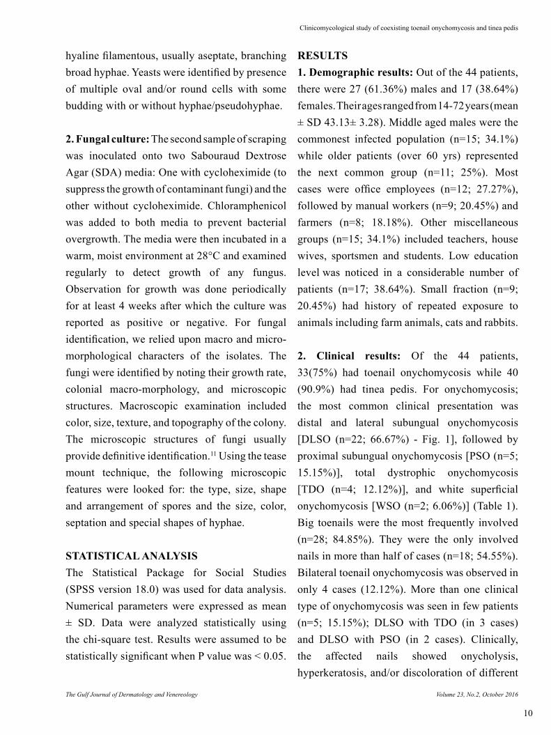
2. Clinical results: Of the 44 patients, 33(75%) had toenail onychomycosis while 40 (90.9%) had tinea pedis. For onychomycosis; the most common clinical presentation was distal and lateral subungual onychomycosis [DLSO (n=22; 66.67%) - Fig. 1], followed by proximal subungual onychomycosis [PSO (n=5; 15.15%)], total dystrophic onychomycosis [TDO (n=4; 12.12%)], and white superficial onychomycosis [WSO (n=2; 6.06%)] (Table 1). Big toenails were the most frequently involved (n=28; 84.85%). They were the only involved nails in more than half of cases (n=18; 54.55%). Bilateral toenail onychomycosis was observed in only 4 cases (12.12%). More than one clinical type of onychomycosis was seen in few patients (n=5; 15.15%); DLSO with TDO (in 3 cases) and DLSO with PSO (in 2 cases). Clinically, the affected nails showed onycholysis, hyperkeratosis, and/or discoloration of different
Volume 23, No.2, October 2016The Gulf Journal of Dermatology and Venereology
11
grades of severity. Concerning tinea pedis; the most common clinical presentation was dry hyperkeratotic “moccasin” type (n=19; 47.5% - Fig. 1) followed by vesiculobullous (n=13; 32.5%) and interdigital varieties (n=8; 20%) (Table 1). Bilateral lesions were observed in 12 cases (30%); 5 interdigital, 4 hyperkeratotic and 3 vesiculobullous. Mixed lesions occurred in some patients (n=10; 25%); hyperkeratotic with interdigital (in 6 cases) and vesiculobullous with interdigital type (in 4 cases).Clinical correlation revealed that both
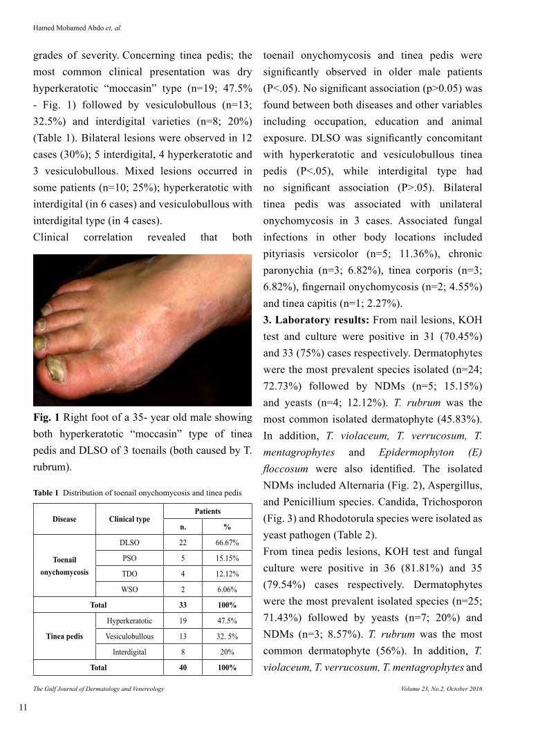
toenail onychomycosis and tinea pedis were significantly observed in older male patients (P<.05). No significant association (p>0.05) was found between both diseases and other variables including occupation, education and animal exposure. DLSO was significantly concomitant with hyperkeratotic and vesiculobullous tinea pedis (P<.05), while interdigital type had no significant association (P>.05). Bilateral tinea pedis was associated with unilateral onychomycosis in 3 cases. Associated fungal infections in other body locations included pityriasis versicolor (n=5; 11.36%), chronic paronychia (n=3; 6.82%), tinea corporis (n=3; 6.82%), fingernail onychomycosis (n=2; 4.55%) and tinea capitis (n=1; 2.27%).3. Laboratory results: From nail lesions, KOH test and culture were positive in 31 (70.45%) and 33 (75%) cases respectively. Dermatophytes were the most prevalent species isolated (n=24; 72.73%) followed by NDMs (n=5; 15.15%) and yeasts (n=4; 12.12%). T. rubrum was the most common isolated dermatophyte (45.83%). In addition, T. violaceum, T. verrucosum, T. mentagrophytes and Epidermophyton (E) floccosum were also identified. The isolated NDMs included Alternaria (Fig. 2), Aspergillus, and Penicillium species. Candida, Trichosporon (Fig. 3) and Rhodotorula species were isolated as yeast pathogen (Table 2).From tinea pedis lesions, KOH test and fungal culture were positive in 36 (81.81%) and 35 (79.54%) cases respectively. Dermatophytes were the most prevalent isolated species (n=25; 71.43%) followed by yeasts (n=7; 20%) and NDMs (n=3; 8.57%). T. rubrum was the most common dermatophyte (56%). In addition, T. violaceum, T. verrucosum, T. mentagrophytes and
Fig. 1 Right foot of a 35- year old male showing both hyperkeratotic “moccasin” type of tinea pedis and DLSO of 3 toenails (both caused by T. rubrum).
Disease Clinical typePatients
n. %
Toenail onychomycosis
DLSO 22 66.67%
PSO 5 15.15%
TDO 4 12.12%
WSO 2 6.06%
Total 33 100%
Tinea pedis
Hyperkeratotic 19 47.5%
Vesiculobullous 13 32. 5%
Interdigital 8 20%
Total 40 100%
Table 1 Distribution of toenail onychomycosis and tinea pedis
Hamed Mohamed Abdo et, al.
Volume 23, No.2, October 2016The Gulf Journal of Dermatology and Venereology
Clinicomycological study of coexisting toenail onychomycosis and tinea pedis
12
Microsporum (M) audouinii were also identified. The yeasts were isolated only from interdigital lesions and included Candida, Rhodotorula and other non-specified species. The isolated NDMs included Alternaria, (from the sole) Aspergillus, and Penicillium species (from web lesions)
(Table 2).Clinicomycological correlation of toenail onychomycosis and tinea pedis revealed that most of the nail and foot lesions were caused by single and the same pathogen, a result which was statistically significant (P<.05). This was true for the dermatophytes of the trichophyton species, namely, T. rubrum (which was the commonest cause of DLSO and hyperkeratotic tinea pedis), T.
Fig. 2 (A) Gross morphology of Alternaria alternata showing olivaceous-black, suede-like colony which was rapidly growing.
Fig. 2 (B) Microscopic morphology of Alternaria alternata showing characteristic conidia. These are ovoid or ellipsoidal, pale brown, smooth-walled conidia present singly or in acropetal chains. They have both transverse and longitudinal septations. Their base is round while they taper towards the apex, giving their typical appearance of short conical or cylindrical beak (water mount ×400).
Fig. 3 (A) Gross morphology of Trichosporon species showing cream-colored, cerebriform colonies which are raised, waxy, with radial furrows and irregular folds.
Fig. 3 (B) Microscopic morphology of Trichosporon species showing budding cells, cylindrical to ellipsoidal arthroconidia and short regular hyphae (water mount×400).

Volume 23, No.2, October 2016The Gulf Journal of Dermatology and Venereology
13
Table 1 Isolation frequencies of dermatophytes, NDMs and yeasts from nail and foot lesions
Isolates
Positive cultures
Toenail onychomycosis
Tinea pedis
n. % n. %
Dermatophytes 24 72.73% 25 71.43%
T. rubrum 11 45.83% 14 56%
T. violaceum 6 25% 6 24%
T. verrucosum 4 16.67% 2 8%
T. mentagrophytes 2 8.33% 2 8%
E. floccosum 1 4.17% - -
M. audouinii - - 1 4%
NDMs 5 15.15% 3 8.57%
Alternaria sp., 3 60% 1 33.33%
Aspergillus sp., 1 20% 1 33.33%
Penicillium sp. 1 20% 1 33.33%
Yeasts 4 12.12% 7 20%
Candida sp., 2 50% 4 57.14%
Trichosporon sp., 1 25% - -
Rhodotorula sp. 1 25% 1 14.29%
Non-specified sp - - 2 28.57%
Total 33 100% 35 100%
violaceum, T. verrucosum and T. mentagrophytes. These fungi were isolated simultaneously from both nail and foot lesions. This also was applied to NDMs (Alternaria, Aspergillus, Penicillium) and, to a lesser extent, yeast fungi (Candida, Rhodotorula) (Table 2). Only 6 cases showed mixed infection (more than one fungal species), which was non-significant (P>.05). This finding was noticed in 4 great toenails (DLSO, 12.12%) and 2 tinea pedis cases (interdigital, 5.7%). From the great toenails, T. rubrum was co-isolated with 2 Candida and 1 Alternaria species, while T. verrucosum was co-isolated with Trichosporon species in the 4th case. The 2 interdigital tinea pedis revealed Candida with non-specified yeast species.
DISCUSSIONMany studies have been done, both retrospectively and prospectively on the prevalence of onychomycosis and tinea pedis but no prospective study aimed to etiologically correlate coexisting toenail onychomycosis and tinea pedis. This study was designed to investigate this association. In this study, presence of toenail onychomycosis and tinea pedis was found to correlate significantly with increasing age and male gender. This coincides with most of the previous studies.12,13,14 Going with this study, Szepietowski et al6 reported that in patients with toenail onychomycosis, tinea pedis was the most frequently concomitant dermatomycoses, and was more often observed in males and in subjects with recurrent and more advanced nail disease. The hyperkeratotic type was significantly more prevalent in older patients. Contrary, Kanth et al15 showed that the onychomycosis was common in females than males. Viegas et al16 found no significant association between gender and tinea pedis and onychomycosis. In addition to influence of age and gender, most cases in this study were office workers (27.27%), followed by manual workers (20.45%) and farmers (18.18%). The increased prevalence of onychomycosis and tinea pedis in these populations could be due to the common wearing of closed shoes and socks for longer times, frequent nail trauma and the more outdoor exposure to fungi particularly in farmers who may walk barefoot during their field activity and exposed frequently to animals.The most common clinical presentation of onychomycosis was DLSO (66.67%) followed by PSO (15.15%), TDO (12.12%) and WSO (6.06%). This frequency of nail fungal infection correlates with the most published studies.17,18,19
Hamed Mohamed Abdo et, al.
Volume 23, No.2, October 2016The Gulf Journal of Dermatology and Venereology
Clinicomycological study of coexisting toenail onychomycosis and tinea pedis
14
The commonest clinical variety of tinea pedis was dry hyperkeratotic type followed by vesiculobullous and interdigital varieties (47.5%, 32. 5% and 20% respectively). This coincides with most of the earlier literature except for the interdigital variety. The interdigital type, although the most common in other reports, was the least in this work because we choose the type of tinea pedis which was only coexisting with nail affection.The etiology of onychomycosis and tinea pedis may be dermatophytes, NDMs and yeasts. Most of the studies identify dermatophytes as the most frequent etiological agents (80-90%), followed by yeasts (5-17%) and NDMs (2-12%).6,13,20 This agrees with our findings in which dermatophytes, NDMs and yeasts were isolated from nails and feet lesions in similar sequence (72.73% and 71.43%; 15.15% and 8.57%; 12.12% and 20% respectively). Lone et al19 reported that the etiological agents were dermatophytes (62.68%) followed by NDMs (29.85%) and yeasts (7.46%). Viegas et al16 identified NDMs as the most frequent etiological agents (44.6%) followed by yeasts (39.8%) and dermatophytes (15.6%). Also Baiu et al21 isolated NDMs from 58% of cases followed by yeasts (26%) and dermatophytes (6%) while 10% revealed mixed fungi. The low frequency of isolation of dermatophytes by Viegas et al16 and Baiu et al21 is questionable. Viegas et al16 explained this by the high number of patients under antifungal treatment which cannot be applied to other results. Some studies, on the other hand, identified dermatophytes and yeasts as the causative fungi,22,23,24 while others revealed only dermatophytes as the sole etiologic agents.25
In this sudy, dermatophytes were the most
common etiological fungi retrieved from nail and foot lesions (72.73%, 71.43% respectively). From these, T. rubrum was the most common isolate from both diseases (45.83%, 56% respectively). This is in concordance with most other international and national studies. Kemna and Elewski26 found that 94.7% of the cases of tinea pedis and 81.9% of onychomycosis cases in the United States were caused by dermatophytes, and T. rubrum was the most commonly isolated organism (78.9%, 76.2% respectively). Summerbell et al27 reported, in Canada, similar results with dermatophytes causing 90.7% of tinea pedis, and 97.7% of onychomycosis cases. In Algeria, Djeridane et al22 reported that T. rubrum was the most common pathogen in onychomycosis (35%) and tinea pedis (17%). In Egypt, Abd El-Glil and Abdul Fattah28 showed that dermatophytes were isolated in 44.4% from onychomycosis samples and T. rubrum was the most common dermatophytes (37.8%). Similarly, Ahmad et al29 reported T. rubrum in 44.5% of onychomycosis cases. In contrast, other workers reported low rate of isolation of dermatophytes. Viegas et al16, isolated dermatophytes in 15.6% in their study. Shoar et al30 from 252 patients with onychomycosis have found yeasts and yeast-like fungi as predominant causative agents of onychomycosis. This difference in fungal isolation rate could be explained by different samples of study population, varied diagnostic procedures, different environmental conditions and degree of exposure to the pathogens. The predominance of certain dermatophytes varies geographically depending on ethnicity and different environmental factors such as climate, humidity, occupation, and different life styles.

Volume 23, No.2, October 2016The Gulf Journal of Dermatology and Venereology
15
Overall, the causative agents vary depending upon geographical location and temporal distribution. T. rubrum is the commonest cause of DLSO in most of the studies19,29 as well as in this work. The high rate of isolation of T. rubrum can be explained by its high capacity to infect the nails, because it can easily colonize on hard keratin.31 It tends to cause chronic infection in all keratinous tissues of the body (skin, hair, nail). Moreover, it does not elicit strong inflammatory response, which may be the cause of delay in seeking medical advice.Although the majority of superficial fungal infections (including nails and feet) are caused by dermatophyes, a large number of NDMs have been implicated in onychomycosis etiology especially of toenails. The recent concept considers the NDMs an emerging and leading cause of onychomycosis affecting specially the toenails. Their isolation rate varies widely in different studies, with rates ranging from 0% up to 60%. In our work, NDMs were isolated from nail and foot lesions in 15.15% and 8.57% respectively. In an Egyptian study, El Batawi et al32 have found NDMs in 59% of the total culture positive cases. Baiu et al21 isolated NDMs from 58% while Viegas et al16 have identified them in 44.6%. Some studies, on the other hand, could not identify NDMs from any case.22,23,24
It is not known whether NDM infections occur as a primary pathogen on healthy nails, or exist as secondary invader in nails already damaged by ischemia, trauma or other conditions. Such infections are considered by some to result following nail trauma, and not due to primary invasion.33,34 However, according to Rosen35 many studies have suggested that NDMs and yeasts are having an increasing role in the
pathogenesis of onychomycosis. In absence of cycloheximide, NDMs are frequently recovered from nail, hair, and skin cultures. However, most researchers agree that these organisms are contaminants. According to Borman and Johnson,36 the decision that a NDM cultured from nail specimens is clinically significant depends upon positive direct microscopy, failure to culture a dermatophyte, isolation of the NDM in pure culture from a large proportion of the clinical sample, and preferably its repeat isolation. By direct microscopy, the presence of a NDM may be indicated by demonstration of intact hyphae (as compared to arthrospores, which are a key feature of dermatophytes) that are frequently distorted by terminal fronding or hyphal swellings. English37 suggests that in order to consider a NDM to be considered clinically significant, it must grow from 5 of 20 inocula.Yeasts are frequently isolated from cultures of nail infections. Although the importance of yeast as a primary pathogen in onychomycosis is not generally accepted, it does appear to have a role in certain individuals especially among elderly diabetics and immunocompromised patients. Candida species can cause nail infections, usually associated with chronic paronychia. Since these organisms are normal commensals of the skin, their recovery in culture from skin or nail specimens is not usually significant unless direct microscopy reveals large amounts of yeast cells/hyphae.36 Like NDMs, the isolation rate of yeast species from onychomycosis is varied widely. In this work, yeast species were isolated from nail and foot lesions in 12.12% and 20% respectively. Viegas et al16 have identified them in 39.8%. Kemna and Elewski26 found Candida albicans in only 4.6% of positive nail cultures. Some
Hamed Mohamed Abdo et, al.
Volume 23, No.2, October 2016The Gulf Journal of Dermatology and Venereology
Clinicomycological study of coexisting toenail onychomycosis and tinea pedis
16
investigators, however, have found yeasts as the most common agents. Mikaeili and Karimi38 have detected them as the most prevalent pathogen (78.5%). This wide difference in isolation rate may be due to the fact that some authors consider nails in chronic paronychia (which is most often caused by yeast) as a type of onychomycosis, while others don’t agree with this concept and recognize these nail changes as a sequel to paronychial inflammation. In this study, we were able to isolate both Trichosporon species (from nail) and Rhodotorula species (from nail and foot). It is noteworthy that Trichosporon species can cause superficial infections including onychomycosis. Some Mexican authors have documented that the isolation of Trichosporon species from tinea pedis and onychomycosis lesions may range from 2.81% to 42.8% of cases.39,40 Rhodotorula species are uncommon agents in etiology of onychomycosis, and very few cases caused by these species have been reported.41,42
There is a general agreement that interdigital web space infection may represent a single or polymicrobial etiology “mixed infection”.43 Polymicrobial etiology, however, cannot simply apply to other forms of tinea pedis or nail infections. In this work, 4 great toenails with DLSO (12.12%) and 2 interdigital tinea pedis cases (5.7%) showed mixed infection. Several studies have focused on mixed infection incidence, but the results are conflicting and disputing. Both dermatophytes and non-dermatophytes, especially Candida albicans, have been identified as sole etiologic agents of onychomycosis. However, the incidence of true mixed infections is difficult to determine accurately.25 Toenail mixed fungal infection can
be explained by repeated traumatic exposure with subsequent traumatic onycholysis, which facilitates the subungual area to harbor different fungi from the patient itself or from the environment. Traumatic onycholysis must, in fact, be considered in the differential diagnosis of onychomycosis.Some investigators believe that claims of an increasing proportion of mixed infections in onychomycosis are exaggerated, and have gone so far as to state that NDMs and yeasts are usually contaminants secondary to dermatophyte onychomycosis. And, that their presence need not affect treatment outcome.44 Generally speaking, all dermatophytes should be considered pathogens. All other isolated organisms are probably laboratory contaminants, unless KOH test indicates they have the atypical frond-like hyphae associated with NDMs or if the same organism is repeatedly isolated.45 To increase the predictive power of a diagnosis of non-dermatophytic invasion of a nail, Summerbell25 suggested that non-dermatophytes identified in nail tissue be categorized as one of the following: I) Contaminant (growing in culture from dormant propagules on the nails); II) Normal surface commensal organism; III) Transient saprobic colonizer (but non-invasive); IV) Persistent secondary colonizer (colonizer of material infected by a dermatophyte but incapable of remaining after the dermatophyte is eliminated); V) Successional invader (can cause infection after gaining entry into a nail via the disruption caused by a primary pathogen); or VI) primary invader (able to infect and cause onychomycosis in a previously non-colonized nail).Several medical and non-medical risk factors can affect a person’s chance of developing
Volume 23, No.2, October 2016The Gulf Journal of Dermatology and Venereology
17
onychomycosis and tinea pedis. Medical factors include diabetes, immunosuppression and conditions contributing to poor peripheral circulation.46 Non-medical conditions may include the habit of wearing occlusive footwear that creates a warm, moist environment for the proliferation of fungi. The spread of foot infections may occur in places such as shower stalls, bathrooms, or locker rooms where floor surfaces often are wet and people are barefoot.9 Other common factors include nail trauma and the advancing age.The Achilles foot screening project (Roseeuw47) revealed that the proportion of patients with foot diseases visiting a dermatologist was high (58%). In the total population, fungal infections were the most prevalent clinically diagnosed foot diseases (35%), especially onychomycosis (23%) and tinea pedis (22%). These data indicated that both disorders are important and constitute a frequent health problem. When clinical tinea pedis and toenail onychomycosis coexist, both may act as a source of the fungal pathogen. However, tinea pedis may be subtle and mistaken by the patient or a clinician to be dry skin.48 It is also worth mentioning that many patients with both diseases are not even aware that they have fungal infection. And, such patients pose a special challenge for physicians to detect and treat the condition. In a study, it has been found that the prevalence of untreated, unsuspected tinea pedis was 25%, and that 59% of those cases were complicated by tinea unguium.49 It is obvious that tinea pedis may precede toenail onychomycosis, and vice versa.
CONCLUSIONCoexistence of toenail onychomycosis and tinea pedis is frequent. Any scaly/dry skin of the foot associated with nail changes should raise suspicion of fungal infection of both. DLSO and hyperkeratotic tinea pedis were the commonest forms of nail and foot lesions. Majority of toenail onychomycosis and tinea pedis were caused by single and the same pathogen. Infected toenails may be a site from which the fungal infections could spread to the foot and vice versa. This may explain chronicity and recurrence of both infections. When both conditions are concurrent, an early and effective nail-targeted treatment plan should be considered.
REFERENCES1. Sahoo AK and Mahajan R. Management of
tinea corporis, tinea cruris, and tinea pedis: A comprehensive review. Indian Dermatol Online J 2016; 7(2):77-86.
2. Havlickova B, Czaika VA and Friedrich M. Epidemiological trends in skin mycoses worldwide. Mycoses 2008; 51:2-15.
3. Lakshmanan A, Ganeshkumar P, Mohan SR, Hemamalini M and Madhavan R. Epidemiological and clinical pattern of dermatomycoses in rural India. Indian J Med Microbiol 2015; 33:134-36.
4. Canavan TN and Elewski BE. Identifying Signs of Tinea Pedis: A Key to Understanding Clinical Variables. J Drugs Dermatol 2015; 14:s42-47.
5. Szepietowski JC. Onychomycosis: prevalence of clinical types and pathogens. In: Kushwaha RS (ed). Fungi in Human and Animal Health. Jodhpur, India Scientific Publishers 2004; pp. 39-54.
6. Szepietowski JC, Reich A, Garlowska E, Kulig M, Baran E; and Onychomycosis Epidemiology Study Group. Factors influencing coexistence of toenail onychomycosis with tinea pedis and other dermatomycoses: a survey of 2761 patients. Arch
Hamed Mohamed Abdo et, al.
Volume 23, No.2, October 2016The Gulf Journal of Dermatology and Venereology
Clinicomycological study of coexisting toenail onychomycosis and tinea pedis
18
Dermatol 2006; 142(10):1279-84.7. Gupta AK, Elewski BE, Rosen T, Caldwell B,
Pariser DM, Kircik LH, Bhatia N and Tosti A. Onychomycosis: Strategies to Minimize Recurrence. J Drugs Dermatol 2016; 15(3):279-82.
8. Ghannoum MA, Hajjeh RA, Scher R, Konnikov N, Gupta AK, Summerbell R, Sullivan S, Daniel R, Krusinski P, Fleckman P, Rich P, Odom R, Aly R, Pariser D, Zaiac M, Rebell G, Lesher J, Gerlach B, Ponce-De-Leon GF, Ghannoum A, Warner J, Isham N and Elewski B. A large-scale North American study of fungal isolates from nails: the frequency of onychomycosis, fungal distribution, and antifungal susceptibility patterns. J Am Acad Dermatol 2000; 34:641-48.
9. Aly R. Ecology and epidemiology of dermatophyte infections. J Am Acad Dermatol 1994; 31:s21-25.
10. Zaias N, Glick B and Rebell G. Diagnosing and treating onychomycosis. J Fam Pract 1996; 42:513-18.
11. Sangeetha J and Thangadurai D. Staining techniques and biochemical methods for the identification of fungi. In: Gupta VK, Tuohy MG, Ayyachamy M, Turner KM, O’Donovan A (eds). Laboratory Protocols in Fungal Biology: Current Methods in Fungal Biology. New York, Springer, Science & Business Media 2013; pp. 237-57.
12. Rogers D, Kilkenny M and Marks R. The descriptive epidemiology of tinea pedis in the community. Australas J Dermatol 1996; 37(4):178-84.
13. Gupta AK, Jain HC, Lynde CW, Macdonald P, Cooper EA and Summerbell RC. Prevalence and epidemiology of onychomycosis in patients visiting physicians’ offices: a multicenter Canadian survey of 15,000 patients. J Am Acad Dermatol 2000; 43:244–48.
14. James A, Mark OO and Nyong AL. Fungal Nail Infections: Spectrum of Aetiologic Agents and Pattern of Lesions. J Microbiol Infect Dis 2016; 6 (1):23-27.
15. Kanth F, Wani T, Manzoor S, Shah IH, Bashir G, Bali N and Mohiuidin G. An Epidemiological Study of Onychomycosis in Kashmir Valley.
BMRJ 2016; 15(1): 1-6.16. Viegas C, Sabino R, Parada H, Brandão J, Carolino E,
Rosado L and Veríssimo C. Diagnosis of tinea pedis and onychomycosis in patients from Portuguese National Institute of Health: a four-year study. Saúde & Tecnologia 2013; 10:36-41.
17. Grover S. Clinicomycological evaluation of onychomycosis at Banglore and Jorhat. Indian J Dermatolol Veneorol Leprol 2003; 69:284-86.
18. Aghamirian MR and Ghiasian SA. Onychomycosis in Iran: epidemiology, causative agents and clinical features. Jpn J Med Mycol 2010; 51:23-29.
19. Lone R, Showkat HI, Bashir D, Khursheed S and Sarmast AH. Clinicomycological Pattern of Onychomycosis: A single center one year study in Kashmir-North India. Eur J Gen Med 2013; 10(3):150-53.
20. Kaur R, Kashyap B and Bhalla P. Onychomycosis: epidemiology, diagnosis and management. Indian J Med Microbiol 2008; 26(2):108-16.
21. Baiu SH, Bridan WMB and Kalfa HMK. Fungi as Pathogens of Onychomycosis among Diabetic Patients. J Appl Environ Microbiol 2016; 4(2):30-33.
22. Djeridane A, Djeridane Y and Ammar-Khodja A. Epidemiological and aetiological study on tinea pedis and onychomycosis in Algeria. Mycoses 2006; 49(3):190-96.
23. Sabadin CS, Benvegnú SA, da Fontoura MMC, Saggin LMF, Tomimori J and Fischman O. Onychomycosis and Tinea Pedis in Athletes from the State of Rio Grande Do Sul (Brazil): A Cross-Sectional Study. Mycopathologia 2011; 171(3):183-89.
24. El Fekih N, Belghith I, Trabelsi S, Skhiri-Aounallah H, Khaled S and Fazaa B. Epidemiological and Etiological Study of Foot Mycosis in Tunisia. Actas Dermosifiliogr 2012; 103(6):520-24.
25. Summerbell RC. Epidemiology and ecology of onychomycosis. Dermatology 1997; 194:32-36.
26. Kemna ME and Elewski BE. A U.S. epidemiologic survey of superficial fungal diseases. J Am Acad Dermatol 1996; 35(4):539-42.
Volume 23, No.2, October 2016The Gulf Journal of Dermatology and Venereology
19
27. Summerbell RC, Kane J and Krajden S. Onychomycosis, tinea pedis and tinea manuum caused by non-dermatophytic filamentous fungi. Mycoses 1989; 32:609-19.
28. Abd El-Glil RR and Abdul-Fattah MI. Mycological Pattern of Onychomycosis in Benha. Egypt J Med Microbiol 2009; 19 (3):37-41.
29. Ahmad M, Gupta S and Gupte S. A Clinicomycological Study of Onychomycosis. Egypt Dermatol Online J 2010; 6(1):4.
30. Shoar MG, Zomorodian K, Emami M, Tarazoei B and Saadat F. Study and Identification of the Etiological Agents of Onychomycosis in Tehran, Capital of Iran. Iran J Publ Health 2002; 31:100-104.
31. Hay RJ. Onychomycosis. Dermatol Clin 1993; 11(1):167.
32. El Batawi MM, Arnaot H, Shoeib S, Bosseila M, El Fangary M and Helmy AS. Prevalence of non-dermatophyte molds in patients with abnormal nails. Egyp Dermatol Online J 2006; 2:12.
33. Campbell CK and Johnson EM. Dermatomycotic moulds. In: Merz WP, Hay RJ (eds). Topley and Wilson’s Microbiology and Microbial infections: Medical Mycology (10th edn), Hodder Arnold ASM Press 2005; pp. 244-55.
34. Borman AM, Campbell CK, Fraser M and Johnson EM. Analysis of the dermatophyte species isolated in the British Isles between 1980–2005 and review of worldwide dermatophyte trends over the last three decades. Med Mycol 2007; 45:131-41.
35. Rosen T. Emerging role of yeasts and NDMs in fungal nail infections. Int J Dermatol 1994; 33:292-99.
36. Borman AM and Johnson EM. Interpretation of Fungal Culture Results. Curr Fungal Infect Rep 2014; 8(4):312-21.
37. English MP. Nails and fungi. Br J Dermatol 1976; 94:697-701.
38. Mikaeili A and Karimi I. The Incidence of Onychomycosis Infection among Patients Referred to Hospitals in Kermanshah Province, Western Iran. Iran J Public Health 2013; 42(3):320-25.
39. Archer-Dubon C, Orozco-Topete R, Leyva-Santiago
J, Arenas R, Carbajosa J and Ysunza A. Superficial mycotic infections of the foot in a native pediatric population: a pathogenic role for Trichosporon cutaneum? Pediatr Dermatol 2003; 20:299-302.
40. Ruiz-Esmenjaud J, Arenas R, Rodriguez-Alvarez M, Monroy E and Felipe Fernandez R. Tinea pedis and Onychomycosis in Children of the Mazahua Indian Community in Mexico. Gac Med Mex 2003; 139:215-20.
41. Uludag Altun H, Meral T, Turk Aribas E, Gorpelioglu C and Karabicak N. A case of Onychomycosis caused by Rhodotorula glutinis. Case Rep Dermatol Med 2014; 2014:563261.
42. Zhou J, Chen M, Chen H, Pan W and Liao W. Rhodotorula minuta as onychomycosis agent in a Chinese patient: first report and literature review. Mycoses 2014; 57(3):191-95.
43. Hassab-El-Naby HM, Mohamed YF, Abdo HM, Kamel MI, Hablas WR and Mohamed OK. Study of the Etiological Causes of Toe Web Space Lesions in Cairo, Egypt. Dermatol Res Pract 2015; 2015:701489.
44. Ellis DH, Watson AB, Marley JE and Williams TG. Non-dermatophytes in onychomycosis of the toenails. Br J Dermatol 1997; 136:490-93.
45. Elewski BE. Onychomycosis: Pathogenesis, Diagnosis, and Management. Clin Microbiol Rev 1998; 11(3):415-29.
46. Scher RK and Baran R. Onychomycosis in clinical practice: factors contributing to recurrence. Br J Dermatol 2003; 149:5-9.
47. Roseeuw D. Achilles foot screening project: preliminary results of patients screened by dermatologists. J Eur Acad Dermatol Venereol 1999; 12:s6-9.
48. Cohen JL, Scher RK and Pappert AS. The nail and fungus infections. In: Elewski B (ed). Cutaneous fungal infections. New York, N.Y: Igaku-Shoin Inc., 1992; pp. 106-22.
49. Ogasawara Y, Hiruma M, Muto M and Ogawa H. Clinical and mycological study of occult tinea pedis and tinea unguium in dermatological patients from Tokyo. Mycoses 2003; 46:114-19.
Hamed Mohamed Abdo et, al.