Clinicobiochemical prediction of biopsy-proven cases of severe ...
8
Clinicobiochemical prediction of biopsy-proven cases of severe hepatic fibrosis in patients with chronic hepatitis C infection Young-Joo Jin, 1 Ju Hyun Shim, 2 Gi Ae Kim, 2 Eunsil Yu, 3 Kang Mo Kim, 2 Young-Suk Lim, 2 Han Chu Lee 2 To cite: Jin Y-J, Shim JH, Kim GA, et al. Clinicobiochemical prediction of biopsy-proven cases of severe hepatic fibrosis in patients with chronic hepatitis C infection. BMJ Open 2014;4:e006255. doi:10.1136/bmjopen-2014- 006255 ▸ Prepublication history for this paper is available online. To view these files please visit the journal online (http://dx.doi.org/10.1136/ bmjopen-2014-006255). Received 31 July 2014 Revised 21 October 2014 Accepted 3 November 2014 1 Department of Internal Medicine, Inha University Hospital, Inha University School of Medicine, Incheon, Republic of Korea 2 Department of Gastroenterology, University of Ulsan College of Medicine, Asan Liver Center, Asan Medical Center, Seoul, Republic of Korea 3 Department of Pathology, University of Ulsan College of Medicine, Asan Liver Center, Asan Medical Center, Seoul, Republic of Korea Correspondence to Professor Ju Hyun Shim; [email protected] ABSTRACT Objective: To evaluate clinicobiochemical factors predicting severe hepatic fibrosis in patients with chronic hepatitis C virus (HCV) infection. Setting: Tertiary institution. Participants: 859 treatment-naïve Korean patients with HCV who underwent liver biopsy. Severe fibrosis was defined as fibrosis stage ≥3 based on the METAVIR system. Primary outcome measures: Clinicobiochemical factors predicting severe hepatic fibrosis. Results: The median serum alanine aminotransferase (ALT) level was 68 IU/L and body mass index (BMI) was 24.2 kg/m 2 . Severe fibrosis was observed in 326 (39.7%) of the 859 patients. The frequencies of severe fibrosis were 0%, 37.8%, 41.9% and 42% in patients with serum ALT concentrations (IU/L) of ≤20, 20–30, 30–40 and >40 (p<0.01), respectively, and 10.7%, 19.8%, 30.5%, 39.2% and 55.6% in patients <30, 30–40, 40–50, 50–60 and ≥60 years old, respectively (p<0.01). Categorised age in years (50–60 (OR 4.26, p=0.03) and ≥60 (OR 7.53, p<0.01) compared with <30), categorised ALT level in IU/L (20–30 (OR 16.76, p<0.01), 30–40 (OR 20.02, p<0.01) and >40 (OR 21.49, p<0.01) compared with ≤20) and BMI >27.5 kg/m 2 (OR 1.65, p=0.03) were independently related to severe fibrosis in patients with chronic HCV. The severe fibrosis rate was 60.6% in patients aged ≥50 years with ALT >20 IU/L and BMI >27.5 kg/m 2 . Conclusions: More advanced age (≥50 years), obesity and serum ALT>20 IU/L are associated with severe fibrosis in patients with chronic HCV. Anti-HCV therapy may be considered for these patients without histological confirmation, regardless of HCV genotype. A wait-and-see policy may be justified for patients with serum ALT ≤20 IU/L. INTRODUCTION Hepatitis C virus (HCV) infection is a leading cause of chronic liver disease. 12 An estimated 70–85% of patients with HCV infection develop chronic hepatitis 3 and approximately 30% of these patients progress to liver cirrhosis over two or three decades following the infection. 4 It is known that the stage of fibrosis observed in the initial liver biopsy can predict the likelihood of progression to cirrhosis in patients with chronic HCV. 5 Furthermore, American Association for the Study of Liver Disease (AASLD) guidelines advise antiviral treatment for patients with severe fibrosis con- firmed by liver biopsy, if serum HCV RNA results are positive. 6 Thus, it is important to differentiate severe hepatic fibrosis from non- severe fibrosis in order to determine whether antiviral treatment should be initiated. Strengths and limitations of this study ▪ Severe hepatic fibrosis was present in almost 40% of the patients with chronic hepatitis C virus with serum alanine aminotransferase (ALT) level greater than 20 IU/L. ▪ However, severe hepatic fibrosis was not observed in any patient with a serum ALT level of 20 IU/L or less. ▪ Furthermore, older age (≥50 years), obesity and elevated levels of serum ALT (>20 IU/L) were closely associated with severe hepatic fibrosis in these patients. ▪ In particular, severe hepatic fibrosis was observed in about 60% of patients aged ≥50 years with serum ALT concentrations >20 IU/L and body mass index (BMI) >27.5 kg/m 2 . ▪ Thus, our findings suggest that the presence of these high-risk factors for severe hepatic fibrosis may justify an active antiviral approach without liver biopsy findings. ▪ Major limitation of the current study is that the data are from a single institution and a single ethnic type. ▪ Because of the differences in procedures for serum ALT measurement in different clinical laboratories and the fact that Asian BMI cut-off points differ from those of Western populations, further multicentre and multiethnic studies are required for external validation and wider use of our estimates. Jin Y-J, et al. BMJ Open 2014;4:e006255. doi:10.1136/bmjopen-2014-006255 1 Open Access Research group.bmj.com on April 2, 2018 - Published by http://bmjopen.bmj.com/ Downloaded from
-
Upload
phunghuong -
Category
Documents
-
view
216 -
download
1
Transcript of Clinicobiochemical prediction of biopsy-proven cases of severe ...

Clinicobiochemical prediction ofbiopsy-proven cases of severe hepaticfibrosis in patients with chronichepatitis C infection
Young-Joo Jin,1 Ju Hyun Shim,2 Gi Ae Kim,2 Eunsil Yu,3 Kang Mo Kim,2
Young-Suk Lim,2 Han Chu Lee2
To cite: Jin Y-J, Shim JH,Kim GA, et al.Clinicobiochemical predictionof biopsy-proven cases ofsevere hepatic fibrosis inpatients with chronic hepatitisC infection. BMJ Open2014;4:e006255.doi:10.1136/bmjopen-2014-006255
▸ Prepublication history forthis paper is available online.To view these files pleasevisit the journal online(http://dx.doi.org/10.1136/bmjopen-2014-006255).
Received 31 July 2014Revised 21 October 2014Accepted 3 November 2014
1Department of InternalMedicine, Inha UniversityHospital, Inha UniversitySchool of Medicine, Incheon,Republic of Korea2Department ofGastroenterology, Universityof Ulsan College of Medicine,Asan Liver Center, AsanMedical Center, Seoul,Republic of Korea3Department of Pathology,University of Ulsan College ofMedicine, Asan Liver Center,Asan Medical Center, Seoul,Republic of Korea
Correspondence toProfessor Ju Hyun Shim;[email protected]
ABSTRACTObjective: To evaluate clinicobiochemical factorspredicting severe hepatic fibrosis in patients withchronic hepatitis C virus (HCV) infection.Setting: Tertiary institution.Participants: 859 treatment-naïve Korean patientswith HCV who underwent liver biopsy. Severe fibrosiswas defined as fibrosis stage ≥3 based on theMETAVIR system.Primary outcome measures: Clinicobiochemicalfactors predicting severe hepatic fibrosis.Results: The median serum alanine aminotransferase(ALT) level was 68 IU/L and body mass index (BMI)was 24.2 kg/m2. Severe fibrosis was observed in 326(39.7%) of the 859 patients. The frequencies of severefibrosis were 0%, 37.8%, 41.9% and 42% in patientswith serum ALT concentrations (IU/L) of ≤20, 20–30,30–40 and >40 (p<0.01), respectively, and 10.7%,19.8%, 30.5%, 39.2% and 55.6% in patients <30,30–40, 40–50, 50–60 and ≥60 years old, respectively(p<0.01). Categorised age in years (50–60 (OR 4.26,p=0.03) and ≥60 (OR 7.53, p<0.01) compared with<30), categorised ALT level in IU/L (20–30 (OR 16.76,p<0.01), 30–40 (OR 20.02, p<0.01) and >40 (OR 21.49,p<0.01) compared with ≤20) and BMI >27.5 kg/m2 (OR1.65, p=0.03) were independently related to severefibrosis in patients with chronic HCV. The severe fibrosisrate was 60.6% in patients aged ≥50 years with ALT>20 IU/L and BMI >27.5 kg/m2.Conclusions: More advanced age (≥50 years), obesityand serum ALT>20 IU/L are associated with severefibrosis in patients with chronic HCV. Anti-HCV therapymay be considered for these patients withouthistological confirmation, regardless of HCV genotype. Await-and-see policy may be justified for patients withserum ALT ≤20 IU/L.
INTRODUCTIONHepatitis C virus (HCV) infection is a leadingcause of chronic liver disease.1 2 An estimated70–85% of patients with HCV infectiondevelop chronic hepatitis3 and approximately30% of these patients progress to liver cirrhosisover two or three decades following the
infection.4 It is known that the stage of fibrosisobserved in the initial liver biopsy can predictthe likelihood of progression to cirrhosis inpatients with chronic HCV.5 Furthermore,American Association for the Study of LiverDisease (AASLD) guidelines advise antiviraltreatment for patients with severe fibrosis con-firmed by liver biopsy, if serum HCV RNAresults are positive.6 Thus, it is important todifferentiate severe hepatic fibrosis from non-severe fibrosis in order to determine whetherantiviral treatment should be initiated.
Strengths and limitations of this study
▪ Severe hepatic fibrosis was present in almost40% of the patients with chronic hepatitis Cvirus with serum alanine aminotransferase (ALT)level greater than 20 IU/L.
▪ However, severe hepatic fibrosis was notobserved in any patient with a serum ALT levelof 20 IU/L or less.
▪ Furthermore, older age (≥50 years), obesity andelevated levels of serum ALT (>20 IU/L) wereclosely associated with severe hepatic fibrosis inthese patients.
▪ In particular, severe hepatic fibrosis was observedin about 60% of patients aged ≥50 years withserum ALT concentrations >20 IU/L and bodymass index (BMI) >27.5 kg/m2.
▪ Thus, our findings suggest that the presence ofthese high-risk factors for severe hepatic fibrosismay justify an active antiviral approach withoutliver biopsy findings.
▪ Major limitation of the current study is that thedata are from a single institution and a singleethnic type.
▪ Because of the differences in procedures forserum ALT measurement in different clinicallaboratories and the fact that Asian BMI cut-offpoints differ from those of Western populations,further multicentre and multiethnic studies arerequired for external validation and wider use ofour estimates.
Jin Y-J, et al. BMJ Open 2014;4:e006255. doi:10.1136/bmjopen-2014-006255 1
Open Access Research
group.bmj.com on April 2, 2018 - Published by http://bmjopen.bmj.com/Downloaded from
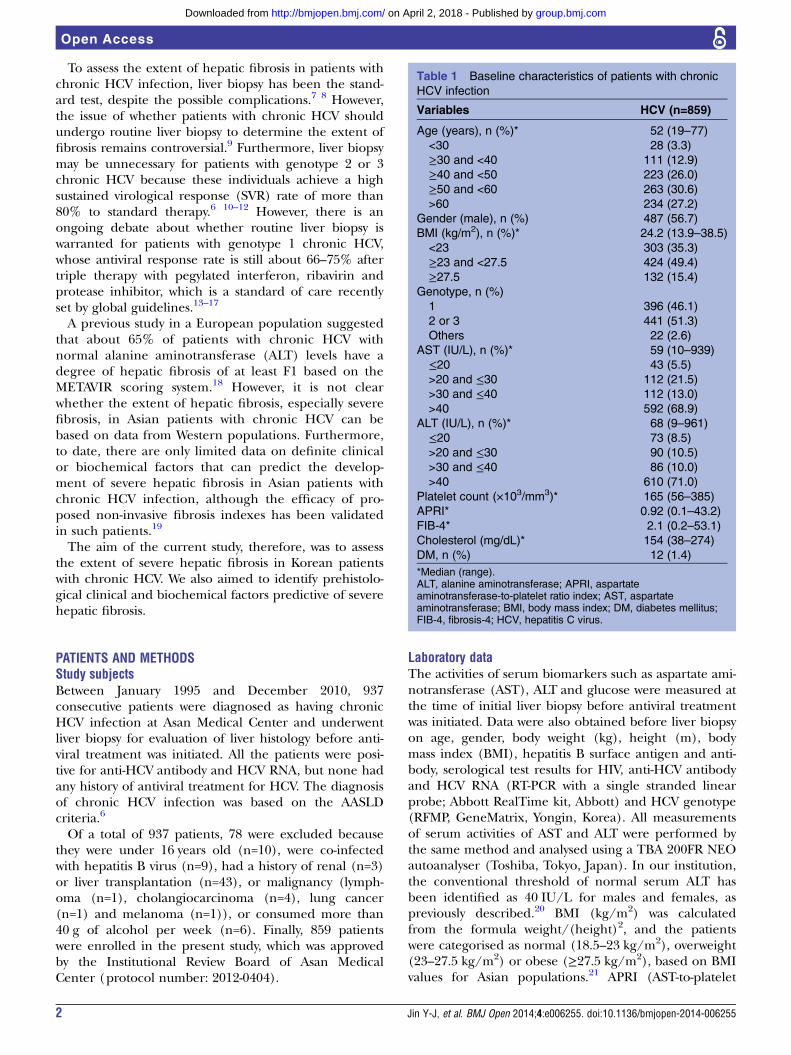
To assess the extent of hepatic fibrosis in patients withchronic HCV infection, liver biopsy has been the stand-ard test, despite the possible complications.7 8 However,the issue of whether patients with chronic HCV shouldundergo routine liver biopsy to determine the extent offibrosis remains controversial.9 Furthermore, liver biopsymay be unnecessary for patients with genotype 2 or 3chronic HCV because these individuals achieve a highsustained virological response (SVR) rate of more than80% to standard therapy.6 10–12 However, there is anongoing debate about whether routine liver biopsy iswarranted for patients with genotype 1 chronic HCV,whose antiviral response rate is still about 66–75% aftertriple therapy with pegylated interferon, ribavirin andprotease inhibitor, which is a standard of care recentlyset by global guidelines.13–17
A previous study in a European population suggestedthat about 65% of patients with chronic HCV withnormal alanine aminotransferase (ALT) levels have adegree of hepatic fibrosis of at least F1 based on theMETAVIR scoring system.18 However, it is not clearwhether the extent of hepatic fibrosis, especially severefibrosis, in Asian patients with chronic HCV can bebased on data from Western populations. Furthermore,to date, there are only limited data on definite clinicalor biochemical factors that can predict the develop-ment of severe hepatic fibrosis in Asian patients withchronic HCV infection, although the efficacy of pro-posed non-invasive fibrosis indexes has been validatedin such patients.19
The aim of the current study, therefore, was to assessthe extent of severe hepatic fibrosis in Korean patientswith chronic HCV. We also aimed to identify prehistolo-gical clinical and biochemical factors predictive of severehepatic fibrosis.
PATIENTS AND METHODSStudy subjectsBetween January 1995 and December 2010, 937consecutive patients were diagnosed as having chronicHCV infection at Asan Medical Center and underwentliver biopsy for evaluation of liver histology before anti-viral treatment was initiated. All the patients were posi-tive for anti-HCV antibody and HCV RNA, but none hadany history of antiviral treatment for HCV. The diagnosisof chronic HCV infection was based on the AASLDcriteria.6
Of a total of 937 patients, 78 were excluded becausethey were under 16 years old (n=10), were co-infectedwith hepatitis B virus (n=9), had a history of renal (n=3)or liver transplantation (n=43), or malignancy (lymph-oma (n=1), cholangiocarcinoma (n=4), lung cancer(n=1) and melanoma (n=1)), or consumed more than40 g of alcohol per week (n=6). Finally, 859 patientswere enrolled in the present study, which was approvedby the Institutional Review Board of Asan MedicalCenter (protocol number: 2012-0404).
Laboratory dataThe activities of serum biomarkers such as aspartate ami-notransferase (AST), ALT and glucose were measured atthe time of initial liver biopsy before antiviral treatmentwas initiated. Data were also obtained before liver biopsyon age, gender, body weight (kg), height (m), bodymass index (BMI), hepatitis B surface antigen and anti-body, serological test results for HIV, anti-HCV antibodyand HCV RNA (RT-PCR with a single stranded linearprobe; Abbott RealTime kit, Abbott) and HCV genotype(RFMP, GeneMatrix, Yongin, Korea). All measurementsof serum activities of AST and ALT were performed bythe same method and analysed using a TBA 200FR NEOautoanalyser (Toshiba, Tokyo, Japan). In our institution,the conventional threshold of normal serum ALT hasbeen identified as 40 IU/L for males and females, aspreviously described.20 BMI (kg/m2) was calculatedfrom the formula weight/(height)2, and the patientswere categorised as normal (18.5–23 kg/m2), overweight(23–27.5 kg/m2) or obese (≥27.5 kg/m2), based on BMIvalues for Asian populations.21 APRI (AST-to-platelet
Table 1 Baseline characteristics of patients with chronic
HCV infection
Variables HCV (n=859)
Age (years), n (%)* 52 (19–77)
<30 28 (3.3)
≥30 and <40 111 (12.9)
≥40 and <50 223 (26.0)
≥50 and <60 263 (30.6)
>60 234 (27.2)
Gender (male), n (%) 487 (56.7)
BMI (kg/m2), n (%)* 24.2 (13.9–38.5)
<23 303 (35.3)
≥23 and <27.5 424 (49.4)
≥27.5 132 (15.4)
Genotype, n (%)
1 396 (46.1)
2 or 3 441 (51.3)
Others 22 (2.6)
AST (IU/L), n (%)* 59 (10–939)
≤20 43 (5.5)
>20 and ≤30 112 (21.5)
>30 and ≤40 112 (13.0)
>40 592 (68.9)
ALT (IU/L), n (%)* 68 (9–961)
≤20 73 (8.5)
>20 and ≤30 90 (10.5)
>30 and ≤40 86 (10.0)
>40 610 (71.0)
Platelet count (×103/mm3)* 165 (56–385)
APRI* 0.92 (0.1–43.2)
FIB-4* 2.1 (0.2–53.1)
Cholesterol (mg/dL)* 154 (38–274)
DM, n (%) 12 (1.4)
*Median (range).ALT, alanine aminotransferase; APRI, aspartateaminotransferase-to-platelet ratio index; AST, aspartateaminotransferase; BMI, body mass index; DM, diabetes mellitus;FIB-4, fibrosis-4; HCV, hepatitis C virus.
2 Jin Y-J, et al. BMJ Open 2014;4:e006255. doi:10.1136/bmjopen-2014-006255
Open Access
group.bmj.com on April 2, 2018 - Published by http://bmjopen.bmj.com/Downloaded from

ratio index) and FIB-4 (fibrosis-4) were also calculatedas non-invasive fibrosis markers.22 23
Preparation and evaluation of liver biopsy specimensThe clinician’s decision for liver biopsy before treatmentwas usually based on HCV genotype and need for theinformation on antiviral prognosis. Before the proced-ure, written informed consent was obtained from allpatients. After liver biopsy, patients were carefully moni-tored every 1 h for the first 4 h, and thereafter every 6 hduring 1 day. Two or more biopsy specimens, eachapproximately 1.5 cm in length, were obtained fromevery patient. All liver biopsy specimens were fixed in10% neutral-buffered formalin. Sections were cut at3–4 μm thickness and stained with H&E, Prussian blueand Masson’s trichrome stain. All pathological findingswere retrospectively obtained by careful review of pathol-ogists’ clinical records under the supervision of onesenior expert pathologist (EY) who confirmed the finalpathological diagnosis. Fibrosis stage and activity gradeof the liver specimens were determined based on previ-ously published guidelines.24 25 Severe fibrosis wasdefined as fibrosis stage ≥3 based on the METAVIR
scoring system,24 25 which is also described in theAASLD guidelines.6 Fatty changes were categorised asnone or minimal (<5%), mild (≥5% and <30%), moder-ate (≥30% and <60%) or severe (≥60%).26
Statistical analysesThe basic clinical characteristics of the patients areexpressed as median (range) and frequency. Differencesbetween categorical or continuous variables were ana-lysed using the χ2 test, Fisher’s exact test, or Student ttest. Multivariate analysis by stepwise linear and logisticregression analysis was performed to assess the predic-tors of severe hepatic fibrosis in patients. A p value ofless than 0.05 (two-tailed) was considered statistically sig-nificant in all analyses, which were performed with SPSSV.18.0 (SPSS Inc, Chicago, Illinois, USA).
RESULTSBaseline clinical and virological characteristicsBaseline characteristics of the 859 patients are shown intable 1. The median age of the patient was 52 years(range 19–77 years) and 487 (56.7%) patients weremale. The most common HCV genotypes were
Figure 1 Histological findings in
patients with chronic hepatitis C
virus infection. The proportion of
individuals with severe fibrosis (A)
and steatosis (B) are shown
according to serum alanine
aminotransferase (ALT) level.
Jin Y-J, et al. BMJ Open 2014;4:e006255. doi:10.1136/bmjopen-2014-006255 3
Open Access
group.bmj.com on April 2, 2018 - Published by http://bmjopen.bmj.com/Downloaded from
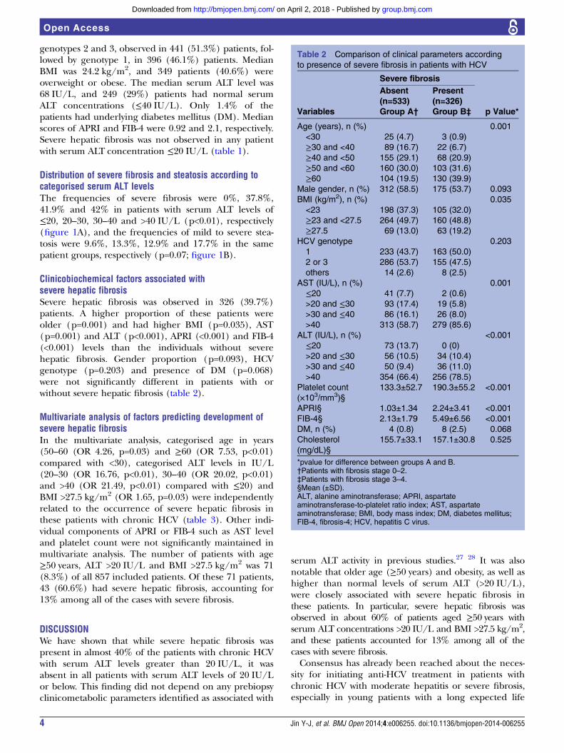
genotypes 2 and 3, observed in 441 (51.3%) patients, fol-lowed by genotype 1, in 396 (46.1%) patients. MedianBMI was 24.2 kg/m2, and 349 patients (40.6%) wereoverweight or obese. The median serum ALT level was68 IU/L, and 249 (29%) patients had normal serumALT concentrations (≤40 IU/L). Only 1.4% of thepatients had underlying diabetes mellitus (DM). Medianscores of APRI and FIB-4 were 0.92 and 2.1, respectively.Severe hepatic fibrosis was not observed in any patientwith serum ALT concentration ≤20 IU/L (table 1).
Distribution of severe fibrosis and steatosis according tocategorised serum ALT levelsThe frequencies of severe fibrosis were 0%, 37.8%,41.9% and 42% in patients with serum ALT levels of≤20, 20–30, 30–40 and >40 IU/L (p<0.01), respectively(figure 1A), and the frequencies of mild to severe stea-tosis were 9.6%, 13.3%, 12.9% and 17.7% in the samepatient groups, respectively (p=0.07; figure 1B).
Clinicobiochemical factors associated withsevere hepatic fibrosisSevere hepatic fibrosis was observed in 326 (39.7%)patients. A higher proportion of these patients wereolder (p=0.001) and had higher BMI (p=0.035), AST(p=0.001) and ALT (p<0.001), APRI (<0.001) and FIB-4(<0.001) levels than the individuals without severehepatic fibrosis. Gender proportion (p=0.093), HCVgenotype (p=0.203) and presence of DM (p=0.068)were not significantly different in patients with orwithout severe hepatic fibrosis (table 2).
Multivariate analysis of factors predicting development ofsevere hepatic fibrosisIn the multivariate analysis, categorised age in years(50–60 (OR 4.26, p=0.03) and ≥60 (OR 7.53, p<0.01)compared with <30), categorised ALT levels in IU/L(20–30 (OR 16.76, p<0.01), 30–40 (OR 20.02, p<0.01)and >40 (OR 21.49, p<0.01) compared with ≤20) andBMI >27.5 kg/m2 (OR 1.65, p=0.03) were independentlyrelated to the occurrence of severe hepatic fibrosis inthese patients with chronic HCV (table 3). Other indi-vidual components of APRI or FIB-4 such as AST leveland platelet count were not significantly maintained inmultivariate analysis. The number of patients with age≥50 years, ALT >20 IU/L and BMI >27.5 kg/m2 was 71(8.3%) of all 857 included patients. Of these 71 patients,43 (60.6%) had severe hepatic fibrosis, accounting for13% among all of the cases with severe fibrosis.
DISCUSSIONWe have shown that while severe hepatic fibrosis waspresent in almost 40% of the patients with chronic HCVwith serum ALT levels greater than 20 IU/L, it wasabsent in all patients with serum ALT levels of 20 IU/Lor below. This finding did not depend on any prebiopsyclinicometabolic parameters identified as associated with
serum ALT activity in previous studies.27 28 It was alsonotable that older age (≥50 years) and obesity, as well ashigher than normal levels of serum ALT (>20 IU/L),were closely associated with severe hepatic fibrosis inthese patients. In particular, severe hepatic fibrosis wasobserved in about 60% of patients aged ≥50 years withserum ALT concentrations >20 IU/L and BMI >27.5 kg/m2,and these patients accounted for 13% among all of thecases with severe fibrosis.Consensus has already been reached about the neces-
sity for initiating anti-HCV treatment in patients withchronic HCV with moderate hepatitis or severe fibrosis,especially in young patients with a long expected life
Table 2 Comparison of clinical parameters according
to presence of severe fibrosis in patients with HCV
Severe fibrosis
p Value*Variables
Absent(n=533)Group A†
Present(n=326)Group B‡
Age (years), n (%) 0.001
<30 25 (4.7) 3 (0.9)
≥30 and <40 89 (16.7) 22 (6.7)
≥40 and <50 155 (29.1) 68 (20.9)
≥50 and <60 160 (30.0) 103 (31.6)
≥60 104 (19.5) 130 (39.9)
Male gender, n (%) 312 (58.5) 175 (53.7) 0.093
BMI (kg/m2), n (%) 0.035
<23 198 (37.3) 105 (32.0)
≥23 and <27.5 264 (49.7) 160 (48.8)
≥27.5 69 (13.0) 63 (19.2)
HCV genotype 0.203
1 233 (43.7) 163 (50.0)
2 or 3 286 (53.7) 155 (47.5)
others 14 (2.6) 8 (2.5)
AST (IU/L), n (%) 0.001
≤20 41 (7.7) 2 (0.6)
>20 and ≤30 93 (17.4) 19 (5.8)
>30 and ≤40 86 (16.1) 26 (8.0)
>40 313 (58.7) 279 (85.6)
ALT (IU/L), n (%) <0.001
≤20 73 (13.7) 0 (0)
>20 and ≤30 56 (10.5) 34 (10.4)
>30 and ≤40 50 (9.4) 36 (11.0)
>40 354 (66.4) 256 (78.5)
Platelet count
(×103/mm3)§
133.3±52.7 190.3±55.2 <0.001
APRI§ 1.03±1.34 2.24±3.41 <0.001
FIB-4§ 2.13±1.79 5.49±6.56 <0.001
DM, n (%) 4 (0.8) 8 (2.5) 0.068
Cholesterol
(mg/dL)§
155.7±33.1 157.1±30.8 0.525
*pvalue for difference between groups A and B.†Patients with fibrosis stage 0–2.‡Patients with fibrosis stage 3–4.§Mean (±SD).ALT, alanine aminotransferase; APRI, aspartateaminotransferase-to-platelet ratio index; AST, aspartateaminotransferase; BMI, body mass index; DM, diabetes mellitus;FIB-4, fibrosis-4; HCV, hepatitis C virus.
4 Jin Y-J, et al. BMJ Open 2014;4:e006255. doi:10.1136/bmjopen-2014-006255
Open Access
group.bmj.com on April 2, 2018 - Published by http://bmjopen.bmj.com/Downloaded from

span ahead.6 However, the necessity for routine liverbiopsy to evaluate fibrosis stage before anti-HCV treat-ment remains controversial. Aside from the fact thatanti-HCV treatment has potential adverse effects, it isnot effective for all patients, and is relatively expensive,29
the biopsy procedure itself is associated with adverseeffects such as pain, bleeding and bowel perforation,8
which may incur additional medical care costs and causepatients distress and anxiety.30 In our series, about one-fourth of the biopsied patients experienced mild ormoderate abdominal pain after liver biopsy, althoughthere were no serious complications requiring a longhospital stay. Indeed, some patients may hesitate toreceive anti-HCV treatment based on histological evi-dence, even despite only the minor chance of seriousadverse events occurring owing to biopsy. Thus, simpleclinicobiochemical factors capable of predicting severehepatic fibrosis without pathological evidence could bepractically helpful in deciding on the treatment forpatients with chronic HCV.Although previous studies have evaluated factors asso-
ciated with histological findings of patients with chronicHCV in Western populations,18 31–34 there is still a lackof data about whether the findings can be confidentlyextrapolated to Asian patients. Interestingly, our Asiandata show that severe hepatic fibrosis was not present inpatients with low normal serum ALT levels (≤20 IU/L),supporting a wait-and-see approach for these patients,
particularly those with clinical or genetic parametersrelated to suboptimal antiviral treatment response.6
In contrast, more advanced age (≥50 years), obesity andserum ALT levels >20 IU/L were independent predictorsof significant hepatic fibrosis. These findings suggestthat immediate anti-HCV treatment without performinga liver biopsy may be beneficial for patients above50 years (albeit not for elderly patients (>65 years),weighing the potential risks and benefits35), especiallyfor obese genotype 2 or 3 patients with serum ALT con-centrations >20 IU/L, because more than 80% ofpatients with HCV with genotype 2 or 3 achieve an SVRto standard-of-care treatment.12 Given the better anti-viral response of Asian patients, who have the favourableIL28B genotype more frequently than Western indivi-duals,36 it may be preferable to initiate antiviral treat-ment for young Asian patients infected with genotype 1HCV without pathology results if serum ALT levels areabove 20 IU/L. Moreover, our results suggest that evenin patients with genotype 1 HCV infection, which is awell-known predictor of negative antiviral treatmentresponse,6 high-risk factors for significant hepatic fibro-sis such as serum ALT levels of >20 IU/L, age ≥50 yearsand obesity may be deemed to justify an active antiviralapproach, preferably with triple regimens, without liverbiopsy findings.We observed severe hepatic fibrosis in about 40% of the
patients with normal ALT levels (ie, less than 40 IU/L).
Table 3 Multivariate analysis of factors predicting severe fibrosis in patients with HCV
VariablesUnivariate analysis Multivariate analysis*OR 95% CI p Value OR 95% CI p Value
Age (years)
<30 (control) – – – – – –
≥30 and <40 2.06 0.57 to 7.45 0.27 1.86 0.49 to 7.07 0.36
≥40 and <50 3.66 1.07 to 12.52 0.04 2.87 0.79 to 10.37 0.11
≥50 and <60 5.37 1.58 to 18.22 0.01 4.26 1.19 to 15.32 0.03
≥60 10.42 3.06 to 34.46 <0.01 7.53 2.09 to 27.11 <0.01
Male gender 1.22 0.92 to 1.61 0.16 – – –
AST (IU/L)
≤20 (control) – – – – – –
>20 and ≤30 4.19 0.93 to 18.82 0.06 0.95 0.22 to 4.41 0.95
>30 and ≤40 6.19 1.40 to 27.38 0.02 0.97 0.22 to 4.22 0.97
>40 18.27 4.38 to 76.24 <0.01 3.36 0.78 to 14.34 0.10
ALT (IU/L)
≤20 (control) – – – – – –
>20 and ≤30 19.55 4.96 to 93.59 <0.01 16.76 3.39 to 82.66 <0.01
>30 and ≤40 20.56 5.88 to 111.07 <0.01 20.02 4.01 to 84.73 <0.01
>40 25.67 6.24 to 105.62 <0.01 21.49 4.34 to 85.43 <0.01
BMI (kg/m2)
<23 (control) – – – – – –
≥23 and <27.5 1.14 0.84 to 1.55 0.39 0.99 0.71 to 1.39 0.97
≥27.5 1.72 1.14 to 2.61 0.01 1.65 1.05 to 2.61 0.03
DM 2.67 0.39 to 18.17 0.32 – – –
Cholesterol (mg/dL) 0.99 0.99 to 1.01 0.38 – – –
*Logistic regression model; patients, n=859; event, severe hepatic fibrosis (n=326).ALT, alanine aminotransferase; AST, aspartate aminotransferase; BMI, body mass index; DM, diabetes mellitus.
Jin Y-J, et al. BMJ Open 2014;4:e006255. doi:10.1136/bmjopen-2014-006255 5
Open Access
group.bmj.com on April 2, 2018 - Published by http://bmjopen.bmj.com/Downloaded from

This rate was similar to that in patients with elevated ALTlevels. This suggests that the decision to initiate anti-HCVtreatment should not be based simply on serum ALTlevels, especially in patients with serum ALT concentra-tions >20 IU/L. Likewise, patients with serum ALT of 20–40 IU/L should not be excluded from antiviral therapysimply because of normal ALT levels. Moreover, liverbiopsy may be required for decision-making regardingantiviral treatment when serum ALT levels are 20–40 IU/Lin older (>50 years) and obese patients who are reluctantto receive treatment.It has been reported that host factors such as age and
obesity are associated with the development of hepaticfibrosis,5 37 and in this respect the outcomes of ourstudy are similar to those of previous studies.5 37
Although non-invasive tests such as elastography, non-alcoholic fatty liver disease fibrosis score, and APRI orthe FIB-4 score have been developed to estimate hepaticfibrosis, their accuracy has not been sufficiently vali-dated.22 23 38 39 Moreover, these tests involve high costand additional calculations. However, we have identifiedinexpensive and simple clinical parameters that are notexpensive to measure and that can aid decision-makingabout severe hepatic fibrosis.Despite the extensive analyses using large scale
pathology-based data sets, a major limitation of thecurrent study is that the data are from a single institu-tion and a single ethnic type. Because of the differencesin procedures for serum ALT measurement in differentclinical laboratories and the fact that Asian BMI cut-offpoints differ from those of Western populations,21 40
further multicentre and multiethnic studies are requiredfor external validation and wider use of our estimates. Inaddition, our cohort did not include patients withHCV-infection who received antiviral treatment withoutresorting to biopsy or who were never treated, whichmay introduce a selection bias. Finally, observational var-iations among pathologists in histological evaluationshould be taken into account when interpreting thepresent results and further applying them in clinicalpractice.In conclusion, advanced age (≥50 years), obesity and
serum ALT levels >20 IU/L are closely associated withthe development of severe hepatic fibrosis in Koreanpatients with chronic HCV infection. These findingscould facilitate clinical decision-making in the manage-ment of patients with HCV-infection.
Acknowledgements This study was supported by an Inha UniversityResearch Grant.
Contributors Y-JJ and JHS were responsible for the concept and design ofthe study, the acquisition, analysis and interpretation of the data, and draftingof the manuscript. GAK, EY, KMK, Y-SL and HCL helped with the acquisition,analysis, interpretation of the data and critical revision of the manuscript forimportant intellectual content.
Funding This research received no specific grant from any funding agency inthe public, commercial or not-for-profit sectors.
Competing interests None.
Provenance and peer review Not commissioned; externally peer reviewed.
Data sharing statement No additional data are available.
Open Access This is an Open Access article distributed in accordance withthe Creative Commons Attribution Non Commercial (CC BY-NC 4.0) license,which permits others to distribute, remix, adapt, build upon this work non-commercially, and license their derivative works on different terms, providedthe original work is properly cited and the use is non-commercial. See: http://creativecommons.org/licenses/by-nc/4.0/
REFERENCES1. Alter MJ. Epidemiology of hepatitis C. Hepatology 1997;26:62S–5S.2. Lauer GM, Walker BD. Hepatitis C virus infection. N Engl J Med
2001;345:41–52.3. Alter MJ, Kruszon-Moran D, Nainan OV, et al. The prevalence of
hepatitis C virus infection in the United States, 1988 through 1994.N Engl J Med 1999;341:556–62.
4. Liang TJ, Rehermann B, Seeff LB, et al. Pathogenesis, naturalhistory, treatment, and prevention of hepatitis C. Ann Intern Med2000;132:296–305.
5. Poynard T, Bedossa P, Opolon P. Natural history of liver fibrosisprogression in patients with chronic hepatitis C. The OBSVIRC,METAVIR, CLINIVIR, and DOSVIRC groups. Lancet1997;349:825–32.
6. Ghany MG, Strader DB, Thomas DL, et al. Diagnosis, management,and treatment of hepatitis C: an update. Hepatology2009;49:1335–74.
7. Bravo AA, Sheth SG, Chopra S. Liver biopsy. N Engl J Med2001;344:495–500.
8. Dienstag JL. The role of liver biopsy in chronic hepatitis C.Hepatology 2002;36:S152–60.
9. Everhart JE, Stolar M, Hoofnagle JH. Management of hepatitis C:a national survey of gastroenterologists and hepatologists.Hepatology 1997;26:78S–82S.
10. Manns MP, McHutchison JG, Gordon SC, et al. Peginterferonalfa-2b plus ribavirin compared with interferon alfa-2b plus ribavirinfor initial treatment of chronic hepatitis C: a randomised trial. Lancet2001;358:958–65.
11. Fried MW, Shiffman ML, Reddy KR, et al. Peginterferon alfa-2a plusribavirin for chronic hepatitis C virus infection. N Engl J Med2002;347:975–82.
12. Shiffman ML, Suter F, Bacon BR, et al. Peginterferon alfa-2a andribavirin for 16 or 24 weeks in HCV genotype 2 or 3. N Engl J Med2007;357:124–34.
13. Poordad F, McCone J Jr, Bacon BR, et al. Boceprevir for untreatedchronic HCV genotype 1 infection. N Engl J Med2011;364:1195–206.
14. Jacobson IM, McHutchison JG, Dusheiko G, et al. Telaprevir forpreviously untreated chronic hepatitis C virus infection. N Engl JMed 2011;364:2405–16.
15. Sherman KE, Flamm SL, Afdhal NH, et al. Response-guidedtelaprevir combination treatment for hepatitis C virus infection.N Engl J Med 2011;365:1014–24.
16. European Association for Study of Liver. EASL Clinical PracticeGuidelines: management of hepatitis C virus infection. J Hepatol2014;60:392–420.
17. Ghany MG, Nelson DR, Strader DB, et al. An update on treatment ofgenotype 1 chronic hepatitis C virus infection: 2011 practiceguideline by the American Association for the Study of LiverDiseases. Hepatology 2011;54:1433–44.
18. Pradat P, Alberti A, Poynard T, et al. Predictive value of ALT levelsfor histologic findings in chronic hepatitis C: a Europeancollaborative study. Hepatology 2002;36:973–7.
19. Ichino N, Osakabe K, Nishikawa T, et al. A new index fornon-invasive assessment of liver fibrosis. World J Gastroenterol2010;16:4809–16.
20. Lee JK, Shim JH, Lee HC, et al. Estimation of the healthy upperlimits for serum alanine aminotransferase in Asian populations withnormal liver histology. Hepatology 2010;51:1577–83.
21. WHO Expert Consultation. Appropriate body-mass index for Asianpopulations and its implications for policy and intervention strategies.Lancet 2004;363:157–63.
22. Vallet-Pichard A, Mallet V, Nalpas B, et al. FIB-4: an inexpensiveand accurate marker of fibrosis in HCV infection. Comparison withliver biopsy and fibrotest. Hepatology 2007;46:32–6.
23. Lackner C, Struber G, Liegl B, et al. Comparison and validation ofsimple noninvasive tests for prediction of fibrosis in chronic hepatitisC. Hepatology 2005;41:1376–82.
6 Jin Y-J, et al. BMJ Open 2014;4:e006255. doi:10.1136/bmjopen-2014-006255
Open Access
group.bmj.com on April 2, 2018 - Published by http://bmjopen.bmj.com/Downloaded from
24. [No authors listed]. Intraobserver and interobserver variations in liverbiopsy interpretation in patients with chronic hepatitis C. The FrenchMETAVIR Cooperative Study Group. Hepatology 1994;20:15–20.
25. Bedossa P, Poynard T. An algorithm for the grading of activity inchronic hepatitis C. The METAVIR Cooperative Study Group.Hepatology 1996;24:289–93.
26. Lee JY, Kim KM, Lee SG, et al. Prevalence and risk factors ofnon-alcoholic fatty liver disease in potential living liver donors inKorea: a review of 589 consecutive liver biopsies in a single center.J Hepatol 2007;47:239–44.
27. Prati D, Shiffman ML, Diago M, et al. Viral and metabolic factorsinfluencing alanine aminotransferase activity in patients with chronichepatitis C. J Hepatol 2006;44:679–85.
28. Kobayashi Y, Kawaguchi Y, Mizuta T, et al. Metabolic factors areassociated with serum alanine aminotransferase levels in patientswith chronic hepatitis C. J Gastroenterol 2011;46:529–35.
29. Levine RA. Treating histologically mild chronic hepatitis C:monotherapy, combination therapy, or tincture of time? Ann InternMed 1998;129:323–6.
30. Regev A, Berho M, Jeffers LJ, et al. Sampling error andintraobserver variation in liver biopsy in patients with chronic HCVinfection. Am J Gastroenterol 2002;97:2614–18.
31. Alberti A, Morsica G, Chemello L, et al. Hepatitis C viraemia andliver disease in symptom-free individuals with anti-HCV. Lancet1992;340:697–8.
32. Gholson CF, Morgan K, Catinis G, et al. Chronic hepatitis C withnormal aminotransferase levels: a clinical histologic study. Am JGastroenterol 1997;92:1788–92.
33. Jamal MM, Soni A, Quinn PG, et al. Clinical features of hepatitisC-infected patients with persistently normal alanine transaminaselevels in the Southwestern United States. Hepatology1999;30:1307–11.
34. Wai CT, Greenson JK, Fontana RJ, et al. A simple noninvasiveindex can predict both significant fibrosis and cirrhosis in patientswith chronic hepatitis C. Hepatology 2003;38:518–26.
35. Puoti C, Bellis L, Guarisco R, et al. HCV carriers with normal alanineaminotransferase levels: healthy persons or severely ill patients?Dealing with an everyday clinical problem. Eur J Intern Med2010;21:57–61.
36. Tanaka Y, Nishida N, Sugiyama M, et al. Genome-wide associationof IL28B with response to pegylated interferon-alpha and ribavirintherapy for chronic hepatitis C. Nat Genet 2009;41:1105–9.
37. Ratziu V, Giral P, Charlotte F, et al. Liver fibrosis in overweightpatients. Gastroenterology 2000;118:1117–23.
38. Huwart L, Sempoux C, Salameh N, et al. Liver fibrosis: noninvasiveassessment with MR elastography versus aspartateaminotransferase-to-platelet ratio index. Radiology2007;245:458–66.
39. McPherson S, Stewart SF, Henderson E, et al. Simple non-invasivefibrosis scoring systems can reliably exclude advanced fibrosis inpatients with non-alcoholic fatty liver disease. Gut 2010;59:1265–9.
40. Dutta A, Saha C, Johnson CS, et al. Variability in the upper limit ofnormal for serum alanine aminotransferase levels: a statewide study.Hepatology 2009;50:1957–62.
Jin Y-J, et al. BMJ Open 2014;4:e006255. doi:10.1136/bmjopen-2014-006255 7
Open Access
group.bmj.com on April 2, 2018 - Published by http://bmjopen.bmj.com/Downloaded from
infectionfibrosis in patients with chronic hepatitis Cbiopsy-proven cases of severe hepatic Clinicobiochemical prediction of
Young-Suk Lim and Han Chu LeeYoung-Joo Jin, Ju Hyun Shim, Gi Ae Kim, Eunsil Yu, Kang Mo Kim,
doi: 10.1136/bmjopen-2014-0062552014 4: BMJ Open
http://bmjopen.bmj.com/content/4/11/e006255Updated information and services can be found at:
These include:
References http://bmjopen.bmj.com/content/4/11/e006255#ref-list-1
This article cites 40 articles, 1 of which you can access for free at:
Open Access
http://creativecommons.org/licenses/by-nc/4.0/non-commercial. See: provided the original work is properly cited and the use isnon-commercially, and license their derivative works on different terms, permits others to distribute, remix, adapt, build upon this workCommons Attribution Non Commercial (CC BY-NC 4.0) license, which This is an Open Access article distributed in accordance with the Creative
serviceEmail alerting
box at the top right corner of the online article. Receive free email alerts when new articles cite this article. Sign up in the
CollectionsTopic Articles on similar topics can be found in the following collections
(220)Gastroenterology and hepatology
Notes
http://group.bmj.com/group/rights-licensing/permissionsTo request permissions go to:
http://journals.bmj.com/cgi/reprintformTo order reprints go to:
http://group.bmj.com/subscribe/To subscribe to BMJ go to:
group.bmj.com on April 2, 2018 - Published by http://bmjopen.bmj.com/Downloaded from