Clinical Urinalysis Review Austin Community College Medical Laboratory Technology Clinical II Spring...
60
Clinical Urinalysis Review Austin Community College Medical Laboratory Technology Clinical II Spring 07
-
Upload
albert-rogers -
Category
Documents
-
view
215 -
download
0
Transcript of Clinical Urinalysis Review Austin Community College Medical Laboratory Technology Clinical II Spring...

Clinical Urinalysis Review
Austin Community College
Medical Laboratory Technology
Clinical II Spring 07

Urine Blood Testing
http://library.med.utah.edu/WebPath/TUTORIAL/URINE/URINE.html

Chemical Exam of Urine

Chemical Exam of Urine
Chemical Exam of Urine
Reagent strip manufactures Bayer Corporation- Diagnostics Division
(formerly Ames) produces Multistix
Boehringer-Mannheim Corporation which produces Chemstrip
Behring Diagnostics which produces Rapignost

Chemical Exam of Urine

Chemical Exam of Urine
Reagent strip precautions and source of errors
Normal dipstick procedure: Dip strip briefly, but completely into well mixed,
room temperature urine sample. Withdraw strip, blot briefly on its side Keeping the strip flat, read results at the
appropriate times by comparing the color to the appropriate color on the chart provided.

Chemical Exam of Urine
Sources of error (& preventions) Testing cold specimens
would result in a slowing down of reactions; test specimens when fresh or bring them to RT before testing
Inadequate mixing of specimen could result in false reduced or negative reactions to
blood and leukocyte tests; mix specimens well before dipping
Over-dipping of reagent strip will result in leaching of reagents out of pads; briefly,
but completely dip the reagent strip into the urine

Chemical Exam of Urine
Inadequate blotting & Failure to keep strip horizontal will result in over-run or mixing of reagents
between the different reaction pads; blot excess urine off the strip and keep strip horizontal. If dipping from the tube, can run the side of the strip along the rim to remove excess urine.
Improper timing of tests over development of reagent pad colors leading
to falsely increased results; follow manufacturer’s recommendations

Chemical Exam of Urine
Inadequate light misinterpretation of results; use good lighting
Mis-using color chart misinterpretation of results; hold strip just over
color chart and match colors as close as possible, consider use of back-up tests, if needed, especially if urine’s color masks reaction colors.

Chemical Exam of Urine
Handling and Storage Keep strips in original container, stored at RT Protect from moisture and volatile fumes Use before expiration date Do not touch reagent pad areas
Chemical Exam of Urine
Quality Control - use appropriate, commercially prepared positive and negative controls. Use commercially prepared pos and neg
controls, at least once per 24 hours, and anytime a new bottle is opened, or question of validity of results. Readings should agree with published results ± one color block.

Urine Glucose Testing
Normal : no glucose detected
Clinical significance of abnormal results (Glucosuria) Plasma glucose level exceeds renal threshold
(160-189 mg/dL) Diabetes mellitus
Renal tubular dysfunction Filtered glucose not being reabsorbed in tubules

Urine Glucose Testing
Dipstick Testing Method Glucose initiates reaction
Coupled reaction Glucose oxidase – oxidizes glucose to gluconic acid and
concurrently reduces oxygen to hydrogen peroxide. Hydrogen peroxide in presence of the enzyme peroxidase
will oxidize an indicator, giving a colored reaction. Chromogens
Potassium iodide or Tetramethylbenzindine
Urine Glucose Testing
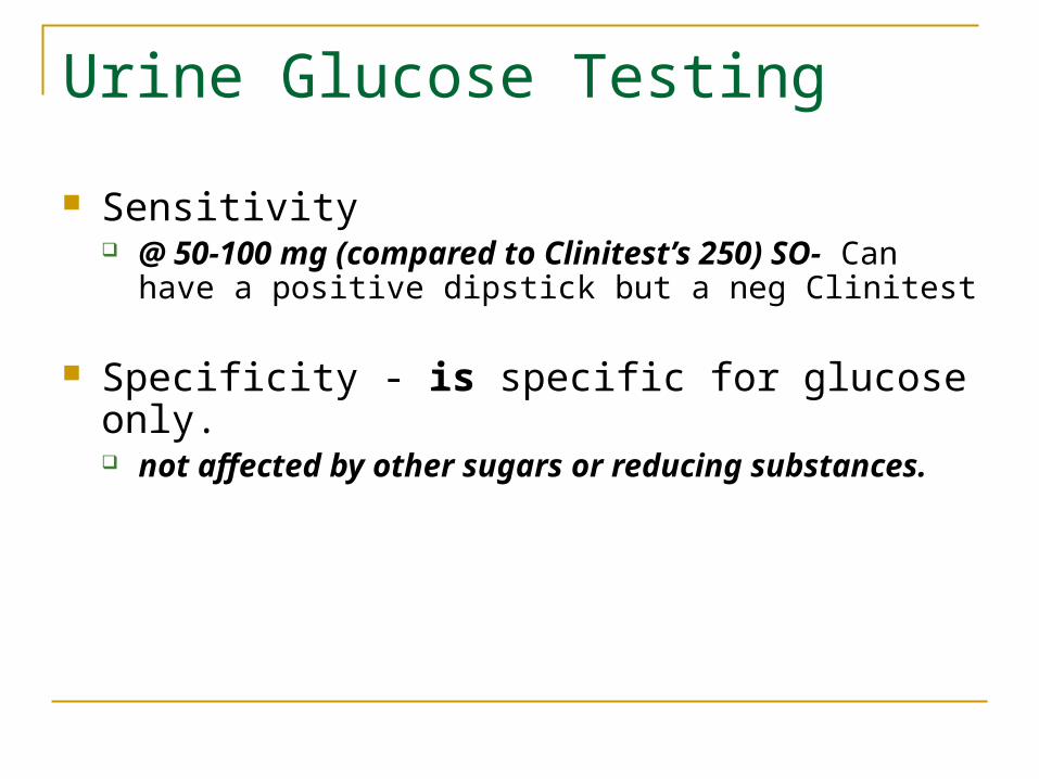
Sensitivity @ 50-100 mg (compared to Clinitest’s 250) SO- Can
have a positive dipstick but a neg Clinitest
Specificity - is specific for glucose only. not affected by other sugars or reducing substances.

Urine Glucose Testing
Interfering substances
High specific gravity and high pH may depress color. Ascorbic acid-false neg Bleach or peroxide may give false positive

Urine Bilirubin Testing
Normal : no bilirubin detected
Clinical significance of abnormal results (Bilirubinuria) Jaundice - Condition when serum bilirubin
becomes greater than the liver can handle, and there is an abnormal collection of bilirubin in the tissues giving them a yellow color

Urine Bilirubin Testing
Prehepatic / Hemolytic jaundice Excessive hemolysis of RBC; beyond what the
liver can process Type of bilirubin? Is bilirubin found in the urine? YES/NO? Explain.

Urine Bilirubin Testing
Prehepatic / Hemolytic jaundice Type of bilirubin? – indirect, insoluble,
unconjugated Is bilirubin found in the urine? – No, the bilirubin
is not water soluble

Urine Bilirubin Testing
Hepatic jaundice Liver’s cells malfunctioning
Ie. viral hepatitis, cirrhosis etc.
Both (direct) bilirubin and urobilinogen found in urine.

Urine Bilirubin Testing
Post hepatic (regurgative or obstructive) hepatitis Obstruction to outflow of bile – some type of
blockage Gall stones Tumor Edema
Conjugated bilirubin backed up into blood (Bilirubinuria) and passes into urine

Urine Bilirubin Testing
Testing method Urine dipsticks for bilirubin – a diazo reaction
Impregnated with stabilized diazotized 2,4 dichloraniline
Color goes from buff to brown also shades of pink – violet
If urine is strongly colored, look for change in pad color after dipping. Use Ictotest for backup.

Urine Bilirubin Testing
Interfering substances
Medication metabolites, pigments and indican may obscure readings
False negatives due to aged specimens, especially those exposed to light and oxidation.

Urine Ketone Testing
Ketone Bodies Origin - not normally present
Products of fat catabolism - breakdown of fat into CO2 and H2O
What are the 3 ketone bodies?

Urine Ketone Testing
Acetone 2%. -Acetone is volatile, & excreted primarily through the
lungs
Diacetic Acid (Acetoacetic) the first formed, 20 % of the total the form detected by most ketone test procedures
Beta hydroxybutyric Acid majority formed, but not detected by routine tests. Only Hart’s test, an old ‘wet chemical’ test will detect this one.

Urine Ketone Testing
Definitions Ketonuria - ketones in the urine Ketonemia - ketones in the blood Ketosis - disease state, when patient has
increased amount of ketones. Acidosis - state when blood pH is decreased,
an accumulation of acids; commonly occurs as a result of ketosis

Urine Ketone Testing Clinical significance
Health – formed in liver and completely metabolized Disease – excessive formation and accumulation
Disturbance of carbohydrate metabolism when there is a decrease of carbohydrate metabolism, then the
body stores of fat must be metabolized to supply energy. As a result of this increased fat metabolism ketones will be found
in the urine. Ex. low carbohydrate diets, diabetes
Starvation Vomiting and diarrhea in children Van Gierke's Disease – glycogen storage disease
High fat diet

Urine Ketone Testing
Clinical effects Metabolic acidosis
Lowering of blood & urine pH Brain toxicity

Urine Ketone Testing
Testing most use nitroprusside
detects diacetic acid and a small amount of acetone, but does not detect β-hydroxybutyric acid.
Produces purple color Can be used on urine or blood

Urine Specific Gravity Testing The specific gravity is a measure of the
weight of urine compared to an equal amount of water.
Specific gravity it proportional to urine osmolality which is a measure of concentration.

Urine Specific Gravity Testing The specific gravity will always be greater
than 1.000 and will increase as more materials are dissolved in the urine.
The value changes throughout the day depending on fluid intake.

Urine Specific Gravity Testing Specific gravity between 1.002 & 1.035 on a
random sample is normal if kidney function is normal. Specific gravity in Bowman’s capsule fluid is @
1.007 Any reading below this indicates hydration Any reading above this indicates some degree of
dehydration
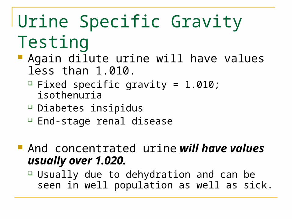
Urine Specific Gravity Testing Again dilute urine will have values less than
1.010. Fixed specific gravity = 1.010; isothenuria Diabetes insipidus End-stage renal disease
And concentrated urine will have values usually over 1.020. Usually due to dehydration and can be seen in
well population as well as sick.

Urine Specific Gravity Testing Increased urine specific gravity may indicate / be seen in: * Dehydration * Diarrhea * Excessive sweating * Glucosuria * Heart failure (related to decreased blood flow to the kidneys) * Renal arterial stenosis * Syndrome of inappropriate antidiuretic hormone secretion
(SIADH) * Vomiting * Water restriction

Urine Specific Gravity Testing Decreased urine specific gravity may indicate
/ be seen in: * Excessive fluid intake * Diabetes insipidus – central or nephrogenic * Renal failure (that is, loss of ability to
reabsorb water) * Pyelonephritis

Urine Specific Gravity Testing Specific gravity > 1.035 (refractometer)
Could have very high glucose levels Could contain radiographic dye

Urine Specific Gravity Testing Testing
Polyelectrolytes , pH indicator (bromthymol blue measures the pH change), and alkaline buffer

Urine Specific Gravity Testing Interfering substances
False elevation of results may be seen in samples with increased protein concentration.
Some reports of reduced specific gravity results on alkaline specimens.
Lipids may also effect results

Urine Blood Testing
Normally not found in urine Hemoglobinuria – free hemoglobin in urine
Circulating free hemoglobin normally picked up by haptoglobin preventing loss in urine
When serum levels of hemoglobin > 100 mg/dL threshold is exceeded
Hematuria – RBCs in the urine Trauma / irritation of renal organs
Urine Blood Testing

Urine Blood Testing
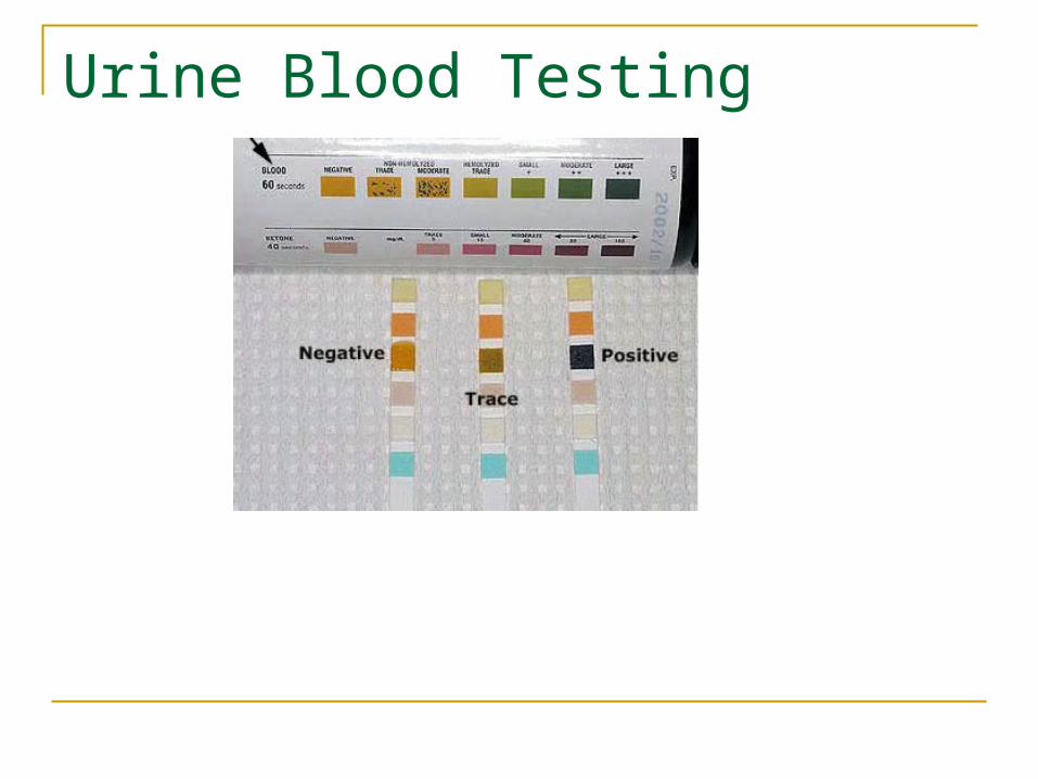
Testing dipstick reaction
HGB H2O2peroxidase OxygenHGB H2O2peroxidase Oxygen
Oxygen + Gum guaiac, benzidine or orthotolidine green or blue
oxidation products

Urine Blood Testing
‘Blood’ test detects Free Hemoglobin RBCs – get lysed on the pad & their hemoglobin
reacts Myoglobin – muscle hemoglobin
Principle based on the peroxidase-like activity of the heme portion of the molecule

Urine Blood Testing
Sensitivity – can detect at levels of 5-10 cells/uL
Interfering substances Ascorbic acid Nitrates Oxidizing agents (ie bleach) Contaminate blood (menstrual)

Urine pH Testing
Normal: kidneys capable of 4.5 – 8.0 Factors effecting pH
Diet – general & specific foods Time of day Metabolic disorders Drugs / medications
Dipstick capable: 4.5 – 9.0

Urine pH Testing
Test method Dipstick indicators – methyl red and bromthymol blue Range 4.5-9.0
Caution – other chemicals on dipstick can effect pH reading

Urine Protein Testing
Normally not found in measurable amounts on dipstick (<150 mg/dL /day) Permeability of glomerulus
Damage to glom capularies Changes in glom blood flow
Albumin excretions may be increased temporarily due to exercise, uti, and acute illness with fever.
Dipstick results of >@ 1+ (30mg/dL) would equal to @ 500 mg/dL (clinical proteinuria)

Urine Protein Testing
Only albumin detectable by dipstick Sensitivity (@15-30 ml/dL)

Urine Protein Testing
New testing for microalbumin & creatinine Results:
Protein 20-200 mg/dL (30-300 mg/dL /24 hr) Creatinine 10-300 mg/dL
Albumin/creatinine ratio Normally albumin in the urine is less than 30 mg/ gram
creatinine
Urine Protein Testing
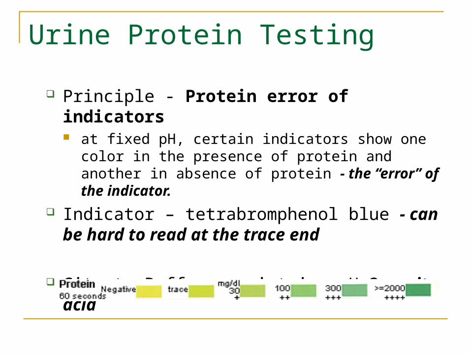
Principle - Protein error of indicators at fixed pH, certain indicators show one color in the
presence of protein and another in absence of protein - the “error” of the indicator.
Indicator – tetrabromphenol blue - can be hard to read at the trace end
Citrate Buffer – maintains pH 3 -quite acid

Urine Protein Testing
Sources of error Sensitive only to albumin Urine with strong / unusual color makes reading
difficult Highly alkaline or buffered urine will neutralize
acid buffer and lead to increased erroneous results.
Urine container contamination would interfere

Urine Protein Testing
Urine back up test 3% sulfosalicylic acid
Added to the supernatant to detect any kind of protein. Urine will turn cloudy if protein is present.

Urine Urobilinogen Testing
Normally found in small amounts, especially in early afternoon
Increased amounts may indicate liver disease or be seen as result of hemolytic disorders
Decreased amounts: If intestinal bacteria destroyed Liver doesn’t conjugate bilirubin Biliary obstruction – failure of bilirubin to reach
small intestine

Urine Urobilinogen Testing
Test principle based on Ehrilich’s reaction
Para-dimethylaminobenzaldehyde = Ehrlich's reagent.
Must protect specimen from light and test immediately

Urine Nitrate Testing
Nitrate Detects presence of certain types of bacteria screening for presence of UTI. Certain species of bacteria convert nitrate (normal
constituent of urine) to nitrite Escherichia - most common cause of UTI Klebsiella Proteus Pseudomonas Enterobacter Citrobacter

Urine Nitrate Testing
Aromatic amine in reagent strip reacts with nitrite; producing a diazonium salt
The diazonium salt reacts with sulfanilic acid and acetic acid to produce a pink azo dye

Urine Nitrate Testing
Limitations reported as positive or negative Not all UTI causing bacteria convert nitrate to nitrite
Haemophilus Staphylococcus Streptococcus

Urine Nitrate Testing
Fresh first morning specimen is preferred - besides being the most concentrated specimen, the urine has been in the bladder longer, allowing bacteria time and opportunity to convert the nitrates to nitrites.
Urine Leukocyte Testing
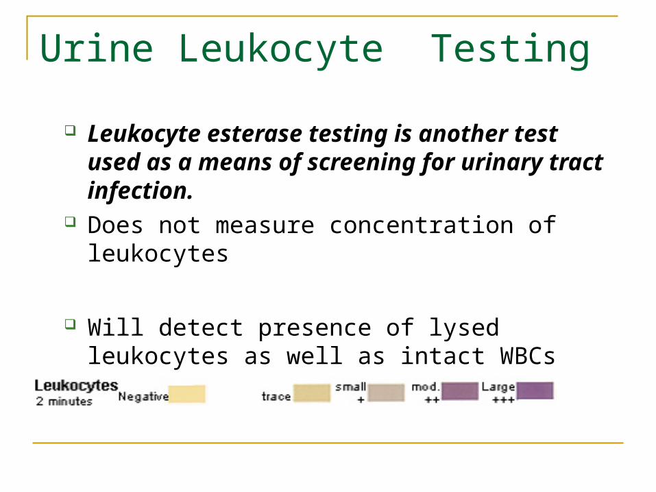
Leukocyte esterase testing is another test used as a means of screening for urinary tract infection.
Does not measure concentration of leukocytes
Will detect presence of lysed leukocytes as well as intact WBCs

Urine Leukocyte Testing
test principle: Leukocyte esterase, an enzyme present in
granulocytes, hydrolyzes indoxylcarbonic acid esterase to produce indoxyl, which reacts with a diazonium salt to create a purple color usually in 2 min.

Urine Leukocyte Testing
Reaction interference False positives - oxidizing detergents False negatives - greatly increased glucose, protein,
or specific gravity- increased sp gr could cause WBC to crenate preventing their releasing their esterase, So it is possible for the dipstick to be negative when there are WBCs present.