CLINICAL STUDY AND COMPARASION OF VARIOUS MODALTIES OF TREATMENT OF VARICOSE VEINS OF LOWER LIMBS
3
Research Paper Medical Science E-ISSN No : 2454-9916 | Volume : 2 | Issue : 3 | March 2016 1 2 *Dr. Laxmana Sastry | Dr. Sandeep Reddy 1 Associate Professor, Department of General Surgery, MRMCW, Hyderabad. 2 Post Grauate , Department of General Surgery, MRMCW & Yashoda Multispecialty Hospital, Hyderabad. 15 International Education & Research Journal [IERJ] INTRODUCTION Varicose veins of lower limb and their treatment are as old as mankind. Hippoc- rates discussed them 2500 years ago. It is not found in other animals and it is the human beings, who had to pay for their erect posture. Varicose veins constitute a progressive disease that becomes steadily worse. There is never remission of the disease except after pregnancy and delivery, when many of dilated varicosities may disappear. During its course, the disease produces complications, which usu- ally induce the patient to seek medical care. Varicose veins, though a common condition many a times remain asymptomatic. It is in the developed countries, where Attire reveals more than it conceals, and patient turn up for cosmetic reasons. In our Indian scenario it is the complications not the cosmetic reasons, bring the patients to the doctor. That is the reason why, though common, varicose veins remains as an iceberg phenomenon. In the INDIAN subcontinent, an estimated 23% of adults have varicose veins, and 6% have more advanced chronic venous disease(CVD), including skin changes and healed or active venous ulcers. Varicose veins have long been con- sidered a cosmetic problem that only affected emotional well-being but were not the source of disability. Varicosities, however, are frequently the cause of dis- comfort, pain, loss of working days, disability, and deterioration of health- related quality of life(QOL).Severe CVD may also lead to loss of limb or loss of life. Evaluation of varicose veins has greatly progressed in the past 2 decades with the wide spread availability of duplex ultrasonography. The treatment of vari- cose veins has also undergone dramatic changes with the introduction of percutaneous endovenous ablation techniques, including endovenous lasert herapy(EVLA), radiofrequency ablation (RFA),and liquid or foam sclerotherapy. Open surgical treatment with stripping of the varicose veins per- formed under general anesthesia, with the associated pain, potential for wound complications, and loss of working days, has been largely replaced by percutaneous office-based procedures that can be performed under local or tumescent anesthesia with similar early and midterm results but with less dis- comfort to the patient, improved early QOL, and earlier return to work. Varicose veins of the lower limbs are dilated subcutaneous veins that are 3mm in diameter measured in the upright position. Synonyms include varix, varices, and varicosities. Varicosity can involve the main axial superficial veins , the great saphenous vein(GSV) or the small saphenous vein(SSV) or any other superficial vein tributaries of the lower limbs.(1) Most varicose veins are due to primary venous disease. The most frequent cause is likely an intrinsic morphologic or biochemical abnormality in the vein wall, although the etiology can also be multifactorial. Labropoulos et al(1) , proposed that the origin of venous refluxinpatients with primary varicose veins can be local or multifocal structural weakness of the vein wall and that this can occur together or independently of proximal saphenous vein valvular incompetence. Varicosities can also develop as a result of secondary causes, such as previous deep vein thrombosis(DVT), deep venous obstruction, superficial thrombophlebitis, or arteriovenous fistula. Varicose veins may also be congenital and present as a venous malformation(3). Varicosities are manifestations of CVD. CVD includes various medical condi- tions of long duration, all involving morphologic and functional abnormalities of the venous system manifested by symptoms or signs(or both)(4). The study of varicose veins in the lower limb has been taken up with two aims 1. To study the distribution in age, sex,occupation, clinical features and sever- ity grade of varicose veins of lower limbs. 2. To study the various modalities of management of varicose veins of lower limb effectively and to prevent its complications. Materials and Methods: The study was a prospective, unicenter study conducted at Yashoda Multispeciality Hospital during 2012-13. 183 limbs studied in 18 to 70 years age group. Other than this range of age group is excluded from study. VCSS (Venous Clinical Severity Score) questionnaire was used to assess benefits of treatment modality. Duplex ultrasound scan was performed at 1wk and 1 month, 5 months post procedure to assess SFJ and GSV occlusion, neovascularization, recanalisation. Pain satisfaction assessment is done by various pain scales- Patient satisfaction was assessed at 12 weeks using a VAS rating of 0cm (com- pletely dissatisfied) to 10cm (completely satisfied). Duplex USG is the investi- gation of choice. Results: In the study varicose veins observed more in young & in long standing occupa- tions & cosmetic appearance is the commonest way of presentation. Long saphenous vein is the common involvement and Duplex USG pre and post opera- tive gave good prognosis. Observations &Discussion: Varicose veins of lower limb is a common clinical manifestation, which starts early in the life but assumes an innocent course for variable length of time. Sex distribution: The male sex appears to be more prone for lower limb varicosities than females. Though the western studies shows the female prepondence (81.88% in Chang C.J. and 73.24% in AHM study) in present study 76 % involved are males only 24 % of patient were females who sought their treatment for complications rather then cosmetic reason. Probably Indian women cover their body with saree hence they are not much bothered about the appearance of dilated veins. Age distribution: varicose veins are more common in the age group of 30- 50years. The youngest was 18 years and oldest was 70 years. This shows that the disease is more common in younger and active phase of life. Occupations: Varicose veins are common in persons whose occupations force them for prolonged standing for long number of hours and violent muscular efforts. In present study approximately 65% of the patients are affected by pro- longed standing. Clinical manifestations: Varicose veins assume an innocent course for variable length of time. All patients involved in this study had prominent veins. But majority of the patients presented with complications and advanced hemodynamical changes (edema, pigmentation & ulcer) and only 1.33% of patients presented with only prominent veins. But in western studies, majority of patients were treated for only prominent veins, (54% in O'Shanghnessy M.Et. al.,) rather then its complications. This shows the cosmetic appearance was the commonest presenting complaint in the western countries but complications of varicose veins seeks medical attention in India. ABSTRACT Varicose veins constitute a progressive disease that becomes steadily worst. In the INDIAN subcontinent,an estimated 23% of adults have varicose veins, and 6% have more advanced chronic venous disease(CVD), including skin changes and healed or active venous ulcers. The study has been taken up to know the distribution & severity of varicose veins of lower limbs & modalities of treatment in prevention of complications. KEY WORDS: Varicose veins, CVD, VAS, VCSS. CLINICALSTUDYANDCOMPARASIONOFVARIOUS MODALTIESOFTREATMENTOFVARICOSEVEINSOF LOWERLIMBS Copyright© 2015, IERJ. This open-access article is published under the terms of the Creative Commons Attribution-NonCommercial 4.0 International License which permits Share (copy and redistribute the material in any medium or format) and Adapt (remix, transform, and build upon the material) under the Attribution-NonCommercial terms.
-
Upload
the-writers-publication -
Category
Documents
-
view
217 -
download
0
description
Varicose veins constitute a progressive disease that becomes steadily worst. In the INDIAN subcontinent,an estimated 23% of adults have varicose veins, and 6% have more advanced chronic venous disease(CVD), including skin changes and healed or active venous ulcers. The study has been taken up to know the distribution & severity of varicose veins of lower limbs & modalities of treatment in prevention of complications.
Transcript of CLINICAL STUDY AND COMPARASION OF VARIOUS MODALTIES OF TREATMENT OF VARICOSE VEINS OF LOWER LIMBS

Research Paper Medical Science E-ISSN No : 2454-9916 | Volume : 2 | Issue : 3 | March 2016
1 2*Dr. Laxmana Sastry | Dr. Sandeep Reddy 1 Associate Professor, Department of General Surgery, MRMCW, Hyderabad.2 Post Grauate , Department of General Surgery, MRMCW & Yashoda Multispecialty Hospital, Hyderabad.
15International Education & Research Journal [IERJ]
INTRODUCTIONVaricose veins of lower limb and their treatment are as old as mankind. Hippoc-rates discussed them 2500 years ago. It is not found in other animals and it is the human beings, who had to pay for their erect posture. Varicose veins constitute a progressive disease that becomes steadily worse. There is never remission of the disease except after pregnancy and delivery, when many of dilated varicosities may disappear. During its course, the disease produces complications, which usu-ally induce the patient to seek medical care.
Varicose veins, though a common condition many a times remain asymptomatic. It is in the developed countries, where Attire reveals more than it conceals, and patient turn up for cosmetic reasons. In our Indian scenario it is the complications not the cosmetic reasons, bring the patients to the doctor. That is the reason why, though common, varicose veins remains as an iceberg phenomenon.
In the INDIAN subcontinent, an estimated 23% of adults have varicose veins, and 6% have more advanced chronic venous disease(CVD), including skin changes and healed or active venous ulcers. Varicose veins have long been con-sidered a cosmetic problem that only affected emotional well-being but were not the source of disability. Varicosities, however, are frequently the cause of dis-comfort, pain, loss of working days, disability, and deterioration of health-related quality of life(QOL).Severe CVD may also lead to loss of limb or loss of life.
Evaluation of varicose veins has greatly progressed in the past 2 decades with the wide spread availability of duplex ultrasonography. The treatment of vari-cose veins has also undergone dramatic changes with the introduction of percutaneous endovenous ablation techniques, including endovenous lasert herapy(EVLA), radiofrequency ablation (RFA),and liquid or foam sclerotherapy. Open surgical treatment with stripping of the varicose veins per-formed under general anesthesia, with the associated pain, potential for wound complications, and loss of working days, has been largely replaced by percutaneous office-based procedures that can be performed under local or tumescent anesthesia with similar early and midterm results but with less dis-comfort to the patient, improved early QOL, and earlier return to work.
Varicose veins of the lower limbs are dilated subcutaneous veins that are 3mm in diameter measured in the upright position. Synonyms include varix, varices, and varicosities. Varicosity can involve the main axial superficial veins , the great saphenous vein(GSV) or the small saphenous vein(SSV) or any other superficial vein tributaries of the lower limbs.(1)
Most varicose veins are due to primary venous disease. The most frequent cause is likely an intrinsic morphologic or biochemical abnormality in the vein wall, although the etiology can also be multifactorial. Labropoulos et al(1) , proposed that the origin of venous refluxinpatients with primary varicose veins can be local or multifocal structural weakness of the vein wall and that this can occur together or independently of proximal saphenous vein valvular incompetence. Varicosities can also develop as a result of secondary causes, such as previous deep vein thrombosis(DVT), deep venous obstruction, superficial thrombophlebitis, or arteriovenous fistula. Varicose veins may also be congenital and present as a venous malformation(3).
Varicosities are manifestations of CVD. CVD includes various medical condi-tions of long duration, all involving morphologic and functional abnormalities of the venous system manifested by symptoms or signs(or both)(4).
The study of varicose veins in the lower limb has been taken up with two aims
1. To study the distribution in age, sex,occupation, clinical features and sever-ity grade of varicose veins of lower limbs.
2. To study the various modalities of management of varicose veins of lower limb effectively and to prevent its complications.
Materials and Methods:The study was a prospective, unicenter study conducted at Yashoda Multispeciality Hospital during 2012-13. 183 limbs studied in 18 to 70 years age group. Other than this range of age group is excluded from study. VCSS (Venous Clinical Severity Score) questionnaire was used to assess benefits of treatment modality. Duplex ultrasound scan was performed at 1wk and 1 month, 5 months post procedure to assess SFJ and GSV occlusion, neovascularization, recanalisation. Pain satisfaction assessment is done by various pain scales-Patient satisfaction was assessed at 12 weeks using a VAS rating of 0cm (com-pletely dissatisfied) to 10cm (completely satisfied). Duplex USG is the investi-gation of choice.
Results:In the study varicose veins observed more in young & in long standing occupa-tions & cosmetic appearance is the commonest way of presentation. Long saphenous vein is the common involvement and Duplex USG pre and post opera-tive gave good prognosis.
Observations &Discussion:Varicose veins of lower limb is a common clinical manifestation, which starts early in the life but assumes an innocent course for variable length of time.
Sex distribution: The male sex appears to be more prone for lower limb varicosities than females. Though the western studies shows the female prepondence (81.88% in Chang C.J. and 73.24% in AHM study) in present study 76 % involved are males only 24 % of patient were females who sought their treatment for complications rather then cosmetic reason. Probably Indian women cover their body with saree hence they are not much bothered about the appearance of dilated veins.
Age distribution: varicose veins are more common in the age group of 30-50years. The youngest was 18 years and oldest was 70 years. This shows that the disease is more common in younger and active phase of life.
Occupations: Varicose veins are common in persons whose occupations force them for prolonged standing for long number of hours and violent muscular efforts. In present study approximately 65% of the patients are affected by pro-longed standing.
Clinical manifestations: Varicose veins assume an innocent course for variable length of time. All patients involved in this study had prominent veins. But majority of the patients presented with complications and advanced hemodynamical changes (edema, pigmentation & ulcer) and only 1.33% of patients presented with only prominent veins. But in western studies, majority of patients were treated for only prominent veins, (54% in O'Shanghnessy M.Et. al.,) rather then its complications. This shows the cosmetic appearance was the commonest presenting complaint in the western countries but complications of varicose veins seeks medical attention in India.
ABSTRACT
Varicose veins constitute a progressive disease that becomes steadily worst. In the INDIAN subcontinent,an estimated 23% of adults have varicose veins, and 6% have more advanced chronic venous disease(CVD), including skin changes and healed or active venous ulcers. The study has been taken up to know the distribution & severity of varicose veins of lower limbs & modalities of treatment in prevention of complications.
KEY WORDS: Varicose veins, CVD, VAS, VCSS.
�����CLINICAL�STUDY�AND�COMPARASION�OF�VARIOUS�MODALTIES�OF�TREATMENT�OF�VARICOSE�VEINS�OF�
LOWER�LIMBS�
Copyright© 2015, IERJ. This open-access article is published under the terms of the Creative Commons Attribution-NonCommercial 4.0 International License which permits Share (copy and redistribute the material in any medium or format) and Adapt (remix, transform, and build upon the material) under the Attribution-NonCommercial terms.
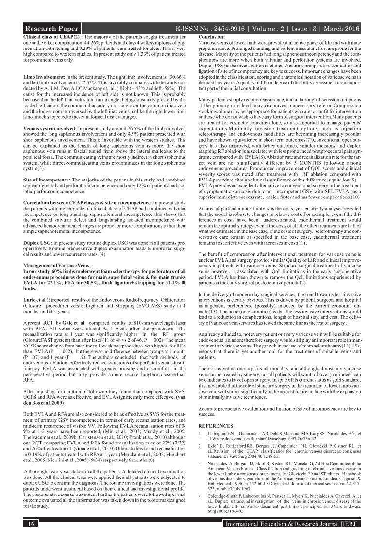
Research Paper E-ISSN No : 2454-9916 | Volume : 2 | Issue : 3 | March 2016Clinical class of CEAP(2) : The majority of the patients sought treatment for one or the other complication, 44.26% patients had class 4 with symptoms of pig-mentation with itching and 9.29% of patients were treated for ulcer. This is very high compared to western studies. In present study only 1.33% of patient treated for prominent veins only.
Limb Involvement: In the present study. The right limb involvement is 30.66% and left limb involvement is 47.33%. This favorably compares with the study con-ducted by A.H.M. Dur, A.I.C Mackaay et., al. ( Right – 43% and left -56%). The cause for the increased incidence of left side is not known. This is probably because that the left iliac veins joins at an angle; being constantly pressed by the loaded left colon, the common iliac artery crossing over the common iliac vein and the longer course traversed by the left iliac veins, unlike the right lower limb is not much subjected to these anatomical disadvantages.
Venous system involved: In present study around 76.5% of the limbs involved showed the long saphenous involvement and only 4.9% patient presented with short saphenous involvement. This is favorable with the western studies. This can be explained as the length of long saphenous vein is more, the short saphenous vein runs in fascial tunnel from above the lateral malleolus to the popliteal fossa. The communicating veins are mostly indirect in short saphenous system, while direct communicating veins predominates in the long saphenous system(3).
Site of incompetence: The majority of the patient in this study had combined saphenofemoral and perforator incompetence and only 12% of patients had iso-lated perforator incompetence.
Correlation between CEAP classes & site on incompetence: In present study the patients with higher grade of clinical class of CEAP had combined valvular incompetence or long standing saphenofemoral incompetence this shows that the combined valvular defect and longstanding isolated incompetence with advanced hemodynamical changes are prone for more complications rather their simple saphenofemoral incompetence.
Duplex USG: In present study routine duplex USG was done in all patients pre-operatively. Routine preoperative duplex examination leads to improved surgi-cal results and lower recurrence rates. (4)
Management of Varicose Veins:In our study, 60% limbs underwent foam sclertherapy for perforators of all endovenous procedures done for main superficial veins & for main trunks EVLA for 27.1%, RFA for 30.5%, flush ligation+ stripping for 31.1% 0f limbs.
Lurie et al (5)reported results of the Endovenous Radiofrequency Obliteration (Closure procedure) versus Ligation and Stripping (EVOLVeS) study at 4 months and at 2 years.
A recent RCT by Gale et al compared results of 810-nm wavelength laser with RFA. All veins were closed At 1 week after the procedure. The recanalization rate at 1 year was significantly higher in the RF group (ClosureFAST system) than after laser (11 of 48 vs 2 of 46, P .002). The mean VCSS score change from baseline to 1 week postprocedure was higher for RFA than EVLA (P .002), but there was no difference between groups at 1 month (P .07) and 1 year (P .9). The authors concluded that both methods of endovenous ablation effectively reduce symptoms of superficial venous insuf-ficiency. EVLA was associated with greater bruising and discomfort in the perioperative period but may provide a more secure longterm closure than RFA.
After adjusting for duration of followup they found that compared with SVS, UGFS and RFA were as effective, and EVLA significantly more effective. (van den Bos et al, 2009)
Both EVLA and RFA are also considered to be as effective as SVS for the treat-ment of primary GSV incompetence in terms of early recanalisation rates, and mid-term recurrence of visible VV. Following EVLA recanalisation rates of 0-9% at 1-2 years have been reported, (Min et al., 2003; Mundy et al., 2005; Theivacumar et al., 2009b, Christenson et al., 2010; Pronk et al., 2010) although one RCT comparing EVLA and RFA found recanalisation rates of 22% (7/32) and 26%after treatment. (Goode et al., 2010) Other studies found recanalisation in 0-19% of patients treated with RFA at 1 year. (Merchant et al., 2002; Merchant et al., 2005; Nicolini et al., 2005) (9/34) respectively 6 months.(6)
A thorough history was taken in all the patients. A detailed clinical examination was done. All the clinical tests were applied then all patients were subjected to duplex USG to confirm the diagnosis. The routine investigations were done. The patients underwent treatment based on their clinical and investigational profile. The postoperative course was noted. Further the patients were followed up. Final outcome evaluated all the information was taken down in the proforma designed for the study.
Conclusion:Varicose veins of lower limb were prevalent in active phase of life and with male preponderance. Prolonged standing and violent muscular effort are prone for the disease. Majority of the patients had long saphenous incompetency and the com-plications are more when both valvular and perforator systems are involved. Duplex USG is the investigation of choice. Accurate preoperative evaluation and ligation of site of incompetency are key to success. Important changes have been adopted in the classification, scoring and anatomical notation of varicose veins in the past few years. A quality of life or degree of disability assessment is an impor-tant part of the initial consultation.
Many patients simply require reassurance, and a thorough discussion of options at the primary care level may circumvent unnecessary referral.Compression stockings alone may be appropriate for patients who are too unfit for intervention or those who do not wish to have any form of surgical intervention.Many patients are treated for cosmetic concerns alone, so it is important to manage patients' expectations.Minimally invasive treatment options such as injection sclerotherapy and endovenous modalities are becoming increasingly popular and have shown equivalence in short term outcomes(7).Conventional open sur-gery has also improved, with better outcomes, smaller incisions and duplex mapping.RF ablation is associated with less pronounced postprocedural pain syn-drome compared with EVLA(8). Ablation rate and recanalization rate for the tar-get vein are not significantly different by 5 MONTHS follow-up among endovenous procedures. Pronounced improvement of QOL scores and clinical severity scores was noted after treatment with RF ablation compared with EVLA procedure, though clinical significance of this difference is quite low(9)EVLA provides an excellent alternative to conventional surgery in the treatment of symptomatic varicosis due to an incompetent GSV with SFJ. EVLA has a superior immediate success rate, easier, faster and has fewer complications.(10)
An area of particular uncertainty was the costs, yet sensitivity analyses revealed that the model is robust to changes in relative costs. For example, even if the dif-ferences in costs have been underestimated, endothermal treatment would remain the optimal strategy even if the costs of all the other treatments are half of what we estimated in the base case. If the costs of surgery, sclerotherapy and con-servative care remain as specified in the base case, endothermal treatment remains cost effective even with increases in cost(11) .
The benefit of compression after interventional treatment for varicose veins is unclear EVLA and surgery provide similar Quality of Life and clinical improve-ments in patients with varicose veins. Standard surgical treatment of varicose veins however, is associated with QoL limitations in the early postoperative period. EVLA has been shown to remove the QoL limitations experienced by patients in the early surgical postoperative period(12).
In the delivery of modern day surgical services, the trend towards less invasive interventions is clearly obvious. This is driven by patient, surgeon, and hospital management preferences, (possibly) imposed by the current economic cli-mate(13). The hope (or assumption) is that the less invasive interventions would lead to a reduction in complications, length of hospital stay, and cost. The deliv-ery of varicose vein services has towed the same line as the rest of surgery .
As already alluded to, not every patient or every varicose vein will be suitable for endovenous ablation; therefore surgery would still play an important role in man-agement of varicose veins. The growth in the use of foam sclerotherapy(14)(15) , means that there is yet another tool for the treatment of suitable veins and patients.
There is as yet no one-cap-fits-all modality, and although almost any varicose vein can be treated by surgery, not all patients will want to have, (nor indeed can be candidates to have) open surgery. In spite of its current status as gold standard, it is inevitable that the role of standard surgery in the treatment of lower limb vari-cose vein will shrink significantly in the nearest future, in line with the expansion of minimally invasive techniques.
Accurate preoperative evaluation and ligation of site of incompetency are key to success.
REFERENCES:
1. LabropoulosN, Giannoukas AD,DelisK,Mansour MA,KangSS, Nicolaides AN, et al.Where does venous refluxstart?JVascSurg 1997;26:736-42.
2. Eklöf B, Rutherford RB, Bergan JJ, Carpentier PH, Gloviczki P, Kistner RL, et al. Revision of the CEAP classification for chronic venous disorders: consensus statement. J Vasc Surg 2004;40:1248-52.
3. Nicolaides A, Bergan JJ, Eklof B, Kistner RL, Moneta G, Ad Hoc Committee of the American Venous Forum. Classification and grad- ing of chronic venous disease in the lower limbs: a consensus state- ment. In: Gloviczki P, Yao JST editors. Handbook of venous disor- ders: guidelines of the American Venous Forum. London: Chapman & Hall Medical; 1996, p. 652-60 J.F.Doyle, Irish Journal of medical science Vol 42, 317-323, number7/july 1967
4. Coleridge-Smith P, Labropoulos N, Partsch H, Myers K, Nicolaides A, Cavezzi A, et al. Duplex ultrasound investigation of the veins in chronic venous disease of the lower limbs: UIP consensus document: part I. Basic principles. Eur J Vasc Endovasc Surg 2006;31:83-92.
16 International Education & Research Journal [IERJ]

5. Lurie T, Brunkwall J. Systematic review and meta-analysis of en- dovenous radio-frequency obliteration, endovenous laser therapy, and foam sclerotherapy for pri-mary varicosis. J Cadiovasc Surg 2008;49:213-33.
6. Mundy L, Merlin TL, Fitridge RA, Hiller JE. Systematic review of endovenous laser treatment for varicose veins. Br J Surg 2005;92:1189-94.
7. Luebke T, Gawenda M, Heckenkamp J, Brunkwall J. Meta-analysis of endovenous radiofrequency obliteration of the great saphenous vein in primary varicosis. J Endovasc Ther 2008;15:213-23.
8. Hoggan BL, Cameron AL, Maddern GJ. Systematic review of en- dovenous laser therapy versus surgery for the treatment of saphenous varicose veins. Ann Vasc Surg 2009;23:277-87.
9. Kundu S, Lurie F, Millward SF, Padberg F Jr, Vedantham S, Elias S, et al. Recom-mended reporting standards for endovenous ablation for the treatment of venous insuf-ficiency: joint statement of the American Venous Forum and the Society of Interventional Radiology. J Vasc Surg 2007;46:582-9925-36.
10. Puggioni A, Kalra M, Carmo M, Mozes G, Gloviczki P. Endovenous laser therapy and radiofrequency ablation of the great saphenous vein: analysis of early efficacy and complications. J Vasc Surg 2005;42:
11. Leopardi D, Hoggan BL, Fitridge RA, Woodruff PW, Maddern GJ.Systematic review of treatments for varicose veins. Ann Vasc Surg 2009;23:264-76.
12. Meissner MH, Gloviczki P, Bergan J, Kistner RL, Morrison N, Pannier F, et al. Primary chronic venous disorders. J Vasc Surg 2007;46 Suppl S:54S-67S.
13. Michaels JA, Campbell WB, Brazier JE, Macintyre JB, Palfreyman SJ, Ratcliffe J, et al. Randomised clinical trial, observational study and assessment of cost-effectiveness of the treatment of varicose veins REACTIV trial. Health Technol Assess 2006;10:1-196.488-93.
14. Jia X, Mowatt G, Burr JM, Cassar K, Cook J, Fraser C. Systematic review of foam sclerotherapy for varicose veins. Br J Surg 2007;94:
15. Mitchel P. Goldman, "Anatomy and histology of venous system of the leg", Sclerotherapy-Treatment of varicose and telangiectatic leg veins, Mosby Year Book, 1991, 1:7-31.
17International Education & Research Journal [IERJ]
Research Paper E-ISSN No : 2454-9916 | Volume : 2 | Issue : 3 | March 2016