CLINICAL SIGNIFICANCE OF GALLOP RHYTHM IN HYPERTENSION · gallop rhythm sooner or later, even...
7
THE CLINICAL SIGNIFICANCE OF GALLOP RHYTHM IN HYPERTENSION BY B. E. MILES From the Department of Medicine, St. Thomas's Hospital Received June 15, 1950 Gallop rhythm is widely held to be a sign of prognostic value, and this opinion has been supported by follow-up studies of patients with gallop rhythm in groups of mixed etiology. This method of assessment is, however, open to criticism; first, because it is so dependent upon the way in which the patients are selected, and secondly, because the prognostic significance of gallop rhythm is clearly not the same in different diseases. This paper reports a clinical and phonocardiographic study of 100 hypertensive subjects, 37 of whom had gallop rhythm. The presence or absence of the various types of gallop has been correlated with the blood pressure, heart size, degree of cardiac failure, and electrocardiogram. DEFINITION AND- NOMENCLATURE Gallop rhythm is here defined as the presence, in a patient with heart disease, of audible vibrations in diastole, homologous either with the physiological third or auricular sounds. A phonocardio- graphic classification is used. The gallop sound coincident with the third sound which occurs during rapid ventricular filling, is called " rapid filling " gallop. That corresponding with the auricular sound and dependent upon auricular systole is called " auricular " gallop. A gallop occurring when diastole is so shortened by tachycardia that the rapid ventricular filling phase merges with auricular systole is called " indeterminate " gallop. The use of the terms " rapid filling " and " auricular " gallop follows the classification used by Lian, Minot, and Welti (1941) and Frost (1949). They seem preferable to " protodiastolic" and " presystolic " gallop, since the former never occurs in protodiastole, and may even occur in the second half of diastole, whereas the latter is at times mid-diastolic. A second alternative, namely, subdivisions of the third sound and fourth sound (Evans, 1943), has the disadvantage that these may be confused with their physiological counterparts; it is also perplexing to call the earliest souind in the cardiac cycle the fourth. At all events, each of these three classifications should be confined to phonocardiographic diagnosis. Clinically, it is best to describe early, mid-, and late diastolic gallops. The term indeterminate gallop has been coined as no other suitable name exists. The summation gallop of Wolferth and Margolies (1931) originally referred to the gallop, that may arise when subaudible protodiastolic and presystolic vibrations merge during tachycardia, and by summation become audible. It remains a valuable term when restricted to this connotation. The epithet " gallop " has been retained; it has survived its centenary. CLINICAL MATERIAL One hundred patients with hypertension were studied: 62 women and 38 men. All had average " casual " diastolic pressures above 95. Their average age was 57 years (range 18-80). The oetio- logical groups were as follows: essential hypertension 91, chronic nephritis 3, chronic pyelonephritis 2, toxemia of pregnancy 3, and coarctation of the aorta 1. 327 on March 29, 2020 by guest. Protected by copyright. http://heart.bmj.com/ Br Heart J: first published as 10.1136/hrt.13.3.327 on 1 July 1951. Downloaded from
Transcript of CLINICAL SIGNIFICANCE OF GALLOP RHYTHM IN HYPERTENSION · gallop rhythm sooner or later, even...

THE CLINICAL SIGNIFICANCE OF GALLOP RHYTHM INHYPERTENSION
BY
B. E. MILESFrom the Department of Medicine, St. Thomas's Hospital
Received June 15, 1950
Gallop rhythm is widely held to be a sign of prognostic value, and this opinion has been supportedby follow-up studies of patients with gallop rhythm in groups of mixed etiology. This method ofassessment is, however, open to criticism; first, because it is so dependent upon the way in which thepatients are selected, and secondly, because the prognostic significance of gallop rhythm is clearlynot the same in different diseases. This paper reports a clinical and phonocardiographic study of100 hypertensive subjects, 37 of whom had gallop rhythm. The presence or absence of the varioustypes of gallop has been correlated with the blood pressure, heart size, degree of cardiac failure, andelectrocardiogram.
DEFINITION AND- NOMENCLATUREGallop rhythm is here defined as the presence, in a patient with heart disease, of audible vibrations
in diastole, homologous either with the physiological third or auricular sounds. A phonocardio-graphic classification is used. The gallop sound coincident with the third sound which occursduring rapid ventricular filling, is called " rapid filling " gallop. That corresponding with theauricular sound and dependent upon auricular systole is called " auricular " gallop. A gallopoccurring when diastole is so shortened by tachycardia that the rapid ventricular filling phase mergeswith auricular systole is called " indeterminate " gallop.
The use of the terms " rapid filling " and " auricular " gallop follows the classification used byLian, Minot, and Welti (1941) and Frost (1949). They seem preferable to " protodiastolic" and" presystolic " gallop, since the former never occurs in protodiastole, and may even occur in thesecond half of diastole, whereas the latter is at times mid-diastolic. A second alternative, namely,subdivisions of the third sound and fourth sound (Evans, 1943), has the disadvantage that these maybe confused with their physiological counterparts; it is also perplexing to call the earliest souindin the cardiac cycle the fourth. At all events, each of these three classifications should be confinedto phonocardiographic diagnosis. Clinically, it is best to describe early, mid-, and late diastolicgallops. The term indeterminate gallop has been coined as no other suitable name exists. Thesummation gallop of Wolferth and Margolies (1931) originally referred to the gallop, that mayarise when subaudible protodiastolic and presystolic vibrations merge during tachycardia,and by summation become audible. It remains a valuable term when restricted to this connotation.The epithet " gallop " has been retained; it has survived its centenary.
CLINICAL MATERIALOne hundred patients with hypertension were studied: 62 women and 38 men. All had average
" casual " diastolic pressures above 95. Their average age was 57 years (range 18-80). The oetio-logical groups were as follows: essential hypertension 91, chronic nephritis 3, chronic pyelonephritis2, toxemia of pregnancy 3, and coarctation of the aorta 1.
327
on March 29, 2020 by guest. P
rotected by copyright.http://heart.bm
j.com/
Br H
eart J: first published as 10.1136/hrt.13.3.327 on 1 July 1951. Dow
nloaded from
Thirteen had angina pectoris and 35 were considered obese. Cases with valvular disease,anmemia, thyrotoxicosis, substantial lung disease, or recent myocardial infarction were excluded.The patients derived from two sources; the cardiovascular clinic (47), and the medical wards (53) ofthe Dunedin Public Hospital, New Zealand. All available cases from the cardiovascular clinic werestudied, and most of the cases from the wards. Patients with marked dyspncea at rest were usuallyexcluded for technical reasons. Otherwise they were unselected. The majority were moderatelysevere or severe cases.
METHODSClinical interpretation of heart sounds. The patients, unless orthopnceic, lay supine on a couch.
The heart was examined with a stethoscope at the apex and near the xiphisternum. A clinicaldiagnosis of any abnormal sound was recorded with the qualifications definite, probable,or possible, and the loudness and site of maximum audibility were noted. No special manceuvreswere employed to try to bring out a gallop sound. The results of previous auscultation wereignored.
Apparatus and recording procedure. The apparatus used was a six-channel oscilloscope designedand constructed in the Department of Medicine of Otago University. The input was derived froma Maico Stethetron pick-up head and tracings were recorded on 35-mm. film. The frequencyresponse curves corresponded closely with the linear, stethoscopic, and logarithmic curves recom-mended by Rappaport and Sprague (1942). Additional information about the instrument and itsacoustic properties have been published elsewhere (Miles, 1950).
As a reference trace for timing phonocardiographic events, a standard lead II electrocardiogramwas used: this was considered adequate in the present group of patients where the main difficultywas in distinguishing auricular gallops from split first sounds. Whilst recording the tracings, firstat the apex and then in the xiphisternal region, the various channels were checked visually on themonitor oscilloscope, and at the same time the heart sounds were heard with the monitor stethoscopefed from the same pick-up head. About 18 inches of film were used for each patient.
Phonocardiographic interpretation of heart sounds. Audible vibrations beginning between 0 I1Iand 0-20 sec. after the onset of the second sound were considered to be rapid filling gallops.Those occurring between the apex of the P wave and the apex of the R wave in the reference electro-cardiogram were considered to be auricular gallops. It is realized that these standards are arbi-trary, and that some of the indeterminate gallops would have been classified as auricular gallopsby other authors (Orias and Braun-Menendez, 1939; Lian et al., 1941). These diagnoses weremade from the logarithmic traces except in the few cases in which the logarithmic channel wasnot recording properly. Even so, it was not always possible to be certain what had actually beenheard. A phonocardiograph can at best only show what is potentially audible. This difficultysometimes arose when a small auricular vibration and also a split first sound were recorded in thesame patient. Three specimen phonocardiograms are shown in Fig. 1.
Final diagnosis of heart sounds. Before gallop rhythm was finally accepted, it had to be bothheard and recorded. The few heard but not recorded and vice versa were excluded.
GRADING OF ASSOCIATED FINDINGSBlood pressure. It was decided to use the more significant diastolic pressure rather than the
more easily determined systolic. The average of several " casual " diastolic pressures was taken,the casual pressure being the lowest recorded after about five minutes with the patient at rest inthe outpatient department or ward. In 64 patients a basal blood pressure was also taken, bythe method recommended by the committee of the British Cardiac Society and the American HeartAssociation.
The patient was first graded according to his casual diastolic pressure as follows: Grade I,96-105; Grade II, 106-115;- Grade III, 116-125; Grade IV, more than 125. If the basal
328 B. E. MILtS
on March 29, 2020 by guest. P
rotected by copyright.http://heart.bm
j.com/
Br H
eart J: first published as 10.1136/hrt.13.3.327 on 1 July 1951. Dow
nloaded from

329GALLOP RHYTHM IN HYPERTENSION
FIG. 1.-Three specimen phonocardio-grams. St = stethoscopic trace.Log. = logarithmic trace. 1 = 1stsound. 2 = 2nd sound. 11' = esplit 1st sound. a = auricular vibra-tions. S = indeterminate gallop.Scale: 1 cm. = 0-2 sec.
(A) Split Ist sound. Auricular vibra-tions (preceding the apex of theelectrocardiogram R wave) arepresent in the stethoscopic butnot in the logarithmic trace.
(B) Auricular gallop. Auricular vibra-tions are present in both stetho-scopic and logarithmic traces.
(C) Indeterminate gallop. Owing totachycardia (rate 130) the gallopsound (S) satisfies the criteria forboth rapid filling gallop (< 0-2sec. after onset, 2nd sound) andauricular gallop (between apex ofP wave and apex of R wave inelectrocardiogram r e f e r e n c etrace).
on March 29, 2020 by guest. P
rotected by copyright.http://heart.bm
j.com/
Br H
eart J: first published as 10.1136/hrt.13.3.327 on 1 July 1951. Dow
nloaded from
diastolic pressure was more than 20 mm. below the lowest figure of the previously assigned casualdiastolic pressure grade, this grade was lowered by 1, provided that the casual pressure was not above135 mm. Hg. Nine were lowered in this way. One patient, with a casual pressure of 210/128and a basal pressure of 128/80 was lowered from Grade IV to Grade II.
Heart size. The size of the heart was judged by eye from a postero-anterior teleradiogram.Thirteen cases were screened as well. The grades (I-IV) were normal limits, slight enlargement,moderate enlargement, and gross enlargement.
Degree of cardiac failure. The following four grades were recognized: no dyspncea on levelwalking; dyspncea on level walking; dyspncea at rest (orthopncea and paroxysmal dyspncea) andright-sided cardiac failure (with raised venous pressure). Breathlessness on hills only, may oftenhave been due to early left ventricular failure, but the significance of minor degrees of this symptomis difficult to assess.
Electrocardiographic changes. An abnormal group was recognized, comprising cases withleft ventricular strain, old myocardial infarction, bundle branch block, and atypical tracings, and anormal which included simple left axis deviation.
RESULTS
The final clinical and phonocardiographic diagnoses of the heart sounds are shown below.
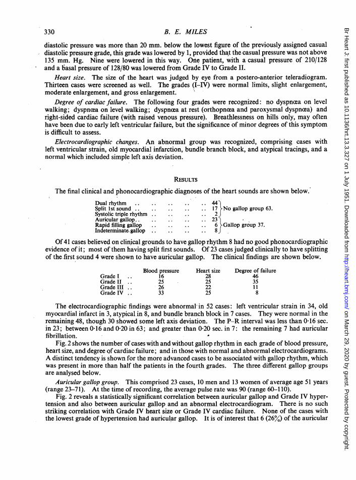
Dual rhythm .. .. .. .. .. 44)Split 1st sound7.... .. .. .. l7No gallop group 63.Systolic triple rhythm .. .. .. 2JAuricular gallop ..........23Rapid filling gallop .. .. .. .. 6 Gallop group 37.Indeterminate-gallop .. .. .. 8)
Of 41 cases believed on clinical grounds to have gallop rhythm 8 had no good phonocardiographicevidence of it; most of them having split first sounds. Of 23 cases judged clinically to have splittingof thefirst sound 4 were shown to have auricular gallop. The clinical findings are shown below.
Blood pressure Heart size Degree of failureGrade I .. 16 28 46Grade II .. 25 25 35Grade III .. 26 22 11Grade IV .. 33 25 8
The electrocardiographic findings were abnormal in 52 cases: left ventricular strain in 34, oldmyocardial infarct in 3, atypical in 8, and bundle branch block in 7 cases. They were normal in theremaining 48, though 30 showed some left axis deviation. The P-R interval was less than 0-16 sec.in 23; between 0-16 and 0-20 in 63; and greater than 0-20 sec. in 7: the remaining 7 had auricularfibrillation.
Fig. 2 shows the number of cases with and without gallop rhythm in each grade of blood pressure,heart size, and degree gf cardiac failure; and in those with normal and abnormal electrocardiograms.A distinct tendency is shown for the more advanced cases to be associated with gallop rhythm, whichwas present in more than half the patients in the fourth grades. The three different gallop g-roupsare analysed below.
Auricular gallop group. This comprised 23 cases, 10 men and 13 women of average age 51 years(range 23-71). At the time of recording, the average pulse rate was 90 (range 60-110).
Fig. 2 reveals a statistically significant correlation between auricular gallop and Grade IV hyper-tension and also between auricular gallop and an abnormal electrocardiogram. There is no suchstriking correlation with Grade IV heart size or Grade IV cardiac failure. None of the cases withthe lowest grade of hypertension had auricular gallop. It is of interest that 6 (26%) of the auricular
B. E. MILES330
on March 29, 2020 by guest. P
rotected by copyright.http://heart.bm
j.com/
Br H
eart J: first published as 10.1136/hrt.13.3.327 on 1 July 1951. Dow
nloaded from
GALLOP RHYTHM IN HYPERTENSION
FIG. 2.-Number of cases with and without gallop rhythm in each grade (see text).
gallop cases had apparently normal sized hearts, 9 (39%) had no breathlessness on walking on theflat, and 7 (30%) had electrocardiograms within normal limits. Three of the cases had neither en-largement of the heart nor, shortness of breath on the flat. One patient, aged 23, with Grade IVhypertension and a loud auricular gallop, was able to enjoy a full day's shooting expedition.
No correlation was observed between the loudness of the auricular gallop and the severity of thecase. Nor was any relation noticed between the severity of the case and the interval between thegallop and first sound. Five of the auricular gallop cases had P-R intervals exceeding 0-2 sec.;they were in other respects typical of the group.
Rapidfilling gallop group. This comprised 6 cases; 2 men and 4 women of average age 63 years(range 52-71). Their average pulse rate at the time ofrecording was 93 (range 75-106). This groupwas unfortunately too smalL to allow firm conclusions to be drawn from it, but 4 of the 6 cases hadgross enlargement of the heart, and 3 of them had right-sided cardiac failure, which is a statisticallysignificant correlation, as there were only 8 cases of right-sided failure in the whole series (X2 = 15-3).One case, on the other hand, had neither cardiac enlargement, breathlessness when walking on thelevel, nor electrocardiographic change.
Two of the three gallops heard- best in the xiphisternal region were rapid filling gallops. Bothwere associated with right-sided cardiac failure.
Indeterminate gallop group. This comprised 8 cases; 5 men and 3 women, of average age 51years (range 31-60). At the time of recording the average pulse rate was 113 (range 100-125).Reference to Fig. 2 shows .that 5 of the 8 cases had gross enlargement of the heart; 1, however, hada normal sized heart on screening and was not breathless on the flat. Six of the 8 cases had loudgallops; the gallop sometimes being the loudest of the three sounds.
331
on March 29, 2020 by guest. P
rotected by copyright.http://heart.bm
j.com/
Br H
eart J: first published as 10.1136/hrt.13.3.327 on 1 July 1951. Dow
nloaded from

The site ofmaximum audibility of the gallop sound is shown below.
Site best heard Auricular gallop Rapid filling IndeterminateSitebestheard cases gallop cases gallop casesApex .. .. .. 17 2 3Xiphisternum .. .. 0 2 1Both equally .. .. 6 2 4
DISCUSSIONAn interesting finding was the commonness of gallop rhythm in hypertension when specially
sought. The best available evidence of its absolute incidence is Garvin's (1943) finding, that 47 percent of hypertensive patients who died a cardiac death had previously had gallop rhythm. That37 per cent. of the present series of unselected hospital patients with hypertension, and as many as63 per cent of those with Grade III and IV cardiac failure, had gallop rhythm when examined on asingle occasion, suggests that more than 50 per cent of hypertensive subjects are likely to developgallop rhythm sooner or later, even when taking into account that some, through obesity or em-physema, will have sounds of low intensity.
If gallop rhythm is to have prognostic value, it must either be a reliable indication that a certainstage of cardiac involvement has been reached, or a sign of a rapidly advancing disease process.The present investigation, while showing a tendency for the severer cases to be associated moreoften with gallop rhythm, demonstrated that in hypertension it occurs over a wide clinicalrange. If, then, we ignore the remote possibility that gallop rhythm is in some way correlatedwith the rate of progress of the hypertension, it follows that gallop rhythm is not a reliable prognosticsign.
There is surprisingly little written reference to the examples, familiar to all clinicians, of galloprhythm occurring in patients with comparatively good cardiac function, though Duchosal (1932)does say that a certain proportion of hypertensive subjects may possess well-defined gallop rhythmfor years.
This finding, that gallop rhythm may be audible under widely differing conditions, is not in theleast unexpected when it is appreciated that the corresponding vibrations in the normal hearts ofyoung people vary enormously in intensity; and, moreover, that in adults great differences occur inthe transmitting properties of the lungs from emphysema and of the chest wall as a result of rigidityor obesity. It is of some interest that 43 per cent of the non-gallop cases were obese, as comparedwith only 22 per cent of the gallop cases, though it is true that the obese and non-obese groups werenot strictly homogeneous.
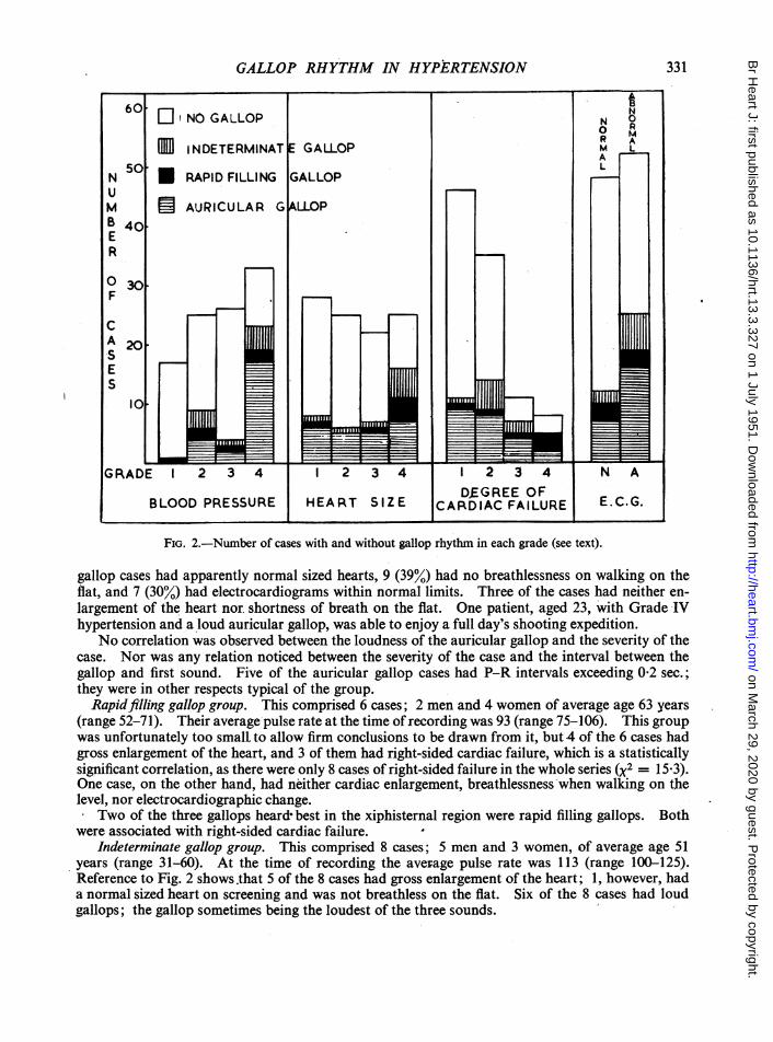
Fig. 3, which compares the clinical findings in the 37 gallop cases and in the 52 with abnormalelectrocardiograms, suggests that gallop rhythm has about the same overall significance as has anyabnormality in the electrocardiogram. It is not doubted that gallop rhythm is a valuable clinicalsign in the detection of heart disease, but it is probably a poor guide to prognosis. There are bettermethods for assessing the cardiac status. If two patients had the same history, blood pressure, heartsize, degree of cardiac failure, and pulse rate, and one of them had gallop rhythm, there is no evidenceto show that the latter would have the worse prognosis.
In addition to the claim that gallop rhythm is useful prognostically, Evans (1943), has assertedthat rapid filling gallop is a sign of right- and auricular gallop of left-sided cardiac failure. In thepresent series, only 2 out of 23 with auricular gallop, and as many as 3 out of 6 with rapid fillinggallop, had right-sided cardiac failure, thus giving some support to Evans' contention. There isalso some corroboration for the view originally propounded by Potain (cited by Holt, 1927), that thegallop may be right-sided (i.e. heard best towards the xiphisternum) in cases of right-sided cardiacfailure. Of the 5 cases with right-sided cardiac failure, 2 were heard best in the xiphisternum region,1 in the apical'region, and 2 equally well at both. The only other right-sided gallop was an indeter-
332 B. E. MILES
on March 29, 2020 by guest. P
rotected by copyright.http://heart.bm
j.com/
Br H
eart J: first published as 10.1136/hrt.13.3.327 on 1 July 1951. Dow
nloaded from

GALLOP RHYTHM IN HYPERTENSION
FIG. 3.-The clinical findings as regards blood pressure, heart size, and degree of cardiac failure are shown to besimilar in the gallop rhythm group (37 cases) and the abnormal electrocardiogram group (52 cases).
minate gallop. It would, however, be rash to assert that right-sided gallop always arose in the rightventricle. Contrary to the findings of Evans (1943), none of the auricular gallop sounds was mostaudible near the xiphisternum.
SUMMARYGallop rhythm as defined was heard and recorded phonocardiographically in 37 of 100 patients
with hypertension.Poor correlation was shown between gallop rhythm and the degree of heart disease as judged by
blood pressure, heart size, degree of cardiac failure and electrocardiographic abnormality.It is suggested that gallop rhythm has little value in prognosis, as it occurs in a wide range of
clinical circumstances.A tendency was shown for" rapid filling " gallop to be assdciated with right-sided cardiac failure.
This paper is taken from a Cambridge M.D. thesis. I wish to thank Professor F. H. Smirk for the generous pro-vision of facilities in his Department at Otago University, and to Professor E. P. Sharpey-Schafer and A. C. Domhorstfor helpful criticism.
REFERENCESDuchosal, P. (1932). Amer. Heart J., 7, 613.Evans, W. (1943). Brit. Heart J., 5, 205.Frost, J. (1949). Acta. med. Scand., 133, 268.Garvin, C. F. (1943). Amer. J. med. Sci., 205, 814.Holt, E. (1927). Amer. Heart J., 2, 453.Lian, C., Minot, G., and Welti, J. J. (1941). Phonocardiographie Auscultation Collective, Paris.Miles, B. E. (1950). M.D. Thesis, Cambridge.Orias, O., and Braun-Menendez, E. (1939). The Heart Sounds in Normal and Pathological Conditions. 1st ed., London'.Rappaport, M. B., and Sprague, H. B. (1942). Amer. Heart J., 23, 591.Wolferth, C. C., and Margolies, A. (1931). Med. Clin. N. Amer., 14, 897.
333
on March 29, 2020 by guest. P
rotected by copyright.http://heart.bm
j.com/
Br H
eart J: first published as 10.1136/hrt.13.3.327 on 1 July 1951. Dow
nloaded from