CLINICAL RECORD I AUTOPSY PROTOCOL (an
8
• A. PA limandbazdrammMos 2140.044Auestlem • Pram Wu. md Durommdthollw4W A —7110441%) AUTOPSY PROTOCOL A63-272 (an: ec) CLINICAL RECORD I • DATE AND HOUR DIED 22 November 1963 1300(CST) P.M IWN : MECITIM (497831) CDR J. J._ _HUMES. EC.. U_SN re-LANICAL DIA0mOWLEI rr r.MW tracamtfaaci PATE AND HOUR MROPSY PIRFORRLD A. M. 22 November 1963 2000(ESOA. ASStUTANT (4896/0 CDR " J " THORNTON BOSWELL,MC,1 SN X LCOL PIERRE A. FINCK,MC,USA (04 043 322) CI-INC* O.. MAI AUTOPSY I HUB ORLY WARM OOLT Ht. - 721/2 inches Wt. - 170 pounds Eyes - blue Hair - Reddish brown PATH oLOO ICAL 01 AONOSICS CAUSE OF DEATH: Gunshot wound, head. - - — ARPROyt - .r, ,YMES, c/R, m e USN MILITARY CRMANIYATION (WRA 'mowed) AGE SEA RACE 1 PRESIDENT, UNITED STATES 46 1 Male 1 Cauc. PA rIENT•• iDENTWiCATIDN (Pat rfp•ai Of ...riffs., entrls• 01041, N•cma—fa•t. *Se, midrib.; ar•do; data; haciatml or otadicat facilit)) KENNEDY, JOHN F. NAVAL MEDICAL SCHOOL 11 , 174TIFICATIO.00. ALP1iPP.WYNO. 1 A63- 272 RXORITIM WARD NO. AUTOPST PROTOCOL Standard Form 8011 P.) - ' CIS . .r.R.VtA VMPskM a , sr.z. ,, LY:Rmod , r (m dlvt-'1 6W ANAWn.mr ,
Transcript of CLINICAL RECORD I AUTOPSY PROTOCOL (an

• A. PA
limandbazdrammMos 2140.044Auestlem • Pram Wu. md
Durommdthollw4W A —7110441%)
AUTOPSY PROTOCOL A63-272 (an: ec) CLINICAL RECORD I
•
DATE AND HOUR DIED
22 November 1963 1300(CST) P.M
IWN:MECITIM (497831)
CDR J. J._ _HUMES. EC.. U_SN re-LANICAL DIA0mOWLEI rr r.MW tracamtfaaci
PATE AND HOUR MROPSY PIRFORRLD A. M.
22 November 1963 2000(ESOA.
ASStUTANT (4896/0
CDR "J" THORNTON BOSWELL,MC,1 SN X
LCOL PIERRE A. FINCK,MC,USA (04 043 322)
CI-INC* O..
MAI AUTOPSY I HUB ORLY WARM OOLT
Ht. - 721/2 inches
Wt. - 170 pounds
Eyes - blue
Hair - Reddish brown
PATH oLOO ICAL 01 AONOSICS
CAUSE OF DEATH: Gunshot wound, head.
- - — ARPROyt -
.r, ,YMES, c/R, me USN
MILITARY CRMANIYATION (WRA 'mowed) AGE SEA RACE 1
PRESIDENT, UNITED STATES 46 1 Male 1 Cauc.
PA rIENT•• iDENTWiCATIDN (Pat rfp•ai Of ...riffs., entrls• 01041, N•cma—fa•t. *Se, midrib.; ar•do; data; haciatml or otadicat facilit))
KENNEDY, JOHN F.
NAVAL MEDICAL SCHOOL
11,174TIFICATIO.00. ALP1iPP.WYNO.
1 A63-272
RXORITIM WARD NO.
AUTOPST PROTOCOL
Standard Form 8011
P.)
- ' CIS ..r.R.VtAVMPskM a,sr.z.,, LY:Rmod,r(mdlvt-'16WANAWn.mr,

."--, .CIDICA1a'i02411ARY:
4 . ,,-1 ' , , According to available iniormatimi'th
: f.i;(- , ' ,7 deceased, President John F. Kennedy, : ....p, . ,,,,,,hs.
1---..17 On 22 November 1963. The President was sitting in the right teat seat with Mrs. '.v4i
i ,„ ,...1111411; riding in an epos car. is a motorcade during au official visit to Dallas, Texas 1:
'Kennedy seated on theisme peat to his left. Sitting directly in front of the :11./ .. 1 i PreSident was Covernor,John B. Connolly of Texas and directly in front of Mrs. Kennedy r.
• O i sat-Hrs. Connolly. Thi vehicle was moving at a slow rate of speed down an incline 1
- into an underpass that leads to a freeway route to theDallas ?ride Mart whcnetbbe ,..., j President was to deliver an address.
Three shots were heard and the President ▪ :!'. fell forward bleeding from the head._.(Governor Connolly was seriously wounded by the ' • same gunfire.) According to newspaper reports ("Washington Poet" November 23, 1963)
Bob Jackson, a Dallas "Times Herald"Photographer, said he looked around as he beard ' ‘114
•
the shoe and saw a rifle. barrel disappearing into a window on an upper floor of the nearby Texas School Book Depository Building.
i
• I
1
.
Shortly following the wounding of the two Men the car was driven to Parkland Hospital in Dallas. In the emergency room of that hospital the President was attended by Dr. Malcolm Perry. Telephone communication with Dr. Perry on November 23, 1963 develops the following information relative to the ob-servations made by Dr. Perry and procodures performed there prior to death. .
/ Dr. Parry noted the massive wound of the
head and a second much smaller wound of the low anterior neck in approximately the• midline. A tracheostomy was performed by extending the latter wound. At this point blOody air was noted bubbling from the wound and an injury to the right lateral wall of the trachea was observed. Incisions ware made in the upper anterior chest wall bilaterally to combat possible subcutaneous emphysema. Intravenous infusions of blood and saline were begun and oxygen was administered. Despite these measures cardiac arrest occurred and closed cheat cardiac massage failed to re-establish cardiac action. The President was pronounced dead approximately thirty to forty minutes after receiving Isis wounds.
The remains were transported via the Presidential plane to Washington, D.C. and subsequently to the Naval Medical School, National Naval Medical Center, Bethesda, Maryland for postmortem examination.
GENERAL DESCRIPTION OF BODY: The body is that of a muscular, well- de developed and well nourished adult Caucasian male measuring 721/2 inches and weighing approximately 170 pounds. There is beginning rigor mortis, minimal dependent livor mortis of the dorsum, and early algor mortis. hair is reddish brown and abundant, the eyes are blue, the right pupil measuring 8 mm. in diameter, the left 4 mm. There is edema and ecchymosis of the inner canthus region of the left eyelid measuring approximately 1.5 cm. in greatest diameter. There is edema and ecchymosia diffusely over the right supra-orbital ridge with abnormal mobility of the underlying bone. (The remainder of the scalp will be described with tho skull.)
•
- • , ..4104%.•
• • 7.4iEl

PATHOLOGICAL EXAMINATION REPORT A63-272 Page 3
There is clotted blood on the external ears but otherwise the ears, flares, and mouth
are essentially unremarkable. The teeth are in excellent repair and there is some
pallor of the oral mucous membrane.
Situated on the upper right posterior
thorax just above the upper border of the scapula there is a 7 x 4 millimeter oval
wound. This wound is measured to be 14 cm. from the tip of the right acromion process
and 14 cm. below the tip of the right mastoid process.
Situated in the low anterior neck at ap-
proximately the level of the third and fourth tracheal rings is a 6.5 cm. long trans-
verse wound with widely gaping irregular edges. (The depth and character of these
wounds wil be further described below.)
Situated on the anterior chest wall in the
nipple line are bilateral 2 am. long recent transverse surgical incisions into the
subcutaneous tissue. The one on the left is situated 11 cm. cephalad to the nipple
and the one on the right 8 cm. cephalad to the nipple. There is no hemorrhage or
ecchymosis associated 1,101 these wounds. A similar clean wound measuring 2 cm. in
length is situated on the antero-lateral aspect of the left mid arm. Situated on the
antero-lateral aspect of each ankle is a recent 2 cm. transverse incision into the
subcutaneous tissue.
There is an old well healed 8 cm. McBurney
abdominal incision. Over the lumbar spine in the midline is an old, well healed
15 cm. scar. Situated on the upper antero-Jateral aspect of the right thigh is an
old, well healed 8 cm. scar.
MISSILE WOUNDS: 1. There is a large irregular defect of Y
the scalp and skull on the right involving
chiefly the parietal bone but extending somewhat into the temporal and occipital
regions. In this region there is an actual absence of scalp and bone producing a
defect which measures approximately 13 cm. in greatest diameter.
From the irregular margins of the above
scalp defect tears extend in stellate fashion into the more or less intact scalp
as follows:
a. From the right inferior temporo-parietal margin anterior to the right ear to
a point slightly above the tragus.
b. From the anterior parietal margin anteriorly on the forehead to approximately
4 cm. above the right orbital ridge.
c. From the left margin of the main defect across the midline antero-laterally
for a distance of approximately 8 cm.
d. From the same starting point as c. 10 cm. postero-laterally.

PATHOLOGICAL EXAMINATION REPORT A63-272 Page 4
Situated in the posterior scalp approximately 2.5 cm. laterally to the right and
slightly above the external occipital protuberance is a lacerated wound measuring
15 x 6 mm. In the underlying bone is a corresponding wound through the skull which
exhibits beveling of the margins of the bone when viewed from the inner aspect of
the skull.
Clearly visible in the above described
large skull defect and exuding from it is lacerated btain tissue which on close
inspection proves to represent the major portion of the right cerebral hemisphere.
At this point it is noted that the falx cerebri is extensively lacerated with dis-
ruption of the superior saggital sinus.
Upon reflecting the scalp multiple complete
fracture lines are seen to radiate from both the large defect at the vertex and the
smaller wound at the occiput. These vary greatly in length and direction, the longest
measuring approximately 19 cm. These result in the production of numerous fragments
which vary in size from a few millimeters to 10 cm. in greatest diameter.
The complexity of these fractures and the
fragments thus produced tax satisfactory verbal description and are better appreciated
in photographs and roentgenograms which are prepared.
The brain is removed and preserved for
further study following formalin fixation.
Received as separate specimens from Dallas,
Texas are three fragments of skull bone which in aggregate roughly approximate the
dimensions of the large defect described above. At one angle of the largest of these
fragments is a portion of the perimeter of a roughly circular wound presumably of
exit which exhibits beveling of the outer aspect of the bone and is estimated to
measure approximately 2.5 to 3.0 cm. in diameter. Roentgenograms of this fragment
reveal minute particles of metal in the bone at this margin. Roentgenograms of the
skull reveal multiple minute metallic fragments along a line corresponding with a line
joining the above described small occipital wound and the right supra-orbital ridge.
From the surface of the disrupted right cerebral cortex two small irregularly shaped
fragments of metal are recovered. These measure 7 x 2 mm. and 3 x 1 mm. These are
placed in the custody of Agents Francir X. O'Neill, .Jr. and James W. Sibert, of the
Federal Bureau of Investigation, who executed a receipt therefor (attached).
2. The second wound presumably of entry
is that described above in the upper right posterior thorax. Beneath the skin there
is eccLymosis of subcutaneous tissue and musculature. The missile path through the
fascia and musculature cannot be easily probed. The wound presumably of exit was
that described by Dr. Malcolm Perry of Dallas in the low anterior cervical region.
When observed by Dr. Perry the wound measured "a few millimeters in diameter", how-
ever it was extended as a tracheostomy incision and thus its character is distorted
at the time of autopsy. However, there is considerable ecchymosis of the strap
muscles of the right side of the neck and of the fascia about the trachea adjacent
to the line of the tracheostomy wound. The third point of reference in connecting
X.0".C1044760,11MMMOWMtrWM"`7.SWkilittkr6WMVM74.1•ZMORMVIMMMMA3WaMS '7ethM,YATCktVM,WWIWW. M5eaMPraEIMIL'NWLftiTt

rATHOLOGiCAL S7[A /NAT-10Y REPORT
&63-272 Page 5
these two wounds is in the apex (supra-clavicular portion) of the right pleural cavity. In this region there is contusion of the parietal pleura and of the extreme apical portion of the right upper lob• of the lung. In both instances the diameter of contusion and ecchymoeia at the point of maximal involvement measures 5 cm. Both the visceral and parietal pleura are intact overlying these areas of trauma.
INCISIONS: The scalp wounds are extended in the coronal plane to examine the cranial content and the
customary (Y) shaped incision is used to examine the body cavities.
THORACIC CAVITY: The bony cage is unremarkable. The thoracic organs are in their normal positions and re-
lationships and there is no increase in free pleural fluid. The above described area of contusion in the apical portion of the right pleural cavity is noted.
LUNGS: The lungs are of essentially similar ap- pearance the right weighing 320 Gm., the
left 290 Gm. The lungs are well aerated with smooth glistening pleural surfaces and gray-pink color. A 5 cm. diameter area of purplish red discoloration and increased firmness to palpation is situated in the apical portion of the right upper lobe. This corresponds to the similar area described in the overlying parietal pleura. Incision in this region reveals recent hemorrhage into pulmonary parenchyma/
HEART: The pericardial cavity is smooth walled and contains approximately 10 cc. of straw-
colored fluid. The heart is of essentially normal external contour and weighs 350 Gm. The pulmonary artery is opened in situ and no abnormalities are noted. The cardiac chambers contain moderate amounts of postmortem clotted blood. There are no gross abnormalities of the leaflets of any of the cardiac valves. The following are the circumferences of the cardiac valves: aortic 7.5 cm., pulmonic 7 cm., tricuspid 12 cm., mitral 11 cm. The myocardium is firm and reddish brown. The left ventricular myocardium averages 1.2 cm. in thickness, the right ventricular myocardium 0.4 cm. The coronary arteries are dissected and are of normal distribution and smooth walled and elastic throughout.
ABDOMINAL CAVITY:
no increase in free and there are a few dominal wall at the
SKELETAL SYSTEM:
abnormalities.
The abdominal organs are in their normal positions and relationships and there is
peritoneal fluid. The vermiform appendix is surgically absent adhesions joining the region of the cecum to the ventral ab-above described old abdominal incisional scar.
Aside from the above described skull wounds there are no significant gross skeletal
PHOTOGRAPHY: Black and white and color photographs depicting significant findings are exposed
but not developed. These photographs were placed in the custody of Agent Roy H. Kellerman of the U. S. Secret Service, who executed a receipt therefore (attached).
~.... , • • .4.. 6. -

• PATHOLOUCAL EXAMINATION REPORT A63-272 Paste 6
ROENTGENOGRAMS: Roentgenograms are made of the entire body and of the separately submitted three fragments of skull bone. These are developed and were placed in the custody 9f Agent Roy H. Kellerman of the U. S. Secret Service, who executed a receipt therefor (attached).
SUMMARY: Based on the above observations it is our opinion that the deceased died as a result of two perforating gunshot wounds inflicted by high velocity projectiles fired by • person or persons unknown. The projectiles were fired from a point behind and some-what above the level of the deceased. The observations and available information do not permit a satisfactory estimate as to the sequence of the two wounds.
The fatal missile entered the skull above and to the right of the external occipital protuberance. A portion of the projectile traversed the cranial cavity in a posterior-anterior direction (see lateral skull roentgenograms) depositing minute particles along its path. A portion of the pro-jectile made its exit through the parietal bone on the right carrying with it portions of cerebrum, skull and scalp. The two wounds of the skull combined with the force of the missile produced extensive fragmentation of the skull, laceration of the superior eaggital sinus, and of the right cerebral hemisphere.
The other missile entered the right superior posterior thorax above the scapula and traversed the soft tissues of the supra-scap-ular and the supra-clavicular portions of the base of the right side of the neck. This missile produced contusions of the right apical parietal pleura and of the apical portion of. the right 2.upper lobe of the lung. The missile contused the strap muscles of the right side of the neck, damaged the trachea and made its exit through the anterior surface of the neck. As far as can be ascertained this missile struck no bony structures in its path through the body.
In addition, it is our opinion that the wound of the skull produced such extensive damage to the brain as to preclude the possibility of the deceased surviving this injury.
A supplementary report will be submitted following more detailed examination of the brain and of microscopic sections. However, it is not anticipated that these examinations will materially alter the findings.
(- r J. J. HUMES CDR, MC, USN (497831)
A' k44
J" THORNTON BOSWELL PIERRE A. FINCK CDR, MC, USN (489878) LT COL, MC, USA
(04-043-322)
CIMAISMM4M4*-1°MitatiMAt..;...,i:TMWAI'k•ikS..

LI. S. NAVAL MEDICAL SCHOOL NATIONAL NAVAL MEDICAL CENTER
BETH1110/, IIAITL YID X•I■ la.shr Mot •
24 Kovvaber 1963
C-1-11-2 •
I, Jamas 1. Puma, certify that *11 marking *tiara',
•Ansociated viiI3 Naval Radical School Autopsy Upon Ad1-241
have romatned Le OF pesiloael cupOO47 Ac a11 OW. ,..*0[011411 .
note and as boll:was& draft of the final report Nara headed
to DIsmandica Officer, U. S. Naval Kodical School, at 1700,
24 November 1963._ No pgparsralatisa to this: cis* rdsait:ia
my possess Lon.
• t•-' .Lk.":1 ...A-4— J. J. MESS COS, NC, USN
tacsioad above wowkiaa paper' this data.
J. a, 8TOV8R JR. CAPt, NC, USN Comoanding Offlcar, O.S. Naval Nodical School National Sisal RsdinalCaator
CUM M ISSION Earner!. 307-4:nntilniett
4-7
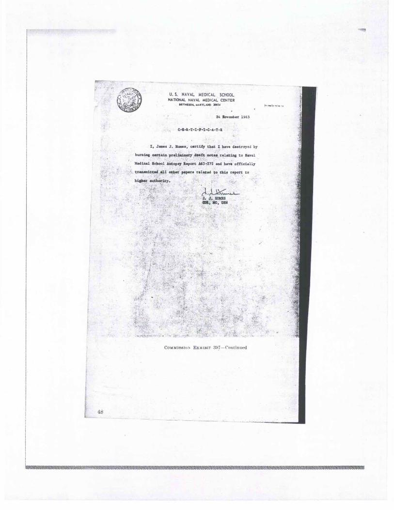
U.S. NAVAL MEDICAL SCHOOL NATIONAL NAVAL MEDICAL CENTER
SI MELBA. .1.11:IL AM) 31101.1
24 Bove:mbar 151
I. Jam.. J. Blass, city that I bay. dostroycJ by
bornine nartain preliminary daft mots. r•latioa to eaval
Medical School Autopsy Report AA3-272 and have officially
transmitted all other paper. relatad to chin report to
Mids.:. author-Sky.
rt . J. J. MIS Cal. MO, USM
COMMISfiIoN EIHISIT 397— Cmitinurd
48
Wel Waf9M VAMMict l$k.-W,Ma,K.̀'VM7VSMM