Clinical recommendations on the use of uterine artery … uterine bleeding/RCOG... · surgical...
22
Report of a Joint Working Party Clinical recommendations on the use of uterine artery embolisation in the management of fibroids Second edition
Transcript of Clinical recommendations on the use of uterine artery … uterine bleeding/RCOG... · surgical...

Report of a Joint Working Party
Clinical recommendations on
the use of uterine artery
embolisation in the
management of fibroids
Second edition

Clinical recommendations on the use of uterine artery embolisation in the management of fibroids. Second edition 3
Contents
Summary of recommendations 41 Introduction and background 52 Literature review 63 Indications for fibroid embolisation 84 Contraindications 95 Infertile patients and those contemplating a subsequent pregnancy 106 Counselling, consent and communication 117 Pretreatment assessment and prophylactic measures 128 The procedure 139 Complications 1410 The individual’s responsibility in clinical practice 1611 Follow-up 1712 Suggested areas for further study 18 References 19 Appendix 1. Post-procedure care and management of complications 21 The Working Party 22

Clinical recommendations on the use of uterine artery embolisation in the management of fibroids. Second edition4
Summary of recommendations
1. The early and mid-term results of uterine artery embolisation (UAE) are promising indicating that it is as least as safe as the surgical alternatives. It provides good symptom relief and is particularly effective for heavy menstrual bleeding.
2. For women with symptomatic fibroids, UAE should be considered as one of the treatment options as well as the conventional surgical treatments such as myomectomy and hysterectomy.
3. UAE as treatment for fibroids in women of child-bearing age who wish, or might wish, to become pregnant in the future should only be offered after fully informed discussion and should be considered on a case-by-case basis. The increased risks of caesarean section and the possibility of increased pregnancy complications should be fully understood.
4. The procedure is contraindicated in women who have evidence of current or recent infection, women who are unwilling to have a hysterectomy in any circumstances and where there is significant doubt about the diagnosis of benign pathology.
5. Patients should be seen by a gynaecologist and accurate pretreatment diagnosis is essential. MRI is recommended but good quality ultrasound is the minimum imaging requirement.
6. The procedure should only be undertaken by radiologists with specialised experience in embolisation who have undergone appropriate training.
7. The particular responsibilities of both gynaecologist and radiologist should be established prior to treatment and be set out in a relevant hospital protocol. The patient must be under a named responsible consultant at all times. Careful follow-up protocols should be established.
8. These recommendations are intended for both the National Health Service and the private sector.
Clinical recommendations on the use of uterine artery embolisation in the management of fibroids. Second edition 5
Arterial embolisation has been used during the last three decades as a method of treating haemorrhage in a variety of clinical situations, including postpartum haemorrhage, bleeding after caesarean section and bleeding following gynaecological surgery. The technique was extended in later years to include its use in the management of arteriovenous malformations of the genital tract and in gestational trophoblastic disease. Ravina et al first used arterial embolisation to treat fibroids in 1991, publishing his first series in 1995.1
In 2000, a Joint Working Party of the Royal College of Obstetricians and Gynaecologists and The Royal College of Radiologists was established to issue guidance on a procedure which at that time was in its infancy with less than 7,000 cases having been undertaken worldwide. Since the publication of that document, the procedure has matured and well over 100,000 cases have now been performed. Some of these cases have been part of national registries and randomised trials. The evidence from these studies has demonstrated the procedure to have good short- and medium-term success rates with acceptable morbidity and very low mortality. This has positioned the procedure in the main stream of treatment for symptomatic fibroids as recommended by the National Institute for Health and Clinical Excellence (NICE) in its heavy menstrual bleeding guideline.2
This document aims to provide information for patients interested in undergoing the procedure and provide guidelines for clinicians involved in their care. This document replaces previous advice given in Clinical Recommendations on the Use of Uterine Artery Embolisation in the Management of Fibroids (2000), which is now withdrawn.
1 Introduction and background

Clinical recommendations on the use of uterine artery embolisation in the management of fibroids. Second edition6
2 Literature review
The initial report of uterine artery embolisation (UAE) in the English literature appeared in 1995 by Ravina et al.1 Since that time it is estimated that in excess of 100,000 procedures have been carried out mainly in the USA and Western Europe.3 Initial evidence was limited to single-centre series but a more secure evidence base has recently developed. A large American registry reported in 20054 and four separate randomised trials have been published between 2003–2008.5–8 A Cochrane review reported in 20069 and a large UK retrospective comparative study (HOPEFUL) published in 2007.10
NICE issued guidance in 2001 and subsequently in 2004 stating latterly that, ‘UAE appeared to be safe for routine use and that the majority of patients have short-term symptomatic relief’.11 The same year (2004) the American College of Obstetricians and Gynecologists recognised UAE as safe and effective.3
From the outset, the differences in the study methodologies and their aims should be understood (see Table 1). The US prospective registry (n=3,160) gives the most information on safety due to the large numbers but has no control group. In summary, it reported a 5.4% major complication rate at 30 days (the majority occurring post-hospital discharge) with three (0.1%) requiring a hysterectomy.4 There were no deaths. In a smaller subgroup, quality of life (QoL) (using a fibroid-specific QoL) was significantly improved over baseline at one year.
The HOPEFUL study (n=1,108) is also a large cohort study and compares UAE with a matched hysterectomy group. It is retrospective and relies on both case record review and direct patient contact.10 The other four studies are randomised controlled trials (RCTs), with comparison against hysterectomy,5,7 myomectomy8 and hysterectomy or myomectomy.6 A Cochrane review was published in 2006 but at this time only two of the above RCTs had reported follow-up beyond six weeks.9
The RCTs all suffer from relatively small numbers and although they use different primary outcome measures, they have collected similar data which might allow further meta-analysis.5–7
Table 1. Randomised UAE trials, US registry and HOPEFUL study
Study Type Year n= Arms Primary outcomePinto7 RCT 2003 57 Hysterectomy Hospital stayMara8 RCT 2008 121 Myomectomy SymptomsEMMY5 RCT 2007 177 Hysterectomy Freedom from hysterectomyREST6 RCT 2007 157 Hysterectomy or myomectomy QoLUS registry4 Voluntary registry 2005 3,160 N/a N/aHOPEFUL10 Retrospective cohort 2007 1,108 Hysterectomy Multiple
In spite of the different study types, the results are very similar with regard to most of the important outcomes which are shown in Table 2 (opposite). In essence, UAE gives equal results to hysterectomy regarding QoL. Hospital stay and recovery of normal milestones are significantly shorter in UAE compared to surgery. Complication rates are broadly similar with both treatments. Some of the discrepancies in complication rates reported may arise due to the way they are recorded and classified. An example is the recording of two coincidental breast cancers in the UAE arm logged as adverse events in one trial. Symptom control following UAE does not match hysterectomy and at one year approximately 10% of UAE patients will require either hysterectomy or repeat embolisation for symptom control. HOPEFUL reported a 23% chance of further intervention following UAE at mean follow-up of 4.6 years. The RCTs continue their long-term follow-up and will report again soon. Cost-effectiveness analysis at one year (even when the additional secondary procedures are included) shows UAE to be significantly more cost-effective by approximately £1,000 per case.6,12 However, cost-effectiveness may reduce longer term as some patients will require further treatment.
In summary, UAE is a very useful proven uterine-sparing procedure that (rather like endometrial ablation for dysfunctional uterine bleeding) spares the majority of women a hysterectomy. The faster, shorter recovery period of UAE with uterine preservation needs to be weighed against the need for further treatment in a minority of patients.
Clinical recommendations on the use of uterine artery embolisation in the management of fibroids. Second edition 7
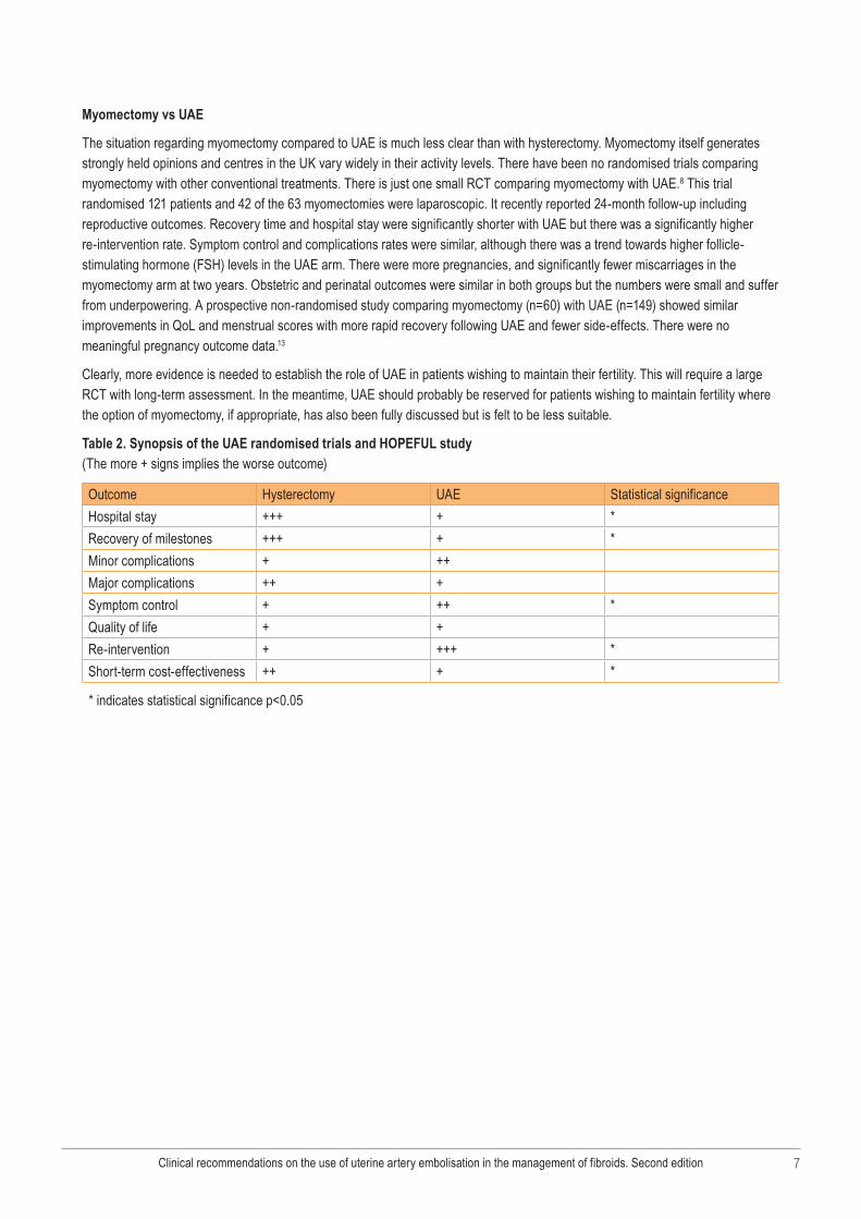
Myomectomy vs UAE
The situation regarding myomectomy compared to UAE is much less clear than with hysterectomy. Myomectomy itself generates strongly held opinions and centres in the UK vary widely in their activity levels. There have been no randomised trials comparing myomectomy with other conventional treatments. There is just one small RCT comparing myomectomy with UAE.8 This trial randomised 121 patients and 42 of the 63 myomectomies were laparoscopic. It recently reported 24-month follow-up including reproductive outcomes. Recovery time and hospital stay were significantly shorter with UAE but there was a significantly higher re-intervention rate. Symptom control and complications rates were similar, although there was a trend towards higher follicle-stimulating hormone (FSH) levels in the UAE arm. There were more pregnancies, and significantly fewer miscarriages in the myomectomy arm at two years. Obstetric and perinatal outcomes were similar in both groups but the numbers were small and suffer from underpowering. A prospective non-randomised study comparing myomectomy (n=60) with UAE (n=149) showed similar improvements in QoL and menstrual scores with more rapid recovery following UAE and fewer side-effects. There were no meaningful pregnancy outcome data.13
Clearly, more evidence is needed to establish the role of UAE in patients wishing to maintain their fertility. This will require a large RCT with long-term assessment. In the meantime, UAE should probably be reserved for patients wishing to maintain fertility where the option of myomectomy, if appropriate, has also been fully discussed but is felt to be less suitable.
Table 2. Synopsis of the UAE randomised trials and HOPEFUL study (The more + signs implies the worse outcome)
Outcome Hysterectomy UAE Statistical significanceHospital stay +++ + *Recovery of milestones +++ + *Minor complications + ++Major complications ++ +Symptom control + ++ *Quality of life + +Re-intervention + +++ *Short-term cost-effectiveness ++ + *
* indicates statistical significance p<0.05

Clinical recommendations on the use of uterine artery embolisation in the management of fibroids. Second edition8
3 Indications for fibroid embolisation
Fibroids (leiomyomas) may be single or multiple, of varying size and occupy a variety of possible situations within the uterus. Many are asymptomatic. Gonadotrophin-releasing hormone (GnRH) analogues have been used to reduce the size of fibroids but such a reduction in size is temporary. Surgical treatment of fibroids has involved either open or laparoscopic myomectomy or hysterectomy or, in the case of submucous fibroids, hysteroscopic resection or thermal ablative techniques in some cases.
UAE for fibroids offers an alternative method of treatment that allows conservation of the uterus. Early and mid-term results are encouraging10 and longer term data is awaited. The safety and efficacy of the technique has been established.6,10,14 The main indication for this technique is women with symptomatic fibroids who require treatment. The NICE heavy menstrual bleeding guidance recommends UAE as a treatment option for patients with symptomatic uterine fibroids.2 Symptoms might include menorrhagia, dysmenorrhoea, dyspareunia and other pressure effects of the fibroid on the urinary or gastrointestinal tract. Suitability of different types of fibroid (position and size) for treatment by UAE is discussed under contraindications. Patients should be made aware of the outcome of the treatment with chances of success and complications. Around 80–90% of patients will be asymptomatic or have significantly improved symptoms at one year with an associated 40–70% reduction in fibroid volume. Women of child-bearing age who wish, or who may wish to become pregnant subsequently, present particular problems. However, there are several case series in the literature documenting successful pregnancies post-UAE,10,14,15 although the miscarriage rate may be higher than in an age-matched population without fibroids. Women who have a medical condition contraindicating surgery, who are unwilling to receive a blood transfusion (such as Jehovah’s Witnesses) or who have had previous unsuccessful surgery for fibroids may also find UAE an acceptable method of treatment. However, patients must recognise that complications of the procedure may lead to hysterectomy in up to 2.9% of cases.14 Premature ovarian failure may occur in 1–2%, although this is largely confined to women over 45 or those approaching the menopause.14 Hysterectomy, even in the presence of ovarian conservation, may also result in early ovarian failure.
Accurate pretreatment diagnosis is essential and should involve the gynaecologist. Appropriate steps should be taken to ensure that bleeding irregularities are not due to pathology other than the fibroids. Magnetic resonance imaging (MRI) is superior to ultrasound in diagnosing fibroids and the technique is more likely to recognise adenomyosis if present.16 There is also evidence that MRI alters management in as many as 22%, with 19% not undergoing UAE.17 Although UAE in the presence of adenomyosis is less efficacious,18 it may still be considered when fibroids and adenomyosis co-exist.
It must be remembered that the procedure will be undertaken in the absence of a tissue diagnosis and the radiologist undertaking the procedure should be watchful for an imaging abnormality with appearances that are not typical of fibroids. There has been at least one case of an apparent fibroid failing to respond to UAE, which proved to be a sarcoma. However, it is recognised that despite the imaging modalities available, differentiating between uterine sarcoma and benign myoma remains suboptimal. It seems that unless the fibroid shows signs of invasion through its capsule, or if there are signs of lymphadenopathy, the value of MRI in looking for sarcomatous changes of fibroids cannot be relied on.19 It is essential that the referring gynaecologist ensures that an up-to-date assessment of the patient is undertaken before the procedure is performed. GnRH analogues should not have been given within the preceding two months prior to the procedure.
The average fibroid shrinkage is very variable but may be as much as 60% at six months, which is maintained in the longer term with further shrinkage up to one year and 100% if the fibroid is expelled. The need for hysterectomy and/or repeat UAE for treatment failure is around 10% at one year, rising to 20–25% within five years.6,10,14

Clinical recommendations on the use of uterine artery embolisation in the management of fibroids. Second edition 9
4 Contraindications
There are very few contraindications to UAE for symptomatic fibroids. The following would be absolute contraindications to performing the procedure.n Any evidence of current or recent infection in the genital tract.n Where a patient would refuse a hysterectomy under any circumstances for social or cultural reasons. In this circumstance, it
would seem sensible to adopt the same guideline that gynaecologists adhere to20 and not subject the patient to UAE. Hysterectomy after UAE is an occasional outcome of all level 1–3 studies.4–7,10,14
n Serious doubt exists as to the diagnosis due to clinical factors or inadequate imaging.With respect to relative contraindications, it is believed that narrow-stalked, pedunculated subserous fibroids might detach and cause significant complications post-embolisation. Though there are no reports as such, this may be because the general rule has been followed and the narrow-stalked subserosal fibroid should remain a relative contraindication.
It has been suggested that the outcome from embolising small fibroids is better than for large fibroids;14 however, most evidence suggests that complication rates are similar. Therefore, large fibroids should not, at this time, be considered a contraindication. However, if a large fibroid is not associated with symptoms then considerable caution must be exercised since a volume reduction might not be sufficient to satisfy patient expectations.

Clinical recommendations on the use of uterine artery embolisation in the management of fibroids. Second edition10
5 Infertile patients and those contemplating a subsequent pregnancy
There is a considerable body of literature supporting the contention that uterine fibroids are a cause of subfertility. Intracavity and intramural fibroids may act primarily by a mechanical effect leading to cavity distortion, but data derived largely from in vitro fertilisation (IVF) studies further suggest that fibroids may exert a negative effect on fertility, even if the cavity appears normal at hysteroscopy, possibly due to an effect on uterine blood flow, impaired embryo implantation or abnormal sperm migration resulting from a hostile environment created by the fibroids.21
However, many women with relatively large fibroids conceive without difficulty. Problems may arise in pregnancy, with reports of an increased risk of first and second trimester miscarriage and difficulties due to fibroid degeneration leading to pain. Premature labour can occur in some cases, as can mechanical complications in labour and postpartum haemorrhage. Effective management of the patient with fibroids who wishes to conceive cannot as yet be determined by an evidence-based approach using randomised controlled trials. However, it is clear that subfertility resulting from fibroids is not absolute and many patients will conceive without intervention. It is therefore sensible to ensure the following. n Other causes of subfertility have been sought and, if found, treated appropriately.n In the absence of other causes of subfertility, the couple is advised to try naturally for pregnancy for at least two years, unless
the woman is 34 years of age or over.n At this time, more rapid intervention becomes advisable, given the negative impact of increasing female age on likelihood of
pregnancy, both naturally and after IVF and embryo transfer (IVF-ET).n If a fertility problem seems likely to be the result of uterine fibroids, a management plan should be devised after careful
discussion with the couple concerned. n Intracavity and submucosal fibroids (when the major portion of the fibroid is projecting into the uterine cavity), depending on
size, may be amenable to hysteroscopic resection, which should be carried out in a specialist centre with experience of treating this condition. Patients should be warned of the possibility of uterine perforation and concomitant effects on fertility and pregnancy.
n Myomectomy remains a sensible option for some women wishing pregnancy. This is most appropriate in patients with a large single fibroid or a small number of fibroids that are easily accessible (such as serosal or intramural). It should be performed in a specialist centre by an experienced surgeon. Laparoscopic myomectomy is a suitable alternative to laparotomy in selected patients and expert hands. The patient should appreciate the small but real risk of excessive haemorrhage during surgery, which might necessitate hysterectomy. This risk might be increased in the presence of very large fibroids.
n Fibroid embolisation carries an obvious appeal for women with large or multiple fibroids who are either subfertile or who repeatedly miscarry. Treatment should reduce fibroid volume and may therefore improve the chance of pregnancy with a successful outcome. There is evidence from several studies that successful pregnancy is possible after UAE but the caesarean section and miscarriage rates may be higher than in an age-matched population of women without fibroids.10,22,23 This may also be true when pregnancy follows myomectomy. Patients must be made aware of the potential complications of the procedure, including the risk of ovarian damage. Theoretically, there may be adverse effects on placental blood supply, and a risk of uterine rupture in pregnancy but there is no hard evidence that these are risks in practice.
The evidence suggests that women who desire pregnancy but experience subfertility or recurrent miscarriage due to fibroids who are unsuitable for hysteroscopic resection or myomectomy, or in whom myomectomy has failed can be offered UAE as a safe effective alternative. What effect UAE has on the success or otherwise of IVF has not been determined.

Clinical recommendations on the use of uterine artery embolisation in the management of fibroids. Second edition 11
6 Counselling, consent and communication
Recent NICE guidance11 suggests women with symptomatic fibroids should be offered UAE as part of their treatment options. The gynaecologist should counsel the patient about the available options and if the patient wishes to consider UAE, she should be referred to an experienced vascular interventional radiologist.
It is recommended that the radiologist should see the patient in the outpatient setting. Notes, letters and previous imaging should be available. The patient should be counselled about the following.n UAE is not as successful as hysterectomy in relieving symptoms, although around 80% of patients will have either complete or
significant relief of symptoms.6,10,14
n A full description of the procedure and early post-procedural complications, including pain and post-embolisation syndrome.n An indication of how long they might spend in hospital and how long the recovery time is likely to be.n A full discussion of complications and their incidence. This should include:
─ Minor complications such as puncture site bruising or, self-limiting discharge which can occur in 20–30% of patients5,6,10
─ Post-embolisation syndrome (see page 14)─ Passage of fibroid material which may require assistance in 6% of patients─ Permanent amenorrhea which overall occurs in between 1.5% and 7% of patients,6,10,14 but that the majority of patients
who have early menopause are over 45 years old─ Urgent hysterectomy due to infection in 2–3%,10,14 which may occur de novo several months after UAE.
n Patients should be informed that they will have a chance of requiring some further treatment in future and that the younger they are, the greater the chance there is of this happening. The risk is 25% below 40 years and 10% between 40 and 50 years old.6,10 Further treatment might include repeat UAE, exploration of uterine cavity, myomectomy or hysterectomy.
n Those patients who desire pregnancy or wish to maintain fertility should be told that successful pregnancy is possible, but that there is a miscarriage rate which may be higher than in an age-matched population who do not have fibroids.8,10,14 They should also be told there is an increased risk of caesarean section.
Time should be allowed for due consideration by the patient. Providing all the above is discussed with the patient, the standard hospital consent form is sufficient. Patient information leaflets should be available which will be influenced by local practice. General practitioners must be kept aware of the details of the patient’s decision, the procedure date and outcome and follow-up arrangements. General practitioners should also be made aware of the possible complications, such as post-embolisation syndrome, the passage of pieces of fibroid, and the management plan (see Appendix 1, page 21).

Clinical recommendations on the use of uterine artery embolisation in the management of fibroids. Second edition12
7 Pretreatment assessment and prophylactic measures
Every effort should be made to avoid fibroid embolisation procedures in the presence of an early pregnancy. Embolisation can be carried out at any stage of the menstrual cycle if the radiologist is confident that the patient has taken adequate contraceptive precautions. However, treatment should only be given in the early to mid-follicular phase of the cycle (equivalent to the ‘ten-day rule’) if adequate contraception has not been used. Patients who present for the procedure later in the cycle should be offered a repeat appointment after a menstrual period and this should be documented in the hospital notes. Alternatively, a pregnancy test should be performed and only proceed if this is negative.
Particular consideration has been given to the risk of infection complicating the procedure. Pretreatment swabbing of the genital tract is done in some centres but its value is uncertain. An intrauterine contraceptive device, if used, should be removed prior to the procedure. It is appreciated that the avascularity induced by arterial embolisation leads to tissue necrosis, which might predispose some patients to a substantial risk of anaerobic and bacterial infection. Likely causal organisms include bacteroides, Escherichia coli and streptococci. Published work suggests that the procedure is associated with a post-treatment infection rate of 2%, even though prophylactic antibiotic therapy has sometimes been used. The data to suggest that perioperative antibiotics are useful is limited.10 Evidence from the use of prophylactic antibiotic therapy in association with vaginal hysterectomy,24 caesarean section25 and colorectal surgery,26 however, makes us believe that single-dose prophylactic antibiotic therapy is reasonable, using a drug combination such as metronidazole with a cephalosporin, a quinolone drug such as ciprofloxacin, gentamicin or amoxicillin. It is appreciated that the time of greatest risk of infection may well be several months after the primary procedure. The use of antibiotics is at the discretion of local hospital or clinician policy. Normal guidance on the use of prophylactic heparin should apply if a patient appears to be at increased risk of thromboembolic disease.27
Guidance on the resumption of sexual activity and the use of tampons post-procedure is not evidence based and again will depend on local practice.
Clinical recommendations on the use of uterine artery embolisation in the management of fibroids. Second edition 13
8 The procedure
The technical details of UAE have developed over the last 12 years and continue to mature. Areas of continuing uncertainty include the type of embolic agent and the so-called ‘embolic endpoint’. The objective of UAE is to completely infarct all the fibroid tissue while preserving both the uterus, ovaries and surrounding pelvic tissues. The procedure will usually be performed by a consultant interventional radiologist with experience in a variety of endovascular procedures and particularly embolisation techniques. It should not be undertaken by a trainee without appropriate supervision. An up-to-date dedicated fixed C-arm fluoroscopic unit is required which will include ‘road mapping’ and dose reduction options such as pulsed fluoroscopy. An appropriately trained radiographer is essential to minimise the radiation burden. Ideally the radiation doses should be audited.
Patients are normally admitted to hospital on the day of the procedure. Although admission to a gynaecological unit is ideal, other models exist (such as a vascular surgical unit) provided protocols are in place clearly identifying named clinical responsibility. The patient should ideally be consented by the operator performing the procedure on the ward prior to the procedure. Pain is an expected initial feature of successful UAE and should be managed proactively. An agreed pain protocol should be in place and adhered to. Analgesia should ideally be instituted prior to the embolisation procedure commencing.
Before the procedure, intravenous cannulation should be established, allowing intravenous conscious sedation and analgesia to be given during and following the procedure. Appropriate monitoring is mandatory, including pulse oximetry, and other vital signs. Oxygen therapy and other resuscitation facilities should be available and the radiologist and other support staff must have had appropriate resuscitation training. It is vital that a dedicated member of staff (usually a nurse) is constantly present to monitor the patient’s status and administer the appropriate medication.
Most operators use a standard percutaneous right-sided femoral artery access, although some find it easier to catheterise both uterine arteries using a bilateral femoral artery approach. The catheter (4 or 5 Fr although some operators use microcatheters routinely to reduce spasm and achieve a more effective flow-directed embolisation) is manipulated under fluoroscopic guidance into the uterine artery via the anterior division of the internal iliac artery. The opposite uterine artery is then embolised in a similar fashion.
There are a variety of embolic agents available and little evidence to support one over another. Two RCTs have been published. The first compared non-spherical polyvinyl alcohol (PVA) with ‘embospheres’ and found no difference between the products. The second compared the newer spherical (calibrated) PVA with ‘embospheres’ but was stopped after an interim analysis showed superiority of ‘embospheres’ over spherical PVA. Most operators use a non-absorbable agent such as PVA, although the newer coated ‘gel spheres’ such as embospheres and other PVA products (Bead block) are gaining in popularity. Gelfoam can also be used, which is much cheaper and a temporary embolic agent. All these agents are also available in a variety of sizes and in general most operators use within the range 300–750 microns with a tendency towards the larger sizes with the newer gel agents. The ‘embolisation endpoint’ remains controversial and agent-specific, although most operators will embolise to complete stasis in the uterine artery. The catheter is then withdrawn and manual compression applied to the femoral artery for 5–10 minutes. Some centres use vascular closure devices for haemostasis. This has the advantage of allowing the patient to adopt a comfortable position in the bed at an early stage. The total procedure time is in the range of 30–90 minutes and routine placement of a bladder catheter is unnecessary and a potential source of infection.
Some advocate imaging the ovarian arteries at the time of the procedure, although embolisation of these vessels would not normally be undertaken at the time of the first embolisation and never without a full frank discussion with the patient in advance warning of the risk of ovarian damage.

Clinical recommendations on the use of uterine artery embolisation in the management of fibroids. Second edition14
9 Complications
Complications related to UAE are best divided as below:n Immediate – occurring during the procedure n Early – within the first 30 days n Late – beyond the first 30 days.
Immediate (peri-procedural)
As with any arteriographic procedure, local complications such as groin haematoma, arterial thrombosis, dissection and pseudoaneurysm can occur. In general, women undergoing UAE have a low incidence of underlying vascular disease and consequently the above complications are rare. In addition, the catheter size is small rarely exceeding 5F.
Significant reactions to iodinated contrast media are rare, but a severe known allergy would be a recognised contraindication to UAE.
Spasm in the uterine artery can be caused by excessive catheter/wire manipulation. It can be minimised by using coaxial catheters and careful technique. It may be reversed by administering vasodilators through the catheter but if persistent may lead to incomplete embolisation. Non-target embolisation relates to particles reaching other vascular beds. This should not occur with good technique but the presence of ovarian-uterine anastomoses can compromise the ovary and may not always be obvious on imaging.
Early (30 days)
Post-embolisation syndrome
This complex consists of pain, nausea, fever and ‘flu-like’ symptoms with raised inflammatory markers and white cell count. It is frequent if not universal to some extent and is usually self-limiting. It is managed by analgesics and anti-inflammatory medications which are given to all patients post-UAE. Prolonged and deteriorating symptoms should raise the suspicion of infection with which it shares many features. Debate continues whether this entity should be considered a complication or an unavoidable component of UAE (and indeed any embolisation procedure) (see literature). Readmission may be required in a minority (3–5%) for parenteral analgesic control and fluid balance control.
Other complications such as urinary tract infection and deep venous thrombosis are very rare.
Late (beyond 30 days)
This is the time period when most recognised complications occur and can occasionally extend beyond the first 12 months. Many of the outcomes listed below occur earlier than 30 days but usually resolve quickly without causing the patient too much alarm.n Vaginal discharge is relatively common (16% at 12 months)4 but is almost always self-limiting. If the discharge is foul smelling
and purulent then infection is likely and should be treated. Persistent discharge with pain raises the suspicion of fibroid expulsion.
n Fibroid expulsion occurs in up to 10% and is more frequent with submucosal fibroids. Management is usually expectant although operative intervention may be required to remove the sloughed material if the cervix has not dilated.
n Amenorrhoea. There is little doubt that UAE can interfere at least temporarily with ovarian blood supply. The US registry quotes a 7.3% incidence of procedural-related amenorrhoea at 12 months.4 The incidence reaches 25% in those aged over 45 years. Studies have been confounded by the natural background incidence of ovarian failure in this group. A recent subgroup analysis from the EMMY trial indicated both UAE and hysterectomy affect ovarian reserve.28
n Sexual function. There has been very little work on this but theoretically sexual function could be impaired through the interruption of blood supply to the clitoris, cervix and uterus. The US registry reported 12% at one year experiencing unwanted changes in sexual function.4
n Infection: endometritis occurs in 0.5% of cases, is associated with fibroid expulsion and usually responds well to antibiotics. Anaerobic organisms are common and dual antibiotic therapy is recommended. Admission for parenteral antibiotics and IV fluids is advised and patients should be assessed by both a gynaecologist and radiologist. Rarely, infection can progress especially if untreated and lead to septicaemia and occasionally multi-organ failure. Emergency hysterectomy may occasionally be indicated for overwhelming sepsis as a life-saving manoeuvre. This may be a technically difficult procedure

Clinical recommendations on the use of uterine artery embolisation in the management of fibroids. Second edition 15
and may need a very experienced gynaecological surgeon. There has been one fatal case of infection reported in the world literature. Hysterectomy has also been performed for infection starting over one year after UAE but without major sepsis.
It should be understood that the temporal profile for complications arising as a result of UAE is very different from those undergoing either hysterectomy or myomectomy. Whereas the first 30 postoperative days capture almost all surgical complications, this is not the case with UAE where complications can occur up to four years later. Patients, medical and nursing staff need to be aware of this and post-UAE patient information sheets are suggested.
MRI imaging should be considered early in the assessment of complications such as sepsis and expulsion. Valuable information regarding tissue viability, fluid collections and partially expelled fragments can be gleaned from MRI.
Clinical recommendations on the use of uterine artery embolisation in the management of fibroids. Second edition16
10 The individual’s responsibility in clinical practice
The primary decision to treat should follow discussion between the patient and gynaecologist. Once the woman has been referred for consideration of embolisation, the method of the procedure is in the hands of the radiologist. During the procedure, the radiologist holds responsibility. Having completed the procedure, the patient is jointly in the care of the radiologist and the gynaecologist. The single most important element in the continuing care of the patient is that the patient should have rapid access to a named individual who will be in a position to allay any anxieties relating to expected sequelae and also to act immediately if there is any question of the development of a problem which could be serious.
We concur with the view of the Academy of Medical Royal Colleges, namely that, ‘Patients admitted to a hospital bed should be under the care of a named consultant at all times during their hospital stay. This principle operates even when more than one consultant, often from different disciplines, provides shared care for the patient; in these circumstances the lead consultant should be clearly identified in the case notes. When the patient requires an invasive procedure such as an interventional radiological technique, an endoscopic procedure or any form of surgery it may be appropriate, depending on the circumstances and the complexity of the procedure, to transfer, temporarily but formally, the responsibility for the patient to the individual specialist performing the procedure. The period of transfer and the arrangements for emergency cover must be formally agreed either by specific instructions in the patient’s hospital notes or by a general protocol. In the case of an interhospital transfer, the referring hospital consultant is responsible for the care of the patient until a formal handover takes place’.29
Clinical radiologists may take principal medical responsibility for the care of a patient for part or all of the hospital stay on the same basis as any other registered medical practitioners, provided that their skills, training and available facilities are sufficient to ensure appropriate care until responsibility is reassumed by the referring medical practitioner.
Where radiologists are working in a hospital without a gynaecological unit, agreed procedures for communication between clinicians allowing rapid recourse to the appropriate specialist are essential. It is anticipated that, normally, patients with complications will be admitted under the care of the gynaecologist.

Clinical recommendations on the use of uterine artery embolisation in the management of fibroids. Second edition 17
11 Follow-up
Patients should have rapid access to a clinician if required. This necessitates GPs being educated and informed about UAE. Routine clinical follow-up is advised; for example, at one, six and 12 months. Imaging is mandatory if symptoms persist or recur. Any complication or suggestion of early infection should be referred early for a gynaecological opinion (Appendix 1, page 21). A clear protocol must be available regarding whose responsibility it is to follow up the patient (gynaecologist or radiologist).
Clinical recommendations on the use of uterine artery embolisation in the management of fibroids. Second edition18
12 Suggested areas for further study
Studies to date have proven the safety and efficacy of uterine artery embolisation in the short and medium term as an effective treatment for symptomatic uterine fibroids. Further research is required into certain technical aspects of the procedure and in fertility.
These should include:n Comparison with myomectomy in terms of symptom relief and pregnancy outcome. It is still unclear what advice to give
patients regarding future pregnancy. In the published series to date, there are many successful pregnancies but the same literature would suggest increased rates of miscarriage, caesarean section and pre-term delivery compared to age-matched populations without fibroids. A randomised trial comparing uterine artery embolisation and myomectomy would be valuable with pregnancy outcome as the primary endpoint. Such a trial needs to be multicentred and properly funded. In addition, further research into the effect of uterine artery embolisation on ovarian function and long-term fertility is required. This could include investigation into the effects of embolisation on ovarian function
n Optimisation of embolisation technique, including identification of the ideal embolic agentn Research into endometrial pathophysiology with regard to the mechanism of successn To increase data on the efficacy of prophylactic antibioticsn To further stratify patient selection on basis of fibroid position, size and number as an indicator of successn Identification of a need to screen for pretreatment infection n In addition, NICE has called for further research on the psychosexual impact of uterine artery embolisation and myomectomyn Ultrasound assessment of ovarian volume and blood flow pre- and at stages post-embolisation. The age at menopause in this
group should be establishedn In the longer term, a study of the outcome of pregnancies following treatment including surveillance of childhood growth and
development would be valuable.

Clinical recommendations on the use of uterine artery embolisation in the management of fibroids. Second edition 19
1. Ravina JH, Herbreteau D, Ciraru-Vigneron N et al. Arterial embolisation to treat uterine myomata. Lancet 1995; 346: 671–672.
2. National Institute for Health and Clinical Excellence. Heavy menstrual bleeding. (NICE guideline CG44). London: NICE, 2007. http://guidance.nice.org.uk/CG44/guidance/pdf/English (last accessed 3/2/09)
3. Committee on Gynecologic Practice, American College of Obstetricians and Gynecologists. ACOG Committee Opinion: Uterine artery embolisation. Obstet Gynecol 2004; 103: 403–404.
4. Worthington-Kirsch R, Spies JB, Myers ER et al. The fibroid registry for outcomes data (FIBROID) for uterine embolization: short term outcomes. Obstet Gynecol 2005; 106(1): 52–59.
5. Volkers NA, Hehenkamp WJK, Birnie E, Ankum WM, Reekers JA. Uterine artery embolization versus hysterectomy in the treatment of symptomatic uterine fibroids: 2 years outcome from the randomized EMMY trial. Am J Obstet Gynecol 2007; 196: 519.e1–11.
6. Edwards RD, Moss JG, Lumsden MA, Wu O, Murray LS, Twaddle S, Murray GD. Committee of the Randomised Trial of Embolisation versus Surgical Treatment of Fibroids. Uterine artery embolisation versus surgery for symptomatic fibroids. N Engl J Med 2007; 356: 360–370.
7. Pinto I, Chimeno P, Romo A, Paul L, Haya J, de la Cal M, Bajo J. Uterine fibroids: Uterine artery embolization versus abdominal hysterectomy for treatment. A prospective, randomized, and controlled clinical trial. Radiology 2003; 226: 425–431.
8. Mara M, Maskova J, Fucikova Z, Kuzel D, Belsan T, Sosna O. Midterm clinical and first reproductive results of a randomized controlled trial comparing uterine fibroid embolisation and myomectomy. Cardiovasc Intervent Radiol 2008; 31: 73–85.
9. Gupta JK, Sinha AS, Lumsden MA, Hickey M. Uterine artery embolisation for symptomatic uterine fibroids. Cochrane Database of Systematic Reviews 2006, Issue 1. Art. No CD005073.
10 Dutton S, Hirst A, McPherson K, Nicholson T, Maresh M. A UK multicentre retrospective cohort study comparing hysterectomy and uterine artery embolisation for the treatment of symptomatic uterine fibroids (HOPEFUL study): main results on medium-term safety and efficacy. BJOG 2007; 114: 1340–1351.
11 National Institute for Clinical Excellence. Uterine artery embolisation for the treatment of fibroids. Interventional Procedure Guidance 94. London: NICE, 2004. http://www.nice.org.uk/nicemedia/pdf/ip/IPG094guidance.pdf (last accessed 3/2/09)
12. Wu O, Briggs A, Dutton S, Hirst A, Maresh M, Nicholson A, McPherson K. Uterine artery embolisation or hysterectomy for the treatment of symptomatic uterine fibroids: a cost-utility analysis of the HOPEFUL study. BJOG 2007; 114: 1352–1362.
13. Goodwin SC, Bradley LD, Lipman JC et al. UAE versus myomectomy study group. Fertil Steril 2006; 85: 14–21.
14. Spies JB, Myers ER, Worthington-Kirsch R, Mulgund J, Goodwin S, Mauro M: Fibroid Registry Investigators. The FIBROID registry: symptom and quality-of-life status 1 year after therapy. Obstet Gynecol 2005; 106: 1309–1318.
15. Walker WJ, McDowell SJ. Pregnancy after uterine artery embolization for leiomyomata: a series of 56 completed pregnancies. Am J Obstet Gynecol 2006; 195: 1266–1271.
16. Cura M, Cura A, Bugnone A. Role of magnetic resonance imaging in patient selection for uterine artery embolization. Acta Radiologica 2006; 47: 1105–1114.
17. Omary R, Vasireddy S, Chrisman HB et al. The effects of pelvic MR imaging on the diagnosis and treatment of women with presumed symptomatic uterine fibroids. J Vasc Interv Radiol 2002; 13: 1149–1153.
18. Lohle PN, De Vries J, Klazen CA et al. Uterine artery embolization for symptomatic adenomyosis with or without uterine leiomyomas with the use of calibrated tris-acryl gelatin microspheres: midterm clinical and MR imaging follow-up. J Vasc Interv Radiol 2007; 18(7): 835–841.
19 Buzaglo K, Bruchim I, Lau SK. Sarcoma post-embolization for presumed uterine fibroids. Gynecologic Oncology 2008; 108: 244–247.
20. Wallach EE. Myomectomy. In: Thompson JD, Rock JA (eds). Te Lind’s Operative Gynaecology, 7th edition. Philadelphia: JB Lippincott; 1992: 47–62.
21. Templeton A, Cooke I, O’Brien PMS (eds). Evidence-based Fertility Treatment. London: RCOG Press, 1998.
22. Walker WJ, Pelage JP. Uterine artery embolisation for symptomatic fibroids: clinical results in 400 women with imaging follow up. BJOG 2002; 109: 1262–1272.
References

Clinical recommendations on the use of uterine artery embolisation in the management of fibroids. Second edition20
23. McLucas B, Goodwin S, Adler L, Rappaport A, Reed R, Perrella R. Pregnancy following uterine fibroid embolization. Int J Gynaecol Obstet 2001; 74: 1–7.
24. Lett JW, Ansbacher R, Davison BL, Otterson WN. Prophylactic antibiotics for women undergoing vaginal hysterectomy. J Reprod Med 1977; 19: 51–54.
25. Smaill F, Hofmeyr GJ. Antibiotic prophylaxis for caesarean section. Cochrane Database Syst Rev 2000; Issue 1.
26. Royal College of Surgeons of England. Guidelines for the Management of Colorectal Cancer. From an Expert Advisory Group composed of the Royal College of Surgeons of England, the Association of Coloproctology of Great Britain and Ireland and representatives from other main bodies. London: RCSE, 1996.
27. Royal College of Obstetricians and Gynaecologists. Report of the RCOG Working Party on Prophylaxis against Thromboembolism in Gynaecology and Obstetrics. London: RCOG Press, 1995.
28. Hehenkamp WJK, Volkers NA, Broekmans FJM et al. Loss of ovarian reserve after uterine artery embolization: a randomized comparison with hysterectomy. Hum Reprod 2007; 22: 1996–2005.
29. Academy of Medical Royal Colleges Statement. Responsibility for the Consultant Care of In-Patients Undergoing Invasive Procedures. Statement from Minutes, item 3.3.11. London: Academy of Medical Royal Colleges, 10 April 2000.

Clinical recommendations on the use of uterine artery embolisation in the management of fibroids. Second edition 21
Appendix 1. Post-procedure care and management of complications
Post-embolisation syndrome is not uncommon in the first two weeks after UAE. It is associated with flu-like symptoms, a mild temperature, and lower abdominal pain. The white count is usually normal or minimally elevated.
Note: It may be difficult to distinguish post-UAE syndrome, from infection. Pyrexia, malaise, lower abdominal pain, and a PV discharge, may be associated with pyometra.
All patients presenting with concerns after UAE need to be investigated in a gynaecology unit to ensure that there is no serious complication. Complications can occur at any time from a few hours to more than 12 months after treatment.
PATIENTS ARE TREATED EMPIRICALLY.
Investigations1. TPR and B/P2. Vaginal examination + high vaginal swabs3. Urinalysis + MSSU4. Bloods: FBC / C-RP / CK / U/Es. If pyrexial – blood cultures.5. Transabdominal USS / Transvaginal USS / +/- MRI. (MRI is preferred radiological investigation; transfer to central unit if MRI is
not available on site.)6. CXR
Symptoms Treatment1. Pyrexia: fever / rigours. General feelings of malaise. If pyrexial with raised WCC, admit for IV antibiotics; eg,
metronidazole + cefuroxime. Rarely, hysterectomy may be necessary.
2. Pain: abdominal +/- uterine tenderness. Use NSAIDs for analgesia to assist with reduction of inflammation, +/- admit.
3. Vaginal discharge +/- offensive discharge. A brownish/ pink discharge is not uncommon for several weeks post UAE. If the discharge is causing vulval irritation or it is offensive, then ? infection.
If patient feels well – no treatment.
Vaginal infection – treat appropriately. Repeat swabs to ensure clear of infection.
If the PV discharge becomes distressingly heavy or persistent, an evacuation of the uterus may be necessary.
4. Passing of fibroid tissue PV may occur, in about 10% of patients after treatment. Passing of a complete fibroid is rarer; associated with fresh bleeding and acute sustained abdominal pain, which does not resolve with simple analgesics.
NSAIDs may need to be taken for several weeks.
Clear os of any debris. Admission for pain control may be necessary. Once fibroid passed, patients usually recuperate quickly.
5. Heavy PV loss / menorrhagia / anaemia. Patients may be at risk from anaemia for 2–3 months before UAE affects symptoms.
FeSO4 +/- blood transfusion.
If patient to be sent home, she must be advised to return immediately if there is any deterioration in her symptoms. Notify both consultant gynaecologist and consultant interventional radiologist. Arrange for follow up at gynaecology clinic.
Reproduced with the kind permission of Jon Moss.

Clinical recommendations on the use of uterine artery embolisation in the management of fibroids. Second edition22
The Working Party
Professor A Watkinson BSc MSc (OXON) FRCS FRCR (Chairman) Professor JG Moss FRCS FRCR Dr AA Nicholson BSc MSc FRCR Professor Mary Ann Lumsden MD FRCOG Mr Michael Maresh MD FRCOG Mr Sanjay Vyas MD FRCOG
With acknowledgments to members of the original working party 2000
Mr KR Peel FRCS (Edin) FRCOG (Chairman) Professor WL Ledger MA DPhil MRCOG Dr JF Reidy FRCP FRCR Professor CJG Sutton FRCOG
Approved by The Royal College of Radiologists: 20 June 2008
Approved by The Royal College of Obstetricians and Gynaecologists: 16 January 2009

The Royal College of Radiologists 38 Portland Place London W1B 1JQ
Tel +44 (0)20 7636 4432 | Fax +44 (0)20 7323 3100 | Email [email protected] | URL www.rcr.ac.uk |
A Charity registered with the Charity Commission No. 211540
Citation details:
The Royal College of Radiologists and the Royal College of Obstetricians and Gynaecologists. Clinical
recommendations on the use of uterine artery embolisation in the management of fibroids. Second
edition. London: The Royal College of Radiologists, 2009.
On publication, this document will be made available at www.rcr.ac.uk and www.rcog.org.uk
ISBN 978-1-905034-37-6 Ref No. BFCR(09)1 © The Royal College of Radiologists, April 2009
All rights reserved. No part of this publication may be reproduced, stored in a retrieval system or
transmitted in any form or by any means, electronic, mechanical, photocopying, recording or otherwise,
without the prior written permission of the Publisher (The Royal College of Radiologists).
For permission to reproduce any of the content contained herein, please email: [email protected]
This material has been produced by The Royal College of Radiologists (RCR) for use internally within
the National Health Service in the United Kingdom. It is provided for use by appropriately qualified
professionals, and the making of any decision regarding the applicability and suitability of the material in
any particular circumstance is subject to the user’s professional judgement.
While every reasonable care has been taken to ensure the accuracy of the material, RCR cannot accept
any responsibility for any action taken, or not taken, on the basis of it. As publisher, RCR shall not be
liable to any person for any loss or damage, which may arise from the use of any of the material. The
RCR does not exclude or limit liability for death or personal injury to the extent only that the same arises
as a result of the negligence of RCR, its employees, Officers, members and Fellows, or any other person
contributing to the formulation of the material.
Design by innov8 graphic design: www.innov8gd.com Printed by Gallpen Colour Print.