
CLINICAL PROFILE AND EVALUATION OF NEW ONSET SEIZURE …
105
CLINICAL PROFILE AND EVALUATION OF NEW ONSET SEIZURE IN ADULTS A Dissertation Submitted to THE TAMILNADU DR. M.G.R MEDICAL UNIVERSITY CHENNAI In Partial Fulfillment of the Regulations for the Award of the Degree of M.D. (GENERAL MEDICINE) - BRANCH – I GOVERNMENT KILPAUK MEDICAL COLLEGE CHENNAI April - 2014
Transcript of CLINICAL PROFILE AND EVALUATION OF NEW ONSET SEIZURE …

CLINICAL PROFILE AND EVALUATION OF NEW ONSET SEIZURE IN
ADULTS
A Dissertation Submitted to
THE TAMILNADU DR. M.G.R MEDICAL UNIVERSITY
CHENNAI
In Partial Fulfillment of the Regulations
for the Award of the Degree of
M.D. (GENERAL MEDICINE) - BRANCH – I
GOVERNMENT KILPAUK MEDICAL COLLEGE
CHENNAI
April - 2014

BONAFIDE CERTIFICATE
This is to certify that “CLINICAL PROFILE AND EVALUATION OF
NEW ONSET SEIZURE IN ADULTS” is a bonafide work performed by
Dr.DHANASEKAR.M., post graduate student, Department of Internal Medicine,
Kilpauk Medical College, Chennai-10, under my guidance and supervision in
fulfilment of regulations of the Tamil Nadu Dr. M.G.R Medical university for the
award of M.D. Degree Branch I (General Medicine) during the academic period
from May 2011 to April 2014.
Prof. Dr. N. Gunasekaran M.D., DTCD Prof. Dr.S.Ushalakshmi M.D.,FMMC., Medical Superintendent & Director INCD Professor and Unit chief,
Professor and HOD, Department of Medicine,
Department of Medicine Kilpauk Medical College,
KMC & GRH Chennai
Chennai.
Prof. P. Ramakrishnan M.D., D.L.O The DEAN
Govt.Kilpauk Medical College
Chennai - 600 010

DECLARATION
I solemnly declare that this dissertation “CLINICAL PROFILE AND
EVALUATION OF NEW ONSET SEIZURE IN ADULTS” was prepared by
me at Government Kilpauk Medical College and Hospital, Chennai, under the
guidance and supervision of Dr.S.Ushalakshmi M.D.,FMMC., Professor of
Internal Medicine, Government Kilpauk Medical College and Hospital, Chennai.
This dissertation is submitted to The Tamil Nadu Dr. M.G.R. Medical
University, Chennai in partial fulfilment of the University regulations for the
award of the degree of M.D. Branch I (General Medicine).
Place: Chennai (Dr.DHANASEKAR.M)
Date:

ACKNOWLEDGEMENT
At the outset, I would like to thank my beloved Dean, Kilpauk Medical
College Prof. Dr. P. Ramakrishnan, M.D., D.L.O., for his kind permission to
conduct the study in Kilpauk Medical College. I would like to express my special
thanks to medical superintendent Prof. Dr.Narayanababu.MD., for permitting to
conduct this study in Kilpauk Medical College Hospital.
It gives me immense pleasure to express my sincere and deep gratitude to
Prof. Dr. N. Gunasekaran M.D., DTCD., Medical Superintendent and Director
INCD, Professor and Head of the Department of Medicine, Kilpauk Medical
College, for rendering permission to do this dissertation.
I would like to thank wholeheartedly, Prof.Dr.S.Ushalakshmi
M.D.,FMMC., my Unit chief and Professor of Medicine for her encouragement
and guidance during the study.
With extreme gratitude, I express my indebtedness to Prof. Dr.Sarala
M.D., D.M., Former Professor and HOD, Department of Neurology, Kilpauk
Medical College Hospital for her continuous motivation, timely advice and
valuable criticism which enabled to complete the dissertation.

I also express my special thanks to Prof. Dr.G.Balan M.D.,
Prof. Dr.T. Ravindran M.D., DNB., Dip Diabetology and
Prof. D. Surendran M.D., I am extremely thankful to Assistant Professor of
Medicine, Dr.M.Bathragiri M.D.,and Dr.A.Marimuthu M.D., for their
assistance and guidance.
I would like to take this opportunity to show gratitude to my Father
Mr.K.Muthusamy, my Mother Smt.M.Shanthamani, my brother
Mr.M.Subramani and my dear brother and best friend Dr. S.Ganesh Aravind,
for their never ending support in finishing this thesis.
I would always remember with extreme sense of thankfulness for the
co-operation and criticism shown by my fellow post graduate colleague and
friends.I pray Almighty God to give me strength to achieve in all my endeavours.
Finally, I wholeheartedly thank all my patients for their active co-operation in this
study, without which this would not have become a reality.

CONTENTS
S.No TOPIC Page No
1 Introduction 1
2 Review of literature 2
3 Aim of study 54
4 Materials and methods 54
5 Observation and Analysis 56
6 Discussion 71
7 Conclusion 76
8 Summary 77
9 APPENDIX
i Bibliography
ii Master chart
iii Ethical committee approval
certificate
iv Abbreviations


ABSTRACT
Background: Seizures beginning in adult life are likely to be an identifiable cause
as compared to those beginning in childhood which are more likely to be
idiopathic.
Objectives: To study the clinical profile and analyze the etiological agents of New
Onset Seizures.
Material and Methods: This Descriptive study done in the KMC hospital to
know the various etiologies in patients presented with new onset seizures.In these
cases history and clinical examination and special investigations like CT BRAIN,
MRI BRAIN,EEG, SEROLOGY, CSF ANALYSIS were done to find out the
etiology.
Results: Out of 100 patients 55% were males,45%were females with male to
female ratio of 1.2:1.Majority of males were in 2nd
decade and females were in 4th
decade.Patients age ranged from 18 yrs to 80 yrs,with the mean of 40.11years with
77% of the patients were in the below 50 yrs. Alcohol withdrawal was the leading
cause of seizures which account for 34% followed by idiopathic seizures
(29%),neuro infection (16%),CVA 12% and metabolic (9%).

Conclusion: Alcohol withdrawal is the most common cause of seizure in new
onset seizure patients who coming to KMCH.
Keywords: New onset seizure, Alcoholwithdrawal,Tuberculoma,
Neurocysticercosis,Meningitis, Metabolic seizure.

1
INTRODUCTION
Epilepsy describes a condition in which a person has recurrent seizures due to
chronic underlying process..
Epilepsy of late onset (Epilepsia tarda, late onset epilepsy) may be simply
defined as epilepsy beginning in adult life.
Epilepsy beginning in adult life is due to progressive brain disease as
compared to idiopathic epilepsy, which has, it’s onset in childhood or adolescence.
With proper history and clinical examination, analysis of etiology is made
with available investigations, the epilepsy can be treated accordingly thus reducing
the morbidity and mortality associated with it.
Hence, this study is aimed to evaluate the clinical profile and etiological
analysis of new onset epilepsy in adults of more than 18 years of age.

2
REVIEW OF LITERATURE
HISTORY OF EPILEPSY :
A seizure (from the Latin sacire, " to take possession of ") is a paroxysmal event
due to abnormal excessive or synchronous neuronal activity in the brain.
From the Greek word ―epilambanein ‖ the term ―epilepsy‖ comes which
means ―to seize‖ or ― to take hold of ‖.
Approximately 500 years before Hippocrates, Atreya, the father of Indian
Medicine, recognized that disturbance of mind is epilepsy, rather than an effect of
supernatural phenomena. Hippocrates, in his book ―on the scared disease‖noted,
―The brain is the seat of epilepsy as it is of every violent disease‖.
The idea that epilepsy was caused by demons, humors, and toxic substances
persisted through the middle ages.
The ideas of supernatural causation slowly died out only to be replaced by
another set of bizarre misnomers. Obstetrician to Queen Victoria, Sir Charles
Locock, Credited crowded teeth, menstruation and masturbation with causing

3
seizures. Dr.Locock pioneered the use of potassium bromide the first modern
antiepileptic drug treatment.He had found that women with ―hysterical‖seizures
associated with menstruation,was thought to curb the sexual desires.
In 1857 after the start of bromide treatments, modern epilepsy treatment
proceeded at a quick pace. In 1912 Phenobarbitone was introduced and 24 years
later to that, pioneer work on phenytoin was carried out. Drugs like
Carbamazepine, sodium valproate and vigabatrin followed in 1954, 1973 and 1990
respectively. Now people with seizures are confronted with an array of confusing
but it often effective, treatments.
We can look forward in future to a deeper understanding of alternative
treatments and advances in diagnosis and treatment through progress in surgery
,biochemistry,and possibly even neural grafts and molecular genetics(1)
.
EPIDEMIOLOGY OF EPILEPSY
PREVALENCE :
It is the proportion of people with seizure in given population at a
specified time. The worldwide prevalence of active seizure is between 4-10 per
1000 population(2)
. The prevalence rate in India is 5.59 per 1000.There is no
statistically different rates between women and men or urban and rural
residence(3)
.
4
INCIDENCE:
The number of new cases of seizure occurring during a given time interval,
usually one year, in a specified population is called as incidence. Incidence rate
varies from 38 to 49.3 per 1, 00,000 population per year from two community
based studies in India.(4)
Type of seizure pattern showed maximum number of cases belonged to
generalized seizures, which is different from western countries where partial
seizure is the commonest variety. (The proportion of generalized seizures and
partial seizures was 58.8% and 30.6% respectively)(5)
.
The prognosis for seizure control is good and over 70% will enter remission.
There is an increased risk of premature death particularly in patients with chronic
epilepsy(6)
.
DIAGNOSIS OF EPILEPSY
It is rare to observe a seizure directly at the first medical examination or at an
outpatient clinic. The confirmation and diagnosis of the seizure type usually based
on the history taken from the patients or caregivers. First we have to distinguish
epileptic seizures from the non- epileptic attacks, such as psychogenic seizures and
syncopal attacks. In distinguishing a syncopal attack from an epileptic seizure, we
must pay attention to the sensation of faintness or feeling of ―blackouts‖
immediately before loss of consciousness and the presence of provoking factors,

5
such as noxious stimuli, sudden unexpected pain or standing for a long time,. Next
we must ask the following questions directly to the patients or indirectly to the care
givers to make a precise seizure diagnosis including the aura, asymmetry of the
seizures, content, clouded consciousness, presence of automatism, deviation of the
head and eyes and a dystonic arm posture(7)
.
A fundamental principle is that seizures may be either focal or generalized.
Generalized seizures involve diffuse regions of the brain simultaneously. Focal
seizures are those in which the seizure activity is restricted to discrete areas of the
cerebral cortex. Generalized seizures may result from biochemical, cellular
abnormality or structural abnormalities that have a more widespread distribution. .

6
In contrast, focal seizures are usually associated with structural abnormalities of
the brain.
Focal Seizure
Focal seizures arise from a neuronal network either discretely localized
within one cerebral hemisphere or more broadly distributed but still within
the hemisphere.The new classification system, described it as focal seizures
with or without dyscognitive features.
Focal seizures can also evolve into generalized seizures. In the past this was
referred to as focal seizures with secondary generalization, but the newer system
relies on specific descriptions of the type of generalized seizures that evolve from
the focal seizure.
Focal Seizures Without Dyscognitive Features
Focal seizures can cause autonomic, motor, sensory or psychic symptoms
without impairment of cognition.The EEG recorded with scalp electrodes during
the seizure may show abnormal discharges in a very limited region over the
appropriate area of cerebral cortex if the seizure focus involves the cerebral
convexity. Seizure activity from deeper brain structures is often not recorded by
the standard EEG, it may require intracranial electrodes for its detection.

7
Three additional features of focal motor seizures are,
1. In some patients the abnormal motor movements may begin in a very
restricted region such as the fingers and gradually progress to include a
larger portion of the extremity. This phenomenon known as a "Jacksonian
march" which represents the spread of seizure activity over a progressively
larger region of motor cortex.
2. Patients may experience a localized paresis (Todd's paralysis) for minutes to
many hours in the involved region following the seizure.
3. In rare instances the seizure may continue for hours or days.It termed
epilepsia partialis continua, also it is often refractory to medical therapy.
Focal seizures may also manifest as changes in vision (flashing lights or
formed hallucinations), equilibrium (sensation of falling or vertigo), or autonomic
function (flushing, sweating, piloerection) somatic sensation (e.g., paresthesias).
Focal seizures arising from the temporal or frontal cortex may also cause
alterations in olfaction, hearing, or higher cortical function sensation of unusual,
intense odors (e.g., burning rubber or kerosene) or an epigastric sensation that rises
from the stomach or chest to the head. Patients describe odd, internal feelings such
as fear, detachment, a sense of impending change, depersonalization, déjá vu, or
illusions that objects are growing smaller (micropsia) or larger (macropsia). These
8
subjective, "internal" events that are not directly observable by someone else are
referred to as auras.
Focal Seizures with Dyscognitive Features
Focal seizures accompanied by a transient impairment of the patient's ability
to maintain normal contact with the environment. These patients unable to respond
appropriately to verbal or visual commands during the seizure and has impaired
awareness or recollection of the ictal phase. Seizures frequently begin with an aura
that is stereotypic for the patient. The start of the ictal phase is a sudden behavioral
arrest or motionless stare. The behavioral arrest is usually accompanied by
automatisms, which are involuntary, automatic behaviors such as chewing, lip
smacking, swallowing, or "picking" movements of the hands, or elaborate
behaviors such as a display of emotion or running. These patients typically
confused following the seizure, and the full recovery of consciousness may range
from seconds up to an hour. Immediately following the seizure may show
anterograde amnesia or, in cases involving the dominant hemisphere, a postictal
aphasia.
Evolution of Focal Seizures to Generalized Seizures: Focal seizures can spread
to involve both cerebral hemispheres and produce a generalized seizure, usually of
the tonic-clonic variety . This evolution is observed following focal seizures

9
arising from a focus in the frontal lobe, but may also be associated with focus
elsewhere in the brain. A focal seizure that evolves into a generalized seizure is
often difficult to distinguish from a primary generalized-onset tonic-clonic seizure.
Generalized Seizures
Generalized seizures are arise at some point in the brain but immediately and
rapidly that engage neuronal networks in both cerebral hemispheres..
Typical Absence Seizures
It characterized by sudden, brief lapses of conscious without loss of postural
control. It typically lasts for only seconds, consciousness returns as suddenly as it
was lost, and there is no postictal confusion. It usually accompanied by
subtle,bilateral motor signs such as chewing movements, rapid blinking of the
eyelids, or small-amplitude, clonic movements of the hands. In typical absence
seizures the onset usually in childhood (ages 4–8 years) or early adolescence and
are the main seizure type in 15–20% of children with epilepsy. Seizures can occur
100 of times per day, but the child may be unaware or unable to convey their
existence. Absence epilepsy is often unexplained "daydreaming" and a decline in
school performance recognized by a teacher. The EEG of typical absence seizures

10
is a generalized, symmetric, 3-Hz spike - and - wave discharges that begins and
ends suddenly, superimposed on a normal EEG background.
Atypical Absence Seizures
The loss of consciousness is usually of longer duration and slow in onset and
cessation.It also associated with motor signs that includes focal or lateralizing
features.EEG shows a generalized,2.5Hz slow spike-and-wave pattern.These are
usually associated with diffuse or multifocal structural abnormalities of the
brain.So associated with other signs of neurologic dysfunction such as mental
retardation.Atypical absence seizures are less responsive to anticonvulsants
compared to typical absence seizures.
Generalized, Tonic-Clonic Seizures
These are the main seizure type in 10% of all persons with epilepsy.Also a
common seizure type resulting from metabolic derangements. The seizure usually
begins abruptly without warning. The initial phase is usually tonic contraction of
muscles throughout the body. Tonic contraction of the muscles of expiration and
the larynx will produce a loud moan or "ictal cry." Contraction of the jaw muscles
may cause biting of the tongue. After 10–20 seconds, the seizure typically evolves
into the clonic phase, produced by the periods of muscle relaxation on the tonic

11
muscle contraction. It progressively increase until the end of the ictal phase. The
postictal phase is characterized by muscular flaccidity, unresponsiveness, and
excessive salivation. Bladder or bowel incontinence may occur. Patients gradually
regain consciousness over minutes to hours, and in this transition there is typically
a period of postictal confusion. The duration of unconsciousness in the postictal
phase can be extremely long in patients with prolonged seizures or underlying
CNS diseases such as alcoholic cerebral atrophy.The EEG during the tonic phase
shows a progressive increase in generalized low-voltage fast activity, followed by
high-amplitude, polyspike discharges. The high-amplitude activity is interrupted
by slow waves to create a spike-and-wave pattern in the clonic phase.
Atonic Seizure
These are characterized by sudden loss of postural muscle tone lasting 1–2
seconds. Brief impairment in Consciousness, but there is usually no postictal
confusion. The EEG shows brief, generalized spike-and-wave discharges followed
by diffuse slow waves that correlate with the loss of muscle tone.
Myoclonic Seizures
Myoclonus is a sudden and brief muscle contraction that may involve one
part of the body or the entire body. A common physiologic form of myoclonus is

12
the sudden jerking movement observed during asleep. Pathologic myoclonus is
commonly seen in association with anoxic brain injury, metabolic disorders and
degenerative CNS diseases. The EEG shows bilaterally synchronous spike-and-
wave discharges synchronized with the myoclonus
Juvenile Myoclonic Epilepsy
It is a generalized seizure disorder of unknown cause that occurs in early
adolescence.It is usually characterized by single or repetitive bilateral myoclonic

13
jerks. The myoclonic seizures can be provoked by sleep deprivation and most
frequent in the morning after awakening. Consciousness is preserved unless the
myoclonus is especially severe.
Lennox-Gastaut Syndrome
It occurs in children and is defined by the following triad: (1) multiple
seizure types (including atonic, generalized tonic-clonic and atypical absence
seizures); (2) impaired cognitive function in most but not all cases,(3) an EEG
showing slow (<3 Hz) spike-and-wave discharges.It is associated with CNS
disease or dysfunction due to developmental abnormalities, trauma, infection,
perinatal hypoxia/ischemia, and other acquired lesions. Many patients have a poor
prognosis due to the underlying CNS disease.
Mesial Temporal Lobe Epilepsy Syndrome
It is the most common syndrome associated with focal seizures with
dyscognitive. High-resolution MRI can detect hippocampal sclerosis that appears
to be essential in the pathophysiology of MTLE for many patients.

14

15
The Causes of Seizures and Epilepsy
Shift in the normal balance of excitation and inhibition within the CNS
causes seizures. Three clinical observations emphasize how a variety of factors
determine why certain conditions may cause seizures or epilepsy in a given patient.
1) The normal brain is capable of having a seizure under the appropriate
circumstances, and individuals vary in the susceptibility or threshold for seizures.
For eg, seizures may be induced by high fevers in children who are otherwise
normal and they may never develop other neuro logic problems, including
epilepsy.Febrile seizures occur only in a relatively small proportion of children.It
implies there are various underlying, endogenous factors which influence the
threshold for having a seizure.
2) There are varieties of conditions that have an extremely high likelihood of
resulting in a chronic seizure disorder. One of the examples of this is severe,
penetrating head injury, which is associated with upto a 45% risk of subsequent
epilepsy.A process is known as epileptogenesis which is, high propensity for
severe traumatic brain injury lead to epilepsy suggests that the injury results in a
long-lasting, pathologic changes in the CNS that transforms a presumably normal
neural network into a abnormally hyperexcitable. Other processes associated with

16
epileptogenesis include infections, stroke, and abnormalities of CNS
development.
3) Seizures are episodic. Patients with epilepsy have seizures
intermittently.Depending on the underlying cause, many patients are completely
normal for months to years between seizures. It shows there are important
precipitating or provocative factors that induce seizures in patients with epilepsy.

17
Causes According to Age

18
aIn benzodiazepine-dependent patients.

19
Mechanisms of Seizure Initiation and Propagation
Focal seizure activity can begin in a discrete region of cortex and then spread to
neighboring regions. There is a seizure initiation phase followed by a seizure
propagation phase.
Initiation phase characterized by the following the two concurrent events,
(1) high-frequency bursts of action potentials and (2) hypersynchronization.
The bursting activity is caused by a long-lasting depolarization of the neuronal
membrane due to influx of extracellular Ca2+
,this leads to the opening of voltage-
dependent Na+channels, influx of Na
+ & generation of repetitive action potentials.
Followed by a hyperpolarizing afterpotential mediated by GABA receptors or K+
channels, depending on the cell type. The spike discharge on the EEG is due to
synchronized bursts from a sufficient number of neurons.Normally, intact
hyperpolarization and a region of "surround" inhibition created by Inhibitors
prevents the spread of bursting activity. There is a recruitment of surrounding
neurons via one of the following four mechanisms, including:
1)An increase in extracellular potassium, which blunts hyperpolarization and
depolarizes neighboring neurons;

20
(2) Enhanced neurotransmitter release due to accumulation of calcium in
presynaptic terminals;
(3) Depolarization-induced activation of the NMDA receptor, which causes
additional calcium influx and neuronal activation;
(4) Changes in tissue osmolarity and cell swelling. Propagation of seizure
activity into contiguous areas via local cortical connections due to recruitment of a
sufficient number of neurons and to distant areas through the long commissural
pathways such as the corpus callosum.
Accidental ingestion of domoic acid, which is an analogue of glutamate
causes profound seizures via direct activation of excitatory receptors throughout
the CNS. Commonly used Penicillin, which can lower the seizure threshold in
humans but in experimental models it reduces inhibition by antagonizing the
effects of GABA and act as a potent convulsant.
Basic mechanisms of other precipitating factors of seizures such as fever, alcohol
withdrawal, infection, sleep deprivation and hypoxia are not as well understood.
The generalized spike-and-wave discharges in absence seizures is due to
oscillatory rhythms which is normally generated during sleep by the circuits
connecting the thalamus and cortex. This involves an interaction between GABAB

21
receptors, T-type calcium channels, and potassium channels located within the
thalamus. This modulation of these receptors and channels can induce absence
seizures.
Alcohol causes intoxication through effects on diverse ion channels and
neurotransmitter receptors, including GABAA receptors—particularly those
containing δ subunits that are localized extrasynaptically and mediate tonic
inhibition and N-methyl-D-aspartate (NMDA) receptors.
Alcohol dependence results from compensatory changes during prolonged
alcohol exposure, including internalization of GABAA receptors, which allows
adaptation to these effects.
Withdrawal seizures are due to reflect unmasking of these changes and may
also involve specific withdrawal-induced cellular events, such as rapid increases in
α4 subunit–containing GABAA receptors that confer reduced inhibitory function.

22
Management of Seizures :
History and Examination
The first goal is to determine whether the event was a true seizure. An in-depth
history is important, for in many cases the diagnosis of a seizure is based solely on
clinical grounds—the examination and laboratory studies are often normal.
Focused on the symptoms before, during, and after the episode of seizure the
questions should be asked, in order to differentiate a seizure from other paroxysmal
events.
The history should also focus on predisposing events and risk factors.
Epileptogenic factors such as prior stroke, head trauma, tumor, or CNS infection
should be identified. History of febrile seizures, brief seizures not recognized as
such,earlier aura and a family history of seizures should be asked to get a clues
regarding predisposition.Ask for developmental milestones in children,it may
provide evidence for underlying CNS disease. Precipitating factors such as
systemic diseases, sleep deprivation, electrolyte or metabolic derangements, drugs
that lower the seizure threshold, acute infection, or illicit drug or alcohol use
should also be identified.

23
On general examination look for signs of infection or systemic illness. Look for
signs of neurocutaneous disorders such as neurofibromatosis, tuberous sclerosis or
chronic liver or renal disease. Presence of organomegaly may indicate a metabolic
storage disease.Presence of limb asymmetry may gives a clue to brain injury early
in development. Look for signs of head trauma, alcohol or illicit drugs uses.
Auscultation of the CVS and carotid arteries may help to identify an abnormality
that predisposes to cerebrovascular disease.
All patients need a complete neurologic examination. Careful assessment of
mental status (including memory, abstract thinking, language function) may
suggest lesions in the anterior frontal, temporal or parietal lobes. Testing of visual
fields will help to screen for lesions in the optic pathways and occipital
lobes.Pronator drift,abnormal deep tendon reflexes, gait, and inco-ordination may
suggest lesions in motor (frontal) cortex, and cortical sensory testing may detect
lesions in the parietal cortex.

24

25
LABORATORY EVALUATION :
Routine blood investigations are indicated to identify the common metabolic
causes of seizures like abnormalities in electrolytes, glucose, magnesium or
calcium, and hepatic or renal disease. A screen for toxins in urine and blood should
also be obtained when no clear precipitating factor has been identified. A lumbar
puncture is indicated in suspicion of meningitis or encephalitis and even in the
absence of symptoms or signs suggesting infection it is mandatory in all patients
infected with HIV.
Electroencephalography(EEG):
By recording from electrodes placed on the scalp, the EEG measures
electrical activity of the brain. The potential difference between pairs of electrodes
is amplified and displayed on a computer monitor, oscilloscope, or paper. The
characteristics of the normal EEG depend on the patient's level of arousal and age.
The recorded activity is the postsynaptic potentials of pyramidal cells in the
cerebral cortex and is characterized by its frequency.
The EEG is best recorded by several different electrode arrangement in turn,
and the following activating procedures like hyperventilation (for 3 or 4 min),
sleep, sleep deprivation on the night pior to the recording and photic stimulation
are usually performed in an attempt to provoke abnormalities.

26
In a patient with suspected epilepsy during evaluation, the presence of
electrographic seizure activity like abnormal, repetitive, rhythmic activity with an
abrupt onset and termination, clearly establishes the diagnosis. But , absence of
electrographic seizure activity does not exclude a seizure disorder. Because simple
or complex seizures may originate from a region of cortex that is not within range
of the scalp electrodes. During generalized tonic – clonic seizures the EEG is
always abnormal.
The EEG may also be supportive of the diagnosis of epilepsy during the
inter-ictal period by showing certain abnormalities. Epileptiform activity consists
of bursts of abnormal discharges containing spikes or sharp waves.The EEG is also
used for classifying seizure disorders and for the selection of anticonvulsant
medications . The routine scalp - recorded EEG is also used to assess the prognosis
of seizure disorders; A normal EEG implies a better prognosis, whereas profuse
epileptiform activity suggests a poor outlook.
Brain Imaging
Patients with new- onset seizures should have a brain imaging study to rule
out an underlying structural abnormality that is responsible . For the detection of
cerebral lesions associated with epilepsy MRI has been shown to be superior to
computed tomography (CT). MRI will identify lesions such as vascular
malformations, tumors, or other pathologies that need immediate therapy. Fluid-

27
attenuated inversion recovery (FLAIR) is the newer MRI method, which has
increased the sensitivity for detection of abnormalities of cortical architecture like
abnormalities of cortical neuronal migration and hippocampal atrophy associated
with mesial temporal sclerosis. In such cases, they do provide an explanation for
the patient's seizures and point to need for possible surgical resection or the need
for chronic anticonvulsant therapy .
ADULT ONSET IDIOPATHIC GENERALIZED EPILEPSY (AIGE)
It is generally thought to have a focal basis and symptomatic
etiology(8)
. However in some patients, IGE is suspected because of typical
generalized tonic - clonic, myoclonic or absence seizures, a family history of
seizures, the generalized spike- wave complexes on EEG, normal brain
imaging(9)
.Two hospital based studies recently reported that 34.8% and 13.4% of
IGE cases had seizure onset in adulthood(10,11)
.The IGE typically appears within
first two decades of life.
The annual age specific incidences of IGE patients aged 15- 24 years were
3.6 per 1,00,000 and 25- 34 years were 3.5 per 1,00,000.Whereas the incidences of
IGE patients aged 5- 9 years were 10.7 per 1,00,000 and 10- 14 years were 15.3
per 1,00,000(12)
.Patients with AIGE have a good prognosis, with good to excellent
seizure control with a single AED
(13) .
28
The patients with AIGE can be divided into three groups on the basis of seizure
type
1) Adult onset absence epilepsy: absence seizures as well as tonic – clonic seizures
occurs in these patients.
2) Adult onset myoclonic epilepsy: myoclonic jerks and tonic- clonic seizures
occurs in these patients.
3) Adult onset tonic – clonic epilepsy: only tonic clonic seizures occurs in these
patients
POSTSTROKE EPILEPSY :
Most frequent causes of seizures in adulthood, particularly in the old age is
stroke. Incidence of seizures after stroke varies from 4.1% to 12.5%(14)
. Post
stroke epilepsy incidence in India is13%(15)
.
Temporal relation of stroke and seizures: Seizure may occur before or at the
onset of or weeks to months after a stroke.
The incidence of epilepsy prior to stroke was 4.5% when compared to 0.6% in
the matched control group(16)
. These seizures occur weeks or even years before the
presenting stroke. This increased incidence of epilepsy in stroke patients may be

29
attributed to subclinical cerebral vascular disease Thus, the onset of seizures in
adult or elderly population may be warning sign for further strokes.
Post stroke seizures classification:
Early onset seizures: seizures occurring within 2 weeks following stroke onset
Late onset seizures: seizures occurring after 2 weeks. This differentiation helps to
determine the need and duration of treatment these patients with an AED’s(14)
.
POST STROKE EARLY SEIZURES:
The incidence of epileptic seizures in acute stroke is 4.4% in patients with
transient ischemic attacks (TIA), lobar infarcts and extensive hemorrhages.
Seizures occur within 24 to 48 hours after the stroke. In a prospective study of
1000 patients with stroke and TIA the incidence of early seizures was (15.4%) in
patients with supratentorial lobar or extensive hemorrhages, followed by
SAH(8.5%), carotid artery cortical infarction (6.5%), and hemispheric TIA’s
(3.7%)(18)
.The commonest site of infarction among the patients with cortical
infarcts and early epilepsy is in the MCA territory. Predictably, subcortical and
deep cerebral, brain stem infarcts, and infratentorial hemorrhages are not
associated with increased risk for seizures. Incidence of seizures is low (1%) in
lacunar infarcts. Cerebral embolism patients experience more seizures than

30
thrombotic infarct patients(19)
. Patients with stroke and early seizures had larger
lesions(>10 mm) on cerebral CT scan. In acute stage, almost 60% of seizures are
focal and 40% are of generalized tonic - clonic type. Of the focal seizures, 75% are
simple focal motor while remaining 25% become generalized. In 0.7 to 1.1% of
patients with stroke early seizures presenting with status epilepticus (SE).
Predictive factors of early seizures:
The presence of precipitating factors like hypoglycemia,
hyperglycemia, hypernatremia ,hyponatremia, hypomagnesemia, hypocalcemia,
infections and renal failure increases the incidence of seizures. Increased risk of
early seizures also been found in patients with history of diabetes and atrial
fibrillation. However, the major predictors of early seizures in new onset stroke
patients are cortical involvement in neuro- imaging studies, initial stroke severity
and acute confusional state at the onset of stroke(20)
. Primary generalized seizure is
common with late onset seizures (56%).But early onset seizures, which are
generally simple partial in nature.Status epilepticus is more common in early onset
than late onset seizures.(24)

31
Pathophysiology of early post stroke seizures:
In infarct, in the ischemic penumbra ionic imbalances, enhanced release of
excitotoxic glutamate, breakdown of membrane phospholipids and release of FFA
play an important role in epileptogenesis(14)
.
Risk of recurrence – There is 11% to 39% risk of recurrent seizures in patients
with early stroke seizures. Lesions involving more than one lobe, patients with
large hemorrhagic strokes, and cortical infarcts are at higher risk of developing
seizures later(21)
.
Management:
Seizures can be controlled with monotherapy alone. (22)
If the stroke patient presents with status epilepticus or if there is early recurrence of
seizures treatment with AED therapy is indicated.
POST STROKE LATE SEIZURES:
It occur after first 2 weeks of stroke.It also may begin months to a year after
a stroke. The incidence is about 15%(23)
. Nearly 24% of seizures occur within
three weeks and 93% in two years.

32
Pathophysiology of late post stroke epilepsy: it occurs due to structural brain
abnormalities leading to the development of an epileptic focus.
Management:
Carbamazepine and Phenytoin have high treatment success in post stroke
epilepsy.
SINGLE SMALL ENHANCING CT LESIONS (SSECTL)
In Indian patients with new onset seizures the commonest imaging
abnormality is spontaneously resolving single, small, enhancing lesion in CT(25)
.
Atleast 26% of Indian patients with focal epilepsy have SSECTL reported by
Wadia et al(26)
. The lesion could have either a disc or a ring like enhancement of
size less than 20mm. The surrounding perifocal oedema may be mild to moderate
and usually there is no mass effect. In 1985 Setiet al
(27), however reported the
spontaneous resolution of these lesions.
By the help of stereotactic brain biopsy Rajshekar et al
(28) have made an
attempt to answer the controversy regarding the etiology of these lesions. He
showed after histopthological diagnosis that, majority of these lesions are
cysticercus granuloma and few of them are tuberculoma. They form the clinical
and radiological criteria of SSECTL to diagnose the lesion to be a solitary

33
cysticercus granuloma.These lesions itself acts as antigen and production of
inflammatory cytokines, causing cytotoxic and vasogenic oedema, which acts as an
epileptogenic foci in dying phase of cyst.
Diagnostic criteria for solitary cerebral cysticercus granuloma (SCCG)(29)
Clinical criteria
1. Seizure as initial symptom (partial or generalized).
2. No features of persistent raised ICT.
3. No evidence of progressive neurologic deficit
4. No evidence of active systemic illness like tuberculosis and/or focus of pyogenic
infections, primary malignancy.
CT criteria:
1. solitary lesion
2. Should enhance after contrast injection
3. <20mm in diameter
4. Absence of severe cerebral edema.

34
Management of SSECTL: AED therapy is the mainstay of treatment. An addition
of short course of oral prednisolone (1mg/kg/day for ten days followed by tapering
over 2 weeks) helps in prevention of seizure recurrence and also in early
disappearance of lesion.(25)
After a period of 10- 12 weeks follow up CT scan
should be done in every patient to document the resolution of granuloma. If the
patient has not had a seizure in the preceding 3 months, soon after a documented
resolution of granuloma early discontinuation of AEDs is recommended.(29)
If the lesion enlarges in size on follow up CT scan Anti tuberculous therapy
(ATT) may be considered.
If clinically malignancy is suspected Surgical excision of the enlarging
lesion for histopathological examination is recommended, as SSECTL in few
patients could be due to meningioma and other primary bone tumours.(29)
NEUROCYSTICERCOSIS
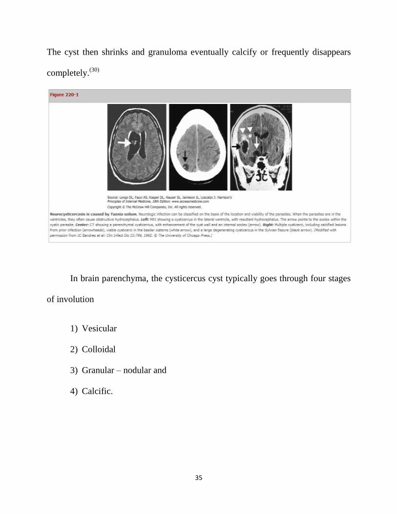
It is a common parasitic disease of the CNS. Neurocysticercosis is caused by
‘cysticercus cellulose’ the encysted larval stage of the tapeworm TaeniaSolium.
For many years the parenchymal cysts may remain dormant and the death of larva
and subsequent intense inflammatory reaction induced by larval antigens produces
symptoms (e.g. seizures). Subsequently, the cyst transforms into active granuloma.
35
The cyst then shrinks and granuloma eventually calcify or frequently disappears
completely.(30)
In brain parenchyma, the cysticercus cyst typically goes through four stages
of involution
1) Vesicular
2) Colloidal
3) Granular – nodular and
4) Calcific.
36
The first 2 stages are considered to represent the live parasite, and last two stages,
the dying or dead forms of the parasite. A live cyst is asymptomatic, evolving no or
minimal host immune response.
In India, majority of patients of neurocysticercosis have single enhancing lesions
but the multiple enhancing CT/MRI lesions are also not uncommon(31)
. These
single or multiple lesions pose a challenge to clinicians and radiologists. The
clinical features and imaging of neurocysticercosis and tuberculoma are
exceedingly similar and it is difficult to differentiate these two conditions.(32)
The distinction between tuberculoma and single cysticercus granuloma is
important because single cysticercus granuloma is a benign and self limiting
condition, but tuberculoma is an active infection which requires prolonged therapy
with potentially toxic drugs.(33)

37
aDiagnosis is confirmed by either one absolute criteria or a combination of 2 major
criteria, one minor criteria, and one epidemiologic criteria. A probable diagnosis is
supported by the fulfillment of (1) one major criteria plus two minor criteria; (2)

38
one major criteria plus one minor criteria and one epidemiologic criteria; or (3)
three minor criteria plus one epidemiologic criteria.
Unequivocal evidence of neurocysticercosis is histopathological demonstration of
the parasite.
Following lesions are highly suggestive of neurocysticercosis in
neuroimaging, solitary cysticercus granuloma ,spontaneous resolution or eventual
calcification after several months.(34)
Electroimmuno transfer blot (EITB)assay is the current serological assay of
choice for the diagnosis of neurocysticercosis. This assay has a specificity of 100%
and a sensitivity of 94% to 98% for patients with 2 or more cystic or enhancing
lesions. But frequent false negative results in patients with a solitary intracranial
cysticercus lesion, in whom less than 50% test positive. The sensitivity and
specificity of EITB is also low in patients with calcified lesions.(35)
CSF ELISA for neurocysticercosis is 95% specific and 87% sensitive . It is a
useful supportive tool for the diagnosis . But in contrast Serum ELISA has a large
number of false negative and false positive results.

39
TUBERCULOMA OF BRAIN
It account for 20 to 30% of intracranial space occupying lesions in
India(36)
.Tuberculoma develop in brain when the initial ―Rich focus‖ does not
rupture into the meninges but expands locally with in the brain parenchyma. It may
also originate in the meninges, and may be found in the superficial cortex. This
meningeal form may resemble a meningioma.(37)
Patients with tuberculoma most often present with seizures (60 to 100%),
signs and symptoms of raised ICT (56- 93%) and focal neurological deficits (33-
68%).Tuberculomas may also be multiple or military.(38)
There may be multiple caseating granulomas in the brain, although most of
the patients (66- 73%) have single or multiple large granulomas with
necrotic centre.(39)
Imaging features:
During the initial phase, edema and necrosis may appear as low attenuating
areas on CT scan., There may be high attenuation, contrast enhancement and
calcification associated with ring enhancement and a surrounding edema when the
granuloma has began to organize. This may be homogenous enhancement or there

40
may be a central radiolucent area which corresponds to a central zone of
necrosis.(40)
MRI is more sensitive than CT in detecting cerebral parenchymal
tubercuoma. Tuberculoma are isointense with gray matter on T1 weighted images.
Lesions show central hyper intensity on T2 weighted images.A hypointense ring is
present within the wall of tuberculoma on T2 weighted MR images in some cases.
Tuberculomas typically ―enhance‖ after administration of IV gadopentetate
dimeglumine in a solid or ring pattern.(41)
SEIZURES IN PATIENTS WITH HIV:
CNS involvement in AIDS patients may first be manifested by seizures(42)
. seizure
threshold is often lower than normal in these patient due to the frequent presence
of electrolyte abnormalities Seizures are seen in 15- 35% of patients with primary
CNS lymphoma 15- 40% of patients with cerebral toxoplasmosis, 7- 50% of
patients with HIV encephalopathy and 8% of patients with cryptococcal
meningitis(43)
. The most common cause of enhancing CT/MRI lesions in patients
with AIDS isToxoplasmosis. Seizures have been reported as an early symptom of
CNS tuberculosis in 50% patients(44)
.

41
Both generalized and focal seizures were noted in patients with focal brain
lesions and in those where no cause for seizures could be identified. Presence of
focal seizure did not necessarily imply the presence of focal brain lesion.(45)
Recurrences of seizures have been observed in 68% of patients despite of
anticonvulsant therapy. It is difficult to predict which patient is likely to have
recurrent seizures. While looking for a cause of seizures in patients with HIV
infection, investigation should include CT/MRI scan, serological test for
toxoplasmosis and CSF examination for cryptococcal antigen, adenosine
deaminase and fungal culture.(45)
Differential Diagnosis of Seizures
In most cases seizures can be distinguished from other conditions by careful
attention to the history and relevant laboratory studies. Additional studies such as
video-EEG monitoring, cardiac electrophysiology, sleep studies, tilt-table analysis
may be required to reach a correct diagnosis. Disorders that may mimic seizure are
listed below,

42

43
Mechanisms of Action of Antiepileptic Drugs
AED’s appear to act primarily by blocking the initiation or spread of
seizures.Through a variety of mechanisms by which they modify the activity of ion
channels or neurotransmitters and these drugs also have pleiotropic effects in most
of the cases. The mechanisms include
1) inhibition of sodium dependent action potentials in a frequency-dependent
manner (e.g., carbamazepine, phenytoin, lamotrigine, rufinamide,
topiramate, zonisamide, lacosamide),

44
2) inhibition of voltage-gated calcium channels (phenytoin,
pregabalin,gabapentin),
3) attenuation of glutamate activity (lamotrigine, felbamate, topiramate),
4) potentiation of GABA receptor function (benzodiazepines and barbiturates),
5) increase in the availability of GABA (valproic acid, tiagabine,gabapentin),
and
6) modulation of release of synaptic vesicles (levetiracetam).
The most effective drugs for absence seizures, valproic acid and ethosuximide
probably act by inhibiting T-type calcium channels in thalamic neurons.
Treatment of Seizure:
When a patient presents shortly after a seizure,we should give first priorities to
vital signs, cardiovascular and respiratory support, and treatment of seizures. Life-
threatening conditions such as metabolic derangement, CNS infection, or drug
toxicity must be managed appropriately.
If patient is not acutely ill,the initial evaluation focus on whether there is a history
of earlier seizures. If this is the first seizure, then the emphasis will be : (1) to
establish whether the reported episode was a seizure or any another paroxysmal

45
event, (2) to determine the cause of the seizure by identifying precipitating events
and risk factors, and (3) to decide whether anticonvulsant therapy is required.
aAs adjunctive therapy.
AED Selection for Focal Seizures
Carbamazepine, lamotrigine and phenytoin is currently the initial drug of
choice for the treatment of focal seizures, including those that secondarily
generalize. Overall, these drugs have very similar efficacy, but differences in
toxicity and pharmacokinetics are the main determinants for use in a given patient.

46
Phenytoin offers the advantage of once or twice daily dosing because it has a
relatively long half- life compared to two or three times daily dosing for
carbamazepine and lamotrigine. An advantage of carbamazepine is that the
relationship between drug dose, serum levels, is linear because of its metabolism
follows first- order pharmacokinetics.By contrast, small increases in dose of
phenytoin above a standard maintenance dose can precipitate marked side effects
because its properties of saturation kinetics. This is the main causes of acute
phenytoin toxicity. The long- term use of phenytoin is associated with cosmetic
effects (e.g., hirsutism, gingival hypertrophy and coarsening of facial features), and
it is often avoided in young patients who require the drug for many years because
of its effects on bone metabolism. Carbamazepine can cause aplastic anemia,
leucopenia, or hepatotoxicity.Therefore it would be contraindicated in patients with
predispositions to these problems. Lamotrigine may cause skin rash during the
initiation of therapy. Sometimes it can be extremely severe and may lead to
Stevens - Johnson syndrome if the symptoms unrecognized and if the medication is
not discontinued immediately. Slow introduction and titration may decreases the
risk.
Valproic acid is an effective alternative for some patients with focal
seizures, especially when the seizures secondarily generalize. GI side effects are

47
fewer when using the valproate semisodium formulation. Valproic acid also rarely
causes hepatotoxicity and reversible bone marrow suppression and laboratory
testing is required to monitor toxicity.In patients with preexisting bone marrow or
liver disease this drug should be avoided.
Topiramate, levetiracetam, tiagabine, zonisamide, gabapentin, and
oxcarbazepine are additional drugs currently used for the treatment of focal
seizures with or without secondary generalization.Previously, phenobarbital and
other barbiturate compounds were commonly used as first - line therapy for most
of epilepsy(58)
.
AED Selection for Generalized Seizures
Currently Valproic acid is the best initial choice for the treatment of
primarily generalized, tonic- clonic seizures. Followed by Lamotrigine, phenytoin
and carbamazepine are suitable alternatives. Valproic acid is also effective in
absence, atonic and myoclonic seizures.Therefore it is the drug of choice in
patients with generalized epilepsy syndromes having mixed seizure types.Both
carbamazepine and phenytoin can worsen certain types of generalized seizures,
including myoclonic, absence, tonic, and atonic seizures.In uncomplicated absence
seizures ethosuximide is a particularly effective drug for the treatment, but it is not

48
useful for tonic- clonic or focal seizures. Periodic monitoring of blood cell counts
is required in ethosuximide therapy because it rarely causes bone marrow
suppression.Zonisamide, topiramate, and felbamate may have similar broad
efficacy.
Initiation and Monitoring of Therapy
Patients should be carefully educated about the approach to therapy because
the response to any antiepileptic drug is unpredictable. The goal is to prevent
seizures and minimize the side effects of therapy; determination of the optimal
dose may take months or longer if the baseline seizure frequency is low. Most
anticonvulsant drugs(59)
need to be introduced relatively slowly to minimize side
effects. Patients should expect that minor side effects such as slight changes in
cognition,mild sedation or imbalance would typically resolve within a few days.
Subsequent increase in dose should be made only after achieving a steady state
with the previous dose.
Monitoring of serum AED levels can be very useful for establishing the
initial dosing schedule.The key determinants are the clinical measures of seizure
frequency and presence of side effects and not the laboratory values. Serum drug
levels measured by Conventional assays measure the total drug (that is both free

49
and protein- bound forms).The concentration of free drug that reflects extracellular
levels in the brain and correlates with efficacy. Patients with decreased levels of
serum proteins (i.e., decreased serum albumin due to impaired liver or renal
function) may have an increased ratio of free to protein bound drug, yet the
concentration of free drug may be adequate for seizure control. These patients may
have a ―subtherapeutic‖ drug level, but the dose should be changed only when
seizures remain uncontrolled, not just to achieve a ―therapeutic‖ level.
If seizures continue despite gradual increases to the maximum tolerated
dose and documented compliance, then it becomes necessary to switch to another
AED. This is usually done by maintaining the patient on(60)
the first drug while a
second drug is added. The second drug should be adjusted to reduce the seizure
frequency and also without causing toxicity. If this is achieved, the first drug can
be gradually withdrawn. The dose of the second drug is then further optimized
based on seizure response and side effects. Anyhow monotherapy should be the
goal.
When to Discontinue Therapy
Overall, about 70% of children and 60% of adults who have their seizures
completely controlled with AED’s can eventually discontinue therapy. The

50
following profile of patient yields the greatest chance of remaining seizure- free
after the drug withdrawal:
(1) complete medical control of seizures for one to five years;
(2) single seizure type, either generalized or partial;
(3) normal neurologic examination;
(4) normal EEG.
The appropriate seizure- free interval is unknown and it varies for different forms
of epilepsy. However, it seems reasonable to attempt withdrawal of therapy after 2
years in a patient who meets all of the above criteria, is motivated to discontinue
the medication. In most cases, it is preferable to reduce the dose of the drug
gradually over two to three months. Most recurrences occur in the first three
months after discontinuing therapy. So patients should be advised to avoid
potentially dangerous situations such as swimming or driving during this period.(61)
Treatment of Refractory Epilepsy
Approximately one - third of patients with epilepsy do not respond to
treatment with a single antiepileptic drug, so it becomes necessary to try a
combination of drugs to control seizures. Patients with multiple seizure types and

51
developmental delay or those who have focal epilepsy related to an underlying
structural lesion are particularly likely to require multiple drugs.The initial
combination therapy combines first- line drugs, i.e., carbamazepine, valproic acid,
phenytoin and lamotrigine. If these drugs are unsuccessful, then add a newer drug
such as topiramate or levetiracetam. Patients with myoclonic seizures resistant to
valproic acid then add clonazepam, and those with absence seizures may respond
to a combination of ethosuximide and valproic acid.
Surgical Treatment of Refractory Epilepsy
Approximately 20% of patients with epilepsy are resistant to medical
therapy. For some, surgery can be extremely effective in substantially reducing
seizure frequency and even providing complete seizure control. Surgical options
are Lesionectomy, Temporal Lobectomy Hemispherectomy Corpus
CallosotomySelective Amygdalohippocampectomy Minimal Access, Stereotactic
ablations, Cerebellar stimulation, and Vagus nerve stimulation.
Status Epilepticus
It refers to continuous seizures or repetitive, discrete seizures with impaired level
of consciousness in the interictal period. It has numerous subtypes, including

52
generalized convulsive status epilepticus (GCSE) and nonconvulsive status
epilepticus. To meet the definition of status epilepticus the duration of seizure
activity has to be15–30 minutes. But, the duration of seizures prompts the acute
use of anticonvulsant therapy in status epilepticus. For GCSE, this is typically
when seizures last beyond 5 minutes.
Generalized convulsive status epilepticus is an emergency and must be treated
immediately, since hyperthermia,cardiorespiratory dysfunction and metabolic
derangements can occur as a consequence of prolonged seizures.These can lead to
irreversible neuronal injury. Furthermore, CNS injury can also occur when the
patient is paralyzed with neuromuscular blockade but continues to have
electrographic seizures. CNS tumors, anticonvulsant withdrawal or
noncompliance, metabolic disturbances, drug toxicity, refractory epilepsy, CNS
infection, and head trauma are most common causes of GCSE.
GCSE is obvious when the patient is having overt convulsions. After 30–45
minutes of uninterrupted seizures, the signs become increasingly subtle.
Sometimes patients may have , fine rapid movements of the eyes and mild clonic
movements of only the fingers. There may be paroxysmal episodes of
hypertension, tachycardia and pupillary dilation. In these cases, the EEG is the
only method of establishing the diagnosis.If the patient stops having overt seizures,

53
yet remains comatose, an EEG must be performed to rule out ongoing status
epilepticus.

54
AIM OF THE STUDY:
To analyse the clinical profile and to evaluate the patients with new onset
seizures who presented to KMCH
MATERIALS AND METHODS
This is a Descriptive study conducted in Government Kilpauk medical college
hospital, Department of medicine in collaboration with Neurology department, for
a period of 9months. A total of 100 cases admitted in medical ward with new
onset seizures were selected for the present study from March 2013 to November
2013.
Materials:
Detailed history, physical examination, ,RFT,Sr.Electrolytes ,EEG, CT
Brain,MRI Brain, CSF, serology and other routine investigations.
METHODOLOGY: In patients with New onset seizures along with history and
clinical examination, special investigations like CT BRAIN, MRI BRAIN,EEG,
SEROLOGY, CSF ANALYSIS will be done to find out the etiology.

55
INCLUSION CRITERIA:
Patients admitted in medical wards of Govt. Kilpauk medical college hospital,
- Age more than 18 years.
- New onset seizures (Provoked and unprovoked )
- Status Epilepticus
EXCLUSION CRITERIA:
- Psychogenic seizures.
- Eclampsia
Data collection
The data of each patient was collected on a proforma specially designed for this
study and included demographic details, detailed history, clinical features, past
medical history. physical examination, RFT, Sr.Electrolytes, EEG, CT Brain ,MRI
Brain and other routine investigations.
Statistical Tools
The information collected regarding all the selected cases were recorded in a
Master Chart. Data analysis was done with the help of computer using statistics
software.

56
Using this software range, frequencies, percentages, means, standard
deviations, chi square and 'p' values were calculated. Kruskul Wallis chi-square
test was used to test the significance of difference between quantitative variables
and Yate’s test for qualitative variables. A 'p' value less than 0.05 is taken to
denote significant relationship.
OBSERVATION AND ANALYSIS :
The results of the study are shown in tables as below. The baseline characteristics
observed are as follows,
Number of cases of new onset seizures studied -100
Table 1: Age and sex distribution
AGE IN YEARS MALE FEMALE TOTAL
1 <20 3 10 13
2 21 - 30 21 5 26
3 31 - 40 13 7 20
4 41 - 50 8 10 18
5 51 - 60 7 9 16
6 61 - 70 3 4 7
TOTAL 55 45 100

57
In the present study patients age ranged from 18yrs to 80 years with the mean of
Majority of patients were in the age group of 21-30yrs(n=26) followed by 31to 40
yrs(n=20).
77% of patients were in 2nd
– 5th
decade. 6%of patients were in the age group
of >60yrs.
FIGURE-1
Majority of males were in 3rd
decade and females were in 5th
decade.
0
5
10
15
20
25
<20 21 - 30 31 - 40 41 - 50 51 - 60 61 - 70
Chart Title
MALE
FEMALE

58
Out of 100 patients 55 were males and 45 were females with male to female ratio
of 1.22 : 1.
Table 2:etiologies and sex distribution
ETIOLOGY MALE (n=55) FEMALE
(n=45)
P value
ALCOHOL
WITHDRAWAL(n=34)
31 3
0.001
IDIOPATHIC (n=29) 13 16
NEURO INFECTION (n=16) 6 10
CVA (n=12) 4 8
METABOLIC ( n=9) 1 8
Among etiologies and sex distribution the P vaue is <0.001. This shows there is
significant correlation between etiologies and sex distribution in this study.

59
FIGURE-2
Table 3: distribution of etiologies in patients with seizures
ETIOLOGIES NUMBER (n=100) P value
1 ALCOHOL WITHDRAWL 34
<0.001
2 IDIOPATHIC 29
3 NEURO INFECTION 16
4 CVA 12
5 METABOLIC 9
0 5 10 15 20 25 30 35
ALCOHOL WITHDRAWAL(n=34)
IDIOPATHIC (n=29)
NEURO INFECTION (n=16)
CVA (n=12)
METABOLIC ( n=9)
Chart Title
FEMALE (n=45)
MALE (n=55)
60
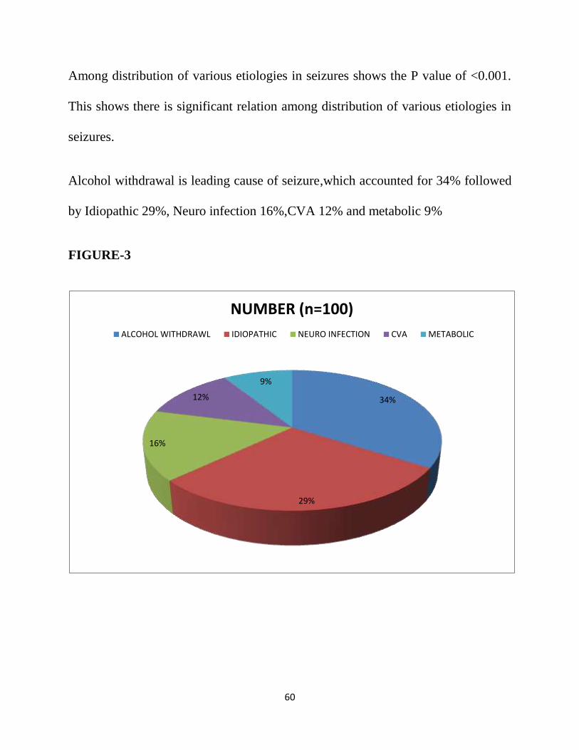
Among distribution of various etiologies in seizures shows the P value of <0.001.
This shows there is significant relation among distribution of various etiologies in
seizures.
Alcohol withdrawal is leading cause of seizure,which accounted for 34% followed
by Idiopathic 29%, Neuro infection 16%,CVA 12% and metabolic 9%
FIGURE-3
34%
29%
16%
12%
9%
NUMBER (n=100)
ALCOHOL WITHDRAWL IDIOPATHIC NEURO INFECTION CVA METABOLIC

61
Table -4:correlation of etiologies with age group
ETIOLOGIES AGE IN YEARS
18-20 21- 30 31-40 41-50 51-60 61- 70 P value
1 ALCOHOL
WITHDRAWAL
0 11 8 8 5 2
<0.001
2 IDIOPATHIC 9 9 7 2 1 1
3 NEURO
INFECTION
4 6 3 0 1 2
4 CVA 0 0 2 5 5 0
5 METABOLIC 0 0 2 2 2 2
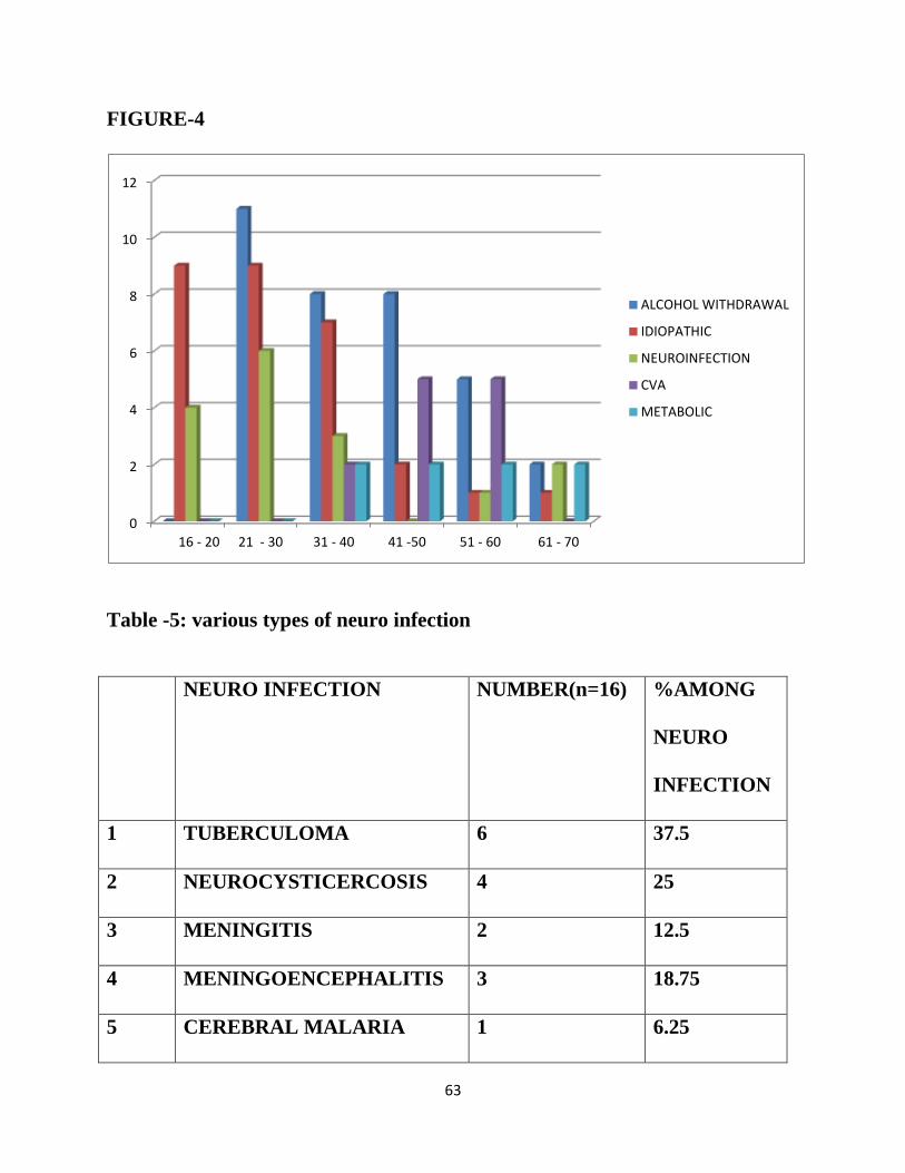
Alcohol withdraeal seizures is more common in 21-30 years.whereas CVA and
metabolic seizures are common in older people.
In 18-20 yrs 69%is due to idiopathic,31%is due to neuro infection.
In 21-30 yrs most common etiology is 42%alcohol withdrawal followed by 35%
idiopathic and 23% neuroinfection.

62
In 31-40 yrs most common etilogy is alcohol withdrawal 36% followed by
idiopathic 31%, neuroinfection 14%, CVA9%, metabolic 9%
In 41-50 yrs most common etiology is alcohol withdrawal 47% followed by CVA
29%,idiopathic 12% and metabolic 12%
In50-60yrs most common etiology is alcohol withdrawal36% and CVA 36%
followed by metabolic 19%, idiopathic%, neuroinfection 7%
In age >60 yrs most commo etiology is neuroinfection and metabolic seizures 29%
followed by 28%alcohol withdrawal and14% idiopathic.
The P value of etiologies and age distribution is <0.001.This shows there is
significant correlation between etiologies and age distribution of this study.
63
FIGURE-4
Table -5: various types of neuro infection
NEURO INFECTION NUMBER(n=16) %AMONG
NEURO
INFECTION
1 TUBERCULOMA 6 37.5
2 NEUROCYSTICERCOSIS 4 25
3 MENINGITIS 2 12.5
4 MENINGOENCEPHALITIS 3 18.75
5 CEREBRAL MALARIA 1 6.25
0
2
4
6
8
10
12
16 - 20 21 - 30 31 - 40 41 -50 51 - 60 61 - 70
ALCOHOL WITHDRAWAL
IDIOPATHIC
NEUROINFECTION
CVA
METABOLIC

64
Among neuro infection Tuberculoma accounts for
37.5%(6),neurocysticercosis4(25%),meningitis 2(12.5%),meningoencephalitis
3(18.75%),cerebral malaria 1(6.25).
FIGURE-5
TUBERCULOMA 37%
NEUROCYSTICERCOSIS
25%
MENINGITIS 13%
MENINGOENCEPHALITIS 19%
CEREBRAL MALARIA 6%
NEURO INFECTION

65
Table 6:Various types of CVA
CVA NUMBER(n=12) % AMONG
CVA
1 INFARCT 5 41.67
2 HAEMORRHAGE 3 25
3 CVT 1 8.33
4 SAH 2 16.67
5 SDH 1 8.33
Among CVA infarct 5( 42%),haemorrhage3(25%),CVT1(8%),SAH2(17%) and
SDH1(8%)
FIGURE-6
42%
25%
8%
17%
8%
CVA
INFARCT
HAEMORRHAGE
CVT
SAH
SDH

66
Table 7: various metabolic causes
METABOLIC CAUSE NUMBER
(n=9)
%AMONG
METABOLIC
CAUSE
1 HYPOGLYCAEMIA 5 55.56
2 HYPERGLYCAEMIA 1 11.11
3 HYPOCALCAEMIA 1 11.11
4 HYPONATRAEMIA 2 22.22
Among metabolic causes 56% is due to hypoglycaemia followed by hyponatremia
22%,hyperglycemia and hypocalcemia 11%
.FIGURE-7
56%
11%
11%
22%
METABOLIC CAUSES
HYPOGLYCAEMIA
HYPERGLYCAEMIA
HYPOCALCAEMIA
HYPONATRAEMIA

67
Table 8:Association for etiology and type of seizures
ETIOLOGY TYPE OF SEIZURE P value
GTCS FOCAL
1 ALCOHOL
WITHDRAWAL
28 6
<0.001
2 IDIOPATHIC 21 8
3 NEURO INFECTION 8 8
4 CVA 9 3
5 METABOLIC 4 5
TOTAL 70 30
The P value of <0.001 shows there is significant relation between etiology and type
of seizures in this study.
Seizures are commmonly present as GTCS.Most of the Alcohol withdrawal
seizures 28(82%) are present as GTCS.

68
FIGURE-8.
Table 9: GTCS distribution
ETIOLOGY NUMBER OF CASES
(n=70)
ALCOHOL WITHDRAWAL 28
IDIOPATHIC 21
CVA 9
NEURO INFECTION 8
METABOLIC 4
28
21
9
8
4
6
8
3
8
5
ALCOHOL WITHDRAWAL
IDIOPATHIC
NEURO INFECTION
CVA
METABOLIC
Chart Title
FOCAL GTCS
69
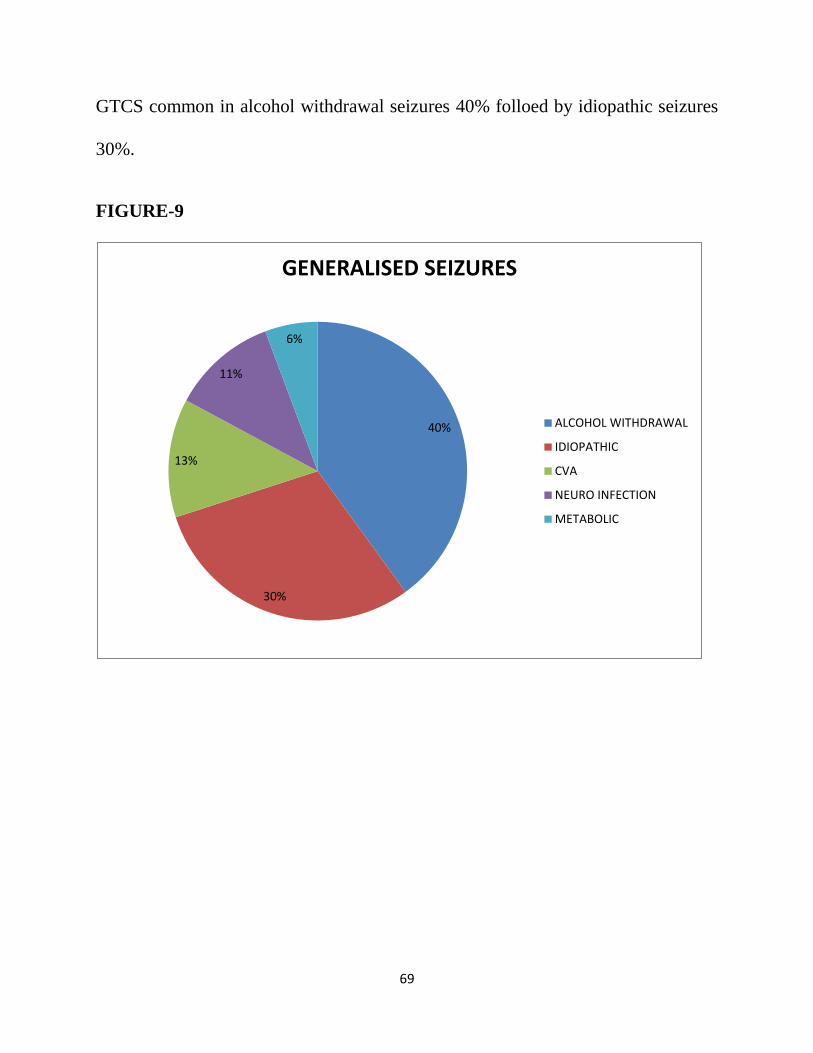
GTCS common in alcohol withdrawal seizures 40% folloed by idiopathic seizures
30%.
FIGURE-9
40%
30%
13%
11%
6%
GENERALISED SEIZURES
ALCOHOL WITHDRAWAL
IDIOPATHIC
CVA
NEURO INFECTION
METABOLIC

70
Table 10: Focal seizures distribution
ETIOLOGY NUMBER OF CASES(n=30)
ALCOHOL WITHDRAWAL 6
IDIOPATHIC 8
CVA 3
NEURO INFECTION 8
METABOLIC 5
Focal seizures are common in neuroinfection and iddiopathic seizures.
FIGURE-10
20%
26%
10%
27%
17%
FOCAL SEIZURES
ALCOHOL WITHDRAWAL
IDIOPATHIC
CVA
NEURO INFECTION
METABOLIC

71
DISCUSSION
In this study, a total of 100 patients with new onset epilepsy were included.
Sex ratio:
There was a slight male preponderance (M: F=1.22:1) in this study as
quoted by other studies on epilepsy in United States and Europe (Granieri et
al,1983).
Age distribution:
Maximum number of patients were in age group of 18- 40 years, the
youngest being 18 years.Alcohol withdrawal seizures and adult onset idiopathic
seizures are more common in this age group. Epilepsy due to cerebral infections
like neurocysticercosis, tuberculoma and brain abscess are common in middle age.
Type of seizures: generalized tonic clonic seizures 70% are more common than
focal seizures 30% in this study.
Etiology of epilepsy
72
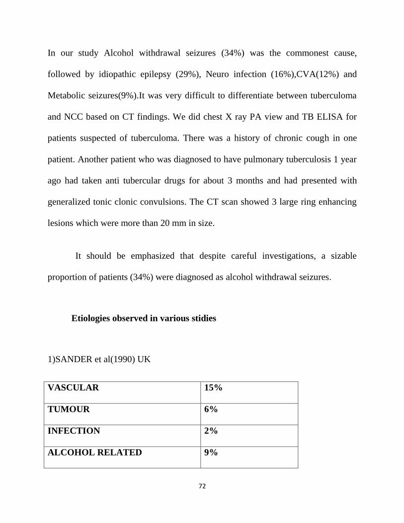
In our study Alcohol withdrawal seizures (34%) was the commonest cause,
followed by idiopathic epilepsy (29%), Neuro infection (16%),CVA(12%) and
Metabolic seizures(9%).It was very difficult to differentiate between tuberculoma
and NCC based on CT findings. We did chest X ray PA view and TB ELISA for
patients suspected of tuberculoma. There was a history of chronic cough in one
patient. Another patient who was diagnosed to have pulmonary tuberculosis 1 year
ago had taken anti tubercular drugs for about 3 months and had presented with
generalized tonic clonic convulsions. The CT scan showed 3 large ring enhancing
lesions which were more than 20 mm in size.
It should be emphasized that despite careful investigations, a sizable
proportion of patients (34%) were diagnosed as alcohol withdrawal seizures.
Etiologies observed in various stidies
1)SANDER et al(1990) UK
VASCULAR 15%
TUMOUR 6%
INFECTION 2%
ALCOHOL RELATED 9%

73
2)HAUSER et al (1995)USA
CVA 18%
NEURO INFECTION 15%
TRAMATIC BRAIN INJURY 13%
TUMOUR 13%
ALCOHOL WITHDRAWAL 11%
METABOLIC 10%
3) MURTHY JMK and RAVI (1999)Hyderabad
NEUROINFECTION 77%
VASCULAR 14%
METABOLIC 3%
TUMOUR 7%

74
4)NARAYANAN JT and JMK (2007)
NEUROINFECTION 32%
VASCULAR 21%
METABOLIC 32%
ALCOHOL 9%
OTHERS 15%
In present study:
ETIOLOGIES NUMBER
(n=100)
1 ALCOHOL WITHDRAWL 34
2 IDIOPATHIC 29
3 NEURO INFECTION 16
4 CVA 12
5 METABOLIC 9

75
GTCS FOCAL
SEIZURES
1)SANDER et al 39% 61%
2)MURTHY JMK and RAVI Y 22% 78%
3)NARAYANAN JT 55% 45%
4)OUR STUDY 70% 30%
In our study:
Most of Alcohol withdrawal seizure patients presented with GTCS(82%)
72% Of idiopathic seizure patients presented with GTCS followed by focal 28%
50% Of neuro infection patients presented with GTCS and focal seizures 50%
75% Of CVA patients presented with GTCS followed by focal seizures25%
54%Of metabolic seizure patients presented with focal seizures followed by
GTCS.

76
CONCLUSION
From the present study ―CLINICAL PROFILE AND EVALUATION OF NEW
ONSET SEIZURES‖the following conclusions were made.
1.Underlying etiologies were made in acute symptomatic seizures which
contributed to 79%.
2.Majority of seizures occurred in patients <50 yrs of age.
3. Etiological spectrum were varied and included alcohol withdrawal,neuro
infection,CVA,metabolic.
4. Alcohol withdrawal accounted for significant number of seizures in all the age
groups.
5.Tuberculoma is most common cause of seizures in neuroinfection.
6.Infarct is most common cause of seizures in CVA patients.
7. Hypoglycemia is an important cause of seizures in metabolic seizures.

77
SUMMARY
This Descriptive study was done in the KMC hospital to know the various
etiologies.100 cases of new onset seizures who fulfilled the criteria as mentioned in
materials and methods were included in the study.
Out of 100 patients 55% were males,45%were females with male to female ratio of
1.2:1.
Majority of males were in 2nd
decade and females were in 4th decade.patients age
ranged from 18 yrs to 80 yrs,with the mean of 40.11yearswith 77% of the patients
were in the below 50 yrs.
Alcohol withdrawal was the leading xcause of seizureswhich account for 34%
followed by idiopathic seizures (29%),neuro infection (16%),CVA 12% and
metabolic (9%).
Among neuro infection tuberculoma acconted for followed by neurocysticrcosis
25%,meningitis13%,meningoencephalitis 19% and cerebral malaria 6%.
Among CVA infarct accounted for 42% followed by haemorrhage 25%,SAH
17%,SDH 8% and CVT 8%.

78
Among metabolic causes hypoglycemia accounted for 56% followed by
hyponatraemia 22% ,hyperglycemia 11% and hypocalcemia 11%.
GTCS was the most common seizure 70%.Most common cause of GTCS was
alcohol withdrawal 40%followed by idiopathic 30%,CVA 13%,neuro
infection11%,metabolic 6%.

79
APPENDIX

80
BIBLIOGRAPHY
1. Orrin Devinsky MD. The falling sickness. Owsei Temkin MD: A guide to
understanding and living with epilepsy, the history of epileptic therapy. DF Scott.
2. Bharucho NE. Epidemiology of epilepsy in India. Epilespia 2003; 44(1):9-11
3. Sridheran R, Murthy BN. Prevalence and pattern of epilepsy in India Epilepsia
1999; 40 (5): 631- 6
4. Radhakrishnan K, Pandian JD. Prevalence, knowledge, attitude and practice of
epilepsy in Kerala, South India. Epilespia 2000; 41(8): 1027-35
5. Ray BK, Bhattacharya S. Epidemiology of epilepsy – Indian perspective. J Indian
Med Assoc 2002; 100(5): 322- 26.
6. Sander JW. The epidemiology of epilepsy revisited. Curr Opin Neurol 2003;
16(2): 165-70
7. Oguni H. Diagnosis and treatment of epilepsy. Epilepsia 2004; 45(Suppl.8): 13-
16
8. Dam A, Fuglsang – Freder, Ksen A, Svaree – Olsen V, et al. Late onset epilepsy:
etiologies, types of seizures, and value of clinical investigation, EEG, and
computerized tomography scan. Epilepsia 1985; 26: 227- 31
9. Luef G, Schauer RS, Baues G. Idiopathic generalized epilepsy of late onset: A
new epileptic syndrome? Epilepsia 1996; 37(4): 4
10. Cutting S, Lauchheimer A, Barr W, et al. Adult onset idiopathic generalized
epilepsy: Clinical and behavioral features. Epilepsia 2001; 42: 1395- 8.
11. Fittipaldi F, Curra A, Fusco L, et al. EEG discharges on awakening: a marker of
idiopathic generalized epileps y. Neurology 2001; 56: 123- 6.

81
12. Zarrelli MM, Behgi E, Rocca WA, et al. Incidence of epileptic syndromes in
Rochester Minesote 1980- 1984. Epilepsia 1999; 40: 1708- 14.
13. Cutting S, Lauchheimer A, Barr W, et al. Adult onset idiopathic generalized
epilepsy: clinical and behavioral features. Epilepsia 2001; 42: 1395- 98.
14. Sitajayalakshmi S, Mani J, Borgohoin, Mohondas S. Post stroke epilepsy. Neurol
India 2002; 50(Suppl.1) S78- S84.,
15. Dhonuka AK, Misra UK, Kalita J. Seizures after stroke: a prospective clinical
study. Neurol India 2001; 49(1): 33- 36.
16. Shinton RA, Gill JS, Zezuulka AV, et al. The freque ncy of epilepsy preceding
stroke; case control study in 230 patients. Lacnet 1987; 1: 11- 13.
17. Kilpatrick CJ, Davis SM, Tress BM, et al. Epileptic seizures in acute stroke.
Arch Neurol 1990; 47: 157- 160.
18. Blodin CF, Alexandrow AV, Bellavance et al. Seizures after stroke: a
prospective multicenter study. Arch Neurol 2000; 57(11): 1617- 22.
19. Arboix A, Garcia – Eroles L, Marsons JB, et al. Predictive factors of early
seizures after acute cerebrovascular disease. Stroke 1997; 28: 1590- 1594.
20. Lancman ME, Golimstok A, Norscini J, et al. Risk factors for developing
seizures after a stroke. Epilepsia 1993; 34(1): 141- 143.
21. Gupta SR, Naheedy MH, Elias D, et al. Post infarction seizures: a clinical study.
Stroke 1988; 19: 1477- 81
22. Gupta RK, Jena A, Sharma A. MR imaging of intracranial tuberculomas.J Compt
Assist Tomogra 1988; 12: 280.

82
23. Paolucci S, Silvestri G, Lubich S, et al. Post stroke late seizures another role in
rehabilitation of in patients. Epilepsia 1997; 38(3): 266-270
24. Horner S, Xiu- Shi N, Duft M, et al. EEG, CT and neuro sonographi cfindings in
patients with postischemic seizures. J Neurol Sci 1995; 132:57-60.
25. Kishore D, Misra S. Short course of oral prednisolone on disappearance of lesion
and seizure recurrence in patients of so litary cysticercol granuloma with single
small enhancing CT lesion: An open Label randomized prospective study. JAPI
2007; 55: 419- 424.
26. Wadia RS, Makhole CN, Kelkar AV, Grant KB. Focal epilepsy in India with
special reference to lesions showing ring or dis c like enhancement on contrast
computed tomography. J Neurol Neurosurg Psychiatry 1987; 50: 1298- 301.
27. Sethi PK, Kumar BR, Madon VS, Mohan V. Appearing and disappearing CT
scan abnormalities and seizures. J Neurol Neurosurg Psychiatry1985; 48: 866-
9.72
28. Rajshekar V. Etiology and management of single small CT lesions in patients
with seizures: understanding a controversy. Acta Neurol Scand 1991; 84: 465-
70.
29. Rajshekar V. Solitary cerebral cysticercus granuloma. Epilepsia 2003;
44(Suppl.1):25- 28.

83
30. Del Brutto OH, Sotelo J. Neurocysticercosis: An update. Rev Infect Dis 1988;
10:1075- 87.
31. Escobar A. In: Polocio E, Rodriguez- Carabazal J, Taveras JM (Ed). The
pathology of neurocysticercosis. Cysticercosis of the central nervous system.
Charles C.Thomas (Illinosis) 1983 .p.27- 54.
32. Singh G. Neurocysticercosis in south- central America and the Indian
subcontinent: A comparative evaluation. Arq Neuro Psiquiatr 1997; 55: 349-55.
33. Shah GV. Central nervous system tuberculosis. Neuroimaging. Clin North Am
2000; 10: 355- 74.
34. Garg RK. Diagnostic criteria for neurocysticercosis: Some modifications are
needed for Indian patients. Neuro l India 2004; 52: 171-77
35. Singh G, Kausal V, Ram S, et al. Cysticercosis immunoblot assay in patients
with single small enhancing CT lesions and multilesional neurocysticercosis.
JAPI. 1999; 47:476-9.
36. Ramamurthy B, Varadarajan MG. Diagnosis of tuberculomas of the brain:
Clinical and radiological correlation. J Neurosurg 1961; 8:1.
37. Garg RK. Diagnosis of intracranial tuberculoma. IndJ Tub 1996; 43:35- 39
38. Gree GT, Bazan III C, Jinkings JR. Miliary tuberculosis involving the brain MR
findings. ARJ 1992; 159: 1075.

84
39. JinkinsJR.Computed tomography of intracranial tuberculosis.Neuroradiology
1991; 33: 186.
40. Gupta RK, Jena A, Sharma A. MR imaging of intracranial tuberculomas.J
Compt Assist Tomogra 1988; 12: 280.
41. DanielTM. New approaches to the radio – diagnosis of TB meningitis.J Infect
Disc 1987; 155: 599.
42. Gerberding JL, Saude AM. Acquired immunodeficiency syndrome. In Stein HJ,
et al Internal Medicine, 5th edn. Mosby, 1998 .p.1478.
43. Fauli S, Lane HC. Human immunodeficiency virus disease, AIDs and related
disorders in Kasper LD, Braunwald E, Fauci SA, Hauser LS, Longo LD, Jameson
LJ. Harrison’s principles of internal disease. 16th edn.McGraw Hill, 2005
.p.1115- 9.
44. Bisburg B, Sundervam G, Rerchman LB, Kapila R. CNS tuberculosis with
AIDS. Ann Intern Med 1986: 210- 3.
45. Chandha DS, Hand a A, Sharma SK, Varadarajulu P, Singh AP. Seizures in
patients with human immunodeficiency virus infection. J Assoc Physician India
2000; 48: 573- 6.
46. Baulac S, Baulac M: Advances on the genetics of mendelian idiopathic
epilepsies. Neurol Clin 27:1041, 2009[PMID: 19853223]

85
47. Bonnett LJ et al: Risk of recurrence after a first seizure and implications for
driving: Further analysis of the Multicentre study of early Epilepsy and Single
Seizures. BMJ 341:6477, 201
48. Brodie MJ et al: Epilepsy in later life. Lancet Neurol 8:1019, 2009[PMID:
19800848]
49. French JA, Pedley TA: Clinical practice. Initial management of epilepsy. N Engl
J Med 359:166, 2008.[PMID: 18614784]
50. ——— et al: Efficacy and tolerability of the new antiepileptic drugs I:
Treatment of new onset epilepsy: Report of the Therapeutics and Technology
Assessment Subcommittee and Quality Standards Subcommittee of the American
Academy of Neurology and the American Epilepsy Society. Neurology 62:1252,
2004
51. ———: Efficacy and tolerability of the new antiepileptic drugs II: Treatment of
refractory epilepsy: Report of the Therapeutics and Technology Assessment
Subcommittee and Quality Standards Subcommittee of the American Academy
of Neurology and the American Epilepsy Society. Neurology 62:1261, 2004
52. Millikan D et al: Emergency treatment of status epilepticus: Current thinking.
Emerg Med Clin North Am 27:101, 2009[PMID: 19218022]
53. Pitkänen A, Lukasiuk K: Molecular and cellular basis of epileptogenesis in
symptomatic epilepsy. Epilepsy Behav 14:16, 2009[PMID: 17023142]

86
54. Rakhade SN, Jensen FE. Epileptogenesis in the immature brain: emerging
mechanisms. Nat Rev Neurol 5:380, 2009[PMID: 19578345]
55. Schuele SU, Lüders HO: Intractable epilepsy: management and therapeutic
alternatives. Lancet Neurol 7:514, 2008[PMID: 18485315]
56. Tomson T et al: Sudden unexpected death in epilepsy: current knowledge and
future directions. Lancet Neurol 7:1021, 2008[PMID: 18805738]
57. Walker SP et al: The management of epilepsy in pregnancy. BJOG 116:758,
2009[PMID: 19432564]
58. Ramamurthy B, Varadarajan MG . Diagnosis of tuberculomas of the brain:
Clinical and radiological correlation. J Neurosurg 1961; 8:1
59. Garg RK. Diagnosis of intracranial tuberculoma. Ind J Tub 1996; 43: 35- 39
60. Gree GT, Bazan III C, Jinkings JR. Miliary tuberculosis involving the brain MR
findings. ARJ 1992; 159: 1075.
61. Jinkins JR Computed tomography of intracranial tuberculosis Neuroradiology
1991; 33: 186.

87
LIST OF ABBREVATIONS USED
AED Antiepileptic Drug
CT Computerized Tomography
EEG Electroencephalo Gram
EITBA Electroimmuno Transfer Blot Assay
ELISA Enzyme Linked Immunosorbent Assay
FNAC Fine Needle Aspiration Cytology
GABA Gamma Amino Butyric Acid
GTCS Generalized Tonic Clonic Seizures
HIV Human Immunodeficiency Virus
MRI Magnetic Resonance Imaging
NCC Neurocysticercosis
PCDD Primary Cerebral Degenerative Disorder
PCR Polymerase Chain Reaction
RBS Random Blood Sugar
SCCG Solitory Cerebral Cysticercal Granuloma
SSECL Single Small Enhancing CT Lesion
TIA Transient Ischemic Attack

88
PROFORMA
NAME : DIAGNOSIS:
AGE/SEX:
OCCUPATION:
ADDRESS:
DETAILS OF PRESENT ILLNESS:
SEIZURES:
Time of onset
Duration
Aura
Any precipitating factors
Partial ( face/ hand/ upper limb/ lower limb/ foot )
Generalised ( LOC, tongue bite, bladder/bowel incontinence, post ictal
confusion, residual neurological deficit )
Number of episodes

89
ASSOCIATED COMPLAINTS:
Fever
Headache
Vomiting
Focal neurological deficit
Head injury
PAST HISTORY: DM-2 / SHT /TB / CVA / Meningitis / Encephalitis / RHD
FAMILY HISTORY: DM-2/SHT/Seizure disorder/Mental retardation
PERSONAL HISTORY: Sleep,Diet, Smoking, Alcoholism,Drug abuse,
Tobacco chewing

90
GENERAL EXAMINATION:
- Built & nourishment:
- Height: weight: BMI:
- P/I/CY/CL/LN/PE
- VITALS- PR.BP,RR,TEMPERATURE
- Cutaneous stigmata of neuroepidermal syndromes
-adenoma sebaceum,
-café-au-lait spots,
-neurofibromatosis,
-ash leaf macules,
-shagreen patch
SYSTEMIC EXAMINATION
Central nervous system
- Higher functions :

91
- Cranial nerves :
- Motor system :
- Sensory system :
- Reflexes :
- Autonomic nervous system :
- Skull & spine :
- Meningeal signs :
Cardiovascular system
Respiratory system
Abdominal system
INVESTIGATIONS:
CBC – TC,DC, ESR, Hb,Platelet count.
BIOCHEMISTRY:-
- RBS,FBS,PPBS
-B.Urea, S.Creatinine.,
-Serum electrolytes( Na, K, Ca)
URINE ROUTINE EXAMINATION---sugar,albumin, deposits
CHEST X-RAY

92
HIV - ELISA
CSF ANALYSIS- appearance, pressure,sugar, protein, cell count, gram
stain, cytology, culture
EEG
CT- BRAIN
MRI- BRAIN
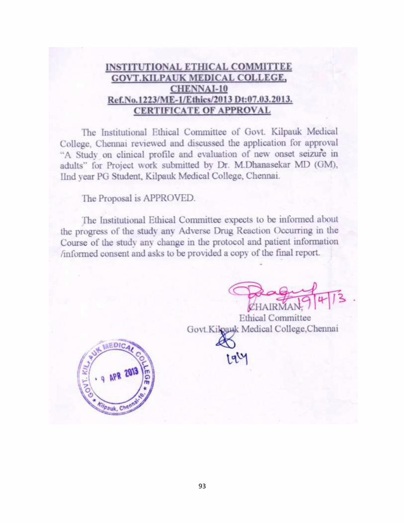
93

94

95


![Automatic detection of epileptic seizure onset and offset ...jitkomut.eng.chula.ac.th/group/poomipat_proposal.pdf7 Proposed method 18 ... [BR07]. An absence seizure which is a generalized](https://static.fdocuments.in/doc/165x107/5f3a6ed6a3b7d32b02482197/automatic-detection-of-epileptic-seizure-onset-and-offset-7-proposed-method.jpg)

![[A_R_EXE] Comparison of Fractal Dimension Estimation Algorithms for Epileptic Seizure Onset Detection](https://static.fdocuments.in/doc/165x107/5695d25e1a28ab9b029a269d/arexe-comparison-of-fractal-dimension-estimation-algorithms-for-epileptic.jpg)
