Clinical Performance of CAD/CAM All-Ceramic Tooth ...
20
materials Review Clinical Performance of CAD/CAM All-Ceramic Tooth-Supported Fixed Dental Prostheses: A Systematic Review and Meta-Analysis Babak Saravi 1, * ,† , Andreas Vollmer 2,† , Maja Hartmann 3 , Gernot Lang 1 , Ralf-Joachim Kohal 4 , Martin Boeker 5 and Sebastian B. M. Patzelt 4,6 Citation: Saravi, B.; Vollmer, A.; Hartmann, M.; Lang, G.; Kohal, R.-J.; Boeker, M.; Patzelt, S.B.M. Clinical Performance of CAD/CAM All-Ceramic Tooth-Supported Fixed Dental Prostheses: A Systematic Review and Meta-Analysis. Materials 2021, 14, 2672. https://doi.org/ 10.3390/ma14102672 Academic Editor: Antonio Scarano Received: 11 April 2021 Accepted: 18 May 2021 Published: 20 May 2021 Publisher’s Note: MDPI stays neutral with regard to jurisdictional claims in published maps and institutional affil- iations. Copyright: © 2021 by the authors. Licensee MDPI, Basel, Switzerland. This article is an open access article distributed under the terms and conditions of the Creative Commons Attribution (CC BY) license (https:// creativecommons.org/licenses/by/ 4.0/). 1 Medical Center—University of Freiburg, Department of Orthopedics and Trauma Surgery, Faculty of Medicine, University of Freiburg, Hugstetter Street 55, 79106 Freiburg, Germany; [email protected] 2 Department of Oral and Maxillofacial Surgery, University Hospital Heidelberg, Im Neuenheimer Feld 400, 69120 Heidelberg, Germany; [email protected] 3 Private Practice, Kantstraße 10, 60316 Frankfurt am Main, Germany; [email protected] 4 Medical Center—University of Freiburg, Center for Dental Medicine, Department of Prosthetic Dentistry, Faculty of Medicine, University of Freiburg, Hugstetter Street 55, 79106 Freiburg, Germany; [email protected] (R.-J.K.); [email protected] (S.B.M.P.) 5 Medical Center—University of Freiburg, Institute of Medical Biometry and Medical Statistics, Faculty of Medicine, University of Freiburg, Hugstetter Street 55, 79106 Freiburg, Germany; [email protected] 6 Private Practice, Am Dorfplatz 3, 78658 Zimmern o.R., Germany * Correspondence: [email protected] † These authors contributed equally. Abstract: Although CAD/CAM ceramics present a promising alternative to metal-ceramic fixed dental prostheses, little is known about their mid- and long-term clinical performance. This systematic review aims to estimate the survival and success rates and describes the underlying complication characteristics for CAD/CAM tooth-supported zirconia- and lithium disilicate-based fixed dental prostheses (FDPs). We systematically searched MEDLINE and Web of Science to find relevant prospective studies with a follow-up of at least one year. We estimated pooled 1-, 5-, and 10-year survival and success rates by combining the collected data in a Poisson regression model. Descriptive statistics were conducted to evaluate the distribution of failures and complications in the included studies. Risk of bias for the included studies was assessed with an adapted checklist for single-arm trials. Pooled estimated 1-, 5-, and 10-year survival rates ranged from 93.80% to 94.66%, 89.67% to 91.1%, and 79.33% to 82.20%, respectively. The corresponding success rates excluding failures, but including any other types of intervention were 94.53% to 96.77%, 90.89% to 94.62%, and 81.78% to 89.25%. Secondary caries was the most frequent cause of failure, followed by chipping of the veneering. The most common cause of complication excluding failures but requiring intervention was chipping of the veneering. Risk of bias was generally acceptable for the included studies, with seven studies associated with low risk of bias, eight studies with a moderate risk of bias, and three studies with serious risk of bias. The current meta-analysis on CAD/CAM-supported FDPs revealed satisfying survival and success rates for up to 10 years of exposure. More prospective studies focusing on long-term performance are needed to strengthen the evidence currently available in the literature. Keywords: CAD/CAM; dental restoration; ceramic; all-ceramic; zirconia; lithium disilicate; survival; fixed dental prosthesis 1. Introduction In the past decades, metal-ceramic restorations have represented the gold standard in fixed prosthetics in dentistry. Zirconium-ceramic crowns are a good alternative to Materials 2021, 14, 2672. https://doi.org/10.3390/ma14102672 https://www.mdpi.com/journal/materials
Transcript of Clinical Performance of CAD/CAM All-Ceramic Tooth ...
materials
Review
Clinical Performance of CAD/CAM All-CeramicTooth-Supported Fixed Dental Prostheses: A Systematic Reviewand Meta-Analysis
Babak Saravi 1,*,† , Andreas Vollmer 2,†, Maja Hartmann 3, Gernot Lang 1 , Ralf-Joachim Kohal 4 ,Martin Boeker 5 and Sebastian B. M. Patzelt 4,6
�����������������
Citation: Saravi, B.; Vollmer, A.;
Hartmann, M.; Lang, G.; Kohal, R.-J.;
Boeker, M.; Patzelt, S.B.M. Clinical
Performance of CAD/CAM
All-Ceramic Tooth-Supported Fixed
Dental Prostheses: A Systematic
Review and Meta-Analysis. Materials
2021, 14, 2672. https://doi.org/
10.3390/ma14102672
Academic Editor: Antonio Scarano
Received: 11 April 2021
Accepted: 18 May 2021
Published: 20 May 2021
Publisher’s Note: MDPI stays neutral
with regard to jurisdictional claims in
published maps and institutional affil-
iations.
Copyright: © 2021 by the authors.
Licensee MDPI, Basel, Switzerland.
This article is an open access article
distributed under the terms and
conditions of the Creative Commons
Attribution (CC BY) license (https://
creativecommons.org/licenses/by/
4.0/).
1 Medical Center—University of Freiburg, Department of Orthopedics and Trauma Surgery, Faculty ofMedicine, University of Freiburg, Hugstetter Street 55, 79106 Freiburg, Germany;[email protected]
2 Department of Oral and Maxillofacial Surgery, University Hospital Heidelberg, Im Neuenheimer Feld 400,69120 Heidelberg, Germany; [email protected]
3 Private Practice, Kantstraße 10, 60316 Frankfurt am Main, Germany; [email protected] Medical Center—University of Freiburg, Center for Dental Medicine, Department of Prosthetic Dentistry,
Faculty of Medicine, University of Freiburg, Hugstetter Street 55, 79106 Freiburg, Germany;[email protected] (R.-J.K.); [email protected] (S.B.M.P.)
5 Medical Center—University of Freiburg, Institute of Medical Biometry and Medical Statistics, Faculty ofMedicine, University of Freiburg, Hugstetter Street 55, 79106 Freiburg, Germany;[email protected]
6 Private Practice, Am Dorfplatz 3, 78658 Zimmern o.R., Germany* Correspondence: [email protected]† These authors contributed equally.
Abstract: Although CAD/CAM ceramics present a promising alternative to metal-ceramic fixeddental prostheses, little is known about their mid- and long-term clinical performance. This systematicreview aims to estimate the survival and success rates and describes the underlying complicationcharacteristics for CAD/CAM tooth-supported zirconia- and lithium disilicate-based fixed dentalprostheses (FDPs). We systematically searched MEDLINE and Web of Science to find relevantprospective studies with a follow-up of at least one year. We estimated pooled 1-, 5-, and 10-yearsurvival and success rates by combining the collected data in a Poisson regression model. Descriptivestatistics were conducted to evaluate the distribution of failures and complications in the includedstudies. Risk of bias for the included studies was assessed with an adapted checklist for single-armtrials. Pooled estimated 1-, 5-, and 10-year survival rates ranged from 93.80% to 94.66%, 89.67%to 91.1%, and 79.33% to 82.20%, respectively. The corresponding success rates excluding failures,but including any other types of intervention were 94.53% to 96.77%, 90.89% to 94.62%, and 81.78%to 89.25%. Secondary caries was the most frequent cause of failure, followed by chipping of theveneering. The most common cause of complication excluding failures but requiring interventionwas chipping of the veneering. Risk of bias was generally acceptable for the included studies, withseven studies associated with low risk of bias, eight studies with a moderate risk of bias, and threestudies with serious risk of bias. The current meta-analysis on CAD/CAM-supported FDPs revealedsatisfying survival and success rates for up to 10 years of exposure. More prospective studies focusingon long-term performance are needed to strengthen the evidence currently available in the literature.
Keywords: CAD/CAM; dental restoration; ceramic; all-ceramic; zirconia; lithium disilicate; survival;fixed dental prosthesis
1. Introduction
In the past decades, metal-ceramic restorations have represented the gold standardin fixed prosthetics in dentistry. Zirconium-ceramic crowns are a good alternative to
Materials 2021, 14, 2672. https://doi.org/10.3390/ma14102672 https://www.mdpi.com/journal/materials
Materials 2021, 14, 2672 2 of 20
metal-ceramic crowns. They achieve similar incidence rates for biological complications(e.g., secondary caries) and technical aspects (e.g., loss of retention) and reveal betteraesthetic properties [1]. There is a steadily increasing demand for alternatives made ofall-ceramic materials. Reasons for this may be a growing awareness of biocompatibility andaesthetics [2]. Systematic reviews could already show promising results for the long-termsurvival of CAD/CAM fabricated lithium disilicate ceramic restorations [3,4]. Studies witha 10-year follow-up period were able to provide survival rates of 96.5% for monolithic aswell as for two-layer disilicate ceramics [5].
Dental ceramics are increasingly finding their way in today’s dentistry. In addition to ahigh degree of stability and aesthetics, they also demonstrate excellent biocompatibility [6].All-ceramics is a generic term for a tooth-colored, mineral material used to manufacturedental restorations without a metal base [7]. Compared to metal-ceramic systems, all-ceramics show clear advantages for aesthetic appearance with rich color stability dueto light-conducting and light-refracting features [8]. In terms of biocompatibility, theyconvince with low plaque accumulation on their glazed surfaces [9,10]. This can beexplained by the fact that ceramics do not dissolve, even in the electrolyte-containingenvironment of the acidic oral milieu, and behave entirely neutrally toward other materials.Their biocompatibility is even higher than that of alloys containing highly noble metals [11].Unlike metals, ceramics have the characteristic of thermal insulation and thus have anotherfundamental advantage. As this prevents thermic transmission, irritation of the vital toothcan be minimized [12].
Similar to the observed progress in dental ceramics, CAD/CAM is increasingly find-ing its way into modern dentistry. Particularly, in the processing of ceramic materials,significant progress has been made. CAD stands for ‘computer-aided design’ and describesa virtual design of the restoration. CAM stands for ‘computer-aided manufacturing’ anddescribes the production of dental restorations by using machine units. This technologyhas become widespread in dentistry and seems indispensable today [13]. In the first step,the three-dimensional anatomy of the respective tooth is recorded with a scanner [14,15].The next step is digital processing and subsequent transfer to the milling unit [14]. Threeproduction systems can be distinguished here: (1) chairside, where everything takes placein the dental practice; (2) laboratory production, where the milling takes place in the dentaltechnician’s laboratory; and (3) centralized production in an external milling center [14].Two scanning methods can be distinguished: direct intraoral scan of the teeth and theextraoral scan on a conventional stone cast [14].
With the help of such systems, it is possible to produce ceramic inlays and crownswithin one session as a ‘chairside’ procedure [16].
One of the most obvious advantages is the reduced number of appointments andsimplified laboratory work compared to conventional restorations [17].
Digitization also provides a decisive advantage in terms of archiving. All clinical datacan be stored electronically and allows the restoration to be remade if damaged withouta clinical appointment. Compared to the conventional method, CAD/CAM methodsreveal a more accurate and reproducible fabrication process [18,19]. The greater accuracyand operator-independent digitalization of the workflow can result in better estheticoutcomes [20]. Due to the shorter production time and reduced number of consultations,less movement of the tooth is likely to occur. This is crucial for accurate and less traumaticrestoration of the tooth [21,22]. For these reasons, it is important to question the currentgold standard and examine the results of CAD/CAM-made ceramic restorations.
There is currently little known about the mid-term and long-term performance ofCAD/CAM fabricated zirconia- and lithium disilicate-based FDPs. Thus, we aim tosummarize the current evidence focusing on this promising therapeutical approach’ssurvival and success rates.

Materials 2021, 14, 2672 3 of 20
2. Materials and Methods2.1. Data Collection
The present systematic review was performed in accordance with the Preferred Report-ing Items for Systematic Reviews and Meta-Analyses (PRISMA) statement [23]. Literaturewas searched from inception up to January 2020. The target of the structured searchapproach were prospective studies, focusing on the clinical performance of CAD/CAMfabricated all-ceramic fixed dental prostheses. We applied language restrictions to ob-tain studies published in English or German. We performed a combined medical-subjectheading (MeSH) and free-text term search in Medline (via OVID) and Web of Science corecollection (via Web of Science). Furthermore, we hand searched the references of retrievedstudies. Truncations were used to retrieve all forms of the search terms. A combinationof search terms was performed by the Boolean operators AND and OR (Figure 1). Theinclusion criteria were adopted based on a modified PICOS process, taking into accountthe missing comparator: (P)opulation were human patients with tooth-borne CAD/CAMmanufactured all-ceramic fixed dental prostheses, (I)nterventions, in which survival ratesand complications as (O)utcomes were assessed within a prospective (S)tudy design.
Materials 2021, 14, x FOR PEER REVIEW 3 of 20
2. Materials and Methods 2.1. Data Collection
The present systematic review was performed in accordance with the Preferred Re-porting Items for Systematic Reviews and Meta-Analyses (PRISMA) statement [23]. Liter-ature was searched from inception up to January 2020. The target of the structured search approach were prospective studies, focusing on the clinical performance of CAD/CAM fabricated all-ceramic fixed dental prostheses. We applied language restrictions to obtain studies published in English or German. We performed a combined medical-subject head-ing (MeSH) and free-text term search in Medline (via OVID) and Web of Science core col-lection (via Web of Science). Furthermore, we hand searched the references of retrieved studies. Truncations were used to retrieve all forms of the search terms. A combination of search terms was performed by the Boolean operators AND and OR (Figure 1). The inclu-sion criteria were adopted based on a modified PICOS process, taking into account the missing comparator: (P)opulation were human patients with tooth-borne CAD/CAM manufactured all-ceramic fixed dental prostheses, (I)nterventions, in which survival rates and complications as (O)utcomes were assessed within a prospective (S)tudy design.
The following exclusion criteria were defined: 1. Metal and metal-ceramic restorations as an intervention. 2. Implant-borne prostheses as an intervention. 3. Hybrid bridges as an intervention. 4. Cantilever bridges as an intervention. 5. Crowns. 6. Animal studies. 7. In vitro studies. 8. Retrospective studies. 9. Case studies. 10. Languages other than German or English.
Figure 1. Search strategy.
Two independent reviewers (M.H. and S.P.) performed the selection of studies in a two-step process. Titles and abstracts were initially screened for relevance, followed by a full-text analysis. Reasons for exclusion were recorded, and any disagreement was re-solved by discussion. Data extraction was based on a standardized data extraction form that included all relevant information: study information (author, study design, year of publication, number of patients, and restorations), intervention-related (material type, CAD/CAM information, framework design, luting agent), and outcome-related (follow-
Figure 1. Search strategy.
The following exclusion criteria were defined:
1. Metal and metal-ceramic restorations as an intervention.2. Implant-borne prostheses as an intervention.3. Hybrid bridges as an intervention.4. Cantilever bridges as an intervention.5. Crowns.6. Animal studies.7. In vitro studies.8. Retrospective studies.9. Case studies.10. Languages other than German or English.
Two independent reviewers (M.H. and S.P.) performed the selection of studies ina two-step process. Titles and abstracts were initially screened for relevance, followedby a full-text analysis. Reasons for exclusion were recorded, and any disagreement wasresolved by discussion. Data extraction was based on a standardized data extractionform that included all relevant information: study information (author, study design, yearof publication, number of patients, and restorations), intervention-related (material type,

Materials 2021, 14, 2672 4 of 20
CAD/CAM information, framework design, luting agent), and outcome-related (follow-up,drop-out, survival data, biological and technical complications, and prognostic factors).Secondary caries, endodontic complications, periodontal pathology, and loss of vitalitywere considered biological complications, whereas fractures of frameworks or veneeringand loss of retention were technical complications.
2.2. Assessment of the Risk of Bias
The methodological quality of included studies could not be assessed with the classicalRisk Of Bias In Non-randomized Studies of Interventions (ROBINS-I) tool [24] due to themissing comparator (single-arm trials). Hence, a quality assessment tool for the single-arm intervention character of the included studies was adapted based on the checklistdeveloped by Moga et al. [25] and adapted previously [26]. We examined seven domains:(1) Study design, (2) Study population, (3) Intervention and co-intervention, (4) Outcomemeasures, (5) Statistical analysis, (6) Results and conclusions, and (7) Competing interestsand sources of support. The following judgments were performed based on the assessmentof these domains: (1) high risk of bias, (2) serious risk of bias, (3) low risk of bias, and (4)no information. Plot visualization was conducted using “robvis” [27].
2.3. Statistics
We included all studies matching our predefined inclusion and exclusion criteria inthe qualitative synthesis results. Studies reporting the necessary amount of survival datawere included in the quantitative results part (meta-analysis). Survival was defined asFDPs were in situ regardless of biological or technical complications. A complication wasdefined as FDPs that had biological or technical events and were still in situ. Complica-tions were defined according to the United States Public Health Service (USPHS) criteriadescribed by Cvar and Ryge and modified by Wilson et al. [28,29]. The following modifiedcriteria were considered: (1) secondary caries, (2) marginal adaption (marginal integrity),(3) marginal discoloration, (4) loss of anatomical form, (5) surface roughness, (6) colormatch, (7) endodontic complications, (8) loss of retention, and (9) fracture. The DigitizeIT(http://www.digitizeit.de, accessed on 15 January 2021) software application was used toextract the required quantitative data not reported in the main text of included studies fromthe Kaplan–Meier curves and graphs. Failure rates (biological and technical failures) werecalculated by dividing the number of events (failures) by the total exposure time (in years).Exposure time for each included study was calculated by taking the sum of exposure timefor all FDPs. Mean exposure times reported by each included study were applied forstatistics if no separate reporting or extractable data was available for individual FDPs. Therespective endpoints were last recall, time to failure, and time to drop-out. Exposure timesuntil last conducted recall for drop-out patients were included when possible. Subsequently,the calculated rates were analyzed in a Poisson regression model. The Pearson goodness-of-fit statistics assessed heterogeneity for the model. Three-year, five-year, and 10-yearsurvival proportions were estimated by assuming constant event rates. A p-value < 0.05was considered significant. Means are shown with their standard deviation (sd), whereasmedian is shown with the interquartile range (IQR). All analyses were performed usingStata Statistical Software Release 15 (StataCorp. 2011, College Station, TX, USA).
3. Results3.1. Study Selection and Study Characteristics
A total of 7104 studies were identified through the database search. After removingduplicates and screening the abstracts, 38 studies remained for full-text analysis. Subse-quently, 20 studies were excluded with reasons based on full-text analysis, mainly becausethe predefined study design criteria were not fulfilled (n = 11) or the follow-up was shorterthan one year (n = 7). Finally, 18 publications matching the eligibility criteria could beincluded in the qualitative synthesis [30–47] (Figure 2). All of these studies provided

Materials 2021, 14, 2672 5 of 20
the outcomes with sufficient extractable quantitative data and could be included in thequantitative synthesis.
Materials 2021, 14, x FOR PEER REVIEW 5 of 20
outcomes with sufficient extractable quantitative data and could be included in the quan-titative synthesis.
Figure 2. PRISMA flow diagram of the selection process.
The study characteristics of the included studies are shown in Table 1. The studies were published between 2005 and 2018. All studies had a prospective design with a me-dian follow-up of five years (range: 1.5–10 years; IQR: 3–9.7 years). One study recruited patients from multiple practices [31], one study in three practices and a university pros-thetic department [39], and all other studies were conducted solely in a university institu-tional environment. One study was a randomized controlled clinical trial focusing on comparing layered versus pressed veneering of 3-unit zirconia-ceramic FDPs [36]. None of the included studies had a controlled or randomized study design comparing the CAD/CAM fabricated all-ceramic FDPs to metal-ceramic FDPs. Overall, 610 patients (range: 15–75) and 669 reconstructions were examined. Available age data of included patients ranged from 19 to 86.4 years. The rate of patients who could not attend the whole observation period (drop-outs) was reported by all studies and ranged from 0% to 21.4%. Eleven studies reported the distribution of maxillary (n = 230) and mandibular (n = 232) FDPs [30–32,34–36,38,40,43,45,47]. Seven studies inserted three-unit bridges [32,34,36,38,39,43,45], five studies three- and four-unit bridges [30,31,37,40,42], two stud-ies three-to-five [41,47], and three studies three-to-six-unit bridges [33,44,46]. One study did not provide information [35]. The majority of the studies reported outcomes of poste-rior FDPs (9/18 studies) or a combination of posterior and anterior FDPs (7/18 studies).
Figure 2. PRISMA flow diagram of the selection process.
The study characteristics of the included studies are shown in Table 1. The stud-ies were published between 2005 and 2018. All studies had a prospective design witha median follow-up of five years (range: 1.5–10 years; IQR: 3–9.7 years). One study re-cruited patients from multiple practices [31], one study in three practices and a universityprosthetic department [39], and all other studies were conducted solely in a universityinstitutional environment. One study was a randomized controlled clinical trial focusingon comparing layered versus pressed veneering of 3-unit zirconia-ceramic FDPs [36].None of the included studies had a controlled or randomized study design compar-ing the CAD/CAM fabricated all-ceramic FDPs to metal-ceramic FDPs. Overall, 610patients (range: 15–75) and 669 reconstructions were examined. Available age data ofincluded patients ranged from 19 to 86.4 years. The rate of patients who could not at-tend the whole observation period (drop-outs) was reported by all studies and rangedfrom 0% to 21.4%. Eleven studies reported the distribution of maxillary (n = 230) andmandibular (n = 232) FDPs [30–32,34–36,38,40,43,45,47]. Seven studies inserted three-unitbridges [32,34,36,38,39,43,45], five studies three- and four-unit bridges [30,31,37,40,42], twostudies three-to-five [41,47], and three studies three-to-six-unit bridges [33,44,46]. Onestudy did not provide information [35]. The majority of the studies reported outcomesof posterior FDPs (9/18 studies) or a combination of posterior and anterior FDPs (7/18studies). One study solely utilized anterior FDPs [44] and one study did not report the

Materials 2021, 14, 2672 6 of 20
examined area [37]. Zircon ceramics were the material of choice in 17/18 studies. Onestudy utilized lithium disilicate (IPS e.max CAD) [39]. The studies utilized eight commer-cially available CAD/CAM systems: InLab Sirona, Lava System, DigiDent, Cerec Sirona,Cercon (DeguDent), iTero System, Procera System, and DCS System Dentform softwarein combination with a Precimill machining center. Three studies did not report on theCAD/CAM system [35,41,42]. Most often, restorations were attached by adhesive systemssuch as RelyX, Panavia, or MultiLink Automix. Five studies utilized glass ionomer cement,whereas two studies used zinc phosphate cement (Table 1). Lops et al. did not provide anyinformation on the cement system [35].
Table 1. Baseline characteristics of included studies.
Study andYear of Publication
No. ofPatients
No. ofFDPs
No. ofFDP Units
Region(Percentage)
Mean Age/Range CAD/CAM Cement Drop-Out
(%)
Beuer et al. 2009 [30] 19 21 3 posterior 50.9 (27–71) InLab SironaGlass
ionomercement
0
Burke et al. 2013 [31] 36 41 3–4
anterior(33%)/
posterior(67%)
NR Lava System RelyX 20
Chaar et al. 2015 [32] 58 65 3 posterior 46.8 InLab SironaGlass
ionomercement
9.2
Edelhoff et al. 2008[33] 18 22 3–6
Anterior(19.1%)/posterior(80.9%)
19–70 DigiDentGlass
ionomercement
5.6
Ioannidis et al. 2016[34] 55 59 3 posterior 52.6 InLab Sirona Panavia 3.6
Lops et al. 2012 [35] 28 28 NRanterior/posterior
(NR)NR NR NR 14.3
Naenni et al. 2015[36] 40 40 3 posterior 52.3
CerecSirona/InLabmilling unit
Panavia 10
Perry et al. 2012 [37] 15 16 3–4 NR NR Lava System RelyX 0Raigrodski et al. 2012
[38] 16 20 3 posterior 48 (36–60) Lava System RelyX 6.3
Reich et al. 2014 [39] 33 38 3anterior/posterior
(NR)
54.8(31.2–86.4)
InLabSystem/MC XL
milling unit(Sirona)
MultilinkAutomix 3
Rinke et al. 2018 [40] 75 99 3–4 posterior 49.4 (26–76) Cercon(DeguDent)
Zincphosphate
cement24.2
Sailer et al. 2006 [31] 45 57 3–5 posterior NR NR Variolink/Panavia 20
Schmitt et al. 2009[42] 30 30 3–4 posterior 52.2 (27–75) NR
Glassionomercement
10
Selz et al. 2015 [43] 24 24 3
Anterior(8.3%)/
posterior(91.7%)
48.5 (30–65)iTero System/ 3 +
1 axes millingunit
MultilinkAutomix 8.3
Solá-Ruiz et al. 2015[44] 27 27 3–6 anterior 30–65 Lava System Multilink
Automix 0
Sorrentino et al. 2012[45] 37 48 3 posterior 45.3
ProceraSystem/Procera
CenterRelyX 0
Teichmann et al. 2018[46] 17 22 3–6
Anterior(15%)/
posterior(85%)
40.1 DigiDentGlass
ionomercement
9.1
Vult von Steyern et al.2005 [47] 18 20 3–5
Anterior(25%)/
posterior(75%)
NR
DCS DentformSoft-
ware/Precimillmachining center
Zincphosphate
cement0
Materials 2021, 14, 2672 7 of 20
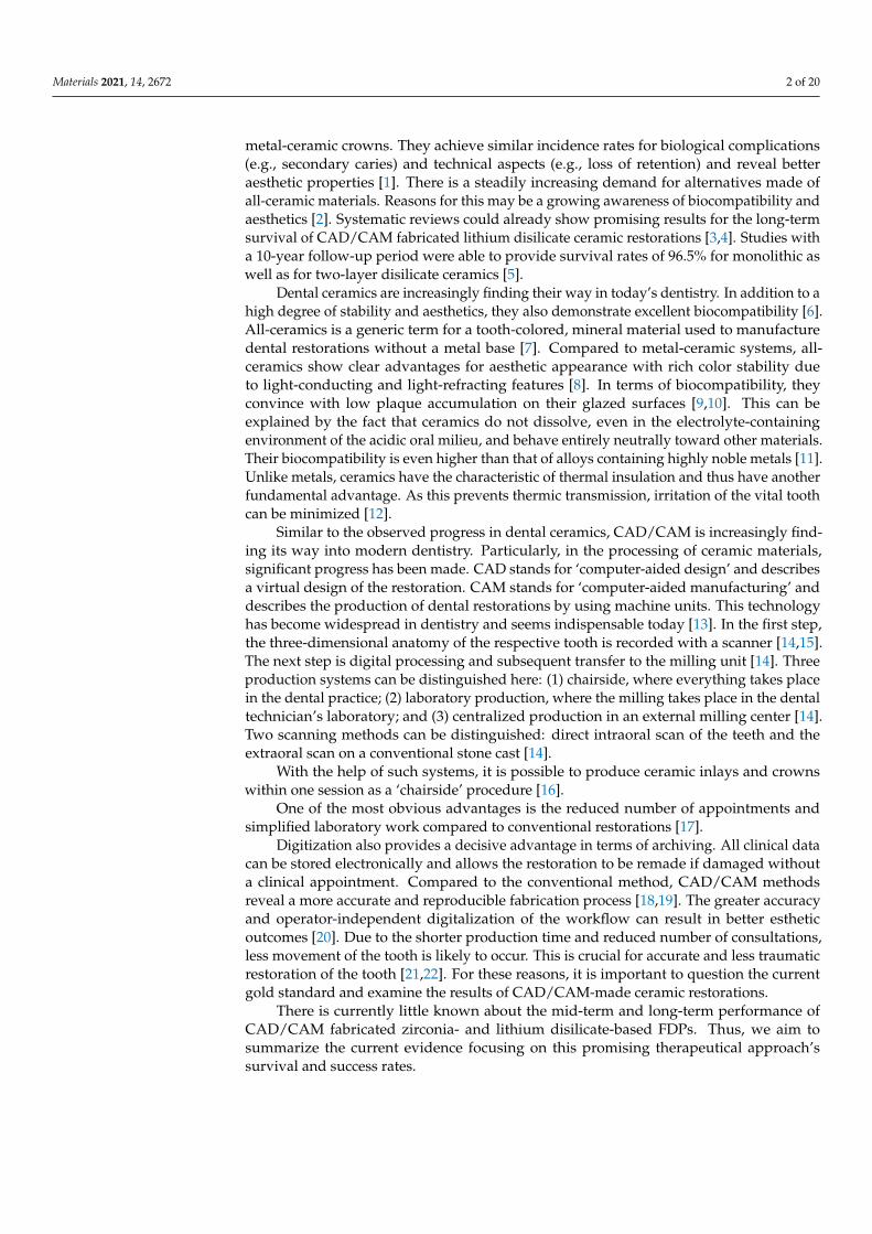
3.2. Qualitative Synthesis of Results
Included studies were divided into three groups for the qualitative synthesis of results,depending on the study follow-ups. Group 1 consisted of nine studies with a follow-upof up to approximately four years (mean: 34.6 months; range: 18 to 46 months), group 2had six studies with a follow-up of up to seven years (mean: 69.3 months; range: 60 to84 months), and group 3 consisted of studies with a follow-up of up to ten years (mean:118.2 months; range: 116.4 to 120 months) (Table 2).
Table 2. Survival characteristics of studies stratified by follow-up time frames.
Group 1
Study andYear of
Publication
No. of FDPsafter
Drop-Outs
Follow-Up inMonths (Years) Type of Material
No. of Failure(Distribution of
Failures)
No. ofComplications
(Distribution ofComplications) *
ReportedSurvival Rate
(%)
Edelhoff et al.2008 [33] 21 39.1 (3.3)
Zirconia withveneeringceramics
0
4(chipping: 3periapical
pathology: 1)
100
Beuer et al. 2009[30] 21 40 (3.3)
Zirconia withveneeringceramics
2(framework fracture:1
loss of retention: 1
1(endodontic
complications: 1)90.5
Vult von Steyernet al. 2005 [47] 20 24 (2)
Zirconia withveneeringceramics
0 3(chipping: 3) 100
Perry et al. 2012[37] 16 24 (2)
Zirconia withveneeringceramics
0 3(chipping: 2) 100
Selz et al. 2015[43] 22 18 (1.5)
Zirconia withveneeringceramics
0
19(chipping: 2
marginal integrity:2
color match: 1surface roughness:
14)
100
Sailer et al. 2006[41] 46 36 (3)
Zirconia withveneeringceramics
7(secondary caries: 3loss of retention: 1
chipping: 1endodontic
complications: 1root fracture: 1)
8(chipping: 6
secondary caries: 2)84.8
Naenni et al. 2015[36] 36 36 (3)
Zirconia withveneeringceramics
0
30(chipping: 12
surface roughness:18)
100
Schmitt et al. 2009[42] 27 34.2 (2.9)
Zirconia withveneeringceramics
0 3(chipping: 3) 100
Reich et al. 2014[39] 32 46 (3.8) Lithium disilicate
ceramics
2(framework fracture:
1persistent pain in
abutment tooth: 1)
5(chipping: 2endodontic
complications: 3)
93

Materials 2021, 14, 2672 8 of 20
Table 2. Cont.
Group 2
Study andYear of
Publication
No. of FDPsafter
Drop-Outs
Follow-Up inMonths (Years) Type of Material
No. of Failure(Distribution of
Failures)
No. ofComplications
(Distribution ofComplications) *
ReportedSurvival Rate
(%)
Lops et al. 2012[35] 24 78 (6.5)
Zirconia withveneeringceramics
2(framework fracture:
1undefined pain inabutment tooth: 1)
3(chipping: 1
loss of retention: 2)88.9
Raigrodski et al.2012 [38] 19 60 (5)
Zirconia withveneeringceramics
3(chipping: 2
root fracture: 1)
4(chipping: 2
marginal integrity:1
endodonticcomplications: 1)
90
Sorrentino et al.2012 [45] 48 60 (5)
Zirconia withveneeringceramics
0
16(chipping: 3
occlusal wear: 6marginal integrity:
3anatomical form: 4)
100
Burke et al. 2013[31] 33 60 (5)
Zirconia withveneeringceramics
1(chipping: 1)
7(chipping: 7) 97
Solá-Ruiz et al.2015 [44] 27 84 (7)
Zirconia withveneeringceramics
3(secondary caries: 2
chipping: 1)
8(chipping: 5
loss of retention: 2periapical
pathology: 1)
88.9
Ioannidis et al.2016 [34] 57 75.6 (6.3)
Zirconia withveneeringceramics
3(root fracture: 2
Secondary caries: 1)
23(chipping: 16
loss of vitality: 3periodontalpathology: 2
secondary caries: 1endodontic
complications: 1)
85
Group 3
Study andYear of
Publication
No. ofFDPs afterDrop-Outs
Follow-Up inMonths (Years) Type of Material
No. of Failure(Distribution of
Failures)
No. ofComplications
(Distribution ofComplications) *
ReportedSurvival Rate
(%)
Rinke et al. 2018[40] 75 119 (9.9)
Zirconia withveneeringceramics
24(framework fracture:
4chipping: 4
loss of retention: 5secondary caries: 6
periodontalpathology: 2
root fracture: 2unknown: 1)
50(chipping: 31
framework fracture:1
loss of retention: 7secondary caries: 6loss of vitality: 5)
75
Teichmann et al.2018 [46] 20 120 (10)
Zirconia withveneeringceramics
1(endodontic
complications: 1)
9(chipping: 8periodontal
pathology: 1)
95
Chaar et al. 2015[32] 59 116.4 (9.7)
Zirconia withveneeringceramics
4(framework fracture:
2secondary caries: 2
29(chipping: 15
loss of retention: 6endodontic
complications: 5secondary caries: 3)
93.6
* Complications were defined according to the modified USPHS criteria and considered as biological or technical complications not leadingto a replacement of the bridge (no failures).

Materials 2021, 14, 2672 9 of 20
Failures were most frequent in Group 3 (failure frequency: 29/186 FDPs; 15.6%).Notably, failure frequencies in Groups 1 (failure rate: 11/268 FDPs; 4.1%) and 2 (failurerate: 12/223 FDPs; 5.4%) were similar. Secondary caries was the leading failure whenpooling failures from all three groups (13/52, 25%), followed by chipping (10/52, 19.2%),and framework fracture (8/52, 15.4%). Framework fractures as failures were seen inGroup 1 (2/11, 18.2%) and Group 3 (6/29, 20.7%) and were absent in Group 2. This wasalso the case with endodontic complications as failures. The most frequent failure inGroup 2 was chipping (4/29, 13.8%), whereas secondary caries was the leading cause offailure in Groups 1 (3/11, 27.3%) and 3 (7/29, 24.1%). One patient lost his FDP alio loco,and this case was characterized as an “unknown” failure in Group 3. Complications notleading to a removal/replacement of the FDPs were observed in 17/18 studies. Perryet al. was the only study not providing information on complications [37]. The mostfrequent complication in all groups was chipping of the veneering. It was observed in44%, 55.7%, and 61.4% of all cases in Groups 1, 2, and 3, respectively. Surface roughness,based on the USPHS criteria, was evident in 42.7% of cases in Group 1. Other cases werenot seen in Groups 2 and 3. In fact, this criterion was only mentioned by two studies inGroup 1 [36,43], reporting a total of 32 affected cases. Other frequent complications wereloss of retention in Groups 2 (6.6% of all group cases) and 3 (14.8% of all group cases) andendodontic complications (Figure 3).
3.2.1. Group 1
The total number of FDPs in Group 1 was n = 241 (range: 16–57). Mean follow-upin Group 1 was 34.6 ± 7.9 (range: 18–46 months). Three studies in Group 1 reportedsurvival rates of 93%, 90.5% and 88.4%, respectively [30,39,41]. All other studies in thisgroup did not observe failures in the given study periods. Beuer et al. observed no chip-ping of veneering porcelain for the 40 months follow-up time frame [30]. A maxillaryFDP framework fracture occurred in the mesial abutment after 30-months of service inone FDP. Another mandibular FDP showed loss of retention and was removed. Oneendodontic treatment was performed due to cold sensitivity. The case was character-ized as an “endodontic complication” for statistics as it did not lead to a failure of theFDP. No significant changes were observed for periodontal parameters such as plaqueindex, bleeding index, and pocket-probing, in the observation period. This led to a sur-vival rate of 90.5% after 40 months. Likewise, Reich et al. observed two failures within46 months of observation: one technical failure after two years due to a framework fracturein a maxillary ten-unit bridge, and one biological failure caused by a root fracture in amandibular four-unit bridge. Sailer et al. described two technical failures: chipping of a4-unit bridge after 38.3 months and one due to loss of retention of a 4-unit bridge after33.3 months [31]. In contrast, there were five biological failures in the examined cohort.Biological failures included persistent periapical inflammation after 42 months (3-unitbridge), root fracture after 21 months (3-unit bridge), one loss of retention, and finally, threecases of secondary caries after 23.3 months (4-unit bridge), 44.1 months (4-unit bridge),and 33 months (5-unit bridge), respectively. Moreover, eight complications were reported,of which six were chipping of the veneering. Despite this, Naenni et al. did not observefailures [36]. They described a total of 12 chippings of the veneerings within 36 months ofservice. Furthermore, 18 cases of surface roughness were reported.

Materials 2021, 14, 2672 10 of 20
Materials 2021, 14, x FOR PEER REVIEW 9 of 20
(6.6% of all group cases) and 3 (14.8% of all group cases) and endodontic complications (Figure 3).
Figure 3. Distribution of complications (A) and failures (B) stratified by follow-up time frames. Group 1: follow-up from 18 to 46 months; Group 2: follow-up from 60 to 84 months; Group 3: follow-up from 116.4 to 120 months.
Figure 3. Distribution of complications (A) and failures (B) stratified by follow-up time frames. Group 1: follow-up from18 to 46 months; Group 2: follow-up from 60 to 84 months; Group 3: follow-up from 116.4 to 120 months.

Materials 2021, 14, 2672 11 of 20
3.2.2. Group 2
Group 2 included a total of 208 FDPs (range: 20–59). Follow-ups ranged from 60 to84 months (mean: 69.3 ± 9.4). Lops et al. reported a survival rate of 88.9% for the 6-yearfollow-up period [35]. One technical failure occurred due to a framework fracture and onebiological failure due to undefined pain in an abutment tooth. In contrast, only a few othercomplications not leading to removal or replacement of FDPs were observed includingtwo loss of retentions and one chipping case. The workgroup of Sola-Ruiz calculated asurvival rate of 88.9% after 84 months [44]. Chipping of the veneering in a six-unit bridgeafter three years was reported as a technical failure. Biological failures included two casesof secondary caries in a three- and four-unit FDP, respectively, after three years. Two morestudies in Group 2 reported chipping as a reason for failures [31,38]. In the cohort ofRaigrodski et al., both chipping cases were observed in the posterior mandibular regionafter 48 and 60 months, respectively [38]. Furthermore, they reported one case of biologicalfailure due to a root fracture of a mandibular second molar, leading to the removal of the3-unit bridge after 60 months. Furthermore, Burke et al. overall reported eight cases ofchipping: five cases occurred in the anterior region, one after one year, and the remainingfour after five years of function [31]. Patients were unaware of the chipping, and the caseswere polished, remained in service, and were not counted as a failure. In two further cases(pontic tooth 14 after two years and pontic tooth 46 after five years), the repair attemptfailed, but the FDPs remained in situ. These cases were also not considered as failures butcomplications. One chipping case involved the mesial-incisal angle of a central incisor(abutment tooth 11), and the bridge was replaced (failure). Sorrentino et al. did not findfailures of FDPs in the 60 month study observation period [45]. However, 16 complicationsnot leading to the removal of FDPs were mentioned. Of these cases, three were chipping ofthe veneering. Ioannidis et al. reported an 85% survival of FDPs after 6.3 years. Biologicalevents caused all three failures. However, 23 complication cases were counted, of which 16were chipping of the veneering.
3.2.3. Group 3
A total of 154 FDPs (range: 22–99) FDPs were included in Group 3. Follow-up rangedfrom 116.4 to 120 months (mean: 118.2 ± 1.4). Rinke et al. revealed a survival rate of 75%for a mean follow-up of 9.9 years [40]. Overall, 24 fatal failures were found, of which 13were caused by technical and 10 by biological events. One FDP was lost alio loco and wascharacterized as an “unknown” failure. Furthermore, 50 complications were reported, ofwhich 31/50 (62%) were chipping of the veneering. In contrast, Teichmann et al. reported ahigher survival rate of 95% after 120 months of service [46]. The only failure was due toan endodontic complication caused by recurrent pain, subsequent endodontic revision,and finally, apicoectomy. Moreover, nine complications were reported, of which themajority were chipping (8/9, 88.9%). Similarly, Chaar et al. described 93.6% survival after116.4 months [32]. In addition to the four failures, 29 complications were observed, ofwhich 15 were chipping of the veneering.
3.3. Quantitative Synthesis of Results
Estimated failure rates, survival rates, and success rates for the different groups arelisted in Table 3. The Pearson goodness-of-fit test to check for heterogeneity and the neces-sity of the random-effects Poisson regression was not significant for any of the individualgroups and rates (p > 0.05), and estimates were determined by standard Poisson regression.The estimated rates were similar across the different follow-up groups, with more varia-tion for the estimated success rates, specifically, the 5-year and 10-year success estimates.Estimated 3-year survival rates ranged between 93.80% to 94.66%, 5-year survival ratesranged from 89.67% to 91.1%, and 10-year survival rates from 79.33% to 82.20%. Successrates considered complications with FDPs excluding failures. Estimated 3-year successrates ranged between 94.53% to 96.77%, 5-year success rates from 90.89% to 94.62%, and10-year success rates from 81.78% to 89.25%.

Materials 2021, 14, 2672 12 of 20
Table 3. Illustration of estimated failure rates, complication rates, and survival rates stratified by follow-up time frames.
Study andYear of
Publication
TotalFDPExpo-sureTime
TotalNo ofFail-ures
EstimatedFailure
Rate Per100 FDP
Years(95% CI)
Estimated3 Year
SurvivalRate (%)
Estimated5 Year
SurvivalRate (%)
Estimated10 Year
SurvivalRate (%)
TotalNo. ofCom-plica-tions
*
EstimatedComplica-tion Rate(Per 100
FDPYears)
Estimated3 Year
SuccessRate
Estimated5 Year
SuccessRate
Estimated10 YearSuccess
Rate
Edelhoffet al. 2008
[33]66.46 0 - 100.00 100.00 100.00 4 6.02 (2.259–
16.037) 81.94 69.91 81.94
Beuer et al.2009 [30] 68.99 2
2.89(0.725–11.589)
91.30 85.51 71.01 1 1.45 (0.204–10.289) 95.65 92.75 95.65
Vult VonSteyern et al.
2005 [47]40.00 0 - 100.00 100.00 100.00 3 7.50 (2.419–
23.254) 77.50 62.50 77.50
Perry et al.2012 [37] 32.00 0 - 100.00 100.00 100.00 2 6.25 (1.563–
24.990) 81.25 68.75 81.25
Selz et al.2015 [43] 36.00 0 - 100.00 100.00 100.00 19
13.89(5.781–33.368)
58.33 30.56 58.33
Sailer et al.2006 [31] 138.60 7
5.05(2.408–10.594)
84.85 74.75 49.49 8 5.77 (2.887–11.542) 82.68 71.14 82.68
Naenni et al.2015 [36] 116.00 0 - 100.00 100.00 100.00 30
10.34(5.875–18.216)
68.97 48.28 68.97
Schmitt et al.2009 [42] 77.00 0 - 100.00 100.00 100.00 3 3.90 (1.257–
12.080) 88.31 80.52 88.31
Reich et al.2014 [39] § 138.87 2
1.44(0.36–5.759)
95.68 92.80 85.60 5 3.60 (1.499–8.650) 89.20 82.00 89.20
Total 713.91 11 75
SummaryEstimate
(95% Ci) #
1.78(1.435–2.216)
94.66 91.10 82.20 1.24 (0.932–1.661) 96.27 93.78 87.56
Study andYear of
Publication
TotalFDPExpo-sureTime
TotalNo ofFail-ures
EstimatedFailure
Rate Per100 FDP
Years(95% CI)
Estimated3 Year
SurvivalRate
Estimated5 Year
SurvivalRate
Estimated10 Year
SurvivalRate
TotalNo. ofCom-plica-tions
EstimatedComplica-tion Rate(Per 100
FDPYears)
Estimated3 Year
SuccessRate
Estimated5 Year
SuccessRate
Estimated10 YearSuccess
Rate
Lops et al.2012 [35] 156.00 2
1.28(0.321–5.126)
96.15 93.59 87.18 3 1.92 (0.620–5.963) 94.23 90.38 94.23
Raigrodskiet al. 2012
[38]94.00 3
3.19(1.029–9.895)
90.43 84.04 68.09 4 4.26 (1.597–11.338) 87.23 78.72 87.23
Sorrentinoet al. 2012
[45]240.00 0 - 100.00 100.00 100.00 16 6.67 (4.084–
10.882) 80.00 66.67 80.00
Burke et al.2013 176.00 1
0.57(0.08–4.034)
98.30 97.16 94.32 7 3.98 (1.896–8.343) 88.07 80.11 88.07
Solá-Ruizet al. 2015
[44]177.00 3
1.69(0.547–5.255)
94.92 91.53 83.05 8 4.52 (2.260–9.038) 86.44 77.40 86.44
Ioannidiset al. 2016
[34]365.10 3
0.82(0.265–2.548)
97.53 95.89 91.78 23 6.30 (4.186–9.480) 81.10 68.50 81.10
Total 1208.10 12 61
SummaryEstimate
(95% CI) #
2.07(1.555–2.746)
93.80 89.67 79.33 1.08 (0.759–1.523) 96.77 94.62 89.25

Materials 2021, 14, 2672 13 of 20
Table 3. Cont.
Study andYear of
Publication
TotalFDPExpo-suretime
TotalNo ofFail-ures
EstimatedFailure
Rate Per100 FDP
Years(95% CI)
Estimated3 Year
SurvivalRate
Estimated5 Year
SurvivalRate
Estimated10 Year
SurvivalRate
TotalNo. ofCom-plica-tions
EstimatedComplica-tion Rate(Per 100
FDPYears)
Estimated3 Year
SuccessRate
Estimated5 Year
SuccessRate
Estimated10 YearSuccess
Rate
Rinke et al.2018 [40] 695.27 24
3.45(2.314–5.15)
89.64 82.74 65.48 50 7.19 (5.451–9.488) 78.43 64.04 78.43
Teichmannet al. 2018
[46]213.50 1
0.47(0.066–3.325)
98.59 97.66 95.32 9 4.22 (2.193–8.102) 87.35 78.92 87.35
Chaar et al.2015 [32] 561.88 4
0.72(0.267–1.897)
97.86 96.44 92.88 29 5.16 (3.587–7.427) 84.52 74.19 84.52
Total 1470.65 29 88
SummaryEstimate
(95% Ci) #
1.82(1.716–1.935)
94.53 90.89 81.78 1.82 (1.479–2.244) 94.53 90.89 81.78
* Surface roughness was not considered as a complication in the statistics for 3-, 5-, and 10-year success; # Estimated with the Poisson-regression model (heterogeneity test based on Pearson goodness-of-fit was not significant (p > 0.05)); § Reich et al. [39] utilized lithiumdisilicate frameworks.
3.3.1. Group 1
A total of 11 FDPs over a 713.91 years cumulative exposure time have failed in Group 1.The estimated failure rate per 100 FDP years was 1.78 (95% CI: 1.435–2.216). This translatesto a 3-year, 5-year, and 10-year survival estimate of 94.66%, 91.1%, and 82.2%, respectively,for Group 1. An estimated complication rate per 100 FDP years of 1.24 (95%CI: 0.932–1.661)was calculated for the n = 75 complications. The respective success rates were 96.27%,93.78%, and 87.56% for 3-, 5-, and 10-years of exposure.
3.3.2. Group 2
The total FDP exposure time in Group 2 was 1208.1 years. Overall, 12 FDPs failed, and61 showed complications excluding failures. Failure rate per 100 FDP years was estimatedto be 2.07 (95% CI: 1.555–2.746). Survival rate dropped from 93.8% for the 3-year-datato an 89.67% 5-year rate, and finally, 79.33% for the 10-year survival rate. A total of 61complications were part of Group 2, leading to a complication rate per 100 FDP years of1.08 (0.759–1.523) with respect to the 1208.1 years of total exposure time. The resultingestimated success rates for 3-, 5-, and 10-year of exposure time were 96.77%, 94.62%, and89.25%, respectively.
3.3.3. Group 3
Group 3 included a total of 29 failures and 1470.65 years of total exposure time forthe three studies, leading to an estimated failure rate per 100 FDP years of 1.82 (95%CI:1.716–1.935). Estimated 3- and 5 years survival rates were 94.53% and 90.89%. A morepronounced drop in survival rates was observed for the 10-year data (81.78%). A total of88 complications were recorded for the three studies. Similar to the survival data results,estimated success rates dropped from 94.53% for the 3-year data to 90.89% and 81.78% forthe 5-year and 10-year data, respectively.
3.4. Assessment of the Risk of Bias
From the 18 included studies, 7/18 had a low risk of bias, 8/18 had a moderate riskof bias, and 3/18 had a serious risk of bias. Most of the studies were judged to have amoderate risk of bias in the “study design” domain due to not reporting whether patientswere included consecutively or not. Five of the studies were judged with “critical risk ofbias” in the “competing interest and sources of funding” domain [36,38,42,44,47]. This was

Materials 2021, 14, 2672 14 of 20
based on the fact that these studies provided neither the conflicts of interest statementnor the funding sources. In fact, this was the main cause leading to the overall judgment“serious risk of bias” for Sola-Ruiz et al. [44] and Vult von Steyern et al. [47] because allother domains but D1 were judged to have a low risk of bias. For Naenni et al. [36], the“critical risk of bias” judgment in D7 caused the overall judgment “moderate risk of bias”,even though all other domains were associated with a low risk of bias. The domainsD2–D7, focusing on the methodology and results section, were overall associated with alow risk of bias throughout most studies. Only Lops et al. [35] had several domains withthe judgment “serious risk of bias” due to lack of information regarding the predefinedinclusion/exclusion criteria and other necessary patient-related information such as meanage. Furthermore, Perry et al. [37] were judged with a “serious risk of bias” in the domain“study population” due to not providing sufficient information regarding patients’ baselinecharacteristics. The overall quality of the included studies was adequate to support therespective studies’ results as assessed with the domain D7 (Figure 4).
Materials 2021, 14, x FOR PEER REVIEW 14 of 20
Figure 4. Risk of bias assessment for the included studies.
Figure 4. Risk of bias assessment for the included studies.

Materials 2021, 14, 2672 15 of 20
4. Discussion
The present study aimed to evaluate the clinical performance of CAD/CAM manu-factured all-ceramic fixed dental prostheses. The available evidence provided a sufficientamount of data to allow for a satisfying conclusion on the success and survival of suchdental therapies. We estimated survival rates of up to 94.66%, 91.1%, and 82.2% for the3-, 5-, and 10-year survival data based on the available evidence in the literature. The lowrange variability between the study groups supports the assumption of good accuracyof survival. Similar satisfying results were seen for the success rates focusing on anyintervention on FDPs after insertion without considering failures as events. The estimatedlowest success rates were up to 96.77%, 94.62%, and 89.25% for the 3-, 5-, and 10-yearsuccess data, respectively. Despite the higher variability of success values than survivalvalues between the different follow-up groups for the 5- and 10-year success rate estimates,these values were still in a satisfying area of success.
Notably, the only three studies observing failures in Group 1 had a follow-up period of3 to 3.8 years [30,31,39] and reported a survival rate of 84.8%, 93%, and 90.5%, respectively.These reported rates were lower than the estimated rates of 93.8% to 94.66% for the 3-yearsurvival based on a meta-analysis of the included studies’ data. Reich et al. (2014) usedlithium disilicate ceramics as a base, the other publications mainly used zirconia [39].Therefore, the survival rates of 93% after 4.7 years described by Reich et al. must beconsidered critically. Furthermore, FDPs in the premolar region were examined, exposedto lower force effects than FDPs in molar regions [39]. The different mechanical propertiesof the two ceramic composites were not taken into account here and likely make a directcomparison inaccurate. However, the study’s overall quality was satisfying as assessedwith the risk of bias tool, allowing the support of the study results provided for lithiumdisilicate bases. In contrast, data of the five studies [31,34,35,38,44] in Group 2 reportingsurvival rates of 85% to 97% for the 5- to 7-year study period were comparable to the5-year survival estimates of 89.67% to 91.1%, calculated for all studies. This translates toa satisfying correlation and reliability for the 5-year data. The outliner with the highestreported survival rates was Burke et al., providing a survival rate of 97% for the 5-yearobservation [31]. The least comparable data were found in Group 3, reporting survivalrates of 75%, 93.6%, and 95% for the approximately 10 years of observation [32,40,46]. Thecorresponding estimates calculated from the meta-analysis were lower, with values from79.33% to 82.2%. In fact, the studies within Group 3 showed high variability in the reportedsurvival rates. The most comparable survival rate was provided by Rinke et al. reportingsurvival rates of 75% for the 10-year observation period [40]. Overall, there seems to be amore pronounced drop in survival rates between the 5-year and 10-year data. Thus, moredata on 10-year survival are warranted to draw a reliable conclusion on the long-termperformance of CAD/CAM all-ceramic FDPs.
The most common reason for failure for the pooled included studies was secondarycaries. The occurrence of secondary caries has been described by five studies [32,34,40,41,44]involving a total of 26 cases, of which 14 led to a failure of the FDP. Interestingly, a sys-tematic review from Sailer et al. did not find a significant difference in caries incidencefor abutment teeth [48]. However, Pjetursson et al. showed a higher caries prevalence onall-ceramic restorations compared to metal-ceramic restorations for multi-unit bridges [49].
The second common reason for failure was chipping. Eight publications showed eitherchipping or cracks of the ceramic, requiring replacement of the restoration [31,38,44,50].This accounts for 10/52 (19.2%) of all observed failure cases. In a systematic review, Heintzeand Rousson et al. were able to show a significantly higher chipping rate of the veneeringceramic in bridges with zirconia frameworks than in conventional metal-ceramic bridgerestorations [2]. No difference was found between 3- and 4-unit bridges. Furthermore,they reported that 24% of all zirconia FDPs examined revealed chipping, which is inaccordance with our results, not considering drop-outs (127/603, 21.06%). In comparison,43% of the metal-ceramic FDPs showed chippings. However, they reported that whencomparing only studies that directly compared zirconia and metal-ceramic FDPs, chipping
Materials 2021, 14, 2672 16 of 20
frequency was higher for zirconia FDPs [2]. Sailer et al. (2009) found chipping in 33% ofall-ceramic bridges after three years. In comparison, chipping was found in 19.4% of themetal-ceramic restorations [51]. However, they found no difference in terms of survivalrates after three years of function [51]. In order to replace metal-ceramic restorations asthe gold standard, an improvement in the bond between the veneer and the frameworkceramic must be achieved. In vitro studies showed that the leading cause of chipping waslying in the veneering ceramic [52], which can often be treated by polishing. The reasonsfor chipping can be the different layer thicknesses and the design of the framework as wellas different temperature expansion coefficients of the ceramics [30,51]. Further attempts toreduce chipping were investigated by Guess et al. (2010) by comparing monolithic lithiumdisilicate crowns with veneered zirconia crowns [52]. Monolithic restorations performedbetter, although further research is needed to be able to assess this conclusively [52]. Insummary, it can be concluded that more focus should be set on framework-veneeringinterfaces and veneering ceramics properties to reduce chipping rates in the future. Oneway to accomplish this task could be the improvement of the design of the framework itselfor strengthening the veneering ceramics.
Regarding framework fractures of all-ceramic restorations, Sulaiman et al. (2020) wereable to provide low failure rates of 1.35% for lithium disilicate as well as for monolithicrestorations in a follow-up period of up to 7.5 years. Similar to the chipping behavior results,monolithic restorations were also found to be at lower risks for framework fractures [53].Focusing on CAD/CAM produced all-ceramics, Belli et al. (2016) was able to showequivalent fracture rates of 1.4% over a period of 3.5 years [54].
Loss of retention as another type of complication leading to either failure or repairwithout replacement of the FDP was addressed by six studies [30–32,35,44,50] and occurredin 22/603 (3.65%) of all cases, not considering drop-outs. Five of these studies reportedthe cementation material and used adhesive [41,44] or conventional [30,32,40] methods.Most of the loss of retention cases were reported by Rinke et al. (12/22, 54.55%) andChaar et al. (6/22, 27.27%), both using conventional cementing methods [32,40]. Similarresults regarding the loss of retention in zirconia frameworks have been described byTinschert et al. for conventional cementing techniques as well as Sailer et al. for adhesivecementation [41,55].
The searched literature was limited to the medical databases MEDLINE and Webof Science, which was considered to be an acceptable limitation, as most of the interna-tional peer-reviewed articles were included. We only included human studies, but didnot consider in vitro examination. There might be other results regarding survival rates ifin vitro-studies were included. However, we wanted to implement real world data includ-ing representative loading situations in the human oral cavity, allowing translation andgeneralization of outcomes. One possible way of further in-depth ex vivo biological andmechanical testing of CAD/CAM restorations could be the application of post-treatmentanalysis using a combination of µ-CT and finite element techniques [56,57]. We includedonly prospective studies, leading to a higher evidence grade compared to the inclusion ofcase series and retrospective studies. Furthermore, this approach allowed us to comparethe data in a meta-analysis reliably. It should be mentioned that none of the publicationshad a control group with metal-ceramic restorations. The focus of our review was restrictedto all-ceramic restorations solely, specifically their clinical performance. Thus, the provideddata did not allow for direct comparisons between metal-ceramics and all-ceramics restora-tions, as this would require an appropriate control group to calculate and compare thenecessary effect sizes in a meta-analysis. Notably, patient diseases and medications canaffect biological parameters such as periodontal bone quantity, and should be considered infuture studies to account for these confounding factors [58–61]. Another limitation was thesmall number of studies focusing on long-term survival rates, which should be addressedin future studies. Furthermore, we restricted this review to all-ceramic restorations only.With this in mind, new materials such as polyetheretherketone (PEEK) can be consideredin the future. In addition to high-temperature and thermoelastic properties, this material is

Materials 2021, 14, 2672 17 of 20
reported to have a high biocompatibility and abrasion resistance and is already findingapplications in dentistry [62,63]. In a recent investigation, PEEK showed a breaking loadforce of 1283 N and thus qualified as a suitable material for restorations. A recent compar-ison of hybrid ceramics, zirconia, and PEEK single-tooth crowns showed no significantdifference between hybrid ceramics and PEEK [62].
In summary, all-ceramic bridge restorations fabricated by the CAD/CAM procedureand supported by natural abutment teeth are promising prosthetics associated with goodshort- and long-term clinical survival and success rates. All-ceramic bridge restorationshave proven to be a robust framework material with a low fracture susceptibility. They arealso a good prosthetic solution for multi-unit bridges in the anterior and posterior regions.Nevertheless, all-ceramic bridge restorations fabricated by the CAD/CAM procedure areassociated with specific problems such as technical (e.g., chipping, loss of retention) orbiological (e.g., secondary caries) complications, which could be linked to a semi-optimalframework design and/or fit, especially in comparison to metal-ceramic restorations. All-ceramic restorations utilizing CAD/CAM manufacturing will increasingly find their wayinto daily clinical practice and might lead to a future shift toward all-ceramic therapyoptions in modern dentistry. Further developments in technology and materials will pavethis way. Finally, we encourage authors to provide more data on the long-term performanceof CAD/CAM manufactured all-ceramic FDPs.
5. Conclusions
CAD/CAM zirconia- and lithium disilicate-based FDPs revealed satisfying survivaland success rates for up to 10 years of exposure. Certain associated complications such aschipping and secondary caries were frequently seen in the included studies, and a future in-depth analysis of their underlying factors would be of clinical relevance. More prospectivestudies focusing on long-term performance are needed to strengthen the evidence currentlyavailable in the literature.
Author Contributions: Conceptualization: S.B.M.P., M.H., B.S., A.V. and R.-J.K.; Methodology:S.B.M.P., M.H., A.V. and B.S.; Validation: G.L., M.B. and R.-J.K.; Formal analysis, B.S., M.H., S.B.M.P.and B.S.; Investigation: M.H., B.S. and S.B.M.P.; Resources: G.L., R.-J.K. and S.B.M.P.; Data curation:M.H., S.B.M.P. and B.S.; Writing—original draft preparation: B.S., A.V. and M.H.; Writing—reviewand editing: G.L., R.-J.K., M.B., S.B.M.P. and M.H.; Visualization: B.S. and G.L.; Supervision: G.L.,S.B.M.P., R.-J.K. and M.B. All authors have read and agreed to the published version of the manuscript.
Funding: The article processing charge was funded by the Baden-Wuerttemberg Ministry of Science,Research and Art, and the University of Freiburg in the funding program Open Access Publishing.
Institutional Review Board Statement: Not applicable.
Informed Consent Statement: Not applicable.
Data Availability Statement: Not applicable.
Acknowledgments: Gernot Lang was supported by the Berta-Ottenstein-Program for AdvancedClinician Scientists, Faculty of Medicine, University of Freiburg.
Conflicts of Interest: The authors declare no conflict of interest. The funders had no role in the designof the study; in the collection, analyses, or interpretation of data; in the writing of the manuscript, orin the decision to publish the results.
References1. Pjetursson, B.E.; Valente, N.A.; Strasding, M.; Zwahlen, M.; Liu, S.; Sailer, I. A Systematic Review of the Survival and Com-
plication Rates of Zirconia-Ceramic and Metal-Ceramic Single Crowns. Clin. Oral Implants Res. 2018, 29 (Suppl. 16), 199–214.[CrossRef] [PubMed]
2. Heintze, S.D.; Rousson, V. Survival of Zirconia- and Metal-Supported Fixed Dental Prostheses: A Systematic Review.Int. J. Prosthodont 2010, 23, 493–502. [PubMed]
3. Aziz, A.; El-Mowafy, O.; Paredes, S. Clinical Outcomes of Lithium Disilicate Glass-Ceramic Crowns Fabricated with CAD/CAMTechnology: A Systematic Review. Dent. Med. Probl. 2020, 57, 197–206. [CrossRef] [PubMed]

Materials 2021, 14, 2672 18 of 20
4. Al-Haj Husain, N.; Özcan, M.; Molinero-Mourelle, P.; Joda, T. Clinical Performance of Partial and Full-Coverage Fixed DentalRestorations Fabricated from Hybrid Polymer and Ceramic CAD/CAM Materials: A Systematic Review and Meta-Analysis.J. Clin. Med. 2020, 9, 2107. [CrossRef]
5. Malament, K.A.; Natto, Z.S.; Thompson, V.; Rekow, D.; Eckert, S.; Weber, H.-P. Ten-Year Survival of Pressed, Acid-Etched e.MaxLithium Disilicate Monolithic and Bilayered Complete-Coverage Restorations: Performance and Outcomes as a Function of ToothPosition and Age. J. Pros. Dent. 2019, 121, 782–790. [CrossRef]
6. Gautam, C.; Joyner, J.; Gautam, A.; Rao, J.; Vajtai, R. Zirconia Based Dental Ceramics: Structure, Mechanical Properties,Biocompatibility and Applications. Dalt. Trans. 2016, 45, 19194–19215. [CrossRef]
7. Kern, M.; Kern, M. So Hält Keramischer Stahl. Dent. Mag. 2008, 1, 28–34.8. Strub, J.R.; Kern, M.; Türp, J.C.; Witkowski, S.; Heydecke, G.; Wolfahrt, S. Currciulum Prothetik, 4th ed.; Quintessenz Verlags-GmbH:
Berlin, Germany, 2011; Volume 2.9. Pospiech, P. Chipping–Systemimmanente Oder Verarbeitungsbedingte Probleme. Quintessenz 2010, 61, 173–181.10. Abboud, M. Stark H Vollkeramische Restaurationen Im Front-Und Seitenzahnbereich. Quintessenz 2003, 54, 1295–1302.11. Kern, M.; Kohal, R.J.; Mehl, A.; Pospiech, P.; Frankenberger, R.; Reiss, B.; Wiedhahn, K.; Kunzelmann, K.H. Vollkeramik
Auf Einen Blick: Leitfaden Zur Indikation, Werkstoffauswahl, Vorbereitung Und Eingliederung von Vollkeramischen Restaurationen;Arbeitsgemeinschaft für Keramik in der Zahnheilkunde: Malsch, Germany, 2010; ISBN 3-00-017195-9.
12. Kunzelmann, K.-H.; Pospiech, P.; Kern, M. Hat Sich Vollkeramik Bewährt. Eine Positionsp. Kons Prothetik. Dentalfresh 2007, 2,16–19.
13. Poticny, D.J.; Klim, J. CAD/CAM in-Office Technology: Innovations after 25 Years for Predictable, Esthetic Outcomes. J. Am.Dent. Assoc. 2010, 141 (Suppl. 2), 5S–9S. [CrossRef]
14. Beuer, F.; Schweiger, J.; Edelhoff, D. Digital Dentistry: An Overview of Recent Developments for CAD/CAM GeneratedRestorations. Br. Dent. J. 2008, 204, 505–511. [CrossRef]
15. Strietzel, R.; Lahl, C. CAD/CAM-Systeme in Labor Und Praxis; Verlag Neuer Merkur GmbH: Planegg, Germany, 2007;ISBN 3-937346-41-4.
16. Harsono, M.; Simon, J.F.; Stein, J.M.; Kugel, G. Evolution of Chairside CAD/CAM Dentistry. Tex Dent. J. 2013, 130,238–244. [PubMed]
17. Kattadiyil, M.T.; Jekki, R.; Goodacre, C.J.; Baba, N.Z. Comparison of Treatment Outcomes in Digital and Conventional CompleteRemovable Dental Prosthesis Fabrications in a Predoctoral Setting. J. Pros. Dent. 2015, 114, 818–825. [CrossRef] [PubMed]
18. Goodacre, B.J.; Goodacre, C.J.; Baba, N.Z.; Kattadiyil, M.T. Comparison of Denture Base Adaptation between CAD-CAM andConventional Fabrication Techniques. J. Pros. Dent. 2016, 116, 249–256. [CrossRef] [PubMed]
19. Janeva, N.M.; Kovacevska, G.; Elencevski, S.; Panchevska, S.; Mijoska, A.; Lazarevska, B. Advantages of CAD/CAM versusConventional Complete Dentures-A Review. Open Access Maced. J. Med. Sci. 2018, 6, 1498–1502. [CrossRef] [PubMed]
20. Cattoni, F.; Teté, G.; Calloni, A.M.; Manazza, F.; Gastaldi, G.; Capparè, P. Milled versus Moulded Mock-Ups Based on the Super-imposition of 3D Meshes from Digital Oral Impressions: A Comparative in Vitro Study in the Aesthetic Area. BMC Oral Health2019, 19, 230. [CrossRef]
21. Goodacre, B.J.; Goodacre, C.J.; Baba, N.Z.; Kattadiyil, M.T. Comparison of Denture Tooth Movement between CAD-CAM andConventional Fabrication Techniques. J. Pros. Dent. 2018, 119, 108–115. [CrossRef]
22. Saponaro, P.C.; Yilmaz, B.; Johnston, W.; Heshmati, R.H.; McGlumphy, E.A. Evaluation of Patient Experience and Satisfactionwith CAD-CAM-Fabricated Complete Dentures: A Retrospective Survey Study. J. Pros. Dent. 2016, 116, 524–528. [CrossRef]
23. PRISMA-P Group; Moher, D.; Shamseer, L.; Clarke, M.; Ghersi, D.; Liberati, A.; Petticrew, M.; Shekelle, P.; Stewart, L.A. PreferredReporting Items for Systematic Review and Meta-Analysis Protocols (PRISMA-P) 2015 Statement. Syst. Rev. 2015, 4, 1. [CrossRef]
24. Sterne, J.A.; Hernán, M.A.; Reeves, B.C.; Savovic, J.; Berkman, N.D.; Viswanathan, M.; Henry, D.; Altman, D.G.;Ansari, M.T.; Boutron, I.; et al. ROBINS-I: A Tool for Assessing Risk of Bias in Non-Randomised Studies of Interventions. BMJ2016, 355, i4919. [CrossRef]
25. Moga, C. Development of a Quality Appraisal Tool for Case Series Studies Using a Modified Delphi Technique: Methodology Paper;Institute of Health Economics: Edmonton, AB, Canada, 2012; ISBN 978-1-926929-04-0. Available online: https://www.semanticscholar.org/paper/Development-of-a-quality-appraisal-tool-for-case-a-Moga-Guo/fd47b9a8e1b91168b3adcb6f3009a59e9078f181 (accessed on 11 April 2021).
26. Sinclair, A.; Peprah, K.; Quay, T.; McInnis, M.; Lang, E.; Severn, M.; Mulla, S.; Weeks, L.; Tsoi, B.; Herrington, E.; et al.Optimal Strategies for the Diagnosis of Acute Pulmonary Embolism: A Health Technology Assessment; CADTH Optimal Use Reports;Canadian Agency for Drugs and Technologies in Health: Ottawa, ON, Canada, 2018.
27. McGuinness, L.A.; Higgins, J.P.T. Risk-of-bias VISualization (Robvis): An R Package and Shiny Web App for VisualizingRisk-of-bias Assessments. Res. Syn. Meth 2021, 12, 55–61. [CrossRef]
28. Cvar, J.F.; Ryge, G. Reprint of Criteria for the Clinical Evaluation of Dental Restorative Materials. Clin. Oral Investig. 2005, 9,215–232. [CrossRef]
29. Wilson, M.A.; Cowan, A.J.; Randall, R.C.; Crisp, R.J.; Wilson, N.H.F. A Practice-Based, Randomized, Controlled Clinical Trial of aNew Resin Composite Restorative: One-Year Results. Oper. Dent. 2002, 27, 423–429.
30. Beuer, F.; Edelhoff, D.; Gernet, W.; Sorensen, J.A. Three-Year Clinical Prospective Evaluation of Zirconia-Based Posterior FixedDental Prostheses (FDPs). Clin. Oral Investig. 2009, 13, 445–451. [CrossRef]

Materials 2021, 14, 2672 19 of 20
31. Burke, F.J.T.; Crisp, R.J.; Cowan, A.J.; Lamb, J.; Thompson, O.; Tulloch, N. Five-Year Clinical Evaluation of Zirconia-Based Bridgesin Patients in UK General Dental Practices. J. Dent. 2013, 41, 992–999. [CrossRef]
32. Chaar, M.S.; Passia, N.; Kern, M. Ten-Year Clinical Outcome of Three-Unit Posterior FDPs Made from a Glass-Infiltrated ZirconiaReinforced Alumina Ceramic (In-Ceram Zirconia). J. Dent. 2015, 43, 512–517. [CrossRef]
33. Edelhoff, D.; Florian, B.; Florian, W.; Johnen, C. HIP Zirconia Fixed Partial Dentures–Clinical Results after 3 Years of ClinicalService. Quint. Int. 2008, 39, 459–471.
34. Ioannidis, A.; Bindl, A. Clinical Prospective Evaluation of Zirconia-Based Three-Unit Posterior Fixed Dental Prostheses:Up-to Ten-Year Results. J. Dent. 2016, 47, 80–85. [CrossRef] [PubMed]
35. Lops, D.; Mosca, D.; Casentini, P.; Ghisolfi, M.; Romeo, E. Prognosis of Zirconia Ceramic Fixed Partial Dentures: A 7-YearProspective Study. Int. J. Prosthodont. 2012, 25, 21–23.
36. Naenni, N.; Bindl, A.; Sax, C.; Hämmerle, C.; Sailer, I. A Randomized Controlled Clinical Trial of 3-Unit Posterior Zirconia-Ceramic Fixed Dental Prostheses (FDP) with Layered or Pressed Veneering Ceramics: 3-Year Results. J. Dent. 2015, 43,1365–1370. [CrossRef]
37. Perry, R.D.; Kugel, G.; Sharma, S.; Ferreira, S.; Magnuson, B. Two-Year Evaluation Indicates Zirconia Bridges AcceptableAlternative to PFMs. Compend. Contin. Educ. Dent. 2012, 33, e1–e5.
38. Raigrodski, A.J.; Yu, A.; Chiche, G.J.; Hochstedler, J.L.; Mancl, L.A.; Mohamed, S.E. Clinical Efficacy of Veneered ZirconiumDioxide-Based Posterior Partial Fixed Dental Prostheses: Five-Year Results. J. Prosthet Dent. 2012, 108, 214–222. [CrossRef]
39. Reich, S.; Endres, L.; Weber, C.; Wiedhahn, K.; Neumann, P.; Schneider, O.; Rafai, N.; Wolfart, S. Three-Unit CAD/CAM-GeneratedLithium Disilicate FDPs after a Mean Observation Time of 46 Months. Clin. Oral Invest. 2014, 18, 2171–2178. [CrossRef]
40. Rinke, S.; Wehle, J.; Schulz, X.; Bürgers, R.; Rödiger, M. Prospective Evaluation of Posterior Fixed Zirconia Dental Prostheses:10-Year Clinical Results. Int. J. Pros. 2018, 31, 35–42. [CrossRef]
41. Sailer, I.; Fehér, A.; Filser, F.; Lüthy, H.; Gauckler, L.J.; Schärer, P.; Franz Hämmerle, C.H. Prospective Clinical Study of ZirconiaPosterior Fixed Partial Dentures: 3-Year Follow-Up. Quint. Int. 2006, 37, 685–693.
42. Schmitt, J.; Holst, S.; Wichmann, M.; Reich, S.; Gollner, M.; Hamel, J. Zirconia Posterior Fixed Partial Dentures: A ProspectiveClinical 3-Year Follow-Up. Int. J. Prosthod. 2009, 22, 597–603.
43. Selz, C.F.; Bogler, J.; Vach, K.; Strub, J.R.; Guess, P.C. Veneered Anatomically Designed Zirconia FDPs Resulting from DigitalIntraoral Scans: Preliminary Results of a Prospective Clinical Study. J. Dent. 2015, 43, 1428–1435. [CrossRef]
44. Solá-Ruíz, M.F.; Agustin-Panadero, R.; Fons-Font, A.; Labaig-Rueda, C. A Prospective Evaluation of Zirconia Anterior PartialFixed Dental Prostheses: Clinical Results after Seven Years. J. Pros. Dent. 2015, 113, 578–584. [CrossRef]
45. Sorrentino, R.; De Simone, G.; Tetè, S.; Russo, S.; Zarone, F. Five-Year Prospective Clinical Study of Posterior Three-UnitZirconia-Based Fixed Dental Prostheses. Clin. Oral Investig. 2012, 16, 977–985. [CrossRef]
46. Teichmann, M.; Wienert, A.L.; Rückbeil, M.; Weber, V.; Wolfart, S.; Edelhoff, D. Ten-Year Survival and Chipping Rates and ClinicalQuality Grading of Zirconia-Based Fixed Dental Prostheses. Clin. Oral Investig. 2018, 22, 2905–2915. [CrossRef]
47. Vult von Steyern, P.; Carlson, P.; Nilner, K. All-Ceramic Fixed Partial Dentures Designed According to the DC-Zirkon Technique.A 2-Year Clinical Study. J. Oral Rehabil. 2005, 32, 180–187. [CrossRef]
48. Sailer, I.; Makarov, N.A.; Thoma, D.S.; Zwahlen, M.; Pjetursson, B.E. All-Ceramic or Metal-Ceramic Tooth-Supported Fixed DentalProstheses (FDPs)? A Systematic Review of the Survival and Complication Rates. Part I: Single Crowns (SCs). Dent. Mater. 2015,31, 603–623. [CrossRef]
49. Pjetursson, B.E.; Sailer, I.; Makarov, N.A.; Zwahlen, M.; Thoma, D.S. All-Ceramic or Metal-Ceramic Tooth-Supported Fixed DentalProstheses (FDPs)? A Systematic Review of the Survival and Complication Rates. Part II: Multiple-Unit FDPs. Dent. Mater. 2015,31, 624–639. [CrossRef]
50. Rinke, S.; Gersdorff, N.; Lange, K.; Roediger, M. Prospective Evaluation of Zirconia Posterior Fixed Partial Dentures: 7-YearClinical Results. Int. J. Pros. 2013, 26, 164–171. [CrossRef]
51. Sailer, I.; Gottnerb, J.; Kanelb, S.; Hammerle, C.H.F. Randomized Controlled Clinical Trial of Zirconia-Ceramic and Metal-CeramicPosterior Fixed Dental Prostheses: A 3-Year Follow-Up. Int. J. Pros. 2009, 22, 553–560.
52. Guess, P.C.; Zavanelli, R.A.; Silva, N.R.F.A.; Bonfante, E.A.; Coelho, P.G.; Thompson, V.P. Monolithic CAD/CAM LithiumDisilicate versus Veneered Y-TZP Crowns: Comparison of Failure Modes and Reliability after Fatigue. Int. J. Prosthodont. 2010, 23,434–442.
53. Sulaiman, T.A.; Abdulmajeed, A.A.; Delgado, A.; Donovan, T.E. Fracture Rate of 188695 Lithium Disilicate and Zirconia CeramicRestorations after up to 7.5 Years of Clinical Service: A Dental Laboratory Survey. J. Pros. Dent. 2020, 123, 807–810. [CrossRef]
54. Belli, R.; Petschelt, A.; Hofner, B.; Hajtó, J.; Scherrer, S.S.; Lohbauer, U. Fracture Rates and Lifetime Estimations of CAD/CAMAll-Ceramic Restorations. J. Dent. Res. 2016, 95, 67–73. [CrossRef]
55. Tinschert, J.; Schulze, K.A.; Natt, G.; Latzke, P.; Heussen, N.; Spiekermann, H. Clinical Behavior of Zirconia-Based Fixed PartialDentures Made of DC-Zirkon: 3-Year Results. Int. J. Prosthodont. 2008, 21, 217–222.
56. Gulati, V. Implementation of Micro CT in CAD/CAM Dentistry for Image Processing and Soft Computing: A Review. J. Phys.Conf. Ser. 2020, 1432, 012079. [CrossRef]
57. Sberna, M.T.; Rizzo, G.; Zacchi, E.; Capparè, P.; Rubinacci, A. A Preliminary Study of the Use of Peripheral Quantitative ComputedTomography for Investigating Root Canal Anatomy. Int. Endod. J. 2009, 42, 66–75. [CrossRef]
Materials 2021, 14, 2672 20 of 20
58. Gherlone, E.F.; Capparé, P.; Tecco, S.; Polizzi, E.; Pantaleo, G.; Gastaldi, G.; Grusovin, M.G. A Prospective Longitudinal Study onImplant Prosthetic Rehabilitation in Controlled HIV-Positive Patients with 1-Year Follow-Up: The Role of CD4+ Level, SmokingHabits, and Oral Hygiene. Clin. Implant Dent. Relat. Res. 2016, 18, 955–964. [CrossRef]
59. Saravi, B.E.; Putz, M.; Patzelt, S.; Alkalak, A.; Uelkuemen, S.; Boeker, M. Marginal Bone Loss around Oral Implants SupportingFixed versus Removable Prostheses: A Systematic Review. Int. J. Implant Dent. 2020, 6, 20. [CrossRef]
60. Saravi, B.; Lang, G.; Ülkümen, S.; Burchard, T.; Weihrauch, V.; Patzelt, S.; Boeker, M.; Li, Z.; Woelber, J.P. The TissueRenin-Angiotensin System (TRAS) and the Impact of Its Inhibition on Inflammation and Bone Loss in the Periodontal Tis-sue. Eur. Cell Mater. 2020, 40, 203–226. [CrossRef]
61. Saravi, B.; Vollmer, A.; Lang, G.; Adolphs, N.; Li, Z.; Giers, V.; Stoll, P. Impact of Renin-Angiotensin System Inhibitors andBeta-Blockers on Dental Implant Stability. Int. J. Implant Dent. 2021, 7, 31. [CrossRef]
62. Tartuk, B.K.; Ayna, E.; Basaran, E. Comparison of The Load-Bearing Capacities of Monolithic PEEK, Zirconia and Hybrid CeramicMolar Crowns. Meandros Med. Dent. J. 2018, 20, 45–50. [CrossRef]
63. Stawarczyk, B.; Beuer, F.; Wimmer, T.; Jahn, D.; Sener, B.; Roos, M.; Schmidlin, P.R. Polyetheretherketone-a Suitable Material forFixed Dental Prostheses? J. Biomed. Mater. Res. B Appl. Biomater. 2013, 101, 1209–1216. [CrossRef]


![All-ceramic front tooth restorations. Perfect Veneer Preparations. · 2020. 6. 30. · adjacent tooth [ see 6 ] . In response to this, special sonic tips are available that perfectly](https://static.fdocuments.in/doc/165x107/609f24a3b4838c661205a0ea/all-ceramic-front-tooth-restorations-perfect-veneer-2020-6-30-adjacent-tooth.jpg)


