Duodenal hematoma following endoscopic duodenal biopsy: A case ...

Clinical Pearls
Gastroenterology
Chad Burski, MD
University of Alabama at Birmingham
Assistant Professor
Gastroenterology and Hepatology
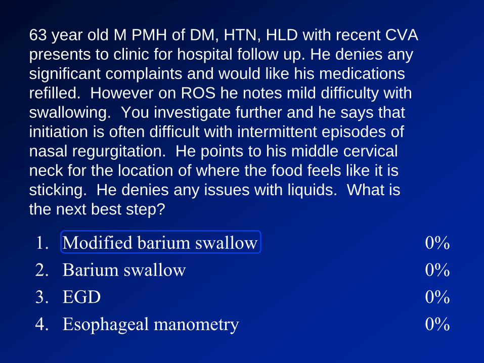
63 year old M PMH of DM, HTN, HLD with recent CVA
presents to clinic for hospital follow up. He denies any
significant complaints and would like his medications
refilled. However on ROS he notes mild difficulty with
swallowing. You investigate further and he says that
initiation is often difficult with intermittent episodes of
nasal regurgitation. He points to his middle cervical
neck for the location of where the food feels like it is
sticking. He denies any issues with liquids. What is
the next best step?
1. Modified barium swallow
2. Barium swallow
3. EGD
4. Esophageal manometry
0%
0%
0%
0%
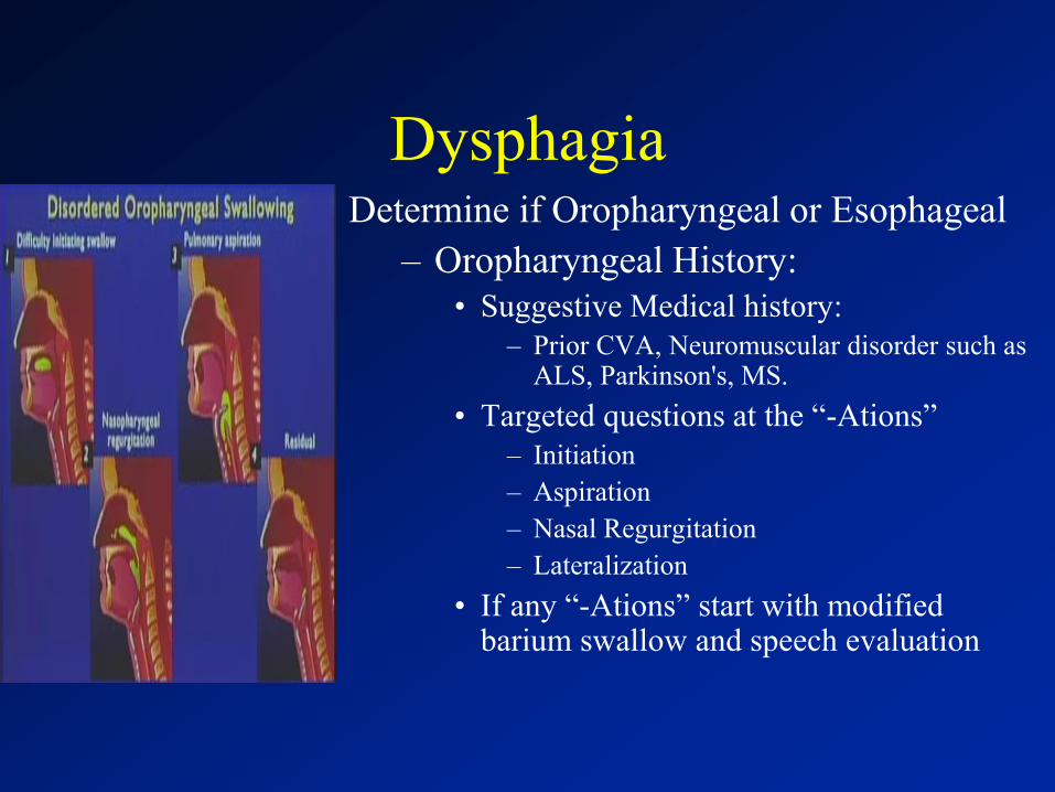
Dysphagia Determine if Oropharyngeal or Esophageal
– Oropharyngeal History:
• Suggestive Medical history:
– Prior CVA, Neuromuscular disorder such as ALS, Parkinson's, MS.
• Targeted questions at the “-Ations”
– Initiation
– Aspiration
– Nasal Regurgitation
– Lateralization
• If any “-Ations” start with modified barium swallow and speech evaluation
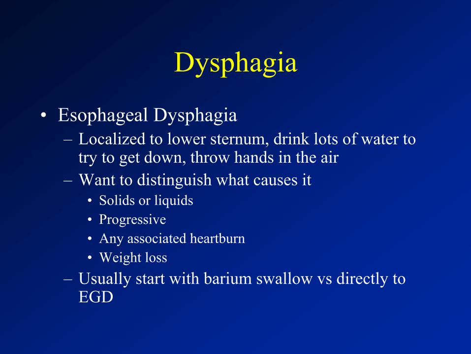
Dysphagia
• Esophageal Dysphagia
– Localized to lower sternum, drink lots of water to try to get down, throw hands in the air
– Want to distinguish what causes it
• Solids or liquids
• Progressive
• Any associated heartburn
• Weight loss
– Usually start with barium swallow vs directly to EGD

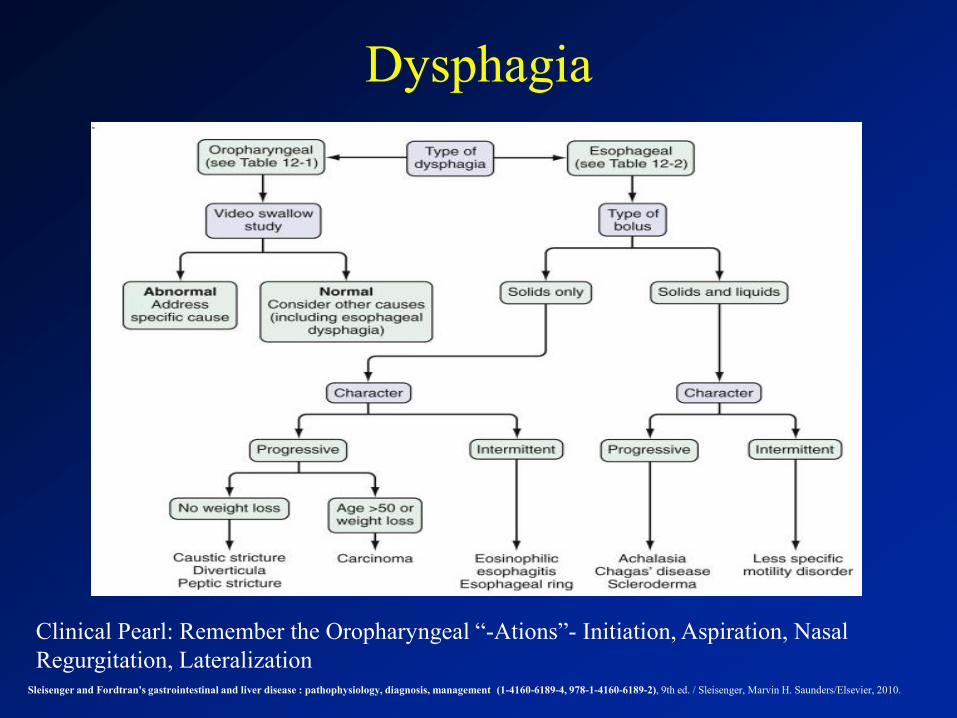
Dysphagia
Oropharyngeal Etiologies
Esophageal Etiologies
Dysphagia
Sleisenger and Fordtran's gastrointestinal and liver disease : pathophysiology, diagnosis, management (1-4160-6189-4, 978-1-4160-6189-2), 9th ed. / Sleisenger, Marvin H. Saunders/Elsevier, 2010.
Clinical Pearl: Remember the Oropharyngeal “-Ations”- Initiation, Aspiration, Nasal
Regurgitation, Lateralization

56 year old Hispanic male presents to office for evaluation of abnormal
lab values after an insurance physical. Patient is well known to you, he
has a past history of DMII, HTN, HLD and obesity. He currently takes
lisinopril, metformin and simvastatin. In the past his AST/ALT have
been mildly evaluated at 56 and 72 respectively. His total bilirubin,
alkaline phosphatase, albumin, INR and ggt were normal. You review
his insurance labs and they are similar to prior values. You order an
liver ultrasound that shows hepatomegaly and extensive steatosis.
Review of your past workup reveals a negative viral panel, negative
autoimmune studies and normal iron studies. What is the next best step
in management?
1. Stop simvastatin and repeat LFT in 6 weeks
2. Stop simvastatin and switch to atorvastatin
3. Recommend weight loss
4. No recommendations at this time but repeat labs in 6 weeks
0%
0%
0%
0%
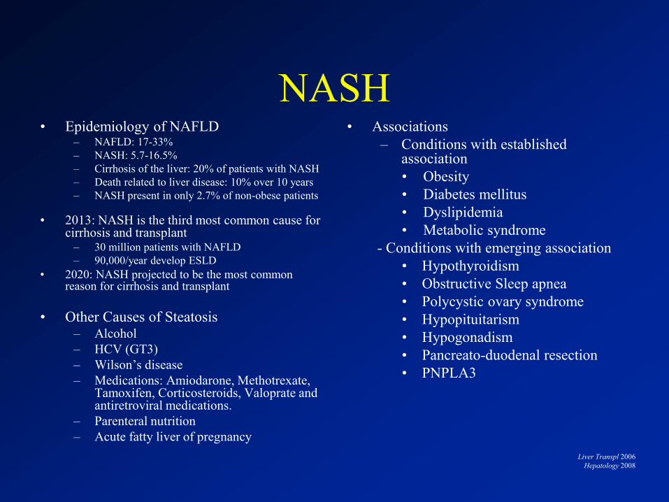
NASH • Epidemiology of NAFLD
– NAFLD: 17-33%
– NASH: 5.7-16.5%
– Cirrhosis of the liver: 20% of patients with NASH
– Death related to liver disease: 10% over 10 years
– NASH present in only 2.7% of non-obese patients
• 2013: NASH is the third most common cause for cirrhosis and transplant
– 30 million patients with NAFLD
– 90,000/year develop ESLD
• 2020: NASH projected to be the most common reason for cirrhosis and transplant
• Other Causes of Steatosis – Alcohol
– HCV (GT3)
– Wilson’s disease
– Medications: Amiodarone, Methotrexate, Tamoxifen, Corticosteroids, Valoprate and antiretroviral medications.
– Parenteral nutrition
– Acute fatty liver of pregnancy
• Associations
– Conditions with established association
• Obesity
• Diabetes mellitus
• Dyslipidemia
• Metabolic syndrome
- Conditions with emerging association
• Hypothyroidism
• Obstructive Sleep apnea
• Polycystic ovary syndrome
• Hypopituitarism
• Hypogonadism
• Pancreato-duodenal resection
• PNPLA3
Liver Transpl 2006
Hepatology 2008

NASH Therapy
• Most effective intervention is Weight loss – Weight loss of ≥7% (over 48 weeks) led to significant
improvements in: • Steatosis (−1.36 vs. −0.41, P < 0.001)
• Lobular inflammation (−0.82 vs. −0.24, P = 0.03)
• Ballooning injury (−1.27 vs. −0.53, P = 0.03)
Vitamin E – 800 IU PO daily showed:
Improvement in the histological findings but not fibrosis.
– Concerns: Increased all-cause mortality
Prostate cancer
Hepatology 2010
N Engl J Med 2010

Statins and NASH
• [2-28-2012] The U.S. Food and Drug Administration (FDA) has approved important safety label changes for the class of cholesterol-lowering drugs known as statins.
• Monitoring Liver Enzymes Labels have been revised to remove the need for routine periodic monitoring of liver enzymes in patients taking statins. The labels now recommend that liver enzyme tests should be performed before starting statin therapy and as clinically indicated thereafter.
• In a large randomized placebo controlled trial studying the effects of statin therapy for dyslipidemia in a cohort of patients known chronic liver disease, patients treated with statin therapy did not have any excess toxicity compared to those taking placebo.
Alqahtani SA, Sanchez W. Statins are safe for the treatment of hypercholesterolemia in patients with chronic liver disease. Gastroenterology 2008; 135(2):702-4.
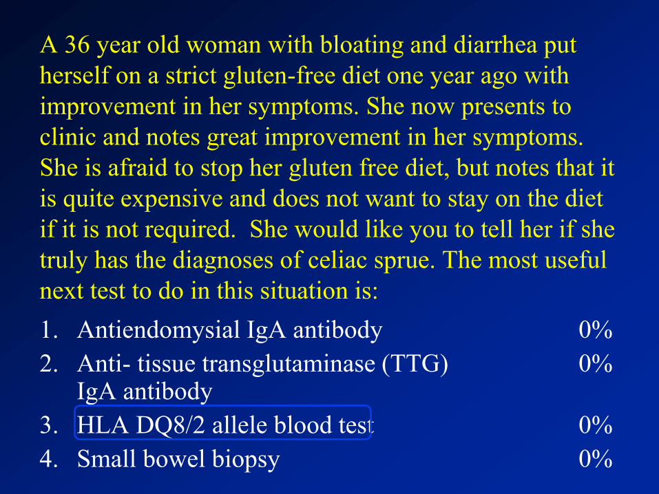
A 36 year old woman with bloating and diarrhea put
herself on a strict gluten-free diet one year ago with
improvement in her symptoms. She now presents to
clinic and notes great improvement in her symptoms.
She is afraid to stop her gluten free diet, but notes that it
is quite expensive and does not want to stay on the diet
if it is not required. She would like you to tell her if she
truly has the diagnoses of celiac sprue. The most useful
next test to do in this situation is:
1. Antiendomysial IgA antibody
2. Anti- tissue transglutaminase (TTG) IgA antibody
3. HLA DQ8/2 allele blood test
4. Small bowel biopsy
0%
0%
0%
0%

Celiac
• Systemic immune-mediated disorder triggered by dietary gluten in genetically susceptible persons.
• Characterized by a broad range of clinical presentations, a specific serum autoantibody response, and variable damage to the small intestinal mucosa.
• Studies range, but affects 0.6-1% of population
• Clinical presentation varies dramatically – Usual presentation- malabsorption – diarrhea, anemia, weight loss,
N/V, abdominal pain, distention, bloating, abnormal Liver Function Tests
– Silent – iron deficiency anemia, Vit D deficiency leading to osteoporosis, infertility.
– Skin Manifestations – Dermatitis Herpetiformis
Fasano, Alessio, MD; Catassi, Carlo, MD, MPH. The New England Journal of Medicine367.25
(Dec 20, 2012): 2419-2426.

Dermatitis Herpetiformis
(images via VisualDx)

Celiac Serologies

Diagnostic algorithm

42 year old male presents with new onset jaundice and ascites. He has
a history of IV drug use and alcohol use. Over the past month he has
increased his alcohol consumption to 10-12 beers daily. He complains
of abdominal distention and abdominal pain. On exam he is febrile to
101, HR:110, BP99/60. He has moderate abdominal distention, pain on
palpation and scleral icterus, mild confusion with asterixis. AST 138
IU/L, ALT 68 IU/L, ALP 135 IU/L, INR 2.1, T.bili 12, wbc 12, hbg 11,
creatnine 2.3. His diagnostic paracentesis is consistent with
spontaneous bacterial peritonitis. He is diagnosed with alcoholic
hepatitis and SBP. He is admitted, started on antibiotics and albumin
and you recommend starting which of the following?
1. Prednisolone 60mg
2. Pentoxifylline 400mg TID
3. Vitamin E 800mg daily
4. Infliximab infusion
0%
0%
0%
0%
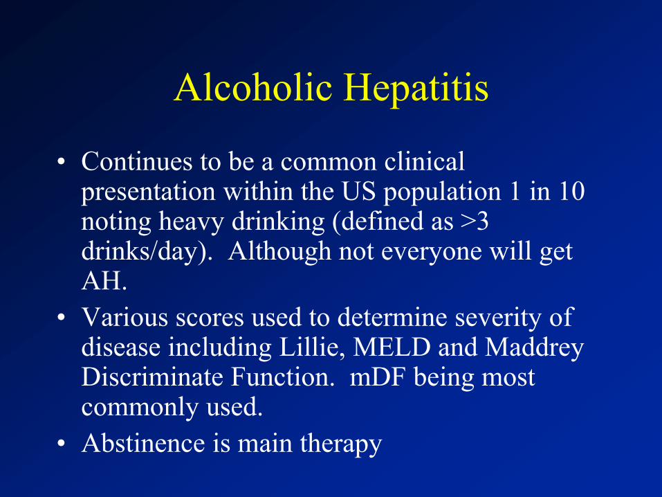
Alcoholic Hepatitis
• Continues to be a common clinical presentation within the US population 1 in 10 noting heavy drinking (defined as >3 drinks/day). Although not everyone will get AH.
• Various scores used to determine severity of disease including Lillie, MELD and Maddrey Discriminate Function. mDF being most commonly used.
• Abstinence is main therapy

Corticosteroids or Pentoxifylline
• Corticosteroids: – Most widely used agent
– Metaanalysis from 5 RCTs that used corticosteroids for severe AH showed an approximately 50% relative survival benefit at 1 month
• With the number needed to treat being 5 patients to reduce 1 death
– When used, oral prednisolone 40 mg daily or parenteral methylprednisolone (for patients unable to take orally) 32 mg per day is the usual initial dose and is administered for 4 weeks.
• About one-fourth of patients develop infection after starting steroids. Therefore, discontinuation of steroids is recommended in patients who are nonresponders after a week of therapy. – Defined as a rise in bilirubin or Lillie score >0.45
• Despite this documented benefit, practice surveys have shown that physicians in the United States prefer pentoxifylline over steroids for managing AH.

Corticosteroids or Pentoxifylline
• Pentoxifylline: – A phosphodiesterase inhibitor, a dose of 400 mg 3 times
daily for 28 days was associated with approximately 50% survival benefit in a pivotal study in 101 patients with severe AH17 and was superior to corticosteroids in another study.55
– However, a meta analysis of 5 RCTs has failed to show any benefit with pentoxifylline therapy.
– Ongoing study that compares corticosteroids to pentoxifylline to corticosteroids and pentoxifylline to no therapy.
– One consistent benefit of pentoxifylline is protection against renal dysfunction.
– Continues to be recommended therapy for severe AH

So How to Choose
Steroid Contraindications:
- Active Infection
- Uncontrollled DM
- Renal Failure
- GI bleeding
Steroid contraindications:
- Active or suspected infection
- Uncontrolled DM
- Renal failure
- Active GI bleeding

33 year old female presents to her primary care doctor for
evaluation of pruritus that she has had for the past 8 weeks.
She does not take any medications, denies any rash, fever or
chills. She denies jaundice, no travel and no recent sick
contact. Her lab data is essentially normal excepted for an
elevated alkaline phosphatase and ggt. Further work up is
negative for viral hepatitis, ANA normal, Total IgG normal,
AMA is positive at 1:1280. Which is the following is a
complication of her disease process?
1. Osteoporosis
2. Glucoma
3. HTN
4. Celiac disease
0%
0%
0%
0%
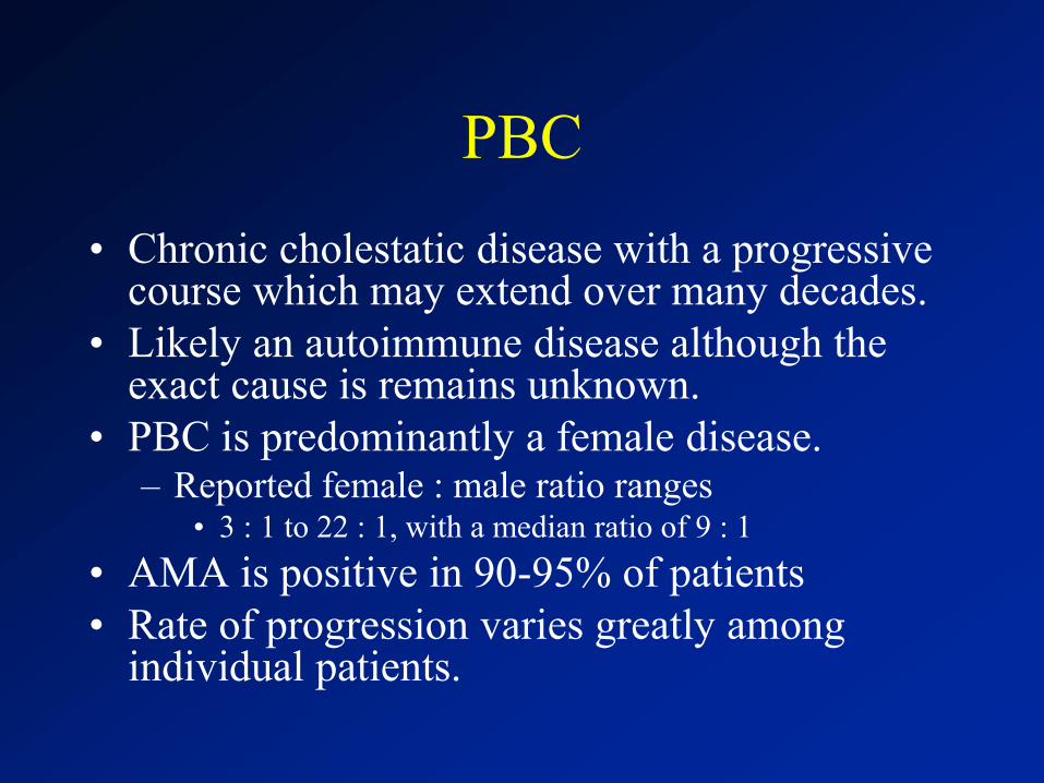
PBC
• Chronic cholestatic disease with a progressive course which may extend over many decades.
• Likely an autoimmune disease although the exact cause is remains unknown.
• PBC is predominantly a female disease. – Reported female : male ratio ranges
• 3 : 1 to 22 : 1, with a median ratio of 9 : 1
• AMA is positive in 90-95% of patients
• Rate of progression varies greatly among individual patients.

Osteoporosis and PBC
• Occurs in up to one-third of patients
• Relative risk for osteoporosis in PBC compared to an age matched and sex-matched healthy population is 4.4
• The cause of osteoporosis in PBC is uncertain. – Vitamin D metabolism is normal in patients with PBC
except for those with jaundice and clinically advanced disease.
• Women with PBC lose bone mass at a rate approximately twice that seen in age-matched controls.
• In a large series of PBC female patients, 21% developed fractures
Feldman: Sleisenger and Fordtran's Gastrointestinal and Liver Disease, 9th ed

PBC therapy
• Mainstay of therapy is and has been ursodeoxycholic acid
• 6a-Ethyl-chenodeoxycolic acid (obeticholic acid) is a novel derivative of the primary human bile acid
– In a study of 140 patients who did not achieve improvement of ALP to less than 1.67 times the upper limit of normal, the addition of obeticholic acid led to an additional 24% improvement in ALP

63 year old male presents to clinic for follow up from hospital
discharge after an upper gastrointestinal bleed. He states that his
upper gastrointestinal bleed was due to an ulcer, but he denies
that he takes any aspirin or NSAID. A biopsy at the time of
endoscopy revealed H.pylori and the patient was treated with 14
days of amoxicillin, clarithromycin and omeprazole. He went to
the lab before coming to your office and his H.pylori stool
antigen is positive. What is the next best step in treatment of his
H.Pylori?
1. Do nothing as it is not uncommon for the stool antigen to remain
positive
2. Repeat the same therapy for 21 days
3. Check H.pylori serology as it is more sensitive in determining
H.Pylori treatment results
4. Treat with Omeprazole, Bismuth, Tetracyline and Metronidazole
5. Treat with Omeprazole, Bismuth, Clarithromycin and Metronidazole
0%
0%
0%
0%
0%
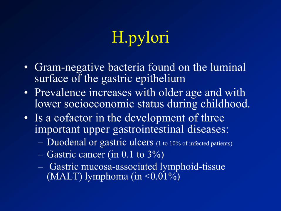
H.pylori
• Gram-negative bacteria found on the luminal surface of the gastric epithelium
• Prevalence increases with older age and with lower socioeconomic status during childhood.
• Is a cofactor in the development of three important upper gastrointestinal diseases: – Duodenal or gastric ulcers (1 to 10% of infected patients)
– Gastric cancer (in 0.1 to 3%)
– Gastric mucosa-associated lymphoid-tissue (MALT) lymphoma (in <0.01%)

H.Pylori therapy • The triple treatment including
PPI-clarithromycin and amoxicillin or metronidazole has become universal with initial therapy.
• However, most recent data show that this combination has lost some efficacy and often allows the cure of only a maximum of 70% of the patients.
• After failure of a PPI-clarithromycin containing therapy:
– Either a bismuth containing quadruple therapy or
– Levofloxacin containing triple therapy are recommended
McColl, Kenneth E L (04/29/2010). "Clinical practice. Helicobacter pylori infection". The New England journal of medicine(0028-4793), 362 (17), p. 1597.

H.Pylori testing
-Remember when confirming eradication to wait:
- 4 weeks after antibiotics are stopped
- 2 weeks after ppi are stopped.
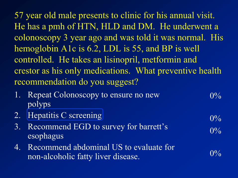
57 year old male presents to clinic for his annual visit.
He has a pmh of HTN, HLD and DM. He underwent a
colonoscopy 3 year ago and was told it was normal. His
hemoglobin A1c is 6.2, LDL is 55, and BP is well
controlled. He takes an lisinopril, metformin and
crestor as his only medications. What preventive health
recommendation do you suggest?
1. Repeat Colonoscopy to ensure no new polyps
2. Hepatitis C screening
3. Recommend EGD to survey for barrett’s esophagus
4. Recommend abdominal US to evaluate for non-alcoholic fatty liver disease.
0%
0%
0%
0%


Epidemiology of HCV
• ≈ 170 M persons infected worldwide
• 2.7 - 4 M Americans infected
• High prevalence rates in USA
– 2.5% of males
– 3.2% of African Americans
– 2.1% of Hispanic Americans
– Peak age of persons born between 1946 - 1964
World Health Organization (WHO). Wkly Epid Rec .1999;74:425-427. WHO.
Hepatitis C: Global Prevalence: Update. 2003. Semin Liver Dis. 2000;20:103-126.
Semin Liver Dis. 2000;20:1-16. Hepatology 2008;47:1128-1135.
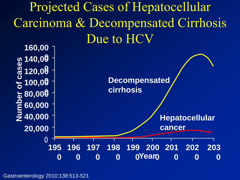
Projected Cases of Hepatocellular
Carcinoma & Decompensated Cirrhosis
Due to HCV
Gastroenterology 2010;138:513-521
195
0
196
0
197
0
198
0
199
0
200
0
201
0
202
0
203
0 Year
Nu
mb
er
of
cases
160,00
0
0
140,00
0 120,00
0 100,00
0 80,000
60,000
40,000
20,000
Decompensated
cirrhosis
Hepatocellular
cancer

Treatment Evolution of HCV
Sustained Viral Response (SVR)
39% 42%
34%
16%
54-56%
6%
0
20
40
60
80
100
IFN
6m
IFN/RBV
6m
Peg-IFN/
RBV 12m
IFN
12m
IFN/RBV
12m
Peg-IFN
12m
1991
1999
2001 2011
Peg-IFN/
RBV + PI
6-12m
70-80%
IFN: Interferon; m: months; RBV: Ribavirin; Peg: Pegylated; PI: Protease inhibitor;
DAA: Direct Acting Antiviral
2014
> 90%
DAA +
RBV +
Peg-IFN/
3-6m

55 year old male with a history of alcoholic cirrhosis presents to
clinic for follow up. He has ascites well controlled on
spironolactone 100mg and Lasix 40mg. He has recently
undergone EGD for variceal screening 3 months ago which was
reported as normal. He is up to date on his vaccinations and his
current MELD is 14. He underwent surveillance for
hepatocellular carcinoma two years ago. In regards to his
cirrhosis what other preventive measures should be ordered?
1. Repeat EGD
2. Liver Ultrasound
3. Echo with bubble study
4. Order AFP
0%
0%
0%
0%

Hepatocellular Carcinoma
• Diagnosed in more than half a million people worldwide every year, including approximately 20,000 new cases in the United States.
• 5-year cumulative risk for the development of hepatocellular carcinoma in patients with cirrhosis ranges between 5% and 30%, depending on:
– Etiology (highest risk those infected with HCV)
– Region or ethnic group (17% in the United States and 30% in Japan)
– Stage of cirrhosis (highest with decompensated disease)
El-Serag, Hashem B (09/22/2011). "Hepatocellular carcinoma". The New England journal of medicine (0028-4793), 365 (12), p.1118.

Underutilization of Surveillance
• 126, 670 patients with HCV
• 13002, with cirrhosis
• Routine surveillance: 12%
• Inconsistent Surveillance: 58.5%
• No Surveillance: 29.5%
Davila, Jessica A (01/18/2011). "Utilization of surveillance for hepatocellular carcinoma among hepatitis C virus-infected veterans in the United States". Annals of
internal medicine (0003-4819), 154 (2), p. 85.

HCC Surveillance
• In 2005 a RCT led to the recommendation of Surveillance by AASLD – RTC of 18,816 patients with AFP and US in HBV with
cirrhosis. Adherence to surveillance was subotimal (<60%) but in the group that did get surveillance the HCC related mortality was reduced by 37%.
– Recommended US and Alpha Fetoprotein every 6 mths.
• In 2011 AASLD altered those recommendations to only an US every 6mths – AASLD noted lack of sensitivity and specificity for
AFP therefore removed it from the guidelines
Modified from AASLD guidelines 2011
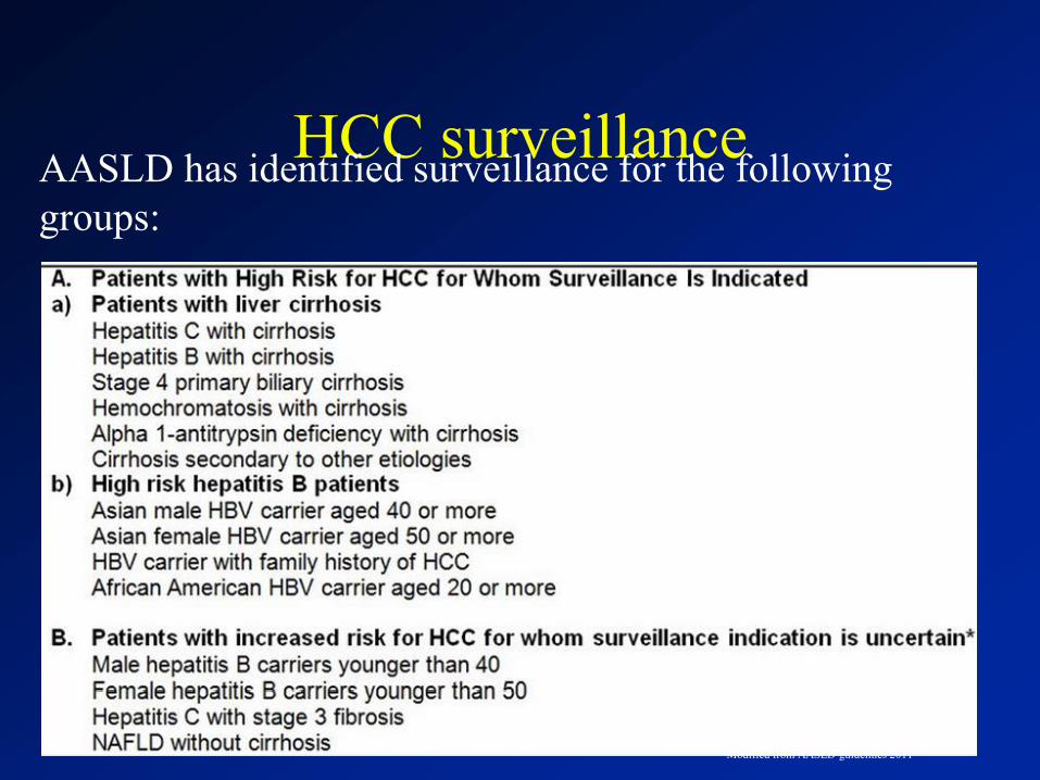
HCC surveillance AASLD has identified surveillance for the following
groups:
Modified from AASLD guidelines 2011
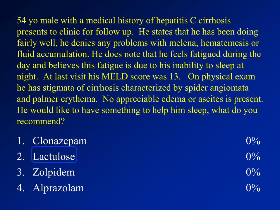
54 yo male with a medical history of hepatitis C cirrhosis
presents to clinic for follow up. He states that he has been doing
fairly well, he denies any problems with melena, hematemesis or
fluid accumulation. He does note that he feels fatigued during the
day and believes this fatigue is due to his inability to sleep at
night. At last visit his MELD score was 13. On physical exam
he has stigmata of cirrhosis characterized by spider angiomata
and palmer erythema. No appreciable edema or ascites is present.
He would like to have something to help him sleep, what do you
recommend?
1. Clonazepam
2. Lactulose
3. Zolpidem
4. Alprazolam
0%
0%
0%
0%
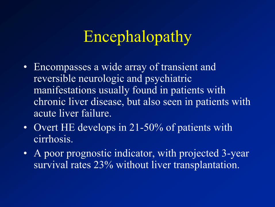
Encephalopathy
• Encompasses a wide array of transient and reversible neurologic and psychiatric manifestations usually found in patients with chronic liver disease, but also seen in patients with acute liver failure.
• Overt HE develops in 21-50% of patients with cirrhosis.
• A poor prognostic indicator, with projected 3-year survival rates 23% without liver transplantation.

Minimal Hepatic Encephalopathy
• MHE has a specific deficit profile on psychomotor testing:
– Attention deficits (major finding)
– Defect in visuo-motor coordination
– Defect in construction ability
– Defect in speed of mental processing
• > 50% progress to Overt HE over 3-4 years
• Affects up to 2/3 of cirrhotic patients

Encephalopathy


Hepatic Encephalopathy Therapy
• Lactulose remains the
mainstay of therapy
• Rifaximin is becoming a
popular choice among
physicians with low side
effect profile and improved
outcomes.
• Limiting issue seems to be
cost

44 year old male with pmh of htn is admitted to the ICU with hypotension, ARF and
diarrhea of 10-14 watery bowel movements daily. Diarrhea has been ongoing for the
past 2 months and this is his second hospitalization for diarrhea and ARF. After
volume resuscitation his ARF and hypotension resolve. His workup for his diarrhea
during included stool studies, c.dif, stool O&P all were negative. His only medication
is olmesartan for his HTN, although has had breaks in therapy due to his ARF with his
prior hospitalization. He underwent several empiric trials as an outpatient including
metronidazole, cholestyramine without success. With fasting in the hospital his
diarrhea improved. Just prior to his ICU admission he underwent an EGD and
colonoscopy which were reported as normal. However duodenal biopsies revealed
total villous atrophy and acute and chronic inflammation in the lamina propria. His
anti-TTG was negative, HLA DQ 2/8 were negative and antienterocyte antibody was
negative. What is best next step?
1. Start gluten free diet
2. Start Oral Vancomycin
3. Send Gastrin level
4. Stop Olmesartan
0%
0%
0%
0%

Olmesartan
• Rubio-Tapia A, Herman ML, Ludvigsson JF, et al. Severe spruelike enteropathy associated with olmesartan. Mayo Clin Proc. Aug 2012;87(8):732-738.
Olmesartan
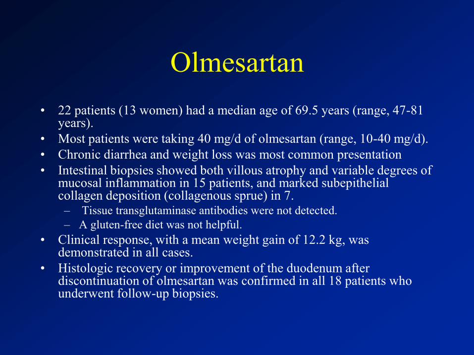
• 22 patients (13 women) had a median age of 69.5 years (range, 47-81 years).
• Most patients were taking 40 mg/d of olmesartan (range, 10-40 mg/d).
• Chronic diarrhea and weight loss was most common presentation
• Intestinal biopsies showed both villous atrophy and variable degrees of mucosal inflammation in 15 patients, and marked subepithelial collagen deposition (collagenous sprue) in 7. – Tissue transglutaminase antibodies were not detected.
– A gluten-free diet was not helpful.
• Clinical response, with a mean weight gain of 12.2 kg, was demonstrated in all cases.
• Histologic recovery or improvement of the duodenum after discontinuation of olmesartan was confirmed in all 18 patients who underwent follow-up biopsies.
Spruelike Enteropathy
• Since publication of this article:
• October 2012, researchers at the American
College of Gastroenterology meeting noted
a link to another 40 cases
• 7-13-2013 FDA required a label change to
olmesartan that included celiac like sprue to
the list of side effects.
A 19 yo male comes to the ER with the complaint of a food
impaction. He states that he was eating fried chicken
approximately 2 hours ago and felt like the food got stuck
midsternum. He states that he has had food “hang up” in the past,
but usually would pass with drinking water. He denies any
complaints of reflux or dyspepsia. He undergoes endoscopy with
successful removal of the food bolus. On endoscopy his
esophagus is described as feline in appearance with no evidence
of a stricture or narrowing. Which of the follow is likely to
improve his symptoms?
1. Calcium channel blockers
2. Nitroglycerin
3. Amitriptyline
4. Oral Fluticasone
0%
0%
0%
0%

Eosinophilic Esophagitis
• A recently recognized, disorder characterized by esophageal mucosal eosinophilia in association with dysphagia.
• Was first described in a few case reports in adults in the 1980s.
• Over the next decade, was mainly viewed as a childhood disease.
• The US prevalence estimate is 56.7/100,000 persons
Dellon, Evan S (04/2014). "Prevalence of eosinophilic esophagitis in the United States". Clinical gastroenterology and hepatology (1542-3565), 12 (4), p. 589.

• Common clinical manifestations seen in adults include – Dysphagia (most common)
– Food impaction
– Chest pain that is often centrally located and does not respond to antacids
– Gastroesophageal reflux disease-like symptoms/refractory heartburn
– Upper abdominal pain
• Diagnosis: – based upon symptoms, endoscopic appearance, and
histological findings.
– Histology >15 eosinophils per high power field

Endoscopy of Eosinophilic
Esophagitis
Treatment
• Pharmacologic:
– Trial of acid suppression with ppi
– Oral fluticasone
– Oral Budesonide
Summary
1. Remember the Oropharyngeal “-Ations”- Initiation, Aspiration, Nasal
Regurgitation, Lateralization.
2. Patients treated with statin therapy with chronic liver disease did not have
any excess toxicity compared to those taking placebo.
3. Osteoporosis occurs in up to one-third of patients with PBC
4. Remember to screen patients that were born between 1945-1965 for HCV.
5. Screen patients with cirrhosis for HCC every 6 months with an ultrasound
6. If a cirrhotic has trouble sleeping think about encephalopathy prior to a sleep
agent.
7. In patients on olmesartan if they start to develop diarrhea or symptoms of
malabsorption, discontinue.

• Questions?
• Thanks