Clinical, histological and therapeutic study regarding … · Clinical, histological and...
5
Romanian Journal of Morphology and Embryology 2009, 50(3):441–445 ORIGINAL PAPER Clinical, histological and therapeutic study regarding the variations of the edentulous ridge’s mucosa MONICA MIHAELA CRĂIŢOIU 1) , MIHAELA PĂUNA 2) , VERONICA MERCUŢ 1) , ŞTEFANIA CRĂIŢOIU 3) , ELENA ANNABEL NIŢULESCU 3) 1) Department of Prosthetic Dentistry, University of Medicine and Pharmacy of Craiova 2) Department of Prosthetic Dentistry, “Carol Davila” University of Medicine and Pharmacy, Bucharest 3) Department of Histology, University of Medicine and Pharmacy of Craiova Abstract The oral mucosa of the edentulous ridges presents many morphological macroscopic and microscopic variations, from one area of the edentulous ridge to another, as well as from one patient to another. Because the mobile prosthetic treatment realizes a direct contact between the dental prosthesis and the mucosa of the denture supporting structures and of the peripheral structures, the prosthetic success depends on establishing a correct and long lasting relation between them. The present article’s purpose is to present the pro-prosthetic and prosthetic treatment possibilities for partial or complete edentulous patients who have variations of the mucosa of the residual ridges, as well as to present the histopathological importance of these variations. The mucosal characteristics essentially influence the attitude in the different treatment phases of a mobile dental prosthesis, the prosthetic success depending on the establishment of a correct and long lasting relation between the dental prosthesis and the oral mucosa. Keywords: denture-bearing area, residual ridge, oral mucosa, morphological variation. Introduction Edentulism represents a pathological state of the stomatognatic system that appears because of the loss of a variable number of teeth [1]. Installation of the edentulism state because of tooth extraction is characterized by a post-extraction healing process that will conform in the end the mucosa of the edentulous ridge [2]. The residual ridge’s mucosa, part of the masticatory mucosa, presents variations from one area of the residual ridge to another and from one patient to another, variations that appear to be caused by exogenous and endogenous factors. Thus, the prosthetic therapeutic attitude will be imposed by the quality of the mucosal layer and will have the purpose of protecting the previously injured, by different factors, mucosa [3]. Patients and Methods The study was realized on a number of 26 partially and totally edentulous patients, of which 12 men and 14 women, who presented modifications of the residual ridge’s mucosa. Twenty patients had previously been wearers of dental prosthetic restorations and six patients had never undergone prosthodontic treatment. The utilized methods were clinical study, histopatho- logy study and prosthetic treatment. The clinical study pursued the examination of the mucosal area, highlighting its thickness, the relationships with the underlying bone structures and its sensitivity. Type, size, duration of evolution, color, texture, confi- guration, consistency and their mobility were marked out at the patients who presented different modifications of the residual ridge’s mucosa. The possible cause– effect relationship between a prosthetic dental treatment and the appearance of the residual ridge’s mucosa modification was also investigated. From patients that required surgical pro-prosthetic treatment, fragments of oral mucosa were obtained through excision of the residual ridge’s modified mucosa. The oral mucosa fragments, after being fixed in 10% formalin, were processed through the paraffin histological technique. The sections obtained were stained with Hematoxylin–Eosin (HE) and Goldner– Szeckelly trichromic (GS). Treatment options were presented for every patient, as well as the way in which the mucosal characteristics influenced the therapeutic decision. Results We present two significant clinical cases. Case no. 1 Patient G.A., aged 68-year, presented to the Prosthetic Dentistry Clinic of the Faculty of Dental Medicine of Craiova requesting prosthetic dental treatment for a Kennedy Class I maxillary edentation.
Transcript of Clinical, histological and therapeutic study regarding … · Clinical, histological and...

Romanian Journal of Morphology and Embryology 2009, 50(3):441–445
OORRIIGGIINNAALL PPAAPPEERR
Clinical, histological and therapeutic study regarding the variations of the edentulous
ridge’s mucosa MONICA MIHAELA CRĂIŢOIU1), MIHAELA PĂUNA2), VERONICA MERCUŢ1),
ŞTEFANIA CRĂIŢOIU3), ELENA ANNABEL NIŢULESCU3)
1)Department of Prosthetic Dentistry, University of Medicine and Pharmacy of Craiova
2)Department of Prosthetic Dentistry, “Carol Davila” University of Medicine and Pharmacy, Bucharest
3)Department of Histology, University of Medicine and Pharmacy of Craiova
Abstract The oral mucosa of the edentulous ridges presents many morphological macroscopic and microscopic variations, from one area of the edentulous ridge to another, as well as from one patient to another. Because the mobile prosthetic treatment realizes a direct contact between the dental prosthesis and the mucosa of the denture supporting structures and of the peripheral structures, the prosthetic success depends on establishing a correct and long lasting relation between them. The present article’s purpose is to present the pro-prosthetic and prosthetic treatment possibilities for partial or complete edentulous patients who have variations of the mucosa of the residual ridges, as well as to present the histopathological importance of these variations. The mucosal characteristics essentially influence the attitude in the different treatment phases of a mobile dental prosthesis, the prosthetic success depending on the establishment of a correct and long lasting relation between the dental prosthesis and the oral mucosa. Keywords: denture-bearing area, residual ridge, oral mucosa, morphological variation.
Introduction
Edentulism represents a pathological state of the stomatognatic system that appears because of the loss of a variable number of teeth [1].
Installation of the edentulism state because of tooth extraction is characterized by a post-extraction healing process that will conform in the end the mucosa of the edentulous ridge [2].
The residual ridge’s mucosa, part of the masticatory mucosa, presents variations from one area of the residual ridge to another and from one patient to another, variations that appear to be caused by exogenous and endogenous factors. Thus, the prosthetic therapeutic attitude will be imposed by the quality of the mucosal layer and will have the purpose of protecting the previously injured, by different factors, mucosa [3].
Patients and Methods
The study was realized on a number of 26 partially and totally edentulous patients, of which 12 men and 14 women, who presented modifications of the residual ridge’s mucosa. Twenty patients had previously been wearers of dental prosthetic restorations and six patients had never undergone prosthodontic treatment. The utilized methods were clinical study, histopatho-logy study and prosthetic treatment.
The clinical study pursued the examination of the mucosal area, highlighting its thickness, the relationships
with the underlying bone structures and its sensitivity. Type, size, duration of evolution, color, texture, confi-guration, consistency and their mobility were marked out at the patients who presented different modifications of the residual ridge’s mucosa. The possible cause–effect relationship between a prosthetic dental treatment and the appearance of the residual ridge’s mucosa modification was also investigated.
From patients that required surgical pro-prosthetic treatment, fragments of oral mucosa were obtained through excision of the residual ridge’s modified mucosa. The oral mucosa fragments, after being fixed in 10% formalin, were processed through the paraffin histological technique. The sections obtained were stained with Hematoxylin–Eosin (HE) and Goldner–Szeckelly trichromic (GS).
Treatment options were presented for every patient, as well as the way in which the mucosal characteristics influenced the therapeutic decision.
Results
We present two significant clinical cases.
Case no. 1
Patient G.A., aged 68-year, presented to the Prosthetic Dentistry Clinic of the Faculty of Dental Medicine of Craiova requesting prosthetic dental treatment for a Kennedy Class I maxillary edentation.
Monica Mihaela Crăiţoiu et al.
442
The patient had a history of arterial hypertension for about four years and was treated by a cardiologist with specific medication. She had undergone prosthetic treatment several years before and had had a metal post at the level of the right upper canine that had repeatedly traumatized the mucosa of the alveolar process. At the time of the patient’s first visit to the Prosthetic Dentistry Clinic, the upper right canine had been extracted, so the trauma cause (the metal post) had been removed.
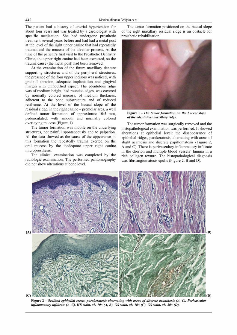
At the examination of the future maxillary denture supporting structures and of the peripheral structures, the presence of the four upper incisors was noticed, with grade I abrasion, adequate implantation and gingival margin with unmodified aspect. The edentulous ridge was of medium height, had rounded edges, was covered by normally colored mucosa, of medium thickness, adherent to the bone substructure and of reduced resilience. At the level of the buccal slope of the residual ridge, in the right canine – premolar area, a well defined tumor formation, of approximate 10/5 mm, pedunculated, with smooth and normally colored overlaying mucosa (Figure 1).
The tumor formation was mobile on the underlying structures, not painful spontaneously and to palpation. All the data showed as the cause of the appearance of this formation the repeatedly trauma exerted on the oral mucosa by the inadequate upper right canine microprosthesis.
The clinical examination was completed by the radiologic examination. The performed pantomography did not show alterations at bone level.
The tumor formation positioned on the buccal slope of the right maxillary residual ridge is an obstacle for prosthetic rehabilitation.
Figure 1 – The tumor formation on the buccal slope of the edentulous maxillary ridge.
The tumor formation was surgically removed and the histopathological examination was performed. It showed alterations at epithelial level: the disappearance of epithelial ridges, parakeratosis, alternating with areas of slight acantosis and discrete papillomatosis (Figure 2, A and C). There is perivasculary inflammatory infiltrate in the chorion and multiple blood vessels’ lumina in a rich collagen texture. The histopathological diagnosis was fibroangiomatosis epulis (Figure 2, B and D).
(A) (B)
(C) (D)Figure 2 – Oralized epithelial crests, parakeratosis alternating with areas of discrete acanthosis (A, C). Perivascular inflammatory infiltrate (A–C). HE stain, ob. 10× (A, B). GS stain, ob. 10× (C). GS stain, ob. 20× (D).

Clinical, histological and therapeutic study regarding the variations of the edentulous ridge’s mucosa
443
Post-surgical removal, on the buccal slope of the right maxillary residual ridge, in the canine–premolar area resulted a mucosal scar area of about 2 cm in length, not capable of supporting the prosthetic pressure (Figure 3).
Figure 3 – The post-excisional scar area.
In the prosthetic choice, along with considerations regarding the topography and type of edentation, the necessity of protecting the scar area was also considered. The partial removable prosthesis allowed the making of prosthetic saddles with reduced dimensionally margins that protect the area and in the same time insure the necessary mechanical resistance (Figures 4 and 5).
Figure 4 – The removal partial prothesis on the model.
Figure 5 – The partial removable prosthesis in the oral cavity.
Case no. 2
Patient S.Z., aged 55-year, came to the Clinic of Prosthetic Dentistry of The Faculty of Dental Medicine of Craiova presenting maxillary and mandibulary total edentation. Anamnesis revealed the fact that the patient had been wearing a complete maxillary denture for ten years and a mandibular partial removable prosthesis, as the lower right canine had been present on the mandibular arch until one month before the patient’s first visit to the Clinic of Prosthetic Dentistry.
The clinical examination of the mandibular edentulous ridge revealed an important bone resorption in the posterior area [4]. In the anterior buccal area, the edentulous mandibular flabby ridge is covered by hyperplastic, slightly sensitive to palpation mucosa, that represents an impediment for the prosthetic treatment (Figure 6A).
The flabby mucosal formation was surgically removed. After a healing period of 14 days, a regular future denture bearing area was obtained, covered by mucosa capable of supporting the prosthetic pressure (Figure 6B). The histopathological examination was performed. It revealed acantosis, acantolisis in the superficial epithelial layers, superficial parakeratosis and papilomatosis (Figure 7, A and B). In the depth of the chorion, a fibrosis process is present, associated with discrete perivascular inflammatory infiltrate.
After the healing, the prosthetic therapy consisted of the realization of two complete dentures with borders molded according to the new morphology (Figure 8).
(A) (B)Figure 6 – Anterior mandibular flabby ridge (A), and the post-excisional aspects (B).

Monica Mihaela Crăiţoiu et al.
444
(A) (B)
(C) (D)Figure 7 – Acanthosis, superficial acatholysis, parakeratosis (A, B), in the lamina propria, areas of fibrosis and lymphoplasmatic inflammatory infiltrate (C, D). HE stain, ob. 10× (A, D) and ob. 20× (B). GS stain, ob. 20× (C).
(A) (B)Figure 8 – Mandibular complete dentures in the oral cavity (A) and the final aspect of the prosthetic treatment (B).
Discussion
The prosthetic therapy with complete dentures realizes a direct contact between the prosthesis and the mucosal layer. Between the mucosal face of the prosthesis and the denture-bearing mucosa, a permanent relationship is established [5].
The mucosal layer of the denture bearing area has a variable macroscopic and microscopic morphology and is, during the entire period of prosthetic pressure [6], under the influence of exogenous and endogenous factors that modify it [7].
The exogenous factors are classified as follows: ▪ mechanical factors, represented by different trauma
types, including the ones caused by incorrect prosthetic restorations, or by old and no longer adequate ones;
▪ chemical factors, represented by drugs such as antibiotics, antimycotics, tonicardiacs, corticosteroids, tranquilizers, neuroleptics, etc.;
▪ biological factors, such as viruses and bacteria. The endogenous factors are represented by diabetes
mellitus, autoimmune diseases that disturb the immuno-logical mechanisms, avitaminosis, sialopathies, etc.
All these factors, together with the process of bone resorption that accompanies the use of a mobile prosthesis, compromise the congruence between the denture’s internal surface and the denture bearing area [8]. Thus, negative effects appear for the denture’s bearing area mucosa, as well as for the maintenance, rest and stability of the denture’s base.
In these conditions, it is necessary to make, during the different treatment phases of a mobile dental

Clinical, histological and therapeutic study regarding the variations of the edentulous ridge’s mucosa
445
prosthesis, a competent study of each area of the mucosal layer of the denture bearing zone and an evaluation of its potential of alteration, with the purpose of initiating an individualized pro-prosthetic and prosthetic therapy [9, 10].
The histopathological study we conducted revealed the changes of the mucosa of the edentulous ridges. The epithelium is seriously altered, presenting the disappearance of epithelial crests, parakeratosis, alter-nating with areas of slight acanthosis and acantholysis. The lamina propria’s connective tissue presents a zonal process of collagen fibrosis, associated with perivas-cular inflammatory infiltrate. Thus, at epithelial level the functionality is compromised, while the chorion is affected only in certain areas and it maintains the recovery capacity, especially the one of the components of the extracellular matrix, as well as the capacity of cellular multiplication. At the same time, the local defense is well established through the macrophages, lymphocytes and plasma cells that are present in a great number in the chorion. The defense process takes place through the phagia process, as well as through the lymphoplasmatic immune process. This process is well sustained by a rich vascularisation that normally insures the bidirectional exchanges between blood and the tissues [11].
Considering the fact that the oral mucosa is only regionally affected and the recovery potential of its connective tissue, the prosthetic therapy can be realized in normal functional conditions, because the oral mucosa of the denture supporting area will be able to support the prosthetic pressures [12, 13].
Certain mucosal modifications produced under the influence of aggressive exogenous and endogenous factors, cause a decrease of the tolerance degree of certain mucosal areas. The success of the prosthetic treatment depends also on a mucosal denture bearing area capable of supporting the prosthetic pressures [14]. The realization of mobile prosthetic dentures through almost standard techniques can only lead to obtaining dentures that will be in conflict with the mucosal layer of the denture supporting area and that will influence the prosthetic therapy’s efficiency [15].
Conclusions
The mucosal characteristics essentially influence the therapeutic attitude during the different treatment phases of a mobile dental prosthesis, the prosthetic treatment’s success depending on establishing a correct, long lasting relation between the dental prosthesis and the oral mucosa.
In the studied cases, the edentulous ridge’s mucosa presented histological modifications at the level of the epithelium, as well as at chorion level. Because the
connective tissue’s modifications are only zonal and they consist of a macrophage-type defense system and of an immune one, the functionality of the lamina propria is not compromised, maintaining its recovery capacity.
The prosthetic treatment was conduced respecting the local mucosal characteristics and individualizing each prosthetic phase accordingly, so that the prosthetic therapy
can be adapted to the quality of the mucosal layer.
References [1] BUDTZ-JORGGENSEN E., Sequelae of wearing complete
dentures. In: ZARB G. A., BOLENDER C. L., CARLSSON G. E. (eds), Boucher’s prosthodontic treatment for edentulous patients, 11th edition, CV Mosby, St. Louis, 1997, 30–45.
[2] IVANHOE J. R, CIBIRKA R. M., PARR G. R., Treating the modern complete denture patient: a review of the literature, J Prosthet Dent, 2002, 88(6):631–635.
[3] ELLIS J. S., PELEKIS N. D., THOMASON J. M., Conventional rehabilitation of edentulous patients: the impact on oral health-related quality of life and patient satisfaction, J Prosthodont, 2007, 16(1):37–42.
[4] TAN K. M., SINGER M. T., MASRI R., DRISCOLL C. F., Modified fluid wax impression for a severely resorbed edentulous mandibular ridge, J Prosthet Dent, 2009, 101(4):279–282.
[5] ZARB G. A., BOLENDER C. L., CARLSSON G. E. (eds), Boucher’s prosthodontic treatment for edentulous patients, Mosby Year Book, St. Louis, Missouri, 1997, 141–181.
[6] KUBO K., KAWATA T., SUENAGA H., YODA N., SHIGEMITSU R., OGAWA T., SASAKI K., Development of in vivo measuring system of the pressure distribution under the denture base of removable partial denture, J Prosthodont Res, 2009, 53(1):15–21.
[7] OGAWA T., TANAKA M., OGIMOTO T., OKUSHI N., KOYANO K., TAKEUCHI K., Mapping, profiling and clustering of pressure pain threshold (PPT) in edentulous oral mucosa, J Dent, 2004, 32(3):219–228.
[8] XIE Q., NÄRHI T. O., NEVALAINEN J. M., WOLF J., AINAMO A., Oral status and prosthetic factors related to residual ridge resorption in elderly subjects, Acta Odontol Scand, 1997, 55(5):306–313.
[9] PĂUNA M., PREOTEASA E., Practical aspects in the prosthetic therapy of complete edentulism, Cerma Publishing House, Bucharest, 2005, 17–59 (in Romanian).
[10] HEYDECKE G., SCHNITZER S., TÜRP J. C., The color of human gingiva and mucosa: visual measurement and description of distribution, Clin Oral Investig, 2005, 9(4):257–265.
[11] HÜE O., BERTERETCHE M. V., Complete dentures. Clinic reality. Therapeutical solutions, Quintessence International, Paris, 2004, 5–15, 27–38.
[12] RIGNON-BRET C., RIGNON-BRET J. M., Complete dentures. Immediate prosthesis. Overdenture, CdP, Langres, France, 2004, 21–60.
[13] HUTU EM., PĂUNA M., BODNAR V., CONSTANTINESCU M. V., ŢÂNCU A., Complete edentulism, National Publishing House, Bucharest, 2005, 16–39, 240–245 (in Romanian).
[14] LELLO G. E., MAKEK M., Submucosal nodular chondro- metaplasia in denture wearers, J Prosthet Dent, 1985, 54(2):237–240.
[15] WALISZEWSKI M., Restoring dentate appearance: a literature review for modern complete denture esthetics, J Prosthet Dent, 2005, 93(4):386–394.
Corresponding author Monica Mihaela Crăiţoiu, Assistant, MD, PhD candidate, Department of Prosthetic Dentistry, University of Medicine and Pharmacy of Craiova, 2–4 Petru Rareş Street, 200349 Craiova, Romania; Phone +40723–629 595, e-mail: [email protected] Received: March 25th, 2009
Accepted: June 15th, 2009