Clinical Guidelines by Consensus Recording a standard …€¦ · Clinical Guidelines by Consensus...
26
Clinical Guidelines by Consensus Recording a standard 12-lead electrocardiogram An approved methodology by the Society for Cardiological Science & Technology (SCST) Issue Date: June 2014 Review Date: June 2017
-
Upload
dinhkhuong -
Category
Documents
-
view
216 -
download
0
Transcript of Clinical Guidelines by Consensus Recording a standard …€¦ · Clinical Guidelines by Consensus...

Clinical Guidelines by Consensus
Recording a standard 12-lead electrocardiogram
An approved methodology by the Society for Cardiological Science & Technology
(SCST)
Issue Date: June 2014 Review Date: June 2017
CS2: Recording a 12-lead ECG v2.1 Page 2
Clinical Guidelines by Consensus Recording a standard 12-lead electrocardiogram
An approved methodology by the Society for Cardiological Science & Technology (SCST)
Document ID CS 2 Lead or Editing Author(s) Jane Eldridge & Dave Richley Lead Authors’ Job titles See acknowledgements Original author(s) Chris Eggett Additional author(s) See acknowledgements Document version number CS 2.1 Ratifying Committee SCST Council Ratification Date Sept 2014 Review Date Sept 2017 Body responsible for review SCST Council Committee for review Clinical Standards Committee Contact for document [email protected] Referencing Included Yes Key Words (for searching) ECG, electrocardiogram, 12-lead Intended users Practitioners recording 12-lead ECGs Equality Impact Assessment Yes
Please cite as: Eldridge MJ, Richley D, Ross C, Cox C, Breen C. (2014) Clinical Guidelines by Consensus: Recording a standard 12-lead electrocardiogram. An approved methodology by the Society for Cardiological Science and Technology (SCST). Available at: http://www.scst.org.uk/resources/CAC_SCST_Recording_a_12-lead_ECG_final_version_2014_CS2v2.1.pdf (include date accessed)

CS2: Recording a 12-lead ECG v2.1 Page 3
Acknowledgements
With many thanks to the following people for help in the document development and review process:
Su Baxter, SCST Chair Head of Cardiology, Good Hope Hospital, Heart of England Foundation Trust Sophie Blackman, SCST Council Member Principal Physiologist, West Herts NHS Trust Cathal Breen, SCST Council Member Lecturer & Course Director Clinical Physiology, University of Ulster Chris Brown, SCST CS Committee Member Principal Physiologist, Shetland Brian Campbell, SCST President Head of Echo & Community Services, Guy’s & St Thomas’s NHS Trust Chris Cox, SCST CS Committee Member Chief Cardiac Physiologist, County Durham & Darlington NHS Trust Jane Eldridge, SCST CS Committee Lead Lead Cardiac Physiologist EP, Papworth Hospital NHS Foundation Trust John Hutchinson, SCST Council Member Lead Cardiac Physiologist, Papworth Hospital NHS Foundation Trust Emma Rees, SCST Council Member Senior Lecturer & Programme Manager, Swansea University Dave Richley, SCST Council Member Cardiac Physiology Lecturer Practitioner, NHS England Catherine Ross, SCST Council Member Specialist Cardiac Physiologist, Craigavon Area Hospital

CS2: Recording a 12-lead ECG v2.1 Page 4
Contents 1. Change history ........................................................................................... 5 2. Introduction ................................................................................................ 5 3. Purpose ..................................................................................................... 6 4. Qualifications and training of staff recording 12-lead ECGs ...................... 6 5. Patient experience, privacy and dignity ..................................................... 7
5.1 Communication .................................................................................... 7 5.2 Level of undress ................................................................................... 7 5.3 Chaperones ......................................................................................... 7
6. Environmental considerations ................................................................... 8 7. Equipment specification ............................................................................. 8 8. Equipment and room preparation .............................................................. 9 9. Infection control ......................................................................................... 9 10. Identification of patient ........................................................................... 10 11. Informed consent ................................................................................... 10 12. Patient preparation ................................................................................ 10
12.1 Patient position ................................................................................ 10 12.2 Skin preparation ............................................................................... 11
13. Electrode placement .............................................................................. 11 13.1 Limb electrode positions .................................................................. 11 13.2 Precordial (chest) electrode positions .............................................. 12
14. Technique for locating chest electrode positions ................................... 13 15. Obtaining a good quality recording ........................................................ 14 16. Documentation, processing, storage and confidentiality of ECG
recordings .............................................................................................. 16 17. Special situations ................................................................................... 17
17.1 Dextrocardia ..................................................................................... 17 17.2 Posterior electrode positions ............................................................ 18 17.3 Electrocardiography on children ...................................................... 18
18. Audit ....................................................................................................... 19 19. Conclusion ............................................................................................. 19 20. References ............................................................................................ 20 Appendix: 12-lead ECG Service Audit Tool ................................................... 24 ECG Standard Electrode Positions Reminder Chart ...................................... 25 ECG Settings Reminder Chart ....................................................................... 26
CS2: Recording a 12-lead ECG v2.1 Page 5
1. Change history Version Date Author Reason Ratification
Required 1 Feb 2010 Consensus Standardisation of
practice Yes: SCST Council
2 Jun 2014 Consensus Update
Yes: SCST Council
2.1 Nov 2015 Consensus Citation added, contributors details updated
Not required
2. Introduction The electrocardiogram (ECG) is an important cardiac diagnostic procedure. It is the first test that most people with suspected heart disease undergo and it has been estimated that up to 300 million ECGs a year are recorded in Europe alone1. The portability, low cost and ease of operation of modern ECG machines means that ECGs can be recorded quickly and easily by a wide variety of personnel in hospitals, doctors’ surgeries, ambulances, sports facilities, patients’ homes and many other environments. The challenge is to achieve effective quality assurance in all environments. There is evidence that many personnel who record ECGs have not been properly trained or assessed in the technique2. Reports and formal studies from a number of countries have repeatedly found that practitioners lack essential knowledge in ECG recording technique and that as a consequence many ECGs are recorded wrongly3,4,5,6. Incorrectly recorded ECGs may result in erroneous diagnoses and inappropriate treatments7,8,9,10,11,12,13.
There is evidence that training results in fewer ECG recording errors14 but it appears that many practitioners are unaware of their need for training; studies have shown that self-confidence is no guide to competence in recording ECGs15,16. SCST recommends that all personnel who record ECGs should be appropriately trained and qualified. It is intended that this document be used as a source of information about correct ECG recording technique to help achieve better practice and improved outcomes for patients. Guidelines provided by SCST are written by expert practitioners in the field, following critical evaluation of the evidence. The intention is to disseminate best practice in order to ensure diagnostic accuracy and improve the patient experience. Where there is limited evidence, consensus decisions have been made. SCST regularly reviews its guidelines to ensure that these continue to reflect current good scientific practice17.

CS2: Recording a 12-lead ECG v2.1 Page 6
3. Purpose This guideline, in promoting excellence in the recording of 12-lead ECGs, addresses the following areas: • Qualification and training of staff recording 12-lead ECGs • Patient experience, privacy and dignity • Investigation environment • Equipment specification • Equipment and room preparation • Patient preparation and infection control • Patient identification and consent • Electrode placement:
§ Limb electrode positions § Chest electrode positions
• Technical skills required for obtaining an accurate artefact-free recording of good clinical quality and diagnostic value
• Optimising settings and making adjustments including: § Use of the filter § Paper speed § Amplitude gain
• Special situations • Documentation, processing, storage and confidentiality of 12-lead ECG
recordings
4. Qualifications and training of staff recording 12-lead ECGs The operator must be competent in the use of the electrocardiograph and in the recording of an ECG16. This should be demonstrated by the possession of a recognised qualification, such as one of those awarded by the Society for Cardiological Science and Technology, including:
• Award in Practical Electrocardiography • Certificate in Electrocardiography • Diploma in Electrocardiography
It is essential that competence in the recording of an electrocardiogram be maintained and this should be demonstrated by periodical review.

CS2: Recording a 12-lead ECG v2.1 Page 7
5. Patient experience, privacy and dignity A key goal of good scientific practice is to ensure that the patient has a positive experience of the procedure17.
5.1 Communication Good communication skills are a key part of enhancing the patient’s experience of any investigative procedure. The patient should be given adequate information about the ECG procedure and this should be clear, precise and communicated at a level consistent with the patient’s needs. Information may be in the form of a booklet, information letter or oral explanation. As a minimum, SCST recommends that before any contact with the patient, operators should introduce themselves, their role and give a brief description of the procedure for recording an ECG. If possible, this should cover the level of undress involved, the use of adhesive electrodes, reassurance that the procedure is brief and painless and a verbal confirmation of consent to continue.
5.2 Level of undress Operators should make every effort to respect the cultural sensitivities of the patient and to minimise embarrassment18. Many patients will feel uncomfortable being touched on their upper torso; operators must be sensitive to this and act in a sympathetic, caring and compassionate manner. Patients should be asked to undress above the waist to allow access to the upper torso for accurate electrode placement. Patients should be allowed to undress in a private environment with minimal risk of interruption. Once the electrodes have been attached the patient may be covered with a gown to preserve his/her modesty. If the patient is comfortable and relaxed it is more likely that the ECG recorded will be free of artefact. Clinical discussions with patients should take place only after re-dressing.
5.3 Chaperones In accordance with the General Medical Council recommendations, a patient should always be given the option to have a chaperone present19. The chaperone must be a health professional with knowledge of the standard practice of recording a 12-lead ECG. Patients may also request a relative or carer to be present.

CS2: Recording a 12-lead ECG v2.1 Page 8
6. Environmental considerations The environment in which a 12-lead ECG is recorded may contribute considerably to the quality of the experience and output. As far as possible the environment should be: • Private: walled, curtained or screened • Quiet • Comfortable • Accessible for disabled and able-bodied patients and staff • Safe • Clean, with appropriate hand-cleaning and clinical waste facilities • Furnished with a height adjustable couch accessible from both sides • Stocked appropriately, with razors, electrodes, ECG paper, etc
7. Equipment specification The electrocardiograph must meet or exceed the requirements of International Electrotechnical Commission standards IEC 6061-2-25:2011i The device should be pre-programmed in accordance with the American Heart Association (AHA) specifications20 as follows: • To avoid distortion of the ST segment the low-frequency cut-off should be
no higher than 0.67Hz in ‘auto’ mode, or 0.05Hz in ‘manual’ mode ii
• To prevent the loss of high frequency information the high frequency cut-off should be no lower than 150Hz in adults and adolescents and no lower than 250Hz in children
• Disposable tab electrodes must meet or exceed the requirements of the
American National Standards Institute/Association for the Advancement of Medical Instrumentation documentiii
i IEC 6061-2-25:2011 – establishes particular requirements for safety, including essential performance of recording and analysing single channel and multichannel electrocardiographs. ii Digital filter design allows for low-frequency filter level of 0.67Hz when recording in ‘auto’ mode. However ST segment distortion may occur when this setting is used in ‘manual’ mode. Fixing the low frequency setting at 0.05Hz in the pre-set should prevent this error occurring. iii ANSI/AAMI EC12:2000(R)2010 – establishes minimum labeling, safety and performance requirements for disposable electrodes used for diagnostic electrocardiography

CS2: Recording a 12-lead ECG v2.1 Page 9
8. Equipment and room preparation Equipment should be safe and ready to use with correct date and time settings. The room and equipment should be clean and orderly with all waste from previous investigations disposed of as clinical waste. All mains leads, cables and connectors should be intact with no evidence of fractures, faults or insulation damage. For battery-operated machines, the battery will need to have sufficient charge. It may be useful for a mains-powered ECG machine to have an easily distinguishable plug if it is to be used in environments, e.g. intensive care units, where a number of items of vital equipment are plugged into wall sockets.
9. Infection control Measures to control the risk of infection transmission must be taken in accordance with local policy. Hands should be washed21 with soap and water or cleansed with alcohol gel, as per local policy, before and after any contact with a patient. It may be reassuring to patients if this is done in their presence. For patients requiring high levels of infection control precautions, personal protective equipment such as gowns and gloves should be worn, in accordance with local policies. Appropriate clinical waste disposal facilities should be available including sharps bins for the disposal of razors.
Recommended recording bandwidths pre-stored in device set-up:
‘Auto’ mode 0.67 – 150Hz
‘Manual’ mode 0.05 – 150Hz
Mains filter (50Hz filter) Off
CS2: Recording a 12-lead ECG v2.1 Page 10
10. Identification of patient It is essential that patients be correctly identified and that appropriate identifying details be displayed on the ECG recording. For patients unable to provide their own identifying details, confirmation of identity must be sought from carers or by using hospital wrist bands. The printed recording must always be checked to ensure it bears the correct patient details. Operators must be aware of potential sources of error if details are not entered digitally for every patient. Some machines retain the information from the last patient and these may be incorrectly printed on the ECG if they have not been altered. Local policy and practice should be developed to ensure that errors do not occur in busy clinical environments.
11. Informed consent Consent must be obtained in accordance with local policy. Practitioners should explain the ECG recording procedure briefly to patients and obtain their oral consent before starting.
12. Patient preparation Whilst it is recognised that 12-lead ECGs are performed in a variety of contexts, environments and states of urgency, attempts to achieve best practice and standard electrode positioning should always be made.
12.1 Patient position The appearance of the ECG can be affected by the angle of incline of the torso at the time of recording. Whilst it is known that an ECG recorded from a supine subject may differ significantly from one recorded when the subject is upright22 and it may make some difference whether a subject is supine or inclined at 60 degrees to the horizontal23, evidence is lacking to suggest that inclinations ranging from supine to 45 degrees affect the ECG recording. Many patients may be uncomfortable lying flat so, for consistency and practicality, a semi-recumbent position of approximately 45 degrees to the horizontal is recommended. Any variation from this position should be noted on the ECG. The limbs should be supported by the bed or couch to minimise artefact due to muscle tension.

CS2: Recording a 12-lead ECG v2.1 Page 11
12.2 Skin preparation Skin preparation is often required to help produce an artefact-free ECG. Care must be taken with patients who have sensitive or broken skin. Various methods are available to minimise the skin-to-electrode impedance, for example: • The skin may require cleansing. There are a variety of methods, including
washing with mild soap and cleaning with an alcoholic wipe. • Exfoliation may be required and should be undertaken with very light
abrasion using a paper towel, gauze swab or proprietary abrasive tape designed for this purpose.
• Chest hair may need to be removed to ensure adequate contact with the
skin. Oral consent should be obtained from the patient and an unused razor should be employed and disposed of in a sharps bin immediately afterwards.
13. Electrode placement Electrodes must be positioned in accordance with AHA recommendations20,24,25. If any of the electrodes need to be sited in non-standard positions the recording must be labelled with this information to avoid misinterpretation of altered ECG waveforms25,26. Each lead wire is generally colour coded to aid identification. However, colour may vary depending on manufacturer. The colours detailed in this document comply with European (IEC) recommendations.
13.1 Limb electrode positions
Limb electrodes should be placed on the wrists and ankles whenever possible. Moving the electrodes up the limbs may alter the appearance of the ECG and should be avoided unless there is a significant tremor or a limb has been amputated. Limb electrodes must not be placed on the torso since this causes significant alteration to wave amplitudes which can invalidate the use of the recording for many diagnostic purposes26,27.

CS2: Recording a 12-lead ECG v2.1 Page 12
13.2 Precordial (chest) electrode positions
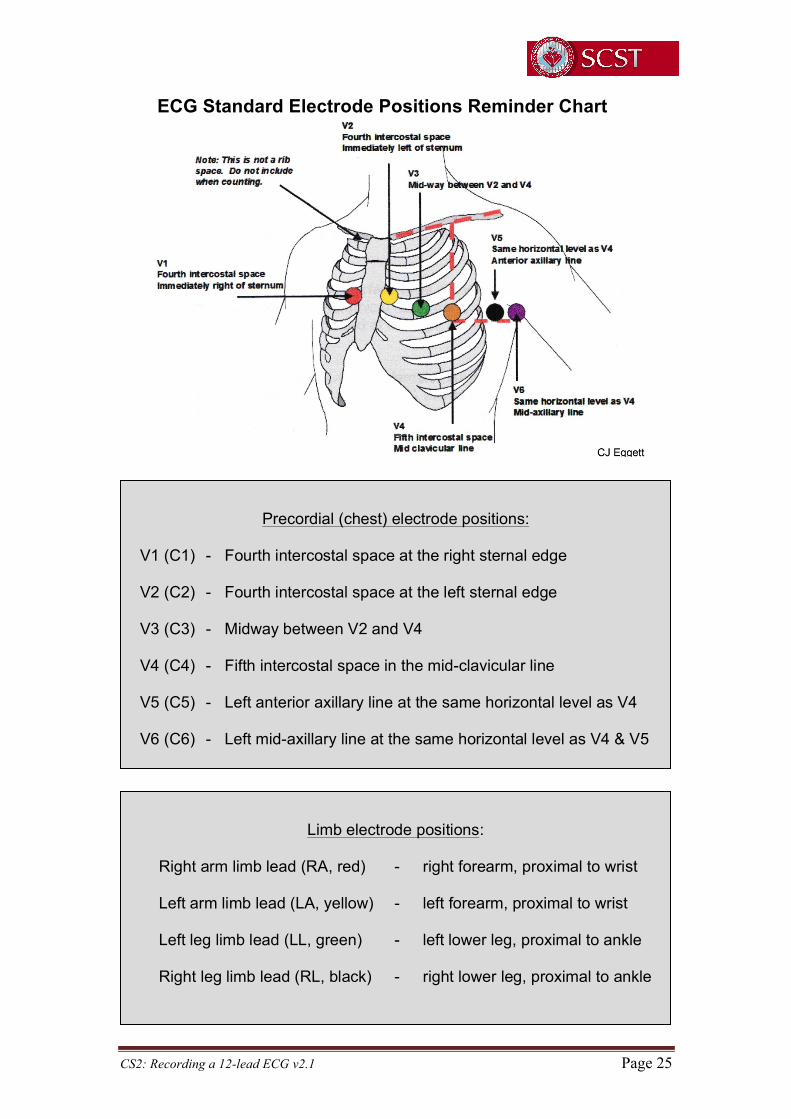
The correct anatomical positions for the chest electrodes have been defined20 (see figure below) and must always be used unless access is not possible. The centre of the active area of the electrode should be aligned with the relevant anatomical landmark.
Previous studies have demonstrated that the V1 and V2 electrodes are frequently placed too high and the V4, V5 and V6 electrodes too low5,28,29,30. These errors can result in diagnostically misleading alterations to the ECG waveform31. Operators should be competent to avoid common errors such as placing V5 and V6 electrodes in the 5th intercostal space, or using incorrect anatomical landmarks.
Limb electrode positions:
Right arm limb lead (RA, red) - right forearm, proximal to wrist Left arm limb lead (LA, yellow) - left forearm, proximal to wrist Left leg limb lead (LL, green) - left lower leg, proximal to ankle Right leg limb lead (RL, black) - right lower leg, proximal to ankle
Precordial (chest) electrode positions:
V1 (C1) - Fourth intercostal space at the right sternal edge V2 (C2) - Fourth intercostal space at the left sternal edge V3 (C3) - Midway between V2 and V4 V4 (C4) - Fifth intercostal space in the mid-clavicular line V5 (C5) - Left anterior axillary line at the same horizontal level as V4 V6 (C6) - Left mid-axillary line at the same horizontal level as V4 & V5

CS2: Recording a 12-lead ECG v2.1 Page 13
14. Technique for locating chest electrode positions Care should be taken when counting the intercostal spaces from the clavicle: the small space between the clavicle and the first rib is not the first intercostal space.
In order to avoid this common error the manubriosternal joint (also called the Angle of Louis) should be used as the main reference point.
To locate the Angle of Louis a finger should be run down the sternum from the top until a bony horizontal ridge is met. Sliding the finger down and to the right side will locate the second intercostal space. From here it is possible to count down to the third and fourth intercostal spaces. Slide the finger towards the sternum until the edge is felt and place the electrode for V1 in this position. This procedure should be repeated on the left side to correctly position V2. Note that the left and right sided rib spaces may be offset, so operators should avoid placing V2 adjacent to V1 without counting the rib spaces. Next, the V4 electrode should be placed in the 5th intercostal space in line with the mid-point of the clavicle. The V3 electrode should then be placed mid-way between the V2 and V4 electrodes.
The V5 and V6 electrodes should then be positioned in horizontal alignment with the V4 electrode. The V5 electrode should be placed on the anterior axillary line; the V6 electrode should be placed on the mid-axillary line.

CS2: Recording a 12-lead ECG v2.1 Page 14
When recording an ECG from female patients it is convention to place the V4, V5 and V6 electrodes beneath the left breast when breast tissue overlies the correct anatomical positions. There is some evidence to suggest that the positioning of these electrodes over the breast may not significantly attenuate the signal31,32, but further supporting evidence is needed to warrant a change in this recommendation. When lifting the breast to place electrodes, care and sensitivity is required. Using the back of the hand to lift the breast can be helpful in minimising contact. In order to achieve accurate ECG electrode positioning, it is usually necessary for all upper torso clothing to be absent.
15. Obtaining a good quality recording Time should be taken to ensure that the patient is relaxed and comfortable. If these conditions are not satisfied the ECG may record somatic muscle potentials as well as cardiac activity. Such interference will make the ECG more difficult to interpret. Some patients cannot relax fully because of pain from conditions such as arthritis, or they may have a condition such as Parkinson’s Disease which causes a tremor. These patients should be made as comfortable as possible and the ECG trace annotated with an appropriate explanation. On occasions it may be necessary to adapt the recommended ECG recording techniques. For example, patients in wheelchairs may need to remain in their wheelchair during the recording process. Any variations to standard recording techniques should be described in writing on the recording. Before recording the ECG checks should be made to ensure the patient’s limbs are still and appear relaxed. If the patient has clenched fists or stiff arms or is moving his/her fingers, it will not be possible to obtain a high quality ECG. The appropriate button should be pressed to initiate a new recording. This is usually labelled as ‘start’ or ‘auto’. A 12-lead ECG and simultaneous rhythm strip is most commonly recorded at 25mm/s with a gain setting of 10mm/mV.
Standard ECG recording:
Paper speed - 25mm/sec Voltage gain - 10mm/mV
CS2: Recording a 12-lead ECG v2.1 Page 15
All filters should be ‘off’ for the initial attempt to record an ECG. The initial recording should be made without the use of the manual filter which alters the upper cut-off frequency to 40Hz. The filter will reduce interference. However, as it also distorts the ECG, it should only be used when absolutely necessary and only after all attempts to eliminate the interference have failed. If, despite efforts to relax the patient and make them comfortable, there is somatic muscle interference on the ECG, the filter may be switched on and the recording repeated. Use of the filter should be clearly identified on the final ECG.
Incorrectly pressing ‘copy’ or ‘reprint’ may on some models initiate a printout of an ECG from a previous patient33. If patient details are not entered into the machine it may not be clear that this ECG relates to a previous patient. Operators must fully understand the equipment they are using and the potential consequences of mistakes. Local practice guidance must minimise the chance of an identity or recording error occurring in this way. If ECG complexes are of such high amplitude that they overlap, then the gain may be adjusted to 5mm/mV to enable clearer visualisation of the complexes and more accurate measurements to be made. Any alteration to the gain settings should be clearly marked on the ECG. Any features on the ECG that might indicate the need for urgent medical attention should be brought to the attention of appropriate staff. If the patient has any symptoms of possible cardiac origin, such as chest pain, palpitations or dizziness, at the time of recording, then this should be noted on the ECG. Confirmation that an ECG of good quality has been recorded should be made by the operator. The recording should be assessed to ensure that all waveforms (such as P waves, QRS complexes and T waves) are clearly visible. The isoelectric line (the baseline between ECG deflections) should be stable, not wandering, and free of interference.
Use of the filter (in auto mode):
Initial recording - filter off - recording made at 0.67-150Hz
Evidence of somatic muscle interference: Repeat recording - filter on - recording made at 0.67- 40Hz
The filter reduces interference but also distorts the ECG

CS2: Recording a 12-lead ECG v2.1 Page 16
The ECG should be correctly labelled with the patient’s identification and any relevant clinical details, as well as any adjustments made. All the electrodes should be removed from the patient and disposed of as clinical waste.
16. Documentation, processing, storage and confidentiality of ECG recordings The Department of Health in England is putting into place a digital strategy with a goal of having a paperless National Health Service by 201834. In the light of this the SCST Clinical Standards Committee is including recommendations on documentation, processing and storage of 12-lead ECG data. All ECG recordings should be digitally stored with the following identifiers to ensure accurate retrieval of clinical data and allow audit: • Patient’s first name and surname (formatted and spelled correctly) • Patient’s date of birth • A unique identifying number if available • The name and position of the referrer • Identity of the person making the recording of the ECG • Date and time of the recording • The name of the institution ECG recordings may be displayed in various formats. The most common is illustrated below:
I aVR V1 V4 II aVL V2 V5 III aVF V3 V6
Rhythm strip – usually lead II or V1
Typically the electronic storage of ECG recordings is made by compressing data. This can speed up the transmission and retrieval of records that are stored in central databases and minimise the memory required for storage. Data compression affects high frequency (short duration) signals more than the smoother low frequency (longer duration) signals. Therefore, compression has greater potential to alter measurements within the QRS complex, such as pacemaker ‘spikes’, Q-wave duration and R-wave amplitude, than to alter other signals such as the ST segment and the T-wave. A non-compressed ECG may differ from its compressed version. This has the potential to affect the comparison of serial ECGs when retrieving ECGs from storage media.

CS2: Recording a 12-lead ECG v2.1 Page 17
For the electronic storage of ECGs it is recommended that compression algorithms should perform in a manner that allows retrieved data to adhere to the fidelity standards established in the 1990 AHA statement with reference to the original signal35. All information pertaining to the patient should be treated in a confidential manner in accordance with local policies and national guidelines on data protection36.
17. Special situations
17.1 Dextrocardia Dextrocardia is the most common form of cardiac malposition and refers to any situation where the heart is located within the right side of the chest rather than the left. It may be associated with the condition situs inversus where other organs are in a mirror image relation to the usual position.
Dextrocardia may be suspected if a resting 12-lead ECG reveals negative P waves and QRS complexes in lead I in the absence of any technical error such as reversal of the right and left arm connections. Poor R-wave progression observed in leads V1 through V6 supports this interpretation. A second ECG should be recorded with the chest electrodes (V3 to V6) positioned on the right side of the chest using the same intercostal spacing and anatomic landmarks as previously described but on the right side. V1 and V2 should remain in the usual position. This approach should provide a ‘true’ ECG representation. The limb lead complexes will continue to appear inverted, demonstrating the abnormal location of the heart. However the repositioned chest leads (V3R to V6R) will now show appropriate R-wave progression. There should be clear annotation on the recording to describe the repositioned electrodes, for example “V3R”, “V4R” etc.
An alternative approach is to swapping the right and left arm connections. This will ‘normalise’ the appearance of the limb leads. If this approach is preferred it is imperative that the ECG be very clearly annotated to prevent the possibility of dextrocardia being overlooked.
CS2: Recording a 12-lead ECG v2.1 Page 18
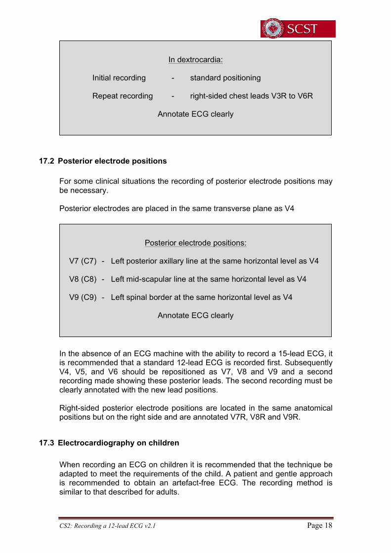
17.2 Posterior electrode positions For some clinical situations the recording of posterior electrode positions may be necessary. Posterior electrodes are placed in the same transverse plane as V4
In the absence of an ECG machine with the ability to record a 15-lead ECG, it is recommended that a standard 12-lead ECG is recorded first. Subsequently V4, V5, and V6 should be repositioned as V7, V8 and V9 and a second recording made showing these posterior leads. The second recording must be clearly annotated with the new lead positions. Right-sided posterior electrode positions are located in the same anatomical positions but on the right side and are annotated V7R, V8R and V9R.
17.3 Electrocardiography on children When recording an ECG on children it is recommended that the technique be adapted to meet the requirements of the child. A patient and gentle approach is recommended to obtain an artefact-free ECG. The recording method is similar to that described for adults.
In dextrocardia:
Initial recording - standard positioning Repeat recording - right-sided chest leads V3R to V6R
Annotate ECG clearly
Posterior electrode positions:
V7 (C7) - Left posterior axillary line at the same horizontal level as V4 V8 (C8) - Left mid-scapular line at the same horizontal level as V4 V9 (C9) - Left spinal border at the same horizontal level as V4
Annotate ECG clearly

CS2: Recording a 12-lead ECG v2.1 Page 19
If possible, the recording should be made with the child semi-recumbent, but the sitting position may be used if this will prevent restlessness or distress. The four limb electrodes are attached as previously described. The chest electrodes are normally positioned as previously described but additional leads, such as V4R, V5R and V7, may be recorded at the request of a clinician or according to local policy. V4R and V3R are right-sided leads recorded from electrodes placed in a mirror image position to the V3 and V4 positions. V7 is a posterior lead with the electrode placed in the posterior axillary line in the same horizontal plane as the V4 electrode. Whilst practice is variable in the routine use of right-sided chest leads when recording ECGs on children, it is not uncommon to find V4R used in infants (up to 1 year old). Practice is determined largely by the indications for the ECG and clinician preference.
18. Audit Audit is a recognised way of assessing and improving practice. The appendix contains a useful checklist intended for use and an audit tool. This checklist can be used to assess how well the guideline is being followed. It may also be used to ensure that any local guidelines developed from this consensus document meet the requirements for competent service delivery.
19. Conclusion To be of maximum diagnostic value an ECG must be recorded correctly by paying proper attention to patient preparation, electrode positioning and the other factors described in this document. This is not difficult to do, and it is hoped that widespread adoption of this guidance will help to ensure a uniformly high standard of practice in ECG recording with consequent benefits to patient care everywhere.

CS2: Recording a 12-lead ECG v2.1 Page 20
20. References 1World Health Organisation (1981) Regional Office for Europe. Uses of the Electrocardiogram. Report on a WHO study; Copenhagen EURO Reports and Studies 37 (project ICP/ATH 003). WHO, Geneva. 2Wolff AR, Long S, McComb J, Richley D, Mercer P. The gap between training and provision: a primary care-based ECG survey in north-east England. Br J Cardiol 2012;19:38-40. 3Rudiger A, Hellerman JP, Mukherjee R, Follath F, Turina J. Electrocardiographic artifacts due to electrode misplacement and their frequency in different clinical settings. Am J Emerg Med 2007;25:174-178. 4Bupp JE, Dinger M, Lawrence C, Wigate S. Placement of cardiac electrodes: written, simulated and actual accuracy. Am J Crit Care 1997;6(6):457-62. 5Rajaganeshan R, Ludlam CL, Francis DP, Parasramka SV, Sutton R. Accuracy in ECG lead placement among technicians, nurses, general physicians and cardiologists. Int J Clin Pract. 2007;62:65-70 6McCann K, Holdgate A, Mahammad R, Waddington A. Accuracy of ECG electrode placement by emergency department clinicians. Emergency Medicine Australasia 2007;19(5):442–448. 7Hill NE, Goodman JS. Importance of accurate placement of precordial leads in the 12-lead electrocardiogram. Heart & Lung 1987;16(5):561-566. 8Batchvarov VN, Malik, M, Camm AJ. Incorrect electrode cable connection during electrocardiographic recording. Europace 2007;9:1081-1090. 9Knight BP, Pelosi F, Michaud GF, Strickberger SA, Morady F. Clinical consequences of electrocardiographic artifact mimicking ventricular tachycardia. N Engl J Med 1999;341:1270–1274. 10Harrigan RA, Chan TC, Brady WJ. Electrocardiographic electrode misplacement, misconnection and artefact.J Emerg Med 2012;43:1038-1044. 11Garcia-Niebla J, Llontob-Garcia P, Valle-Racero JI, Serra-Autonell G, Batcharov, VN, Bayes de Luna A. Technical mistakes during the acquisition of the electrocardiogram. Ann Noninvasive Electrocardiol 2009;14(4):389-403. 12Garcia-Niebla J, Serra-Autonell G, Bayes de Luna A. Brugada syndrome electrocardiographic pattern as a result of improper application of a high pass filter. Am J Cardiol 2012; 110(2):318-320. 13Castellanos A, Pastor JA, Zambrano JP, Myerburg RJ (2002). Left bundle-branch block with technical right-axis deviation. Circulation;106:2288-2289.

CS2: Recording a 12-lead ECG v2.1 Page 21
14Thaler T, Tempelman V, Maggiorini M, Rudiger A. The frequency of electrocardiographic errors due to electrode cable switches: a before and after study. J Electrocardiol 2010;43:676-681. 15Barnsley L, Lyon PL, Hibbert EJ, Cunningham I, Gordon FC, Field MJ. Clinical skills in junior medical officers: a comparision of self-reported confidence and observed competence. Medical Education 2004;38:358-367. 16Richley D, Wolff A, Eggett C, Ashton J, Corrigan J. ECG Recording in primary care: is it done correctly? Prim Care Cardiovasc J. 2013; 6:25-27 17Good Scientific Practice Booklet published by the Academy of Healthcare Sciences, Institute of Physics in Engineering in Medicine (IPEM): http://www.ipem.ac.uk/Portals/0/Documents/News/AHCS_Good_Scientific_Practice.pdf (accessed April 2014) 18Keogh B. Review into the quality of care and treatment provided by 14 hospital trusts in England: overview report. NHS England. 2013: http://www.nhs.uk/NHSEngland/bruce-keogh-review/Documents/outcomes/keogh-review-final-report.pdf (accessed April 2014 19General Medical Council. Intimate examinations and chaperones.2013: http://www.gmc-uk.org/Intimate_examinations_and_chaperones.pdf_51449880.pdf (accessed June 2014) 20Kligfield P, Gettes LS, Bailey JJ, Childers R, Deal B, Hancock W, van Herpen G, Kors JA, Macfarlane P, Mirvis DM, Pahlm O, Rautaharju P, Wagner GS. Recommendations for the Standardization and Interpretation of the Electrocardiogram: Part I: The Electrocardiogram and Its Technology A Scientific Statement From the American Heart Association Electrocardiography and Arrhythmias Committee, Council on Clinical Cardiology; the American College of Cardiology Foundation; and the Heart Rhythm Society Endorsed by the International Society for Computerized Electrocardiology. J Am Coll Cardiol. 2007; 49(10):1109-1127. 21Guidelines on Hand Hygiene in Healthcare published by the World Health Organisation (2009): http://whqlibdoc.who.int/publications/2009/9789241597906_eng.pdf (accessed April 2014) 22Baevsky RH, Haber MD, Blank FS, Smithline H. Supine vs semirecumbent and upright 12-lead electrocardiogram: does change in body position alter the electrocardiographic interpretation for ischaemia? Am J Emerg Med 2007;25:753-756. 23Bergman KS, Stevenson WG, Tillisch JH, Stevenson LW. Effect of body position on the diagnostic accuracy of the electrocardiogram. Am Heart J 1989;117(1):204-206.

CS2: Recording a 12-lead ECG v2.1 Page 22
24Kossman CE, Brody DA, Burch GE, Hecht H, Johnston FD, Kay C, Lepeschkin E, Pipberger HV, Baule G, Berson AS, Briller SA, Geselowitz DB, Horan LG, Schmitt OH. Recommendations for standardization of leads and of specifications for instruments in electrocardiography and vectorcardiography. Circulation 1967; 35:583-601. 25Pipberger HE, Arzbaecher RC, Berson AS. American Heart Association Committee on Electrocardiography: recommendations for standardization of leads and of specifications for instruments in electrocardiography and vectorcardiography. Circulation 1975;52:11-31. 26Pahlm O, Haisty WK, Edenbrandt L, Wagner NB, Sevilla DC, Selvester RH, Wagner GS. Evaluation of changes in standard electrocardiographic QRS waveforms recorded from activity-compatible proximal limb lead positions. Am J Cardiol. 1992; 35:583-601. 27Sevilla DC, Dohrmann ML, Somelofski CA, Wawrzynski RP, Wagner NB, Wagner GS. Invalidation of the resting electrocardiogram obtained via exercise electrode sites as a standard 12-lead recording. Am J Cardiol. 1989; 63:35-39. 28Wenger W, Kligfield P. Variability of precordial electrode placement during routine electrocardiography. J Electrocardiol. 1996; 29:179-184. 29Zema MJ, Luminais SK, Chiaramida S, Goldman M, Kligfield P. Electrocardiographic poor R wave progression III: the normal variant. J Electrocardiol. 1980; 13:135-142 30August T, Mazzeleni A, Wolff L. Positional and respiratory changes in precordial lead patterns simulating acute myocardial infarction. Am Heart J. 1958; 55:706-714. 31Colaco R, Reay P, Beckett C, Aitchison TC, Macfarlane PW. False positive ECG reports of anterior myocardial infarction in women. J Electrocardiol. 2000; 33:239-244
32Macfarlane PW, Colaco R, Stevens K, Reay P, Beckett C, Aitchison T. Precordial electrode placement in women. Neth Heart J. 2003; 11:118-122 33Medical and Healthcare Products Regulatory Agency (MHRA) medical device alert reference number MDA/2010/056 34Department of Health Policy: Making the NHS more efficient and less Bureaucratic. Speech made by J.Hunt MP on 16/1/13 https://www.gov.uk/government/news/jeremy-hunt-challenges-nhs-to-go-paperless-by-2018--2 (accessed April 2014)

CS2: Recording a 12-lead ECG v2.1 Page 23
35Bailey JJ, Berson AS, Garson A Jr, Horan LG, Macfarlane PW, Mortara DW, Zywietz C. Recommendations for standardization and specifications in automated electrocardiography: Bandwidth and digital signal processing. Circulation 1990; 81:730-739. 36Data protection act 1998: http://www.legislation.gov.uk/ukpga/1998/29/contents
CS2: Recording a 12-lead ECG v2.1 Page 24
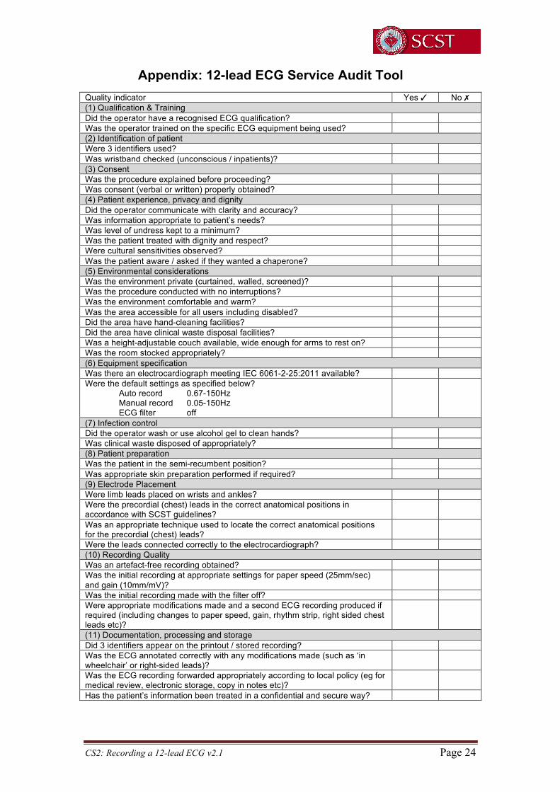
Appendix: 12-lead ECG Service Audit Tool Quality indicator Yes ✓ No ✗ (1) Qualification & Training Did the operator have a recognised ECG qualification? Was the operator trained on the specific ECG equipment being used? (2) Identification of patient Were 3 identifiers used? Was wristband checked (unconscious / inpatients)? (3) Consent Was the procedure explained before proceeding? Was consent (verbal or written) properly obtained? (4) Patient experience, privacy and dignity Did the operator communicate with clarity and accuracy? Was information appropriate to patient’s needs? Was level of undress kept to a minimum? Was the patient treated with dignity and respect? Were cultural sensitivities observed? Was the patient aware / asked if they wanted a chaperone? (5) Environmental considerations Was the environment private (curtained, walled, screened)? Was the procedure conducted with no interruptions? Was the environment comfortable and warm? Was the area accessible for all users including disabled? Did the area have hand-cleaning facilities? Did the area have clinical waste disposal facilities? Was a height-adjustable couch available, wide enough for arms to rest on? Was the room stocked appropriately? (6) Equipment specification Was there an electrocardiograph meeting IEC 6061-2-25:2011 available? Were the default settings as specified below? Auto record 0.67-150Hz Manual record 0.05-150Hz ECG filter off
(7) Infection control Did the operator wash or use alcohol gel to clean hands? Was clinical waste disposed of appropriately? (8) Patient preparation Was the patient in the semi-recumbent position? Was appropriate skin preparation performed if required? (9) Electrode Placement Were limb leads placed on wrists and ankles? Were the precordial (chest) leads in the correct anatomical positions in accordance with SCST guidelines?
Was an appropriate technique used to locate the correct anatomical positions for the precordial (chest) leads?
Were the leads connected correctly to the electrocardiograph? (10) Recording Quality Was an artefact-free recording obtained? Was the initial recording at appropriate settings for paper speed (25mm/sec) and gain (10mm/mV)?
Was the initial recording made with the filter off? Were appropriate modifications made and a second ECG recording produced if required (including changes to paper speed, gain, rhythm strip, right sided chest leads etc)?
(11) Documentation, processing and storage Did 3 identifiers appear on the printout / stored recording? Was the ECG annotated correctly with any modifications made (such as ‘in wheelchair’ or right-sided leads)?
Was the ECG recording forwarded appropriately according to local policy (eg for medical review, electronic storage, copy in notes etc)?
Has the patient’s information been treated in a confidential and secure way?
CS2: Recording a 12-lead ECG v2.1 Page 25
ECG Standard Electrode Positions Reminder Chart
Precordial (chest) electrode positions:
V1 (C1) - Fourth intercostal space at the right sternal edge V2 (C2) - Fourth intercostal space at the left sternal edge V3 (C3) - Midway between V2 and V4 V4 (C4) - Fifth intercostal space in the mid-clavicular line V5 (C5) - Left anterior axillary line at the same horizontal level as V4 V6 (C6) - Left mid-axillary line at the same horizontal level as V4 & V5
Limb electrode positions:
Right arm limb lead (RA, red) - right forearm, proximal to wrist Left arm limb lead (LA, yellow) - left forearm, proximal to wrist Left leg limb lead (LL, green) - left lower leg, proximal to ankle Right leg limb lead (RL, black) - right lower leg, proximal to ankle

CS2: Recording a 12-lead ECG v2.1 Page 26
ECG Settings Reminder Chart
Standard ECG recording:
Paper speed - 25mm/sec Voltage gain - 10mm/mV
Use of the filter:
Initial recording - filter off - recording made at 0.67-150Hz
Evidence of somatic muscle interference: Repeat recording - filter on - recording made at 0.67- 40Hz
The filter reduces interference but also distorts the ECG
In dextrocardia:
Initial recording - standard positioning Repeat recording - right-sided chest leads V3R to V6R
Annotate ECG clearly