CLINICAL GUIDELINE FOR THE USE OF … · CLINICAL GUIDELINE FOR THE USE OF INTRAVASCULAR CATHETERS...
40
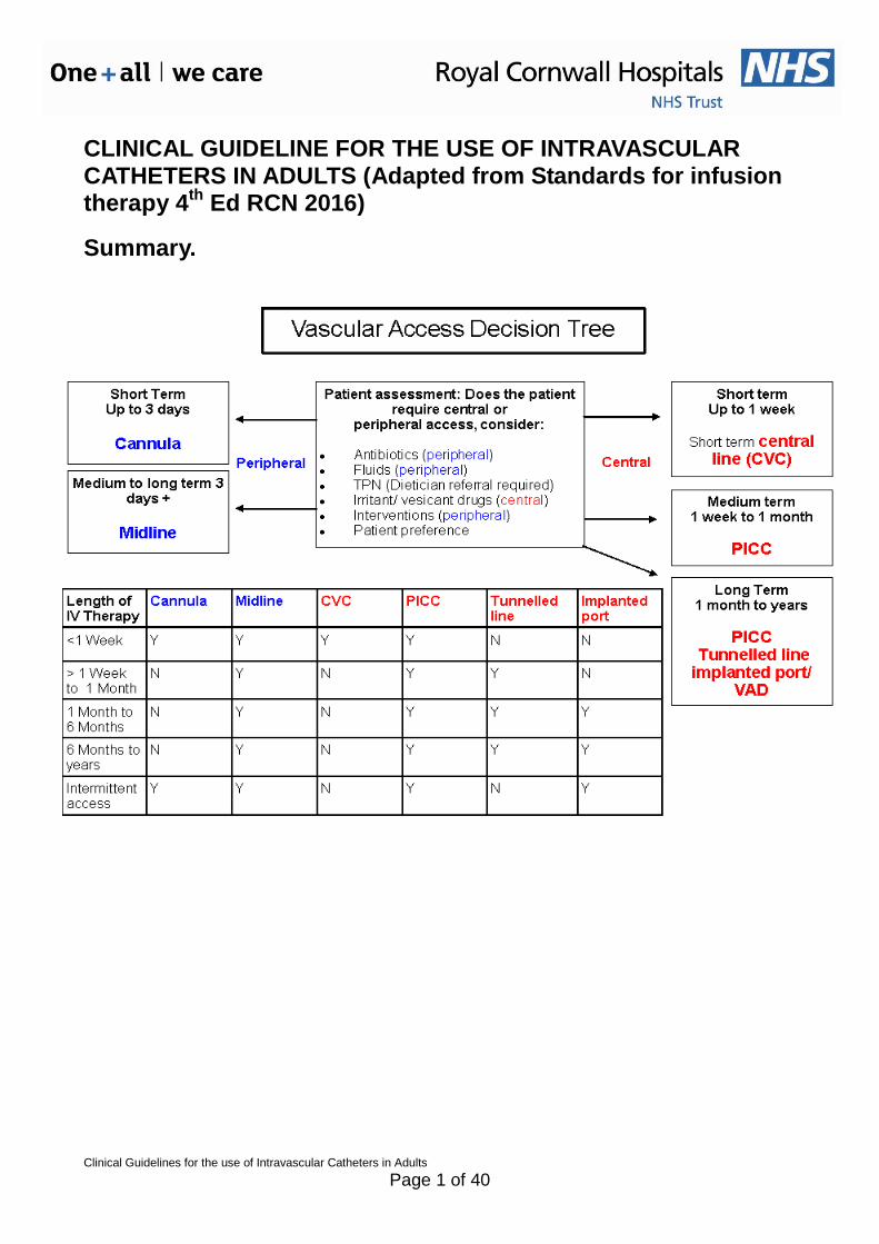
Clinical Guidelines for the use of Intravascular Catheters in Adults Page 1 of 40 CLINICAL GUIDELINE FOR THE USE OF INTRAVASCULAR CATHETERS IN ADULTS (Adapted from Standards for infusion therapy 4 th Ed RCN 2016) Summary.
Transcript of CLINICAL GUIDELINE FOR THE USE OF … · CLINICAL GUIDELINE FOR THE USE OF INTRAVASCULAR CATHETERS...
Clinical Guidelines for the use of Intravascular Catheters in Adults
Page 1 of 40
CLINICAL GUIDELINE FOR THE USE OF INTRAVASCULAR CATHETERS IN ADULTS (Adapted from Standards for infusion therapy 4
th Ed RCN 2016)
Summary.

Clinical Guidelines for the use of Intravascular Catheters in Adults
Page 2 of 40
Quick Reference Guide
Line Type Skin Prep Type of Flush Volume of flush Frequency of
flush, when not in use.
Type of dressing
Frequency of changing dressing
Frequency of recording VIP
Peripheral line 2% Chlorhexidine
Chloraprep
0.9% sodium chloride
10mls Twice daily Transparent, semi-
permeable polyurethane
dressings
If dressing not intact or clean.
Twice daily
Midline 2% Chlorhexidine
Chloraprep
0.9% sodium chloride
10mls Weekly Transparent film plus
securement device. (Sterile gauze for 24
hours)
1st dressing within 24 hours
(Remove gauze) then weekly or
sooner if soiled/ loose
Twice daily
Short term central line
2% Chlorhexidine
Chloraprep
0.9% sodium chloride
10mls Twice daily Transparent, semi-
permeable polyurethane
dressings plus securement
device.
If dressing not intact or clean.
Twice daily
PICC 2% Chlorhexidine
Chloraprep
0.9% sodium chloride
10mls Weekly Transparent film plus
securement device. (Sterile gauze for 24
hours)
1st dressing within 24 hours
(Remove gauze) then weekly or
sooner if soiled/ loose
Twice daily
Skin tunnelled catheter ie Hickmann
2% Chlorhexidine
Chloraprep
0.9% sodium chloride
10mls into each lumen using a
10ml luer lock syringe (The volume
used should be equal to at least
twice the
Weekly (every 7 days)
Transparent, semi-
permeable polyurethane
dressings
Weekly (every 7 days) or sooner if
dressing is no longer intact, if
moisture collects under the dressing or
Twice daily
Clinical Guidelines for the use of Intravascular Catheters in Adults
Page 3 of 40
catheter volume. This is
normally between 5-
10mls)
if the VIP score indicates site
should be cleaned
(Loveday et al 2014)
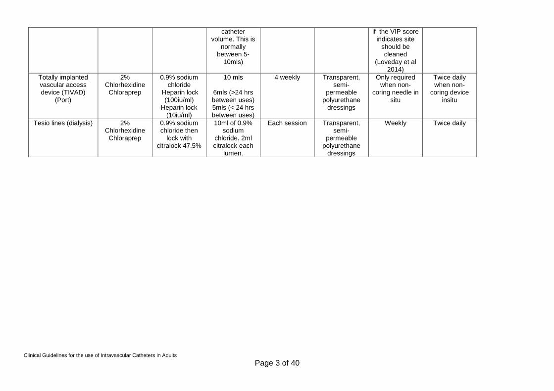
Totally implanted vascular access device (TIVAD)
(Port)
2% Chlorhexidine
Chloraprep
0.9% sodium chloride
Heparin lock (100iu/ml)
Heparin lock (10iu/ml)
10 mls
6mls (>24 hrs between uses) 5mls (< 24 hrs between uses)
4 weekly Transparent, semi-
permeable polyurethane
dressings
Only required when non-
coring needle in situ
Twice daily when non-
coring device insitu
Tesio lines (dialysis) 2% Chlorhexidine
Chloraprep
0.9% sodium chloride then
lock with citralock 47.5%
10ml of 0.9% sodium
chloride. 2ml citralock each
lumen.
Each session Transparent, semi-
permeable polyurethane
dressings
Weekly Twice daily

Clinical Guidelines for the use of Intravascular Catheters in Adults
Page 4 of 40
Table of Contents
CLINICAL GUIDELINE FOR THE USE OF INTRAVASCULAR CATHETERS IN ADULTS (Adapted from Standards for infusion therapy 4th Ed RCN 2016) .................................. 1
Summary. ............................................................................................................................ 1
Quick Reference Guide........................................................................................................ 2
1. Aim/Purpose of this Guideline ...................................................................................... 5
2. The Guidance ............................................................................................................... 5
3. Site and device selection and placement ...................................................................... 5
4. Site care and maintenance ......................................................................................... 12
5. Specific devices .......................................................................................................... 16
6. Infusion-related complications .................................................................................... 18
7. Monitoring compliance and effectiveness ................................................................... 23
8. Equality and Diversity ................................................................................................. 24
Appendix 1. Governance Information ................................................................................ 25
Appendix 2. Initial Equality Impact Assessment Form ....................................................... 27
Appendix 3. Algorithm for persistent withdrawal occlusion ................................................ 29
Appendix 4: Complications ................................................................................................ 30
Appendix 5: Maintaining Patency ....................................................................................... 35
Appendix 6 References...................................................................................................... 38

Clinical Guidelines for the use of Intravascular Catheters in Adults
Page 5 of 40
1. Aim/Purpose of this Guideline 1.1. This document is for use by all practitioners who are requesting, caring for or removing intravascular catheters within the Royal Cornwall Hospitals Trust (RCHT).
2. The Guidance 2.1. It is the responsibility of the practitioner requesting the intravenous catheter to ensure that the device is appropriate to the patient and the pharmaceuticals involved, see vascular access decision tool above. 2.2. The practitioner inserting the line must ensure this is done in keeping with all relevant RCHT Procedures. They must be competent to undertake the procedure through RCHT approved training and maintain standards to practice. 2.3. The clinical area manager is responsible for ensuring that staff caring for patients with intravenous catheters have received appropriate training. 2.4. The individual is responsible for ensuring that they remain competent in their practice.
2.5. An explanation of the full procedure including potential complications and aftercare must be outline to all patients for the placement of any intravascular access device and consent obtained.
2.6. Prior to insertion, care of or removal of any line the patient must be identified using three points of identification and consent (written and/or verbal) must be obtained in accordance to the RCHT Consent Policy.
2.7. ANTT should be used for all procedures relating to intravenous catheters as per ANTT Policy this includes: insertions, dressing changes, IV/drug administration, changing of add on devices and removals of lines.
2.8. Parenteral Nutrition should be administered by a dedicated peripheral line i.e. midline or central line (PICC/ CVC/ Hickman) and via a dedicated lumen if the device is multi lumen. The lumen should be labelled/ identified. Please contact the nutrition team for further advice as device will dictate PN formulation and appropriate nutritional delivery.
2.9. Any areas not covered in this policy The Royal Marsden Hospital Manual of Clinical Nursing Procedures should be followed. 2.10.Before discharge from hospital, patients with intravascular catheters and their carers should be taught any techniques they may need to use to prevent infection.
3. Site and device selection and placement 3.1. Site and device selection

Clinical Guidelines for the use of Intravascular Catheters in Adults
Page 6 of 40
Standards relating to all sites and devices.
3.1.1. Initial assessment should include whether or not infusion therapy is required and have other routes been considered and excluded (Hallam et al., 2016) 3.1.2. If infusion therapy is required, site and device selection for vascular access should then include assessment of the patient‟s condition, age and diagnosis; vascular condition; infusion device history; and the type and duration of the therapy as well as the potential complications associated with vascular access devices (Hallam et al., 2016). 3.1.3. The vein/vessel should accommodate the gauge and length of the device required by the prescribed therapy (INS, 2016). Patient‟s lifestyle, body image, any known abnormalities, relevant past medical history (PMH) patient preference, and therapy duration and setting should all be considered for site and device selection (Hallam, et al., 2016). 3.1.4. Using the same criteria, any device initially placed should be reviewed after 48 hours (Hallam et al., 2016). 3.1.5. Placement of any vascular access device, particularly central vascular access devices, is an aseptic procedure that should only be undertaken by staff who have had appropriate training (Loveday et al., 2014; NMC, 2015a). 3.1.6. Prior to making a peripherally inserted central catheter (PICC) insertion, anatomical measurements should be taken to determine the length of the catheter required to ensure full advancement of the catheter to achieve catheter tip placement in the superior vena cava/right atrium. 3.1.7. The length of the central vascular access catheter will be selected in order to ensure that the distal tip of the catheter lies in the lower third of the superior vena cava or right atrium. (Bodenham et al., 2016). 3.1.8. A multiple-lumen device will not be routinely placed unless the patient‟s condition/intended treatment necessitates one (Loveday et al., 2014). 3.1.9. All catheters must be radiopaque.
General guidance for all sites and devices
3.1.10. The HCP should have the necessary knowledge and competence to select the most appropriate site and device for the patient and the intended therapy. This should include: knowledge of the patient and environment, knowledge of the product in regard to insertion technique, potential complications, and appropriateness linked to prescribed therapy and medication and manufacturers‟ guidelines (INS, 2016). 3.1.11. Central venous catheters should be of single lumen configuration

Clinical Guidelines for the use of Intravascular Catheters in Adults
Page 7 of 40
unless additional therapies are required (Loveday et al., 2014).
3.2. Peripheral devices: cannulae and midline catheters A peripheral cannula is defined as one that is less than or equal to 3 inches (7.5cm) in length. Peripheral cannulae should be selected for short term therapy of 3–5 days and for bolus injections or short infusions in the outpatient/day unit setting.
A midline catheter for adults is defined as one that is between 3 and 8 inches (7.5cm–20cm) in length. Midline catheters are used for the administration of blood, fluid and medication when the therapy is expected to last between 1-4 weeks. They may be used where patients present with poor peripheral venous access and when the use of a central venous catheter is contraindicated. The midline catheter provides venous accessibility along with an easy, less hazardous insertion at the antecubital fossa.
Specific guidance relating to peripheral devices:
3.1.12. Veins that should be considered for peripheral cannulation are those found in the forearm or hands (O‟Grady et al., 2011). 3.1.13. Site selection should be routinely initiated in the distal areas of the upper extremities; subsequent cannulation should be made proximal to the previously cannulated site.
3.1.14. Peripheral cannulae should only be inserted by Registered Healthcare Professionals, Assistant practitioners, Trainee assistant practitioners and designated Band 3 staff where cannulation is an essential element of their role and scope of practice.
3.1.15. Where possible, use non dominant forearm for peripheral cannulation following policy and procedure. 3.1.16. Veins in the lower extremities should not be used routinely in adults due to the risk of thrombosis, thrombophlebitis and increased infection risk. 3.1.17. Patients with diabetes should not generally be cannulated in their feet. 3.1.18. Site selection should involve assessment for previous venepuncture and subsequent damage to the vein. Aids to assist in cannulation including ultrasound and infrared imaging should be considered (Hallam et al., 2016). 3.1.19. Choice of an alternative site due to infiltration/extravasation of solutions into the extremity should require assessment of the type of solution, its pH, osmolarity, the estimated volume of the infusate/duration of infusion and the condition of the vein (Hallam et al., 2016). 3.1.20. Site selection should avoid areas of flexion although this may not always be possible in an emergency situation.

Clinical Guidelines for the use of Intravascular Catheters in Adults
Page 8 of 40
3.1.21. Arterial flow should not be compromised when pressure is applied to produce venous distension. 3.1.22. Blood pressure cuffs and tourniquets should be avoided if possible on an extremity where a peripheral device has been placed. 3.1.23. Cannulation of fistulae and grafts for infusion therapy requires specialist approval and organisational policies and procedures must be followed. 3.1.24. Peripheral devices should not be routinely used for blood sampling but blood can be taken immediately following insertion (WHO, 2010). Exceptions include patients requiring endocrine testing on the medical day case unit and those recruited to diabetes research. 3.1.25. Do not take blood from an existing peripheral venous access site because this may give false results. (WHO, 2010). 3.1.26. Consider the use of an extension set between the peripheral catheter and needleless connector to reduce catheter manipulation (INS, 2016) 3.1.27. A relevant HCP should be consulted, and the decision documented, prior to cannulation of the arm of a patient who has undergone axillary node dissection/radiotherapy with risk of lymphoedema (for example, following a mastectomy) or who may have existing AV fistula access or other contraindications; for example, they require future fistula formation (RA, 2015b). 3.1.28. The basilic, cephalic or brachial veins of the patient‟s arm are used for the insertion of a midline catheter, with the basilic vein generally being the vein of choice due to its diameter and position away from artery and median nerve (Alexandrou et al., 2011). 3.1.29. Placement of the midline should be just above or below the fold of the antecubital area so as to aid the patient‟s comfort when flexing their arm. This will also minimise the potential for catheter kinking. Ultrasound should be used to assist with location (Alexandrou et al., 2011). 3.1.30. As the tip of the midline catheter does not extend beyond the axillary vein, X-ray confirmation of tip placement is not required prior to use (INS, 2016). 3.1.31. Use vascular visualisation technology to aid vein identification for midlines (INS, 2016). 3.1.32. Midline catheters can be used as an alternative to peripheral cannula for longer term IV infusion therapy (up to <4 weeks) dependant on type of infusate, duration and individual risk assessment and following local policies (Alexandrou et al., 2011). 3.1.33. Midline catheters can be considered as an alternative to
Clinical Guidelines for the use of Intravascular Catheters in Adults
Page 9 of 40
subcutaneous infusion in palliative care depending on the infusate/medicine to be infused, volume and duration of infusion therapy and individual patient needs and dependant on local policy and procedure (Bortolussi et al., 2015). 3.1.34. Therapies which are not appropriate for certain peripheral cannulae and midlines include continuous vesicant chemotherapy, parenteral nutrition solutions and/or medications with osmolarity greater than 900 Osm/L (INS, 2016). These aspects should not be considered in isolation and a risk assessment that includes vein assessment, duration and environment of therapy as well as pH and osmolarity is important prior to any site and device selection (Hallam et al., 2016; Gorski et al., 2015). 3.1.35. Ideally, peripheral devices should be equipped with a safety device with engineered sharps injury protection. (Loveday et al., 2014; DH, 2013a).
3.3. Central venous access devices
Specific guidance relating to central venous access devices:
3.3.1. The choice of veins for non-tunnelled, tunnelled or implantable device should balance the risks for infection against the risks of mechanical complications and include the internal jugular and subclavian veins (Loveday et al., 2014). Unless medically contraindicated, use the subclavian site in preference to the jugular site for non-tunnelled catheter placement (Parienti et al., 2015) except for haemodialysis catheters.
3.3.2. Use of 2D ultrasound imaging is recommended for all routine placements of all central venous access devices (Bodenham et al., 2016; Lamperti et al., 2012). 3.3.3. Central catheters should have the distal tip dwelling in the lower third of the superior vena cava or the upper right atrium (Bodenham et al., 2016; Frykholm et al., 2014). 3.3.4. The femoral vein should be avoided where possible due to the higher risk of infection. If used the rationale for using the femoral vein must be clearly recorded in the medical record. If used in an emergency situation, then it should be replaced as soon is practically possible (Loveday et al., 2014).
A peripherally inserted central catheter (PICC) is a catheter that is inserted via the upper arm veins and is advanced into the central veins, with the tip located in the superior vena cava (usually the lower third) (INS, 2016). The cephalic, basilic or median cubital veins of the adult patient‟s arm can be used for the insertion of a PICC.
A short-term central venous catheter is a device that typically enters the vein from a skin puncture site over the vein.
Antimicrobial central venous catheters should be considered in high-risk patients to minimise the risk of catheter-related bloodstream infection (Loveday et al., 2014).

Clinical Guidelines for the use of Intravascular Catheters in Adults
Page 10 of 40
A skin-tunnelled catheter is a long-term catheter that lies in a subcutaneous tunnel before entering a central vein. These catheters have a cuff which is surgically implanted. The cuff embeds into the tissue of the patient providing additional protection against central line infection.
An implanted port is a totally implanted vascular access device made of two components; a reservoir with a self-sealing septum, which is attached to a catheter.
3.4. Hair removal
Clippers should be used if hair removal is required and should have disposable heads for single-patient use (NICE, 2013).
Shaving with a razor should not be performed because of the increased risk of infection (NICE, 2013).
3.5. Local anaesthesia
Use of injectable anaesthetic should be monitored because of the potential for allergic reaction, tissue damage and inadvertent injection of the drug into the vascular system (EMC, 2015).
Local anaesthetics should not be injected into inflamed or infected tissues.
Other types of local anaesthesia, such as iontophoresis or topical transdermal agents, should be considered and used according to organisational policies and procedures, and manufacturers‟ guidelines.
The HCP administering the local anaesthesia should have demonstrated competency and knowledge of the drug, method of administration used and management of complications (NMC, 2015c).
3.6. Insertion site preparation
2% chlorhexidine gluconate in 70% alcohol must be used with awareness of potential chlorhexidine allergy and an alternative used for example povidone iodine in alcohol (Loveday et al., 2014).
Application technique should be as per manufacturer‟s instructions and RCHT ANTT policy, allowing for appropriate cleaning and drying time.
3.6.1 Peripheral cannulae
Decontaminate the skin as per guidance in 3.6 with emphasis on cleaning and drying time.
Wear clean non-sterile gloves for insertion of the cannula (Loveday et al., 2014).
3.6.2 Midlines and central venous access devices
Maximal barrier precautions including sterile gown, sterile gloves and large sterile drapes should be used for arterial, central and peripherally inserted central catheter insertions and midlines in order to minimise the risk of infection to the patient (Loveday et al., 2014).
Decontaminate the skin as per guidance in Section 3.6, with emphasis on cleaning and drying time.
After initial site preparation, unless the skin decontamination process involves a non-touch technique, sterile gloves should be changed prior to midline, arterial, central and peripherally inserted central catheter placement (INS, 2016).

Clinical Guidelines for the use of Intravascular Catheters in Adults
Page 11 of 40
3.7. Intravascular device placement
The HCP placing any vascular access device should have a comprehensive understanding of anatomy and physiology, vascular assessment techniques and insertion techniques appropriate to the specific device.
The HCP should be aware of the manufacturers‟ advice relating to the particular vascular access device, preparation and placement, connections and administration set dwell time and compatibility with other fluids to ensure safe use of the device (Loveday et al., 2014).
Aseptic technique must be used and standard precautions must be observed during vascular access device placement. This includes the appropriate use of hand hygiene and PPE selection/use (Loveday et al. 2014).
A device designated as „single-use‟ must not be reused. Only one vascular access device should be used for each insertion attempt for that particular device and patient (MHRA, 2013a).
Caution should be employed when stylets, needles and/or wires are used to facilitate vascular access device placement because of the risk of needlestick injury.
Stylets, which are part of the catheter product, should never be reinserted due to the risk of severing and/or puncturing the catheter (INS, 2016).
Peripheral and central vascular access device placement, including gauge and length, product name, batch and lot number, number of attempts, anatomical location and patient‟s response to the placement, must be included in the patient‟s nursing and medical records (INS, 2016).
Radiological confirmation of the tip location or a recognised alternative procedure (for example, ECG guidance for PICCs) must be obtained in the following clinical situations: prior to use of the central vascular access device; difficulty with catheter advancement; pain or discomfort after catheter advancement; inability to obtain positive aspiration of blood; inability to flush the catheter easily; difficulty in removing guide wire or guide wire bent on removal. (Bodenham et al., 2016; Frykholm et al., 2014).
The distal tip of a central venous access device should dwell in the lower third of the superior vena cava or upper right atrium. Catheter tip location should be determined radiographically or use of ECG guidance for some devices; for example PICCs, depending on local protocols and included in the patient‟s medical record prior to initiation of the prescribed therapy.
3.8. Device stabilisation
Products employed to stabilise peripheral cannulae, midlines or central venous catheters include: transparent film dressings, sutures, engineered stabilisation devices and sterile wound closure strips.
Device stabilisation must be performed using an aseptic technique (Loveday et al., 2014).
Devices should be stabilised in a manner that does not interfere with assessment and monitoring of the access site, that does not impede delivery of the prescribed therapy, and that is acceptable to the patient (INS, 2016).
Stabilising devices should be placed so as not to impede circulation or impede infusion through the access device (INS, 2016).
For stabilisation of CVCs, and particularly midlines and PICCs, consider use of an engineered stabilisation device (INS, 2016).
As use of sutures is associated with needlestick injury, where practicable their use should be avoided for VAD securement.
Clinical Guidelines for the use of Intravascular Catheters in Adults
Page 12 of 40
When a specific securement device is used for stabilisation, placement should be in accordance with manufacturers‟ guidelines (INS, 2016).
A catheter which has migrated externally should not be re-advanced prior to re-stabilisation.
3.9. Dressings
Transparent film dressings must be used to cover intravascular insertion sites where possible (Loveday et al., 2014; NICE, 2012).
In some circumstances a sterile gauze dressing may have to be used; for example, if the patient has profuse perspiration or the insertion site is leaking or bleeding. In these instances the intravascular site should be checked regularly and the gauze dressing replaced as soon as possible (preferably within 48 hours) with a transparent film dressing (Loveday et al., 2014; NICE, 2012).
Transparent film dressings should be changed every seven days or sooner if the integrity of the dressing is compromised or moisture collects under the dressing (Loveday et al., 2014).
Dressings used on tunnelled implantable ports should be changed every seven days until the insertion site has healed, unless there is a clinical indication to change earlier. Once the insertion site has healed there may no longer be a requirement for a dressing to be in place (Loveday et al., 2014).
For central venous access devices, the optimal time interval for changing transparent film dressings will depend on the dressing material, age and condition of the patient, environmental conditions and manufacturer‟s guidelines, but these should be assessed at least on a daily basis, not remain in place longer than seven days (after initial 24 hour post-insertion dressing) and must be changed if the integrity of the dressing has been compromised (Loveday et al., 2014; NICE, 2012).
The insertion site must be visually inspected at a minimum during each shift and, a visual infusion phlebitis (VIP) score (Jackson, 1998) must be recorded twice daily (Loveday et al., 2014).
An aseptic technique must be used for each dressing change and any contact with the insertion site or catheter (for manipulation of the device for access/administration of fluids/blood/blood components/medications and so forth) (Loveday et al., 2014).
3.10 Documentation
All documentation related to insertion, assessment, dressing care and maintenance must be recorded on the appropriate care plan.
4. Site care and maintenance
4.1. Care/access of vascular access device sites
Following hand hygiene, an aseptic technique must be used when performing site care for central venous access devices including sterile gloves (Loveday et al., 2014).
Cleansing of the peripheral venous access site must be carried out at dressing change using a single application of 2% chlorhexidine gluconate in 70% isopropyl alcohol (or povidone iodine in alcohol for those with an allergy to chlorhexidine) and allowed to air dry (Loveday et al., 2014).

Clinical Guidelines for the use of Intravascular Catheters in Adults
Page 13 of 40
Cleansing of the central venous access site must be carried out at dressing change using a single application of 2% chlorhexidine gluconate in 70% isopropyl alcohol (or povidone iodine in alcohol for those with an allergy to chlorhexidine) and allowed to air dry (Loveday et al., 2014).
Antimicrobial solutions should be used in accordance with manufacturer‟s guidelines and ensure allergy status of the patient has been established (Loveday et al., 2014).
Consider daily cleansing of the patient with chlorhexidine wash solution for patients with a central venous access device as a measure to recue catheter related blood stream infections (Loveday et al., 2014; O‟Horo et al., 2014).
When performing site care, observation and evaluation of the device and surrounding tissue, the integrity of the device and security of the connections/add on devices should be checked and documented at least every shift (Loveday et al., 2014).
Documentation of catheter site care should reflect the condition of the catheter site; any specific actions/interventions taken to resolve or prevent adverse reactions should be documented in the patient‟s medical records.
4.2. Maintaining patency of vascular access devices
The patency of the vascular access device must be checked prior to administration of medications and/or solutions.
For peripheral cannulae, it may be necessary to remove the device if patency cannot be established.
The HCP should aspirate midlines and central venous access devices to check blood return to confirm patency, assess catheter function and prevent complications prior to administration of medications and/or solutions (INS, 2016).
In the absence of a blood return for midlines and central venous access devices, an attempt should be made to flush the device; if resistance is met force should not be applied. For midlines and all central venous access devices, the HCP should take further steps to assess patency of the device prior to administration of medications and/or solutions, for example diagnostic tests (INS, 2016). The relevant algorithm should be followed for checking blood return from a central venous access device (see Appendix 1).
Sterile 0.9% sodium chloride should be used to flush and lock catheter lumens that are accessed frequently (Loveday et al., 2014; NICE, 2012).
The volume of the flush solution can vary depending on the patient, device, catheter size and nature and type of infusion/medication. A minimum is at least twice the volume of the catheter (INS, 2016).
Flushing with 0.9% sodium chloride solution to ensure and maintain patency should be performed before, between and after the administration of incompatible medications and/or solutions (INS, 2016).
0.9% saline flushes should be prescribed unless provided in a pre-filled syringe and classed as a medical device.
Follow manufacturer‟s guidance on the flushing of open-ended catheter lumens and implanted ports.
Systemic anticoagulants should not be used routinely to prevent catheter-related blood stream infections (Loveday et al., 2014; NICE, 2012) refer to appendix 5.
Positive pressure within the lumen of the device should be maintained by a
Clinical Guidelines for the use of Intravascular Catheters in Adults
Page 14 of 40
specifically designed injection caps or positive displacement caps or by the HCP using a pulsated push pause method.
4.3. Catheter clearance This must only be performed by an appropriately trained HCP who should assess the catheter for a potential cause of the occlusion – thrombotic, non-thrombotic or mechanical
4.3.1. Thrombotic occlusions Thrombolytic agents specifically indicated for dissolving clots must be prescribed and administered in line with local policies. The instilled volume of thrombolytic agents must not exceed the volume capacity of the catheter.
4.3.2. Non-thrombotic occlusions Agents specifically indicated for dissolving medication and/or solution precipitate should be administered and must be prescribed in line with local policies. The instilled volume of precipitate clearance agents must not exceed the volume capacity of the catheter.
4.3.3. Mechanical causes of occlusion Kinking or pinch-off syndrome can impair the patency of the device and the HCP must have the knowledge to recognise early signs and act accordingly. This may involve the patient undergoing a chest X-ray or consideration of device removal, in line with local policies and procedures.
Guidance
The HCP using a thrombolytic agent or precipitate clearance agent should have knowledge of dosage, contraindications, side effects and mechanism of instillation (NMC, 2015c).
Thrombolytic agents specifically indicated for catheter clearance only should be administered; and used in accordance with manufacturer‟s guidelines.
The HCP‟s responsibilities should include assessment for appropriateness of use, documentation of outcome and continued surveillance of the patient.
Instillation, aspiration and flushing of vascular access devices should be performed using a method that is within the catheter manufacturer‟s maximum pressure limits in pounds per square inch (PSI).
The syringe size used for this procedure should be in accordance with the catheter manufacturer‟s guidelines, as excessive pressure may cause complications such as catheter separation and/or rupture, resulting in loss of catheter integrity. It is recommended that a syringe smaller than 10ml is not used.
Should the procedure using these thrombolytic agents or precipitate clearance agents not restore catheter patency, the appropriate HCP should be notified.
The procedure should be documented in the patient‟s records (NMC, 2015a). 4.4. Vascular access device removal
Any vascular access device may be removed by a HCP provided that they have the appropriate experience, knowledge and skills and have been assessed as competent to undertake the procedure.

Clinical Guidelines for the use of Intravascular Catheters in Adults
Page 15 of 40
If removal is related to actual or suspected catheter-related blood stream infection the catheter tip should sent to the microbiology laboratory for culture and antimicrobial sensitivity. This action should be documented in the patient‟s records (INS, 2016).
When the device is removed the tip should be checked to ensure it is intact. If the tip is not complete it should be reported and the appropriate patient observation and actions taken. It should also be documented in the patient‟s medical and nursing records.
Any device defect should be reported to the organisation‟s risk management department, the manufacturer, and the MHRA (MHRA, 2015a).
4.4.1. Peripheral devices
Peripheral cannula should be re-sited if still required at 72 hours. Where difficulties in cannulation have been identified and the line is still required, re-siting is not recommended if the line does not show any signs of inflammation or occlusion.
Document the reason for the removal and condition of the site, by using the Visual Infusion Phlebitis (VIP) score (Jackson, 1998), to record evidence of phlebitis; (Loveday et al., 2014).
A peripheral cannula inserted in an emergency situation or outside the hospital setting, where aseptic technique has been compromised, should be replaced within 24 hours.
The optimal dwell time for the removal of midline catheters is unknown; on-going and frequent monitoring of the access site should be performed (O‟Grady et al., 2011) alongside manufacturer‟s guidelines.
A midline catheter must be removed if the tip location is no longer appropriate for the prescribed therapy (Hallam et al., 2016;).
Removal of a midline to insert another peripheral vascular access device may increase the risk of infection, therefore risk assessment and clinical indications for the need for a further peripheral vascular access device should be used when removing midlines (Loveday et al., 2014).
4.4.2. Central vascular access devices
Central venous access devices should not be routinely changed; instead they should be monitored at least every shift and catheter assessed if any signs of inflammation, infiltration or blockage (Loveday et al., 2014).
Caution should be used in the removal of central venous catheters, including precautions to prevent air embolism (Bodenham et al., 2016).
Following removal of the catheter and application of digital pressure, the CVC site wound should be covered with an occlusive dressing and assessed regularly until healed. The condition of the site should be documented in the patient‟s notes with the relevant HCP informed of adverse concerns.
If resistance is encountered when the catheter is being removed, the catheter should not be removed and the relevant HCP should be notified immediately and/or local policies followed.
4.5. Catheter malposition External catheters should be secured appropriately to prevent catheter malposition and associated complications.
Clinical Guidelines for the use of Intravascular Catheters in Adults
Page 16 of 40
If catheter malposition is suspected, the catheter should not be used for the administration of medication, solutions or chemotherapy until the catheter tip position has been confirmed.
4.6. Catheter exchange Standard Exchange should only be performed if there is no evidence of infection at the catheter site or proven bloodstream infection (Loveday et al., 2014).
A non-tunnelled central catheter can be exchanged over a guide wire and only if there is no infection (Loveday et al., 2014). Maximal barrier precautions to aid asepsis should be observed during the exchange of the catheter following manufacturer‟s instructions (INS, 2016). Gloves should be changed after removing the old catheter and before touching the new catheter (O‟Grady et al., 2012).
The HCP undertaking the exchange of a catheter should have a comprehensive understanding experience and skill of the technique involved for the particular device (INS, 2016).
The patient should be positioned as for catheter insertion to prevent air embolism (INS, 2016).
The HCP should inspect the catheter for product integrity prior to placement.
The manufacturer‟s guidelines for product use should be considered in the preparation and placement of the device.
When the device is removed it should be checked to ensure it is intact; if it is not, it should be reported and the appropriate patient observation and actions taken.
Any defect in the retrieved catheter should be reported via the organisation‟s risk management system, the manufacturer, as well as the MHRA (MHRA, 2016b).
Radiographic confirmation of the correct tip location should be performed prior to using the catheter (INS, 2016 ).
A record of the procedure and any complications and or actions should be documented in the patient‟s records (NMC, 2015a).
4.7. Catheter repair This can only be undertaken on Tunnelled catheters.
5. Specific devices
5.1. Arteriovenous fistulae, grafts and haemodialysis catheters Standard The construction or removal of an arteriovenous (AV) fistula (AVF) or AV graft (AVG) is considered to be a surgical procedure and should be undertaken by those with the necessary experience knowledge and skills to undertake the procedure (INS, 2016). The insertion of a haemodialysis catheter should be performed by a HCP, with the

Clinical Guidelines for the use of Intravascular Catheters in Adults
Page 17 of 40
necessary experience knowledge and skills to undertake the procedure (INS, 2016). Administration of medicines and/or solutions through an AV fistula, graft or haemodialysis catheter is not preferred practice due to risks of infection, sclerosis and impeded flow rates and should only be undertaken by specifically-trained personnel (RA, 2015b). Guidance
AV fistulae are generally the preferred form of vascular access device for adults receiving haemodialysis (NICE, 2014) due to longevity and reduced complications. An AV fistula should be first choice, AV synthetic graft second choice, tunnelled central venous catheter third choice and a non-tunnelled temporary catheter as an emergency measure (RA, 2015a).
Catheters should only be placed as a last resort or in an emergency when other alternatives are not available (RA, 2015a).
AV fistulae, shunts and haemodialysis catheters should not be used for routine administration of parenteral medication and/or solutions (INS, 2016).
Aseptic technique, standard precautions and appropriate PPE must be used for all procedures relating to haemodialysis access devices (Loveday et al., 2014).
Clinical assessment and where necessary imaging of the upper arms for potential vessel suitability should be performed (RA, 2015a).
The AVF should be placed as distally as possible and radiocephalic and brachiocephalic AVF are preferential to brachiobasilic AVF (RA, 2015a).
Haemodynamic monitoring and venepuncture should not be performed on the extremity containing an AV fistula or graft except in an emergency and where there is no alternative.
Tunnelled and non-tunnelled catheters should be inserted with ultra-sonographic guidance. The right internal jugular vein is recommended for catheter placement due to lower risk of complications such as venous stenosis; catheter related infection; catheter related thrombosis and other perioperative complications. Subclavian external jugular and femoral veins should be avoided due to high risk of stenosis (RA, 2015a).
All vascular access devices used in long-term haemodialysis should have their device monitored and maintained to minimise failure, detect complications and allow for the planning of replacement with definitive vascular access and avoid need for emergency access (RA, 2015a).
To minimise the potential for catheter-related complications, consideration should be given to the gauge and length of the haemodialysis catheter.
In haemodialysis catheters an antibiotic or antimicrobial lock solution should be considered (RA, 2015a) but this should be in line with the antibiotic policy.
When removing the guide wire from the catheter, or removing the needle from the fistula, techniques should be employed to reduce the potential for bleeding and promote haemostasis.
Protocols for the removal of haemodialysis catheters should be in accordance with manufacturer‟s guidelines.
The optimal dwell time for a haemodialysis catheter is unknown; on-going and frequent monitoring of the access site should be performed. Depending on the type of catheter, it will usually be removed at seven days. If it is not, it should be assessed every 24 hours thereafter until it is removed.
The optimal dwell time for the removal of a non-tunnelled haemodialysis catheter is unknown; on-going and frequent monitoring of the access site should

Clinical Guidelines for the use of Intravascular Catheters in Adults
Page 18 of 40
be performed. Depending on the type of catheter and the clinical risk factors it will usually be removed at seven days. If it is not, it should be assessed every 24 hours thereafter until it is removed.
The haemodialysis catheter must be removed immediately when contamination or a complication is suspected, or when therapy is discontinued (RA, 2015b).
Radiographic confirmation should be obtained prior to the initiation of therapy.
Caution should be used in the removal of a haemodialysis catheter, including precautions to prevent air embolism; digital pressure should be applied until haemostasis is achieved; then a sterile, occlusive dressing should be applied to the access site.
The occlusive dressing should remain in situ for 72 hours to prevent delayed air embolism. The dressing should be assessed regularly during this time to ensure that it remains intact and effective.
Further information may be found on the Renal Association website at: www.renal.org/guidelines/ modules/vascular-access-for-haemodialysis
6. Infusion-related complications 6.1. Phlebitis
All vascular access sites should be assessed twice daily as a minimum for signs and symptoms of phlebitis using the VIP score (Loveday et al., 2014).
Any incident of phlebitis should be investigated by the appropriate HCP to identify the cause and possible steps for future prevention. Removal of the device should be in line with the VIP scale.
Any incident of phlebitis along with the intervention, treatment and corrective action, should be documented in the patient‟s records (NMC, 2015a).
The most suitable device, site and vein should be chosen to prevent phlebitis; for example, for PICC lines a proximal valve polyurethane (PVP) PICC should be used as opposed to a distal valve silicone (DVS) PICC (Ong et al., 2010).
For peripheral catheters, closed system peripheral intravenous catheters are less likely to cause phlebitis than open system peripheral intravenous catheters (Gonzalez-Lopez et al., 2014).
6.2. Infiltration Infiltration is defined as the inadvertent administration of non-vesicant medication or solution into the surrounding subcutaneous or subdermal tissue instead of in to the intended vascular pathway.
Standard Any infiltration should be identified and assessed by the HCP, and appropriate interventions implemented immediately to minimise the effects of the infiltration. All information related to the event should be reported and documented in the patient‟s records (NMC, 2015a). Guidance
An infiltration scale should be used to support staff to recognise and report its occurrence accurately.

Clinical Guidelines for the use of Intravascular Catheters in Adults
Page 19 of 40
The infiltration scale should be standardised and used in documenting the infiltration; infiltration should be graded according to the most severe presenting indicator (INS, 2016;).
Observation of an infiltration occurrence should prompt immediate discontinuation of the infusion.
Treatment should be dependent upon the severity of the infiltration (INS, 2016).
On-going observation and assessment of the infiltrated site and any clinical outcomes should be performed as well as the presence and severity of the infiltration, and any actions performed and documented (INS, 2016).
Organisations should monitor infiltration rates and initiate quality improvement programmes if necessary.
6.3. Extravasation Extravasation should be defined as the inadvertent administration of vesicant medication or solution into the surrounding subcutaneous or subdermal tissue instead of into the intended vascular pathway. Standard Precautions should be taken to avoid extravasation. Medication likely to cause extravasation injury should be given through a central line. Attention should also be given to the manufacturer‟s recommendations for administration of the medication (BNF, 2016a). An extravasation should be identified and assessed by the HCP and appropriate interventions/actions should be implemented to minimise the effects of the extravasation (Fidalgo et al., 2012). Extravasation should prompt immediate discontinuation of the infusion and should require immediate intervention (BNF, 2016a; MHRA, 2013b).
Guidance
Treatment should be dependent on the pharmaceutical manufacturer‟s guidelines, the properties of the extravasated agent and the severity of the extravasation (BNF, 2016a).
If a vesicant medication has extravasated, treatment should be determined prior to catheter removal, as this may be used to remove some of the drug (BNF, 2016a).
On-going observation and assessment of the extravasated site should be performed and documented in the patient‟s medical records (MHRA, 2013b).
An extremity should not be used for subsequent vascular access device placement when extravasation of a vesicant agent has occurred.
The doctor should be notified when an extravasation occurs. Specialist treatment may be required depending on the drug and the extent of the extravasation injury (BNF, 2016a).
A critical incident form and specific extravasation documentation should be completed. All information related to the event should be documented in the patient‟s medical and nursing records (Fidalgo et al., 2012).
Clinical Guidelines for the use of Intravascular Catheters in Adults
Page 20 of 40
6.4. Prevention and management of infusion/device-related bloodstream infections Catheter-related blood stream infections (CR-BSIs) are potentially the most dangerous complications associated with health care‟ (Loveday et al, 2014,). These are frequently associated with the use of IV devices and can result in secondary infections such as osteomyelitis and endocarditis. Guidance
HCPs must use the Trust recognised pre-insertion bundles/quality improvement interventions for the insertion and maintenance of any vascular access device.
When selecting the most appropriate intravascular insertion site, HCPs should assess risk of infection against the risk of mechanical complication and patient comfort (Loveday et al., 2014).
Avoiding the femoral site can assist in the reduction of CR-BSIs (Hsu et al., 2014).
Routine intranasal and or prophylactic systemic antimicrobials before or during the use of an intravascular device must not be used to prevent catheter colonisation or blood stream infections (Loveday et al., 2014).
Routine antimicrobial lock solutions must not be used to prevent CR-BSI (Loveday et al., 2014; NICE, 2012).
Routine systemic anticoagulants must not be used to prevent CR-BSIs. Sterile sodium chloride 0.9% should be used to flush and lock catheter lumens that are accessed on a frequent basis (Loveday et al., 2014).
Antimicrobial impregnated central venous access devices should be used for all patients whose catheter is expected to remain in place for >5 days if CR-BSI rates remain above the locally agreed benchmark despite implementation strategies to reduce CR-BSI (Loveday et al., 2014; Lai et al., 2013).
Standard precautions and aseptic technique must be adopted when accessing any component of the device, site or line (Loveday et al., 2014).
The use of a chlorhexidine impregnated dressing should be considered for patients with a CVAD to assist in the reduction of CR-BSI; consideration should be given to chlorhexidine allergy (Loveday et al., 2014).
When accessing the vascular access device and add on devices the site must be cleaned with 2% chlorhexidine gluconate in 70% alcohol, providing that the patient is not sensitive to chlorhexidine and/or the manufacturer‟s guidelines do not preclude the use of alcohol (Loveday et al., 2014; NICE, 2012). The hub should be cleaned for a minimum of 15 seconds and allowed to dry (Loveday et al., 2014).
Where line related infection is suspected blood cultures should be taken through the suspected device as well as via peripheral venepuncture.
When intrinsic contamination is suspected, the pharmacy, the manufacturer and the MHRA should be notified via the yellow card scheme (MHRA, 2016b; MHRA, 2015a).
6.5. Thrombosis Guidance
The HCP must demonstrate knowledge of the anatomy associated with peripheral and central venous access devices (INS, 2016).

Clinical Guidelines for the use of Intravascular Catheters in Adults
Page 21 of 40
The HCP must demonstrate knowledge of the causative factors related to the development of a thrombosis such as underlying disease, obesity, hypertension, previous history of DVT, catheter material, tip location and vesicancy and osmolarity of the medication (Maneval and Clemence, 2014).
The HCP must be aware of the strategies to minimise the risk of peripheral and central venous thrombosis and treatment options.
The HCP must observe for secondary effects of thrombosis; for example, pulmonary embolism, limb perfusion and report and signs symptoms immediately.
The HCP must be aware of the increased risk of thrombosis in patients with peripherally inserted central catheters (PICCs) and give consideration to other risk factors as discussed above (causative factors) and consider alternatives if there is a high risk (Chopra et al., 2013; Maneval and Clemence, 2014). Femoral insertion of PICCs are also at higher risk of thrombosis compared to centrally inserted central venous catheters (Mitchell et al., 2013).
All information relating to the event and any actions should be documented in the patient‟s records (NMC, 2015a).
6.6. Haematoma
Guidance
The HCP should perform a risk assessment in order to identify individuals who may be particularly susceptible to haematoma formation, including older people and those having anticoagulation therapy.
Strategies to minimise the risk of haematoma should be employed. These should include the use of optimal pressure to the puncture site following a failed procedure or removal of a vascular access device. The HCP should have the appropriate level of expertise for insertion of the device.
The HCP should have knowledge of the management of haematoma including the use of pharmacological methods such as heparinoid cream (Medicines Complete, 2016b) and observing limb perfusion to avoid perfusion injury following an arterial haematoma.
Incidence of haematoma, together with cause and its treatment, should be noted in the patient‟s records (NMC, 2015a), so that possible steps for future prevention can be identified.
6.7. Haemorrhage
Guidance
Assessment of the risk of haemorrhage should be made. Risk factors include, but are not limited to, the patient‟s health status, anticoagulant therapy and the chosen access site (Hunt et al., 2015).
Observation of haemorrhage occurrence should prompt immediate treatment to arrest bleeding/minimise blood loss whilst adhering to standard precautions. Treatment should be dependent on the cause/site of the bleeding.
On-going observation and assessment of the haemorrhage site should be performed and documented, and details of the cause and action taken should be documented in the patient‟s records (NMC, 2015a).
An incidence of haemorrhage should be reported as an adverse patient outcome. The practitioner must be competent to identify haemorrhage and employ appropriate strategies to minimise blood loss/arrest bleeding (Bodenham et al.,

Clinical Guidelines for the use of Intravascular Catheters in Adults
Page 22 of 40
2016; Frykholm et al., 2014).
All information relating to the event should be documented in the patient‟s records (NMC, 2015a).
6.8. Air embolus
An HCP with the appropriate training, experience, knowledge and skills must be responsible for the insertion and removal of venous access devices.
HCPs caring for patients with vascular access devices must be aware of the potentially lethal complications of air embolus associated with the use of central venous catheters.
HCPs must know how to recognise an air embolism and the action to be taken to manage air embolism.
To avoid air embolism during PICC insertion, the patient‟s arm should be kept below the level of the heart.
Central venous access devices placed in the large veins in the upper part of the body should be removed with the patient supine or in the Trendelenburg position. The catheter should be removed while the patient performs the Valsalva manoeuvre (forced expiration with the mouth closed) or following inspiration if the patient is unable to perform this technique.
Caution should be used in the removal of vascular access devices, including precautions to prevent air embolism; gentle digital pressure should be applied to the exit site and vein entry site until haemostasis is achieved (INS, 2016) and a sterile occlusive, airtight (air-impermeable) dressing should be applied to the access site immediately on catheter removal. The dressing should be managed in line with local policies.
Air-in-line detectors must be used to monitor for air bubbles in administration sets when delivered via an electronic infusion device.
Air must be „purged‟ from administration sets and extension tubing prior to attachment to a vascular access device.
All equipment used with vascular access devices must have luer-locking connections, equipment with safety features designed to detect and or prevent air embolism, for example electronic infusion devices with air sensors/alarms and administration sets with eliminating filters (INS, 2016).
The in-line clamp or an external clamp should be used to close the catheter when changing equipment; for example, end caps and administration sets (INS, 2016).
Infusion bags and containers should not be allowed to run dry/empty during an infusion (INS, 2016).
6.9. Pneumothorax and haemothorax
The HCP should demonstrate knowledge of the relevant anatomy for the insertion of central venous catheters.
Strategies to minimise the risk of pneumothorax/haemothorax should be employed including, but not limited to, choice of venous access site, optimal patient positioning and respiratory pause and use of ultrasound imaging to aide insertion (NICE, 2012).
Radiological determination or other recognised determination as per local guidelines of the catheter placement, following insertion, should be made and documented.
Treatment should be dependent on the needs of the individual patient.

Clinical Guidelines for the use of Intravascular Catheters in Adults
Page 23 of 40
Information relating to the cause, action taken and outcome of the event should be documented in the patient‟s record (NMC, 2015a). An incident of pneumothorax/haemothorax associated with vascular access should be reported as an adverse patient outcome (MHRA, 2015a).
6.10.Speed shock/fluid overload and electrolyte imbalance
The HCP administering the medication and/or infusion should have the knowledge of the speed or rate over which to perform administration of infusions to prevent fluid overload and/or electrolyte imbalance (NICE, 2013).
The HCP should be able to recognise and detect the signs and symptoms of speed shock and overloading and electrolyte imbalance (NICE, 2013).
Should either occur, the HCP must be able to act accordingly and seek medical advice immediately.
6.11.Cardiac tamponade The HCP should be competent to identify the acutely ill patient following a possible tamponade and take appropriate action (NICE, 2007).
Assessment of the risk of tamponade should be carried out by a skilled professional. Risk factors include, but are not limited to, the patient‟s health status, anticoagulant therapy, and the procedure being performed. Tamponade is associated with central venous catheters and can occur on insertion or subsequently, particularly if the catheter is placed in the heart chambers.
The practitioner should demonstrate knowledge of the signs and symptoms of tamponade.
Observation of the signs and symptoms of tamponade occurrence should prompt immediate treatment to relieve cardiac compression.
On-going observation and assessment of the patient should be performed and documented.
Information relating to the cause, action taken and outcome of the event should be documented in the patient‟s records (NMC, 2015a).
Incidence of tamponade, together with the cause, should be recorded so that possible steps for future prevention can be identified.
An incidence of cardiac tamponade associated with vascular access should be reported as an adverse patient outcome.
All information relating to the event should be documented in the patient‟s nursing and medical notes (NMC 2015a).
7. Monitoring compliance and effectiveness
Element to be monitored
Line infection rates and line care.
Lead IPAC team initially
Tool Vascular access Surveillance tool
Frequency Surveillance will be conducted for one quarter
Reporting arrangements
The results will be reviewed by the vascular access working group and the Infection Prevention and Control Steering Group.
Acting on recommendations and Lead(s)
The Vascular access working group will make recommendations as
necessary.
Change in Required changes to practice will be identified and actioned within

Clinical Guidelines for the use of Intravascular Catheters in Adults
Page 24 of 40
practice and lessons to be shared
a specific time frame determined as necessary according to findings. A lead member of the team will be identified to take each change forward where appropriate. Lessons will be shared with all
the relevant stakeholders
8. Equality and Diversity 8.1. This document complies with the Royal Cornwall Hospitals NHS Trust service Equality and Diversity statement which can be found in the 'Equality, Diversity & Human Rights Policy' or the Equality and Diversity website. 8.2. Equality Impact Assessment The Initial Equality Impact Assessment Screening Form is at Appendix 2.

Clinical Guidelines for the use of Intravascular Catheters in Adults
Page 25 of 40
Appendix 1. Governance Information
Document Title Clinical guideline for the use of intravascular catheters in Adults.
Date Issued/Approved: 8th May 2017
Date Valid From: 13th May 2017
Date Valid To: 12th May 2020
Directorate / Department responsible (author/owner):
Vascular Access Working Group
Contact details: Chair, Louise Dickinson 01872 254969
Brief summary of contents
Includes insertion, care and removal guidelines for peripheral lines, Midlines, Hickman lines, PICC lines, short term central lines Implanted ports,.
Suggested Keywords:
Vascular access, Cannula, Short term central venous catheter, Subclavian, jugular, vascath, Midline, PICC, skinned tunelled catheters, Hickman line, Ports,
Target Audience RCHT PCH CFT KCCG
Executive Director responsible for Policy:
Chief Nurse
Date revised: 12th April 2017
This document replaces (exact title of previous version):
Clinical guideline for the use of intravascular catheters in adults V1
Approval route (names of committees)/consultation:
Vascular Access Working Group Hospital Infection Prevention and Control Committee.
Divisional Manager confirming approval processes
Louise Dickinson Joint DIPC
Name and Post Title of additional signatories
„Not Required‟
Name and Signature of Divisional/Directorate Governance Lead confirming approval by specialty and divisional management meetings
{Original Copy Signed}
Name:
Signature of Executive Director giving approval
{Original Copy Signed}
Publication Location (refer to Policy on Policies – Approvals and
Internet & Intranet Intranet Only

Clinical Guidelines for the use of Intravascular Catheters in Adults
Page 26 of 40
Ratification):
Document Library Folder/Sub Folder Clinical / Infection Prevention & Control
Links to key external standards CQC Regulation 12
Related Documents: See Appendix 6
Training Need Identified? Yes to be determined by each departmental manager
Version Control Table
Date Version
No Summary of Changes
Changes Made by (Name and Job Title)
10.02.14 V1
New policy – amalgamation of Procedure for peripheral intravenous cannulation and continuing care of cannula and Central venous catheter (CVC) guidelines (excluding dialysis catheters)
Louise Dickinson, Consultant Nurse, Infection Prevention
14.04.17 V2 Complete re-write based on RCN guidelines Vascular Access Working Group – Louise Dickinson Chair
All or part of this document can be released under the Freedom of Information Act 2000
This document is to be retained for 10 years from the date of expiry.
This document is only valid on the day of printing
Controlled Document
This document has been created following the Royal Cornwall Hospitals NHS Trust Policy on Document Production. It should not be altered in any way without the
express permission of the author or their Line Manager.

Clinical Guidelines for the use of Intravascular Catheters in Adults
Page 27 of 40
Appendix 2. Initial Equality Impact Assessment Form
Name of Name of the strategy / policy /proposal / service function to be assessed (hereafter referred to as policy) (Provide brief description):
Directorate and service area: Corporate
Is this a new or existing Policy? Existing
Name of individual completing assessment: Louise Dickinson
Telephone: 01872 254969
1. Policy Aim* Who is the strategy / policy / proposal / service function aimed at?
This document is for use by all practitioners who are requesting, inserting, caring for patients with intravascular catheters
2. Policy Objectives* To provide evidence based, best practice to practitioners involved with midlines.
3. Policy – intended Outcomes*
Clear guidance Best practice guidance
4. *How will you measure the outcome?
Through completion of surveillance over one quarter
5. Who is intended to benefit from the policy?
Staff & patients
6a) Is consultation required with the workforce, equality groups, local interest groups etc. around this policy? b) If yes, have these *groups been consulted? C). Please list any groups who have been consulted about this procedure.
Yes Yes Vascular Access Working Group Hospital Infection Prevention and Control Committee

Clinical Guidelines for the use of Intravascular Catheters in Adults
Page 28 of 40
Are there concerns that the policy could have differential impact on: Equality Strands: Yes No Rationale for Assessment / Existing Evidence
Age x There is no impact upon this group. This is not a barrier to accessing this service/ care.
Sex (male, female, trans-
gender / gender reassignment)
x There is no impact upon this group. This is not a barrier to accessing this service/ care.
Race / Ethnic communities /groups
x There is no impact upon this group. This is not a barrier to accessing this service/ care.
Disability - Learning disability, physical disability, sensory impairment and mental health problems
x There is no impact upon this group. This is not a barrier to accessing this service/ care.
Religion / other beliefs
x There is no impact upon this group. This is not a barrier to accessing this service/ care.
Marriage and civil partnership
x There is no impact upon this group. This is not a barrier to accessing this service/ care.
Pregnancy and maternity x There is no impact upon this group. This is not a barrier to accessing this service/ care.
Sexual Orientation, Bisexual, Gay, heterosexual, Lesbian
x There is no impact upon this group. This is not a barrier to accessing this service/ care.
You will need to continue to a full Equality Impact Assessment if the following have been highlighted:
You have ticked “Yes” in any column above and
No consultation or evidence of there being consultation- this excludes any policies which have been identified as not requiring consultation. or
Major service redesign or development
8. Please indicate if a full equality analysis is recommended. Yes No
9. If you are not recommending a Full Impact assessment please explain why.
No impact on any of the groups above.
Signature of policy developer / lead manager / director Date of completion and submission 25th April 2017
Names and signatures of members carrying out the Screening Assessment
1. Louise Dickinson
Keep one copy and send a copy to the Human Rights, Equality and Inclusion Lead, c/o Royal Cornwall Hospitals NHS Trust, Human Resources Department, Knowledge Spa, Truro, Cornwall, TR1 3HD A summary of the results will be published on the Trust‟s web site.
Signed Louise Dickinson
Date 25th April 2017
7. The Impact Please complete the following table.

Clinical Guidelines for the use of Intravascular Catheters in Adults
Page 29 of 40
Appendix 3. Algorithm for persistent withdrawal occlusion
Clinical Guidelines for the use of Intravascular Catheters in Adults
Page 30 of 40
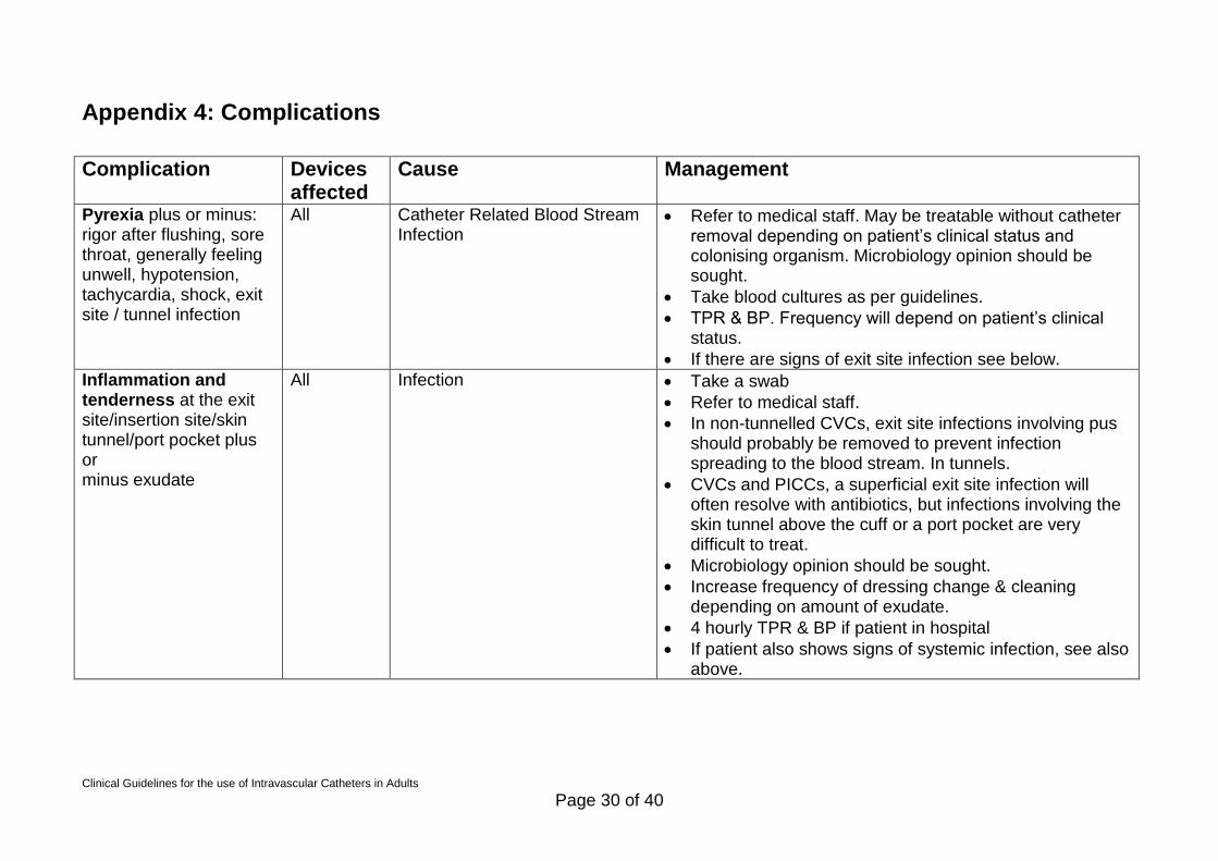
Appendix 4: Complications
Complication Devices
affected Cause Management
Pyrexia plus or minus: rigor after flushing, sore throat, generally feeling unwell, hypotension, tachycardia, shock, exit site / tunnel infection
All Catheter Related Blood Stream Infection
Refer to medical staff. May be treatable without catheter removal depending on patient‟s clinical status and colonising organism. Microbiology opinion should be sought.
Take blood cultures as per guidelines.
TPR & BP. Frequency will depend on patient‟s clinical status.
If there are signs of exit site infection see below.
Inflammation and tenderness at the exit site/insertion site/skin tunnel/port pocket plus or minus exudate
All Infection Take a swab
Refer to medical staff.
In non-tunnelled CVCs, exit site infections involving pus should probably be removed to prevent infection spreading to the blood stream. In tunnels.
CVCs and PICCs, a superficial exit site infection will often resolve with antibiotics, but infections involving the skin tunnel above the cuff or a port pocket are very difficult to treat.
Microbiology opinion should be sought.
Increase frequency of dressing change & cleaning depending on amount of exudate.
4 hourly TPR & BP if patient in hospital
If patient also shows signs of systemic infection, see also above.

Clinical Guidelines for the use of Intravascular Catheters in Adults
Page 31 of 40
The catheter is sluggish, or there is no flashback of blood, or there is complete blockage .
All Clotted blood in catheter
Fibrin sheath (which may be diagnosed using fluoroscopy)
Malpositioned catheter
Build up of lipids (Parenteral Nutrition)
Drug Precipitation
NB: In Implantable Ports needle may be incorrectly positioned: check before taking any other action.
See maintaining patency Appendix 2
Pain or visible swelling when catheter is used or fluid leaks from exit site when catheter is flushed.
Midline PICC Hickman Ports
Malposition of catheter
Internal catheter fracture
Fibrin Sheath (which may be diagnosed using fluoroscopy)
Separation of port and catheter (Implantable ports)
NB: In Implantable Ports needle may be incorrectly positioned: check before taking any other action.
Stop using the catheter.
Refer to the central venous access team and / or medical staff: a malpositioned catheter
should usually be removed. Internal fracture cannot be repaired.
If there is a fibrin sheath severe enough to cause leakage the catheter should be removed.
If catheter is fractured or faulty complete Adverse Incident Form.
Do not retain the catheter but if possible record the
Leakage from external portion of catheter when flushed.
Midline PICC Hickman
External catheter fracture Clamp or fold catheter between the exit site and the leak to prevent air entry. If using a clamp (eg artery forceps) pad with gauze to avoid trauma to the catheter.
Catheter must be repaired or removed as soon as possible. Some catheters can be repaired if equipment & expertise available. The advisability of repair will depend on the patient‟s clinical status as it carries a risk of
Clinical Guidelines for the use of Intravascular Catheters in Adults
Page 32 of 40
infection.
Complete Adverse Incident Form.
Do not retain the catheter but if possible record the make and lot number on the incident form.
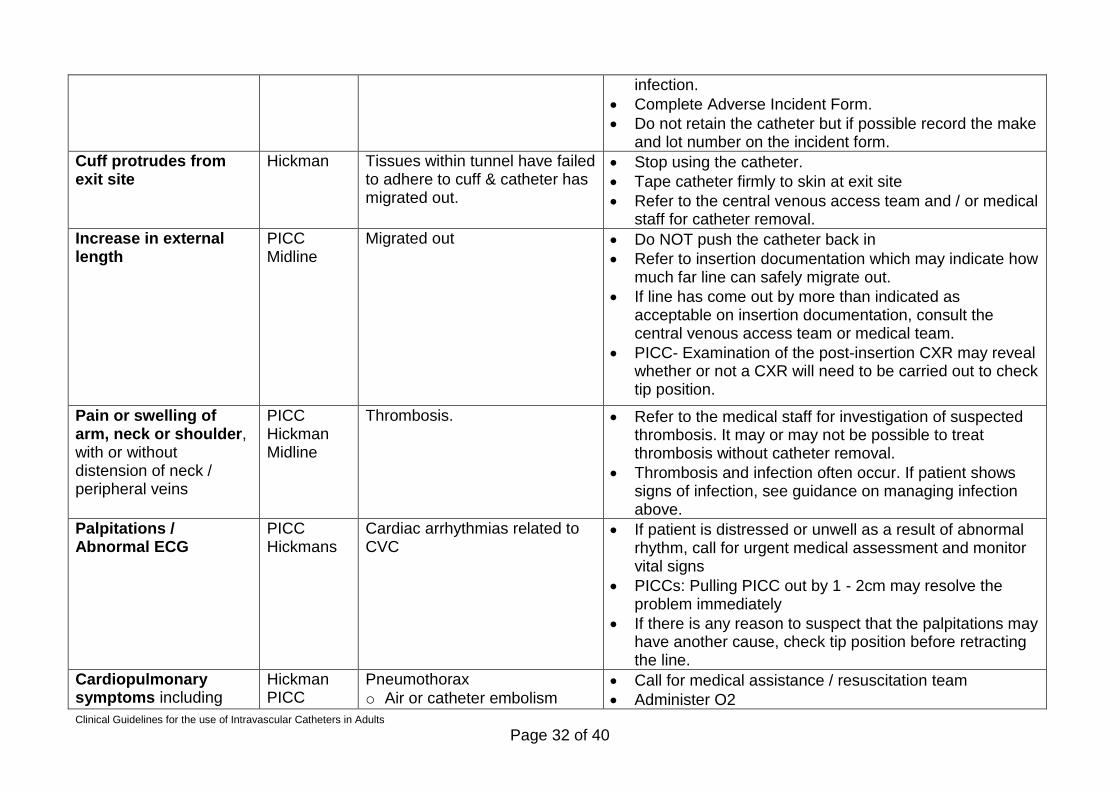
Cuff protrudes from exit site
Hickman Tissues within tunnel have failed to adhere to cuff & catheter has migrated out.
Stop using the catheter.
Tape catheter firmly to skin at exit site
Refer to the central venous access team and / or medical staff for catheter removal.
Increase in external length
PICC Midline
Migrated out Do NOT push the catheter back in
Refer to insertion documentation which may indicate how much far line can safely migrate out.
If line has come out by more than indicated as acceptable on insertion documentation, consult the central venous access team or medical team.
PICC- Examination of the post-insertion CXR may reveal whether or not a CXR will need to be carried out to check tip position.
Pain or swelling of arm, neck or shoulder, with or without distension of neck / peripheral veins
PICC Hickman Midline
Thrombosis. Refer to the medical staff for investigation of suspected thrombosis. It may or may not be possible to treat thrombosis without catheter removal.
Thrombosis and infection often occur. If patient shows signs of infection, see guidance on managing infection above.
Palpitations / Abnormal ECG
PICC Hickmans
Cardiac arrhythmias related to CVC
If patient is distressed or unwell as a result of abnormal rhythm, call for urgent medical assessment and monitor vital signs
PICCs: Pulling PICC out by 1 - 2cm may resolve the problem immediately
If there is any reason to suspect that the palpitations may have another cause, check tip position before retracting the line.
Cardiopulmonary symptoms including
Hickman PICC
Pneumothorax
o Air or catheter embolism Call for medical assistance / resuscitation team
Administer O2

Clinical Guidelines for the use of Intravascular Catheters in Adults
Page 33 of 40
any of the following: respiratory distress / failure apnoea, reduced o2 saturation levels, tachycardia, bradycardia, hypotension, pallor, cyanosis, anxiety, chest pain, loss of consciousness
Ports o Pulmonary embolism
o Cardiac tamponade /
pericardial effusion
Monitor vital signs
Extravasation of Fluids / Drugs due to Incorrect Needle Position or Needle Dislodgement (in Implantable Ports)
Ports Needle is not inserted far enough into the port or if the needle
The needle may become dislodged if it is inadequately secured with dressing tape, if there is tension on the extension tubing or if the needle used is of insufficient length, causing the patient's normal movements to loosen the needle.
If extravasation has occurred or is suspected, the needle should be removed and a fresh needle used to access the port correctly.
If vesicant or irritant solutions (eg chemotherapy) are extravasated, seek medical
Extravasation of Fluids Cannulas Is the leakage or accidental infiltration of intravenous drugs into the surrounding tissues from the vein. This can lead to an immediate inflammatory painful reaction and with some drugs may result in local tissue destruction (necrosis) and other complications.
Determine risk of drug that has extravasated and act according to table in appendix 4
Extravasation of Fluids PICC Midlines
Is the leakage or accidental infiltration of intravenous drugs
Determine risk of drug that has extravasated and act according to table in appendix to be inserted no in final

Clinical Guidelines for the use of Intravascular Catheters in Adults
Page 34 of 40
into the surrounding tissues from the vein. This can lead to an immediate inflammatory painful reaction and with some drugs may result in local tissue destruction (necrosis) and other complications.
draft
For vesicants or large volumes extravasations of any drugs referral to plastic team should be made for further advice
Extravasation of Fluids Hickman
Is the leakage or accidental infiltration of intravenous drugs into the surrounding tissues from the vein. This can lead to an immediate inflammatory painful reaction and with some drugs may result in local tissue destruction (necrosis) and other complications.
Determine risk of drug that has extravasated and act according to table in appendix to be inserted no in final draft
For vesicants or large volumes extravasations of any drugs referral to plastic team should be made for further advice
Catheter Fracture PICC Midline Hickman line
This may occur externally or internally and may result from over-forceful flushing, trauma to the catheter or incorrect position Internal fracture will usually result in patency impairment and / or pain, redness and swelling when the catheter is flushed..
Externally
The line must be clamped or folded over on itself immediately to prevent air embolism.
Sometimes the catheter can be repaired or replaced over a guidewire but the advisability of this will depend on the patient‟s clinical status.
Unless the correct equipment and expertise are available for a repair to be carried out, the catheter should be removed immediately.
Internally
There is a risk that the catheter itself will embolise. If this occurs there may be no symptoms at all or there may be signs of pulmonary embolism
Remove line
Clinical Guidelines for the use of Intravascular Catheters in Adults
Page 35 of 40
Appendix 5: Maintaining Patency
Patency problems are common in Central Venous Catheters and include:
no flashback of blood
sluggish flow
complete blockage
Possible causes
clotted blood in the catheter (most likely cause)
fibrin sheath
malpositioned catheter
drug precipitation
build up of lipids (parenteral nutrition)
Incorrectly positioned needle in an implantable port: check before taking any other action.
Preventing Patency Problems: good flushing techniques Use a brisk „push-pause‟ flushing technique routinely when flushing the catheter –
i.e.flush briskly, pausing briefly after approximately each ml of fluid.
Use a “positive pressure finish” when you lock the catheter – i.e. clamp the line while you are flushing in the final ml. If there is no clamp remove the syringe from the Bionector
(or similar) while you are still injecting the final ml. Managing Patency Problems
No flashback of blood o Ask the patient to take deep breaths and try different positions. Flush briskly using
10mls saline*. If this fails use a thrombolytic (see below). o If lipids/drug precipitation suspected consult pharmacy advice for suitable agent to
dissolve occlusion
Catheter flow is sluggish o Ask the patient to take deep breaths and try different positions. Flush briskly with
10mls saline*. If this fails use a thrombolytic (see below). o If lipids/drug precipitation suspected consult pharmacy advice for suitable agent to
dissolve occlusion
Catheter is completely blocked o Use a 3-way tap technique to instil thrombolytic into catheter (see below).
What is a thrombolytic?
A thrombolytic is a drug capable of breaking up a thrombus.
Urokinase is the thrombolytic used for unblocking CVCs in Cancer Services: use 5000 units in 2mls per lumen.
A thrombolytic should always be prescribed.
Heparin and Hepsal are NOT thrombolytics: they are capable only of inhibiting thrombus formation.

Clinical Guidelines for the use of Intravascular Catheters in Adults
Page 36 of 40
When should you use a thrombolytic?
Use a thrombolytic to improve patency in the following situations: o flashback of blood is absent o free-flow of fluids is sluggish or intermittent o resistance is felt when flushing o the catheter/lumen is completely blocked
What if the thrombolytic fails to restore function?
If a thrombolytic used correctly (see below) fails to restore function, contact the central venous access team and / or medical team. A chest x-ray may need to be carried out to check the position of the line. If a chest x-ray shows that the catheter is correctly placed, it may be worth investigating further using fluoroscopy which may reveal a fibrin sheath.
If the cause could be a build up of lipids from Parenteral Nutrition or drug precipitation consult pharmacy advice for a suitable agent to dissolve occlusion.
How to use a thrombolytic
Arrange prescription. (Caution if patient‟s clotting is severely deranged or if high doses of an anticoagulant are being given concurrently.)
Draw up the thrombolytic as per manufacturer‟s instruction eg for Urokinase: reconstitute10,000 unit vial with 2mls water for injection and dilute further to 4 mls. Use 2mls (5000 units) per lumen.
Instil the thrombolytic into the catheter and wait 2 hours (or preferably longer if possible). But note that if the lumen is completely blocked do NOT force the thrombolytic into the catheter: see Using a Thrombolytic in a Completely Blocked Catheter (below).
Assess the catheter again. NB It is best to flush the catheter with 0.9% saline (must be prescribed) prior to attempting to obtain flashback, otherwise there is a risk of creating a further blockage in the line before you have cleared it. NB There is no need to worry that you are flushing the thrombolytic into the patient: small doses can be flushed into the patient without danger unless the patient has exceptionally deranged clotting.
If full function has not returned instil the thrombolytic again and leave in for longer – several hours or overnight if possible. NB the fact that urokinase has a half life of 20 minutes once it is in the patient‟s system is irrelevant to how long it carries on working while still in the catheter. This explains why leaving it in longer is often more effective.
If the procedure fails to restore function consider whether lipids / drug precipitation could be causing a blockage. If not, refer to the central venous access team and / or medical staff: a chest x-ray may reveal malposition of the line.

Clinical Guidelines for the use of Intravascular Catheters in Adults
Page 37 of 40
Using a Thrombolytic in a Completely Blocked Catheter.
Attach 3-way-tap & syringes (NB 3-way taps are now contraindicated for routine IV use but are still available for this procedure.
Always use a 3-way tap without an extension set) o Open clamp (if there is one). o Open stopcock to the empty syringe and the blocked catheter. o Pull back on the plunger of the empty syringe to create a vacuum in the
catheter. You will need to pull quite forcibly. o Maintain suction with one hand and with the other hand turn stopcock so it is
closed to the empty syringe and open to the syringe containing thrombolytic, which will be sucked into the catheter. Don‟t worry if it seems that very little thrombolytic is sucked in: even a tiny volume will reach several cm into the catheter.
Three-way Tap Technique – to be performed only by suitably trained staff
Leave for several hours or overnight. DO NOT CLAMP CATHETER as this will prevent the thrombolytic from penetrating into the line.
After this time, assess the catheter by attempting to flush it catheter using 0.9% saline* in a 10ml syringe. Do not use excessive force. (It is best NOT to try aspirating before flushing at this stage as you may block the catheter again).
If the catheter is still completely blocked, repeat the procedure: sometimes you will need to do it several times before it works. Sometimes leaving the thrombolytic in overnight seems to
Don‟t worry about overdosing the patient: if the catheter is blocked none of the drug will actually have been flushed into the blood stream.
Once the catheter can be flushed, and only then, check for flashback. If flashback is absent, administer thrombolytic as described above.
· If the procedure fails despite repeated attempts consult the central venous access team and / or medical team with a view to removing the catheter.

Clinical Guidelines for the use of Intravascular Catheters in Adults
Page 38 of 40
Appendix 6 References Alexandrou E, Ramjan LM, Spencer T, Frost SA,Salamonson Y, Davidson PM and Hillman KM (2011) The use of midline catheters in the adult acute caresetting – clinical implications and recommendations for practice, Journal of the Association for VascularAccess, 16(1), pp.35-41. Bodenham A, Babu S, . Bennett J, Binks R, . Fee P, Fox B, Johnston A J, Klein AA, Langton JA, Mclure H and Tighe S Q M. (2016) Association of Anaesthetists of Great Britain and Ireland Safe vascular access 2016. Anaesthesia, 2016, 71, 573–585 (V) available: www.aagbi.org/sites/default/files/Safe%20vascular%20access%202016.pdf [accessed 03 Nov 2016] Bortolussi R, Zotti P, Conte M, Marson R, Polesel J, Colussi A, Piazza D, Tabaro G and Spazzapan S (2015) Quality of life, pain perception, and distress correlated to ultrasound-guided peripherally inserted central venous catheters in palliative care patients in a home or hospice setting, Journal of Pain and Symptom Management, 50(1), pp.118-123. (IV) British National Formulary (2016a) Extravasation [online], available: www.evidence.nhs.uk/formulary/bnf/current/10-musculoskeletal-and-jointdiseases/103-drugs-for-the-treatment-of-soft-tissuedisorders-and-topical-pain-relief/extravasation [accessed: 28 August 2016].
Chopra V, Anand S, Hickner A, Buist M, Rogers MA, Saint S and Flanders SA (2013) Risk of venous thromboembolism associated with peripherally inserted central catheters: A systematic review and meta- analysis, The Lancet, 382(9889), pp.311-325. Electronic Medicines Compendium (2015) Lidocaine Chloride Injection BP Minijet 1% [online], available: www.medicines.org.uk/emc/medicine/4888 [accessed:30 August 2016]. Fidalgo JA, Fabregat LG, Cervantes A, Marguiles A, Vidall C and Roila F (2012) Management of chemotherapy extravasion: ESMO clinical practice guidelines, Annals of Oncology, 23(supp. 7), pp. vii167-vii173. Frykholm P, Pikwer A, Hammarskjöld F, Larsson AT, Lindgren S, Lindwall R, Taxbro K, Oberg F, Acosta S, Akeson J (2014) Clinical guidelines on central venous catheterisation. Swedish Society of Anaesthesiology and Intensive Care Medicine, Acta Anaesthesiol Scand, 58(5):508-24. doi: 10.1111/aas.12295. Epub 2014 Mar 5. González López JL, Arribi Vilela A, Fernández del Palacio E, Olivares Corral J, Benedicto Marti C and Herrera Portal P (2014) Indwell times, complications and costs of open vs closed safety peripheral intravenous catheters: A randomized study, Journal of Hospital Infection, 86(2), pp.117-26. Gorski LA, Hagle ME and Bierman S (2015) Intermittently delivered IV medication and pH: Reevaluating the evidence, Journal of Infusion Nursing, 38(1), pp. 27-46. Hallam, Ce, Weston, V, Denton, A, Hill, S, Bodenham, A, Dunn, H, Jackson, T (2016) Development of the UK Vessel Health and Preservation (VHP) framework: a multi-organisational collaborative. Journal of Infection Prevention. ISSN 1757-1774
Hsu Y, Weeks K, Yang T, Sawyer MD and Marsteller JA (2014) Impact of self-reported guideline compliance: Bloodstream infection prevention in a national collaborative, American Journal of Infection Control, 42(Supplement), pp.S191-S196.

Clinical Guidelines for the use of Intravascular Catheters in Adults
Page 39 of 40
Hunt BJ, Allard S, Keeling D, Norfolk D, Stanworth SJ and Pendry K (2015) A practical guideline for the haematological management of major haemorrhage, British Journal of Haematology, (170), pp.788-803. Infusion Nurses Society (2016) Infusion therapy standards of practice, Journal of Infusion Nursing, 39(1S), available: INS Digital Press. Jackson A (1998) Infection control: a battle in vein infusion phlebitis, Nursing Times, 94(4), pp.68-71. Lai NM, Chaiyakunapruk N, Lai NA, O‟Riordan E, Pau WS and Saint S (2013) Catheter impregnation, coating or bonding for reducing central venous catheter-related infections in adults, The Cochrane database of systematic reviews, (6) CD007878, doi:10.1002/14651858. Lamperti, M., Bodenham, A. R., Pittiruti, M., Blaivas, M., Augoustides, J. G., Elbarbary, M., Pirotte, T., Karakitsos, D., LeDonne, J., & Doniger, S. (2012).International evidence-based recommendations on ultrasound-guided vascular access, Intensive care medicine 38(7),1105–1117. Loveday HP, Wilson JA, Pratt RJ, Golsorkhi M, Tingle A, Bak A, Browne J, Prieto J and Wilcox M (2014) Epic3: National evidence-based guidelines for preventing healthcare-associated infections in NHS hospitals in England, Journal of Hospital Infection, 86(Supp.1), pp. S1-S70. Maneval RE and Clemence BJ (2014) Risk factors associated with catheter-related upper extremity deep vein thrombosis in patients with peripherally inserted central venous catheters: A prospective observational cohort study: Part 2, Journal of Infusion Nursing, 37(4), pp.260-268.
Medicines and Healthcare Products Regulatory Agency (2013a) Single-use medical devices: implications and consequences of reuse, London: MHRA. Medicines and Healthcare Products Regulatory Agency (2013b) Infusion systems, London: MHRA. Medicines and Healthcare Products Regulatory Agency (2015a) Adverse drug reporting [online], available: https://yellowcard.mhra.gov.uk/ [accessed: 1 September 2016]. Medicines and Healthcare Products Regulatory Agency (2016b) Yellow card [online], available: https://yellowcard.mhra.gov.uk/ [accessed: 1 September 2016]. Medicines Complete (2016 b) Heparinoid cream [online], available: www.medicinescomplete.com/mc/bnf/current/DMD3158411000001101.htm?q=Hirudoid&t=search&ss=text&p=1#DMD3158411000001101 [accessed: 1 September 2016]. Mitchell MD, Agarwal R, Hecht TE and Umscheid CE (2013) Nonpharmacologic interventions for prevention of catheter-related thrombosis: A systematic review, Journal of Critical Care, 28(3), pp.316.e9-.e16.
National Institute for Health and Care Excellence (2007) Quick reference guide. Acutely ill patients in hospital: recognition of and response to acute illness inadults in hospital, CG50, London:NICE. National Institute for Health and Care Excellence (2012) Healthcare-associated infections: prevention and control in primary and community care [online], available: www.nice.org.uk/guidance/cg139/chapter/1-Guidance#vascular-access-devices [accessed: 1 September 2016].
National Institute for Health and Care Excellence (2013) CG174: Intravenous fluid therapy in adults in hospital [online], available: www.nice.org.uk/guidance/cg174?unlid=7240841622016876654 [accessed: 1September 2016].

Clinical Guidelines for the use of Intravascular Catheters in Adults
Page 40 of 40
Nursing and Midwifery Council (2015a) The Code:Professional standards of practice and behaviour fornurses and midwives, London: NMC. Nursing and Midwifery Council (2015c) Standards formedicines management, London, NMC. O‟Grady NP, Alexander M, Burns LA, Dellinger EP,Garland J, Heard SO, Lipsett PA, Masur H, Mermel LA,Pearson ML, Raad II, Randolph AG, Rupp ME and Saint S (2011) Guidelines for the prevention of intravascularcatheter-related infections, Centers for Disease Control and Prevention [online], available: www.cdc.gov/hicpac/pdf/guidelines/bsi-guidelines-2011.pdf [accessed: 1 September 2016]. O‟Horo JC, Maki DG, Krupp AE and Safdar N (2014)Arterial catheters as a source of bloodstream infection: A systematic review and meta-analysis, Critical Care Medicine, 42(6), pp.1334-1339. Parienti J, Mongardon N, BMegarbane B, Mira J, KalfonP, Gros A, Marque S, Thuong M, Pottier V, Ramakers M,Savary B, Seguin A, Valette X, Terzi N, Sauneuf B,Cattoir V, Mermel LA, and du Cheyron D (2015)Intravascular Complications of Central Venous Catheterization by Insertion Site, New England Journal of Medicine, (373), pp.1220-1229. Renal Association (2015a) Summary of audit measuresrecommended for dialysis access for adults with endstage kidney disease [online], available: www.renal.org/guidelines/modules/vascular-access-forhaemodialysis#sthash.MsYoKEmh.dpbsicaotion [accessed: 2 September 2016]. Renal Association (2015b) Vascular Access forHaemodialysis, [online], available: www.renal.org/guidelines/modules/vascular-access-forhaemodialysis#sthash.aVJF7o3D.dpbs [accessed 8 November 2016] World Health Organization (2010) WHO Guidelines ondrawing blood: best practices in phlebotomy, Geneva:WHO.