Clinical Examples Which Questions may be answered Advantages … · 2008-06-13 · Noninvasive...
52
Noninvasive cardiac imaging by CT & MR Change of Diagnostic Workflow Interdisciplinary Cardiac Imaging Center [IC] 2 Med.University Graz/Austria Clinical Examples Which Questions may be answered Advantages for Patient / Referral Place in Diagnostic Algorithm Berlin
Transcript of Clinical Examples Which Questions may be answered Advantages … · 2008-06-13 · Noninvasive...

Noninvasive cardiac imaging by CT & MR
Change of Diagnostic Workflow
Interdisciplinary Cardiac Imaging Center [IC]2 Med.University Graz/Austria
Clinical ExamplesWhich Questions may be answeredAdvantages for Patient / Referral
Place in Diagnostic Algorithm
Berlin

R. Rienmüller (1983) Thesis EBT 2001
CT in the Clinical Diagnostic of Cardiac Disease
„CTCA is a valuable clinical tool, thathas the potential to improve the investigation of cardiac patients.
CTCA will undoubtedly become bothmore prevalent and more useful as access and technology improves.“
A. Baumbach, M.CK.Hamilton: ESC, E-Journal – Vol 5. N 20, Feb. 2007

Spatial resolution 1.5 mmTime resolution < 50 ms
Interdisciplinary Cardiac Imaging Center [IC]2 Med.University Graz/Austria

64
Interdisciplinary Cardiac Imaging Center [IC]2 Med.University Graz/Austria
Spatial resolution < 0.7 mmTime resolution ≅ 165 ms
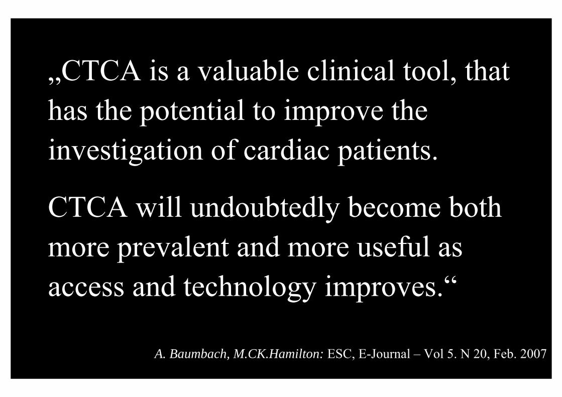
Dual Source CT
www.siemens.com/medical
Spatial resolution < 0.7 mmTime resolution ≅ 83 ms

CT– Technologien
Isotrope Bild Voxel165 ms(84 ms)
Dual Source
Schichtdicke 1,5 mm50-100 msEBT (UFCT)(1. Multislice & MultidetectorSystem)
Isotrope Bild Voxelca 330-500 ms(165 ms)
Multislice, Multidetector(16, 32, 64 - 256)
ca 500-700 msSpiralSchichtdicke ca 1mmca 1000 ms Single SliceBemerkungenScanzeitModalität
Benötigt wird eine Scanzeit von ca 20 ms, ca 10 Mal während einer Herzaktion für die Dauer von ca 40 Herzaktionen als Volumenscan
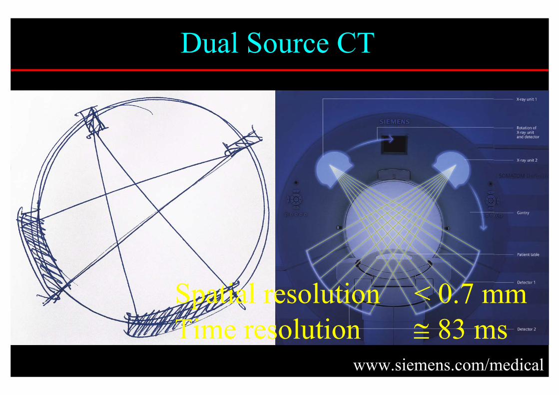
Spatial resolution < 0.5 mmTime resolution < 30 ms
Interdisciplinary Cardiac Imaging Center [IC]2 Med.University Graz/Austria

34 y male with Known Coarctation of the Aorta
Present History: disorder of sleep with dyspnoe and episodes of choking, peripheral edema, acute cardiac decompensation
Risk factors: Hypertension (RR:r 150/60 mm Hg, RR:l 135/60 mm HG),Bicuspid valve, Aortic insufficiency, Nicotin,
Medication: Seloken ret. plus, Lasix 20 mg
Diagnosticevaluation: Catheterisation, CTA, MR

34 y male with Coarctationof the Aorta
MSCT 64

34 y male with Coarctationof the Aorta
MSCT 64

34 y male with Coarctationof the Aorta
MSCT 64

W.S., ♂, 37 YO

34 y male with Known Coarctation of the AortaFindings
MSCT 64: Bicuspid valve, Dilatation of ascending aorta and of theanomalous supraaortic vessels, Severe (afew mm) stenosis, Bypassing circulation by dilated internal thoracic arteries,
by dilated intercostal arteries (III), by dilated lateral thoracic arteries, by dilated rete scapulare
Normal origin of LCA and RCA,thin LAD, CX and RCA
EBT: (Borderline) myocardial perfusion of 63 ml/100g/min at HR of 78 min-1, RR 180/80 mm HG

34 y male with Coarctationof the Aorta
Cardiac MR
Work in Progress. G. Reiter, U. Reiter, R. Rienmüller

MR: EDV – 232 ml, ESV – 138 ml, SV – 93 ml, EF – 40,3%, LVMM – 176 gAsc. Aorta: Forward-Flow
Reverse-FlowNet Forward-Flow
Poststenotic Desc. Aortar. Intercostal arteryl. Intercostal arteryDesc. Aorta prox.Desc. Aorta dist.
Suggestedtherapy: short term: interventional treatment of the stenosis
long term: aortic valve and ascending aorta surgery
80 ml35 ml45 ml
0,078 ml 5 ml8 ml
15 ml14 ml
34 y male with Known Coarctation of the Aorta

„... is the Manifestation of Atherosclerosisin the Coronary Arteries. As the Disease is a Multifactorial Process Leading toMyocardial Ischemia, it may Appear asAngina Pectoris, Myocardial Infarction,Cardiac Dysrhythmia, Sudden Death orCardiac Insufficiency. The Course of theDisease may be Silent.“
Coronary Heart Disease
J. Meyer. Klinische Kardiologie, Edd. E. Erdmann, G. Riecker. Springer Verlag 1996

Clinical Question1) Does the patient have coronary atherosclerosis?
(Stage, Localization, Kind of sclerosis, Degree of stenosis)
2) Does the patient have myocardial ischemia?3) How big is the individual myocardial perfusion
(ml/100g/min)?4) Which mechanism may prevent or reduce myocardial
ischemia?5) Does the patient have myocardial infarction?6) Which parts of the myocardium are viable?7) How big is the individual coronary reserve?

74 y anxious female with CAD and HypertensionPresent History:Angina at night for two years
Past History:3 y ago: suspected MI and stroke,Non-conclusive CA
Medication: TASS 100 mg, Plavix, Nomexor, Iterium, Dancor, Temesta, Plendil ret., Tebonin ret.
Hospitalisation: July 6-11, 2005Coronary Angiography rejectedErgo- and Tl-Scint. not doneCT-Angiography recommendedBlood pressure allways in „lower normal range“

Traditional approach in patients with suspected CHD
Patients historyPhysical findingsChest X-rayECGEchocardiographyCardiac ScintigraphyLabor parametersCoronary angiography

Traditional approach in patients with suspected CHD
Chest X-rayECGEchoSPECTLabor parameters
Indirect methods, No visualization of coronary arteries and No measuring of myocardial perfusion in (ml/100g/min)
Coronary angiography ≃ Luminography No visualization of coronary walls and myocardial perfusion No relationship between extend of luminal stenosis and myocardial perfusion

„Angiographic Evidence of The Severityof Coronary Stenosis is not Correlatedwith Physiological and Clinical Effects.
„There is no Apparent Relation betweenthe Severity of Coronary Lesions and their Propensity to Cause Future CardiacEvents. Most Lesions Resulting in Cardiac Events are not Severely Stenotic.“
As cited by G. Levine et al. N Engl J Med, 1995; 332:512-521

EDVESVEFCOLVMMHRRRCa++ Score
Myocardial Perfusion global = 54 [75 ± 10] ml/100g/min ant.wall = 49 [75 ± 10] ml/100g/min
Perf/HR = 1.00 [1 ± 0.3]Perf/RPP = 0.0083 [0.009]
10029713,67054120/7567
[105 ± 10] ml[30 ± 5] ml[>70] %[5 ± 0.5] l/mingmin-1mm Hg[0]
74 y anxious female with CAD and Hypertension
LAD > 90 % → 3,5 cm> 50 % → 5-7 cm
Diagonal > 50 % → 0,5 cm

74 y anxious female with CAD and HypertensionProcedure after CTA:
→ Referring physian informed by phone + letter→ Patient referred to EBA (Emergency)→ Patient came to Radiology→ Patient referred to Dep. of Cardiology→ ECG – Stress test negative → Patient send home→ Patient continues to complain→ Cardiology LKH-West→ Coronary angiography – CTA-Diagnosis confirmed and by
Drug eluting Stents dilatation performed, and antihypertensive therapy adapted to present Hypotension

W.S., m 63y Symptoms: Chest pain
Findings: LAD – hard & softplaques,Stenosis > 50%RCA – hypoplastic
Ca Score: 110
Perfusion: globally reducedto 41ml/100g/min at HR of 52 /min, RR of 119/76 mmHg
Morphology& Function: EDV – 162 ml,
LVMM – 169, EF – 65%

Calcium Scoring
“Increased Calcium Score adds to the risk profile and can trigger preventive treatment”
De Bacher G, et all: Eur Heart J. 24 (17): 1601-10; 2003
“The routine assessment of the Coronary Calcium Score is still under debate”
Hamilton MCK, et all: BMJ 2003; 326: 1045-1046

Clinical Question
- What is the smallest soft and calcified plaque to beidentified by CT?
- How does Ca++ score correlate
with soft plaque?with stenotic lesions?with age?

Courtessy of R.Vliegenthart PhDLecture at ESCR Meeting Berlin 2004

Logistic Regression Analysisin Patients with Suspected Coronary Heart Disease
00.10.20.30.40.50.60.70.80.9
1
0 1000 2000 3000 4000Total Calcium Score
Like
lihoo
dfo
rSte
nosi
s>5
0 %
0
680
90 % Likelihood= Score 1300
Score
Score=
=
Likelihood
Likelihood
50 %
10 %

Calcium Score vs Age

Co-factors of Ca++ Score
Age lvmm syst. RPP CO SV HR Perfusion
Aortic
Compliance
Ca++ Score
p
r²
r
n
00.440.60.940.020.12000
0.04000000.020.030.14
-0,13-0,02-0,0100,020,040.080.150.25
80514121474131113081459146214101472
AorticCompliance
PerfusionHRSVCORPPSyst.lvmmAge

M.S., m 73y Symptoms: post Stent, occlusion?
Findings: LAD – hard & softplaques,with stenosis > 50% beforeopen stent,R.intermedius – multipl.stenosis > 50%
Ca Score: 673
Perfusion: globally reduced – 47 ml/100g/minat HR of 49 /min, RR of 164/95 mmHg
Morphology& Function: EDV – 120 ml, LVMM – 98 g,
EF – 66%


CT – Coronary Angiography (CTCA)CTCA tends to overestimate coronary stenosis
compared to invasive coronary angiography pos. pred. value – ca.80%neg. pred. value – ca.95%
Achenbach et al. Eur J Radiol 2006
CT Bypass Sensitivity for occlusion up to 100% Sensitivity and specificity for stenosis ca. 95/ 89% (Cave Metal clip)
Pache et al. EHJ 2006
CT Stent Specificity and sensitivity ca. 98/ 83%(Cave geometry should be above 2 mm, right reconstruction Kernel)
Cademartiri et al. AJC 2005

Diagnostic Accuracy of EBT / MSCTvs. Angiography for Stenotic Lesions > 50%
NPV (%)PPV (%)Specificity (%)Sensitivity (%)
96NPV (%)73PPV (%)96Specificity (%)73Sensitivity (%)
All segments
n = 222 patients, 2220 segments,
disease prevalence 45%
92237467
92237467
All segments
n = 20 patients, 200 segments,
disease prevalence 10%
Interdisciplinary Cardiac Imaging Center [IC]2 Med.University Graz/Austria

1. Planning and Overview2. Function3. Morphology4. Coronary Arteries5. Perfusion
6. Cine Short Axes7. Aortic Flow8. Late Enhancement
TrueFISP breathhold
Lung
Right and Left Ventricle
Myocardial Sectors
Standard MR Protocol: Suspected CHD

Stilcke Hans-Gottfried
Late Enhancement: Flash 2D Cine SA True-Fisp
Berlin

T1 T2 T2FS
Late Enhancement Cine TrueFISP
Intima Sarcoma

TI
Aortenklappenersatz
ASD

Change of Diagnostic Workflow by
Cardiac CT and MR
Interdisciplinary Cardiac Imaging Center [IC]2 Med.University Graz/Austria
Clinical ExamplesWhich Questions may be answeredAdvantages for Patient / Referral
Place in Diagnostic Algorithm
Berlin

Indication for Cardiac CT/MR- studies?
ARVD7) Anomaly of coronary vesselsShunt8) Bypass-stenosis/-occlusion
9) Stent-occlusion
Cardial Thrombus/ Tumor6) Pericardial diseasesPericardial Constriction5) Myocardial ischemiaValve vitium4) Myocardial infarctionMyocarditis3) Coronary stenosis > 50%Myocardial scare2) Chronic Coronary SyndromInfarction/ „Vitality“1) „Subclinical“ CHD
MRCT
Interdisciplinary Cardiac Imaging Center [IC]2 Med.University Graz/Austria

Change of Diagnostic Workflow by
Cardiac CT and MR
Interdisciplinary Cardiac Imaging Center [IC]2 Med.University Graz/Austria
Clinical ExamplesWhich Questions may be answeredAdvantages for Patient / Referral
Place in Diagnostic Algorithm
Berlin

Benefit for the Patient
Objective, non invasive, reproducible, fast method for exclusion or for early recognition and staging of coronary, cardiac and pulmonary diseases as One Stop Shop.

Benefit for the Referrals Objective, non invasive, quantitative, reproducible, fast method for exclusion or for early recognition and staging of coronary, cardiac and pulmonary diseases as One Stop Shop.
Therapy recommendation and control of its individual effectiveness (evidence based medicine).
Benefit for CardiologistsReduction of unnecessary coronary catheterization.
Increase capacity for interventional procedures.
Objective, non invasive, quantitative, reproducible, fast method for exclusion or for early recognition and staging of coronary, cardiac and pulmonary diseases as One Stop Shop.
Therapy selection and control of its individual effectiveness (evidence based medicine).

Change of Diagnostic Workflow by
Cardiac CT and MR
Interdisciplinary Cardiac Imaging Center [IC]2 Med.University Graz/Austria
Clinical ExamplesWhich Questions may be answeredAdvantages for Patient / Referral
Place in Diagnostic Algorithm
Berlin

Risk Profile of CHD
CT enables noninvasive direct visualization of soft- and calcified coronary arterial plaques – “The potential of this information for risk stratification has sparked intense interest in plaque imaging to identify patients at high risk of a coronary event”
0% low intermediate high 100%
CT / MR
0 Catheter
asymptomatic symptomatic
S. Achenbach 2007

New Concept for Evaluation of patients with suspected or known CHD
MSCT, (DSCT, EBT) and MR
Interdisciplinary Cardiac Imaging Center [IC]2 Med.University Graz/Austria
2. Step: Evaluation of etiology and accompanying individual health issues as history, physical findings, HR and RR, laboratory parameters, ECG – Arhythmia (Sympath./Para-sympath.), Echo – Valvular disease
(Cyclotron-PET) – Metabolism, Perfusion (ml/100g/min)
1. Step: Visualization of micro-pathology and patho-physiology by

Non-Invasive In Vivo Visualisation Of OrganMicro-Pathology and Patho-Physiology
Conclusion I:
Will create new diagnostic work-up → new work-flow:
- “First look Doctor” in whole body One-Stop-Shop withComputed assisted diagnostic devices (pattern recognition) with therapeutical options
- Target history taking and target physical examinationwith additional tests dependent on individual patient’s situation
Interdisciplinary Cardiac Imaging Center [IC]2 University of Graz/Austria

Non-Invasive In Vivo Visualisation Of OrganMicro-Pathology and Patho-Physiology
Will demand for:
- New match of the (imaging) diagnostician and (therapeutic) clinicians
- Reform of medical education (radio-patho-anatomy and radio-patho-physiology)
- Implication of “autopsy” by CT/MR technologies with target biopsy and adequate evaluation.
Interdisciplinary Cardiac Imaging Center [IC]2 University of Graz/Austria
Conclusion II:

Non-Invasive In Vivo Visualization Of OrganMicro-Pathology and Patho-Physiology
Conclusion III:
Will result in:
Effective, science-based, transparent, economic, individual patient orientated
new Health-Care Systemopen for genetic and molecular biology implementation
Interdisciplinary Cardiac Imaging Center [IC]2 University of Graz/Austria

Change of Diagnostic Workflow by
Cardiac CT and MR
Interdisciplinary Cardiac Imaging Center [IC]2 Med.University Graz/Austria
Berlin
Thank you for your attention!
R.RienmüllerB.Schröttner H.Mächler J.TeublU.Reiter W.Klein P.LyszczM.Kutateladze N.Gagarina K.KovalevaM.Zink O.Waltersdorfer R. Reiter
![NONINVASIVE ECG IMAGING [ ECGI ] OF CARDIAC ARRHYTHMIAS · electrocardiographic measurements, noninvasively • Noninvasive imaging is a corner stone of the practice of modern medicine](https://static.fdocuments.in/doc/165x107/5f022a177e708231d402e402/noninvasive-ecg-imaging-ecgi-of-cardiac-arrhythmias-electrocardiographic-measurements.jpg)


![Noninvasive surface imaging.ppt [โหมดความเข้ากันได้] surface imaging.pdf · 1 Noninvasive Surface Imaging Supenya Varothai, M.D. Department of](https://static.fdocuments.in/doc/165x107/5e0571a85dfeb539200c59cf/noninvasive-surface-aaaaaaaaaaaaaaaaaa-surface.jpg)