Clinical Approach to Parkinson’s Disease: Features ...
15
Clinical Approach to Parkinson’s Disease: Features, Diagnosis, and Principles of Management Joa ˜ o Massano 1,2 and Kailash P. Bhatia 1 1 Sobell Department of Motor Neuroscience and Movement Disorders, Institute of Neurology, University College London, Queen Square, London, United Kingdom 2 Movement Disorders and Functional Surgery Unit, Department of Neurology Hospital de Sa ˜o Joa ˜o and Faculty of Medicine University of Porto, Porto, Portugal Correspondence: [email protected] Parkinson’s disease (PD) is one of the most common neurodegenerative disorders. The con- dition causes a heavy burden both on those affected, as well as their families. Accurate diagnosis is critical and remains founded on clinical grounds as no specific diagnostic test is available so far. The clinical picture of PD is typical in many instances; however, features distinguishing it from other disorders should be thoroughly sought. Monogenic forms of PD also have some distinctive characteristics in many cases. This text is a roadmap to accurate diagnosis in PD, as it approaches clinical features, diagnostic methodology, and leading differential diagnoses. Therapeutic issues are also briefly discussed. N early 200 years have passed since the pub- lication of James Parkinson’s succinct ob- servations in An Essay on the Shaking Palsy (Par- kinson 1817, reprinted in Parkinson 2002; for more details on the history of PD see the text by Goetz 2011). Remarkably, the original clinical description of the disease remains a landmark reference, especially with regard to the motor features. Parkinson’s disease (PD), as Jean Mar- tin Charcot called it a few decades later (Lees 2007; Lanska 2010), is a common and often disabling disorder seen in people from all races and geo- graphical locations, with clinical signs emerging also in a wide age range, including the young (Marsden 1994; Tolosa et al. 2006; Lees et al. 2009). The epidemiological figures pertaining to PD are not accurately determined, as different studies were variable in diagnostic criteria, meth- odology, and study population. A prevalence of 1%–2% in the population older than 60–65 yr, or 0.3% in the general population, is now commonly accepted (de Lau and Breteler 2006; Hirtz et al. 2007; Alves et al. 2008), with prev- alence rates ranging from 65.6 to 12,500 per 100,000 population (von Campenhausen et al. 2005). In 2005, more than 4 million PD patients existed in the world (Dorsey et al. 2007). Annual incidence rates range from 8.6 to 19 per 100,000 (Twelves et al. 2003; de Lau and Breteler 2006; Alves et al. 2008). PD is the second most com- mon neurodegenerative disorder after Alzhei- Editor: Serge Przedborski Additional Perspectives on Parkinson’s Disease available at www.perspectivesinmedicine.org Copyright # 2012 Cold Spring Harbor Laboratory Press; all rights reserved; doi: 10.1101/cshperspect.a008870 Cite this article as Cold Spring Harb Perspect Med 2012;2:a008870 1 www.perspectivesinmedicine.org
Transcript of Clinical Approach to Parkinson’s Disease: Features ...

Clinical Approach to Parkinson’s Disease:Features, Diagnosis, and Principles ofManagement
Joao Massano1,2 and Kailash P. Bhatia1
1Sobell Department of Motor Neuroscience and Movement Disorders, Institute of Neurology,University College London, Queen Square, London, United Kingdom
2Movement Disorders and Functional Surgery Unit, Department of Neurology Hospital de Sao Joaoand Faculty of Medicine University of Porto, Porto, Portugal
Correspondence: [email protected]
Parkinson’s disease (PD) is one of the most common neurodegenerative disorders. The con-dition causes a heavy burden both on those affected, as well as their families. Accuratediagnosis is critical and remains founded on clinical grounds as no specific diagnostic testis available so far. The clinical picture of PD is typical in many instances; however, featuresdistinguishing it from other disorders should be thoroughly sought. Monogenic forms of PDalso have some distinctive characteristics in many cases. This text is a roadmap to accuratediagnosis in PD, as it approaches clinical features, diagnostic methodology, and leadingdifferential diagnoses. Therapeutic issues are also briefly discussed.
Nearly 200 years have passed since the pub-lication of James Parkinson’s succinct ob-
servations in An Essay on the Shaking Palsy (Par-kinson 1817, reprinted in Parkinson 2002; formore details on the history of PD see the text byGoetz 2011). Remarkably, the original clinicaldescription of the disease remains a landmarkreference, especially with regard to the motorfeatures. Parkinson’s disease (PD), as Jean Mar-tin Charcot called it a few decades later (Lees 2007;Lanska 2010), is a common and often disablingdisorder seen in people from all races and geo-graphical locations, with clinical signs emergingalso in a wide age range, including the young(Marsden 1994; Tolosa et al. 2006; Lees et al.2009).
The epidemiological figures pertaining toPD are not accurately determined, as differentstudies were variable in diagnostic criteria, meth-odology, and study population. A prevalence of�1%–2% in the population older than 60–65yr, or 0.3% in the general population, is nowcommonly accepted (de Lau and Breteler 2006;Hirtz et al. 2007; Alves et al. 2008), with prev-alence rates ranging from 65.6 to 12,500 per100,000 population (von Campenhausen et al.2005). In 2005, more than 4 million PD patientsexisted in the world (Dorsey et al. 2007). Annualincidence rates range from 8.6 to 19 per 100,000(Twelves et al. 2003; de Lau and Breteler 2006;Alves et al. 2008). PD is the second most com-mon neurodegenerative disorder after Alzhei-
Editor: Serge Przedborski
Additional Perspectives on Parkinson’s Disease available at www.perspectivesinmedicine.org
Copyright # 2012 Cold Spring Harbor Laboratory Press; all rights reserved; doi: 10.1101/cshperspect.a008870
Cite this article as Cold Spring Harb Perspect Med 2012;2:a008870
1
ww
w.p
ersp
ecti
vesi
nm
edic
ine.
org

mer’s disease, with a male-to-female ratio ofabout 3:2 in most studies (de Lau and Breteler2006; Alves et al. 2008). It occurs infrequentlyunder 40 years of age, with early onset increas-ing the probability that genetic causes might beinvolved (de Lau and Breteler 2006; Schrag andSchott 2006).
The cause of PD is unknown, although com-plex interactions between genetic and environ-mental factors are probably involved. Variousrisk factors have been found for sporadic PD,including exposure to pesticides and other tox-ics, positive family history, and oophorectomy,but age remains the most important one docu-mented so far (de Lau and Breteler 2006; Elbazand Moisan 2008; Bronstein et al. 2009). Thus,disease prevalence is expected to increase dra-matically in the next decades as the populationages, which might raise serious issues at a world-wide level with regard to social security andhealth care systems. This might be particular-ly true for developing countries such as Chinaor India (Dorsey et al. 2007). Conversely, therehave been data showing that protective factorsmight exist, the most robust association withsmoking, but also coffee or black tea drinking,and possibly nonsteroidal anti-inflammatorydrugs (Elbaz and Moisan 2008; Bronstein et al.2009; Gao et al. 2011). In this regard, an inter-esting finding concerns high uric acid levels,which seem to protect from PD, and are ap-parently also associated with decreased rates ofdisease progression (Elbaz and Moisan 2008;Schlesinger and Schlesinger 2008).
CLINICAL FEATURES
Motor Symptoms
From the motor standpoint PD is characterizedby a clinical syndrome universally known asparkinsonism, which includes four cardinal fea-tures: bradykinesia, rest tremor, rigidity, andpostural and gait impairment. One should bearin mind that these are not always observed inevery patient, at least in a given time frame.
1. Bradykinesia refers to slowness of move-ments with a progressive loss of amplitudeor speed during attempted rapid alternating
movements of body segments (Marsden 1982;Edwards et al. 2008a; Jankovic 2008; Rodrı-guez-Oroz et al. 2009). It is crucial to distin-guish true bradykinesia from simple slow-ness, which is frequently seen in patientswith decreased muscle power (paresis), spas-ticity, or reduced motivation (e.g., depres-sion). In fact, failing to acknowledge this isa major source of misdiagnosis. Clinically,bradykinesia can be assessed by asking thepatient to perform some repetitive move-ments as quickly and widely as possible,namely, opening and closing the hand, tap-ping thumb and index fingers, or tapping thefoot on the ground (see online Movies 1 and2 at www.perspectivesinmedicine.org). Theexaminer must pay attention to the emer-gence of progressive slowness and/or lossof amplitude, which might ultimately bringthe movement to full arrest ( freezing, seeonline Movie 2). Bradykinesia can also besearched for globally by observing the pa-tient’s spontaneous movements while sit-ting, standing up from a chair, or walking(see online Movies 3 and 4 at www.perspec-tivesinmedicine.org). Other clinical displaysof bradykinesia are hypomimia (decreasedfacial expression and eye blinking, termed“poker face” in milder stages, see onlineMovie 2), hypophonia (softer voice), micro-graphia (progressively smaller handwriting),and difficulty swallowing.
2. Rest tremor (sometimes also called parkinso-nian tremor) is a rhythmic oscillatory invol-untary movement that comes about whenthe affected body part is relaxed and sup-ported by a surface, thus removing the actionof gravitational forces (Deuschl et al. 1998;Bain 2007; Edwards et al. 2008b). It vanisheswith active movement, and typically can re-appear after a few seconds when the arms areheld outstretched (reemergent tremor). InPD, rest tremor frequency is usually in thelow to mid-range (3–6 Hz), whereas the am-plitude is quite variable, from less than 1 cmto .10 cm wide (see online Movies 1, 3, 5,and 6 at www.perspectivesinmedicine.org).The most distinguishing tremor in this dis-
J. Massano and K.P. Bhatia
2 Cite this article as Cold Spring Harb Perspect Med 2012;2:a008870
ww
w.p
ersp
ecti
vesi
nm
edic
ine.
org

order is the so-called “pill-rolling” type, avisual portrayal resulting from the simulta-neous rubbing movements of thumb and in-dex fingers against each other (see onlineMovie 1). Other forms of tremor movementscan be seen, such as finger flexion-extensionor abduction-adduction. Tremor can also bepresent in the lower limbs, jaw (see onlineMovie 5), and tongue. Head tremor is nottypical of PD; in fact, it should prompt care-ful diagnostic reconsideration. An additionalform of tremor, postural (e.g., occurs imme-diately on stretching out the arms), faster(6–8 Hz), can be occasionally seen in PD,but this is noncontributory to the diagnosis.In clinical practice, tremor is best observedwhile the patient is focused on a particularmental task (e.g., countdown from 100 witheyes closed), which facilitates limb musclerelaxation.
3. Rigidity refers to an increased muscle tone feltduring examination by passive movement ofthe affected segment (limbs or neck), involv-ing both flexor and extensor muscle groups(Edwards et al. 2008a; Jankovic 2008; Rodrı-guez-Oroz et al. 2009). This resistance is feltthroughout the full range of movement, anddoes not increase with higher mobilizationspeed, which distinguishes it from spasticityowing to upper motor neuron lesions. Whenresting tremorcoexists the classical “cog wheelrigidity” can be felt during passive limb mo-bilization, especially in the wrist. Rigidity inthe examined segment is very typically in-creased by voluntary movement of other bodyparts (Froment’s maneuver), and this is a use-ful way to detect mild rigidity in many cases.
4. Postural and gait impairment. Parkinsonianpatients tend to adopt a stooped posture (seeonline Movie 7 at www.perspectivesinmedi-cine.org), owing to the loss of postural re-flexes, a major contributor to falls (Edwardset al. 2008a; Jankovic 2008; Sethi 2008). Insome cases extreme anterior truncal flexionmay supervene (camptocormia). Parkinso-nian gait is slow, occurs on a narrow base,and is characterized by short shuffling steps,which gives the observer the impression that
the patient is chasing his own center of grav-ity. There is decreased arm swing, turningaround is slow and performed with multi-ple small steps (see online Movies 3 and 5),whereas freezing of gait can occur (see onlineMovie 8 at www.perspectivesinmedicine.org),especially in crowded or narrow places (Ed-wards et al. 2008a; Sethi 2008). In certaincircumstances there is festination, in whicha very fast succession of steps is seen, withthe patient at times only able to stop whenmeeting some sort of obstacle. Walking andturning becomes more difficult or even im-possible in parkinsonian patients if an addi-tional cognitive load is imposed (e.g., dualtasking) (Sethi 2008; Spildooren et al. 2010;Plotnik et al. 2011). Clinically, one shouldobserve posture and gait both on an opencorridor and while passing through narrowdoorways or spaces. The “pull test” is per-formed in order to assess postural stability;the examiner stands behind the supine pa-tient who is previously warned of the “pull”applied to his/her shoulders, then allowinghim/her to step back in order to regain bal-ance—some patients will fall without anysort of postural response.
Nonmotor Symptoms and the PremotorPhase of PD
PD has been traditionally regarded as a motordisorder, perhaps because the original accountof the clinical features emphasized these symp-toms, while failing to recognize the importantnonmotor aspects of the disease. In addition,motor symptoms often meet the eye straight-away, even for untrained observers. However,in recent years there has been an increasing in-terest in nonmotor symptoms of PD (Table 1),because their recognition is useful for diagnos-tic purposes, but also because they are a majorsource of deterioration in quality of life, andwarrant specific management (Poewe 2008;Chaudhuri and Schapira 2009; Lim et al. 2009;Gallagher et al. 2010).
The work from Braak and coworkers showedthat disease symptoms correlate with the exten-sion of the pathology affecting the nervous sys-
Clinical Approach to Parkinson’s Disease
Cite this article as Cold Spring Harb Perspect Med 2012;2:a008870 3
ww
w.p
ersp
ecti
vesi
nm
edic
ine.
org

tem (Braak et al. 2003; Hawkes et al. 2010). Be-cause of long-term pathological progression,some of these nonmotor features may be presentbefore any of the classical motor signs are no-ticeable, sometimes for years or decades, whichconfers them potential diagnostic utility in ear-ly disease stages, such as hyposmia, rapid eyemovement (REM) behavior disorder, constipa-tion, and depression (Lim et al. 2009; Tolosa etal. 2009; Hawkes et al. 2010; Savica et al. 2010;Schapira and Tolosa 2010). Some patients willhave unexplained shoulder pain or fatigue be-
fore overt motor symptoms emerge. On the oth-er hand, features like dementia and hallucina-tions occur late in the course of disease, which isuseful for distinguishing PD from other disor-ders. Mild cognitive dysfunction is apparent inmany cases from early stages, but recent data hasshown that frank dementia will occur in .80%of patients after 20 years of disease (Healy et al.2008).
DIAGNOSIS OF PD AND DIFFERENTIALDIAGNOSIS
How to Diagnose PD
The diagnosis of PD is still largely a clinical one,as there is no definitive test able to confirm thediagnosis during life, with the exception of genetesting in a reduced number of cases. PD is adisease combining clinically defined parkinson-ism with specific pathological findings, name-ly, dopaminergic neuron loss in the region ofsubstantia nigra pars compacta, as well as thepresence of intraneuronal Lewy bodies (Mars-den 1994; Lees et al. 2009), although there are afew notable exceptions to this with regard to thepathological diagnosis. From a practical per-spective, the first step for the diagnosis of PDis careful history taking. Thorough questioningof the patient and family should be performed,trying to define which symptoms emerged andtheir sequence, as well as perceived anatomicalinvolvement. Inquiry about the presence of pre-motor symptoms including sleep-related REMsleep behavior, loss of smell, and constipationcan be helpful if present. Drug intake history,both past and present, especially concerningdrugs able to cause parkinsonian symptoms, isparamount. Likewise, possible exposure to en-vironmental toxics should also be searched for(e.g., manganese in welders). Past and presentmedical disorders should be systematically re-corded. Family history is also an importantstage, and should include neurological disor-ders in other family members, as well as inquiryabout ethnic ancestry as monogenic forms ofPD are more prevalent in some (e.g., AshkenaziJewish and North African Arabs who have ahigher frequency of LRRK2 genetic PD).
Table 1. Nonmotor symptoms in Parkinson’s disease
Neuropsychiatric featuresApathyAnxiety, panic attacksMood disorders, especially depressionHallucinations, illusions, delusionsCognitive deterioration, ranging from mild
impairment to dementia
DysautonomiaOrthostatic hypotensionConstipationUrinary dysfunction (urgency, retention)Sexual dysfunctionExcessive sweatingSeborrheaSialorrhea (i.e., drooling, also attributable to
decreased swallowing movements)
Sleep disordersInsomniaREM behavior disorderRestless legs syndromePeriodic limb movements in sleepExcessive daytime sleepiness
Sensory dysfunctionHyposmia (i.e., loss of sense of smell)Decreased visual contrast and color discriminationDecreased visual motion perceptionAbnormal sensations, such as paresthesias (i.e.,
tingling)
PainFatigue
Data from Silva et al. 2005; Emre et al. 2007; Poewe
2008; Castelo-Branco et al. 2009; Chaudhuri and Schapira
2009; Lim et al. 2009; and Gallagher et al. 2010.
Abbreviation: REM, rapid eye movement.
J. Massano and K.P. Bhatia
4 Cite this article as Cold Spring Harb Perspect Med 2012;2:a008870
ww
w.p
ersp
ecti
vesi
nm
edic
ine.
org

Clinical examination follows: this should bethorough and systematic. A note should be madeif there is a typical resting pill-rolling tremorand bradykinesia. If this is present in an asym-metric fashion then it is almost pathognomon-ic for PD. Notably, PD parkinsonism is usuallydifferent from that seen in other parkinsoniandisorders, because it emerges and progressesasymmetrically (e.g., one side of the body ismore affected), with gait and balance being af-fected later in the course of disease. Moreover,it is important to confirm that parkinsonianfeatures are the only clinical signs—implyingthat pyramidal, sensory, and cerebellar deficitsshould be excluded, as well as dementia (early indisease course), and other movement disorders(e.g., chorea, myoclonus, tics, unexpected typeof tremor), with the exception of dystonia as thiscan be seen in some cases, particularly the young-onset forms of PD. Eye movements in PD shouldbe full range and display normal latency (e.g.,immediate movement after command), speed,and accuracy. Some findings should raise doubtsabout the diagnosis of PD, and other disordersshould be considered instead, whenever any ofthese are observed (Table 2). In some cases whohave received levodopa treatment, the presence oftypical limb choreiform dyskinesias is also a use-ful sign suggesting IPD.
In typical circumstances, a restricted num-ber of investigations are necessary to establishthe clinical diagnosis of PD. A few treatable con-ditions, which might cause asthenia or “slow-ness” (not true bradykinesia), such as anemiaand hypothyroidism, should be ruled out byappropriate laboratory testing. Other specificsuspicions guide further investigations. Brainstructural imaging, either by computed tomog-raphy(CT)ormagnetic resonance imaging (MRI)should always be performed; where available thelatter is preferred, because some positive find-ings occasionally reveal other diagnostic entities(Massano et al. 2008; Sitburana and Ondo2009). CT scan should be used whenever calci-um deposits are being searched for (e.g., Fahr’sdisease). Dopamine functional imaging mightbe considered to confirm that degenerativeparkinsonism is the cause of symptoms. Posi-tron emission tomography (PET) with fluoro-
dopa is one of the technologies available, butthe costs and limited accessibility make it diffi-cult to use. In this regard, dopamine transporter(DAT) imaging with single-photon emissionCT (DAT-SPECT) is a very useful approach, be-cause it is sensitive for the detection of presyn-aptic dopaminergic neuron degeneration in thestriatum (Kagi et al. 2010a). None of thesemethods is able to distinguish PD from othercauses of degenerative parkinsonism, but pre-synaptic dopamine imaging is normal in es-sential tremor, dystonic tremor, drug-induced,psychogenic tremor, and psychogenic parkin-sonism (for more on details on functional im-aging in PD please refer to the text by Nietham-mer et al. 2012). In appropriate circumstances,genetic testing might be considered; whenever apathogenic mutation is found, a definitive diag-nosis of PD is achieved in vivo. Another way ofsanctioning the diagnosis of PD is through theobservation of the clinical benefit gained froman acute challenge of oral levodopa or subcuta-neous apomorphine, which should markedly
Table 2. “Red flags” for an incorrect diagnosis of PD
Absence of symptom asymmetrySevere axial or lower limb involvement, especially in
early stagesFrequent falls, especially in early stagesFast disease progression (e.g., Hoehn and Yahr stage 3
in less than 3 years)Eye movement disorders (e.g., supranuclear palsy,
dysmetric or slow saccades)Other unexpected movement disorder, such as
myoclonus, tics, and choreaPyramidal or cerebellar dysfunctionBulbar or pseudobulbar featuresParietal associative sensory disturbances
(agraphesthesia, astereognosis)ApraxiaAlien limbSevere cognitive deterioration or psychosis early in
disease courseMarked autonomic dysfunction in early stagesInsufficient clinical benefit gained from adequate
trial of levodopa or apomorphine
The features listed here are not typical of Parkinson’s
disease, and should raise suspicion about alternative
diagnoses. Hoehn and Yahr staging is described in Goetz
et al. 2004.
Clinical Approach to Parkinson’s Disease
Cite this article as Cold Spring Harb Perspect Med 2012;2:a008870 5
ww
w.p
ersp
ecti
vesi
nm
edic
ine.
org

improve the clinical symptoms—otherwise, oth-er diagnostic possibilities should be considered,although some PD patients will only respond tolong-term high doses of levodopa. Dopaminergicdrugs confer a sustained benefit in PD. The UKParkinson’s Disease Society Brain Bank clinicaldiagnostic criteria (the Queen Square BrainBank criteria) are routinely used to make the di-agnostic process as objective and accurate as pos-sible. Three major steps are required for the diag-nosis of PD, as depicted in Table 3.
Currently, MRI is preferred over CT, andfamily history is not regarded as an exclusioncriterion, because a number of Mendelian formsof PD have been described.
Differential Diagnosis
PD can be confused with many disorders, anddiagnostic accuracy improves with increasingclinical experience. The entities most common-ly confused with PD are (Edwards et al. 2008c;Jankovic 2008; Lees et al. 2009)
1. Vascular parkinsonism: In this situation par-kinsonian symptoms predominate in thelower limbs, and gait tends to be quite affect-ed, hence the designation “lower body par-kinsonism”; rest tremor is uncommon. Oth-er signs of brain vascular lesion might bepresent, such as spasticity, hemiparesis, andpseudobulbar palsy, whereas response to levo-dopa is usually scarce (Winikates and Jan-kovic 1999; Edwards et al. 2008c; Lees et al.2009; Kalra et al. 2010). Structural brain im-aging is especially important to rule out orsupport this diagnosis.
2. Drug-induced parkinsonism (DIP): Parkin-sonian signs tend to present symmetricallyand a coarse postural tremor is often present.Other drug-induced disorders might be pres-ent, such as orolingual dyskinesias, tardivedystonia, or akathisia, especially in thosecases in which the culprit drug is an antipsy-chotic. For the diagnosis of DIP it is impor-tant that symptoms have emerged after thedrug has been introduced (Alvarez and Evi-dente 2008). In true DIP symptoms improve
Table 3. The UK Parkinson’s Disease Society BrainBank clinical diagnostic criteria
Step 1: Diagnosis of parkinsoniansyndrome
Bradykinesia (slowness of initiation of voluntarymovement with progressive reduction in speed andamplitude or repetitive actions)
And at least one of the following:Muscular rigidity4–6 Hz rest tremorPostural instability not caused by primary visual,
vestibular, cerebellar, or proprioceptivedysfunction
Step 2: Exclusion criteria for Parkinson’sdisease
History of repeated strokes with stepwise progressionof parkinsonian features
History of repeated head injuryHistory of definite encephalitisOculogyric crisesNeuroleptic treatment at onset of symptomsMore than one affected relativea
Sustained remissionStrictly unilateral features after 3 yearsSupranuclear gaze palsyCerebellar signsEarly severe autonomic involvementEarly severe dementia with disturbances of memory,
language, and praxisBabinski signPresence of a cerebral tumor or communicating
hydrocephalus on CT scanNegative response to large doses of levodopa
(if malabsorption excluded)MPTP exposure
Step 3: Supportive positive criteria of Parkinson’sdisease
Three or more required for diagnosis of definiteParkinson’s disease:
Unilateral onsetRest tremor presentProgressive disorderPersistent asymmetry affecting the side onset mostExcellent response (70%–100%) to L-dopaSevere levodopa-induced choreaLevodopa response for 5 years or moreClinical course of 10 years or moreHyposmiaVisual hallucinations
aThis criterion is no longer used (Hughes et al. 1992;
Litvan et al. 2003; Lees et al. 2009).
J. Massano and K.P. Bhatia
6 Cite this article as Cold Spring Harb Perspect Med 2012;2:a008870
ww
w.p
ersp
ecti
vesi
nm
edic
ine.
org

markedly or remit a few months after com-plete drug withdrawal, but symptoms re-main at least partially in those patientswith a concomitant cause for parkinsonism(e.g., PD).
3. Tremor disorders: Essential tremor (ET) is of-ten confused with tremulous PD (Schrag et al.2000; Jain et al. 2006), but careful observationwill result in a correct diagnosis, because thecharacteristics of ET are quite distinct; it isa largely symmetric postural or kinetic handtremor reaching a frequency of up to 12 Hz,infrequently observed at rest, and unaccom-panied by any parkinsonian signs or abnor-mal posturing (Bain et al. 1994; Deuschl et al.1998; Edwards et al. 2008b). In a large series,Bain and coworkers (1994) found that auto-somal-dominant inheritance was archetypal,with the mean age at onset of tremor being 15years old; half of the patients displayed alcoholresponsiveness, and head tremor was mildwhen present. A few interesting and mindprovocative reflections about ET have recentlybeen published (Quinn et al. 2011). Anothergroup of tremor patients frequently misdiag-nosed as PD were more recently characterized(Schneider et al. 2007; Schwingenschuh et al.2010)—These patients have SWEDDs (“scanswithout evidence of dopaminergic deficits”),hence named owing to the fact that DATimaging is normal, thus ruling out striatalpresynaptic degeneration. In these patients,tremor at rest or asymmetry are frequent, aswell as head tremor, but no true akinesia isseen, although decreased arm swing may beapparent. This group shows clinical and elec-trophysiological characteristics resemblingdystonia, which should be actively searchedfor clinically (Schneider et al. 2007; Schwin-genschuh et al. 2010).
4. Dementia with Lewy bodies (DLB): Demen-tia is the fundamental feature of this dis-order, whereas parkinsonism is seen eitherearly or along the course of the disease, instriking contrast with PD. These patients,usually elderly, suffer from marked dailyfluctuations in alertness and cognition, aswell as very detailed and colorful visual hal-
lucinations, involving human figures (chil-dren is a very typical motif ) and animals.Other features frequently seen are REM sleepbehavior disorder, extreme sensitivity to theeffects of neuroleptic drugs, and dysautono-mia (Geser et al. 2005; Weisman and Mc-Keith 2007). Because of common clinical andpathological findings, a lively debate still re-volves around the fact that DLB and PDcould belong to the same spectrum of a com-mon disease or, in contrast, represent trulyseparate disorders.
5. Multiple system atrophy: This is one of themost common causes of degenerative par-kinsonism, with age at onset of symptomsusually in the late 6th or early 7th decades.Classically, patients present with a core com-bination of dysautonomia, cerebellar features,and parkinsonism; in most patients the latterpredominates, except in Japanese popula-tions. A jerky postural tremor is frequentlyseen, as well as pyramidal signs, such as gen-eralized hyperreflexia and extensor plantar re-flexes. Parkinsonism will respond to levodopain up to roughly a third of patients, but this isusually a suboptimal and short-lived benefit(Edwards 2008c; Gilman et al. 2008; Stefa-nova et al. 2009). Other suggestive featuresof this disorder are severe dysarthria or dys-phonia, orofacial dystonia, marked antecollis,and inspiratory sighing (Gilman et al. 2008;Stefanova et al. 2009). MRI may help in thediagnosis, by disclosing findings such as cer-ebellar and pontine atrophy, the “hot crossbun” sign, or a hypertense rim surroundingthe putamen in T2-weighted sequences (Mas-sano et al. 2008; Sitburana and Ondo 2009).
6. Progressive supranuclear palsy: The classicalphenotype (Richardson syndrome, RS) willbe hardly confused with PD, as patients pre-sent with a largely symmetric akinetic-rigidsyndrome, with predominant axial involve-ment, including impairment of gait and bal-ance, with falls occurring as early as the firstyear of symptoms. Tremor is infrequentlyseen in these patients. Other typical signsof RS are vertical gaze supranuclear palsy(only slowing of vertical saccades is apparent
Clinical Approach to Parkinson’s Disease
Cite this article as Cold Spring Harb Perspect Med 2012;2:a008870 7
ww
w.p
ersp
ecti
vesi
nm
edic
ine.
org
in early stages), pseudobulbar symptoms,retrocollis, and continuous activity of thefrontalis muscle, with eyes permanently wideopen (e.g., “staring eyes”). Frontal-subcor-tical cognitive deficits are usually evident.Levodopa is usually of no benefit (Warrenand Burn 2007; Williams and Lees 2009).However, other patients present with a par-kinsonian syndrome resembling PD: symp-toms are asymmetric, at times with resttremor, with few axial involvement, delayedonset, or no eye movement disorder, anddisplaying levodopa responsiveness, even ifpartial. This syndrome has been called PSP-P(“PSP-parkinsonism”) and survival is longerthan in RS (Williams et al. 2005).
7. Fragile X-tremor ataxia syndrome (FXTAS):This is a late-onset (usually .50 years ofage) neurodegenerative disorder seen in pa-tients (especially men) who carry an abnor-mal number of CGG repeats in the FMR1gene, in the premutation range (55–200 re-peats). Core symptoms are cerebellar gaitataxia and postural/intention tremor, vari-ably accompanied by parkinsonism, dysau-tonomia, cognitive decline of the frontaltype, and peripheral neuropathy. Diseaseprogression is usually slow. Parkinsoniansymptoms resembling the classical pictureof PD have been described. Women carryingthe premutation tend to display mitigatedsymptoms, as well as premature ovarian fail-ure and menopause. MRI may be a usefuladjunct to the diagnosis, as many patientswill show T2 hyperintensities in the middlecerebellar peduncles (“MCP sign”), especial-ly affected males. Confirmation of diagnosisis achieved through molecular testing. Chil-dren in the family of those affected withFXTAS may have the classical fragile-X syn-drome, owing to meiotic repeat expansion tothe full mutation range (Berry-Kravis et al.2007; Jacquemont et al. 2007).
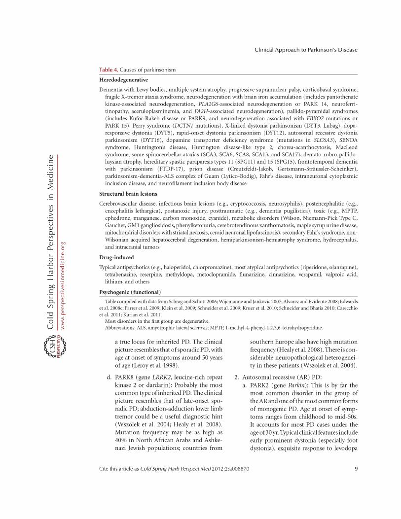
Many other parkinsonian disorders have beendescribed (Table 4), and some of them are attimes confused with PD, although they tendto present specific clinical features, frequentlywith complex phenotypes.
HEREDITARY FORMS OF PARKINSON’SDISEASE AND THEIR CLINICAL FEATURES
A small but significant number of PD patientshave a family history compatible with Mende-lian autosomal inheritance (10%–15%), eitherdominant or recessive. Many of these are classi-fied as young-onset (,40 yr) or juvenile-onsetPD (,21 yr) (Schrag and Schott 2006). A num-ber of levodopa-responsive parkinsonian syn-dromes have been described and linked to aspecific locus or gene in the last few years, andsome of them have been classified as PARK syn-dromes (Gasser 2007; Klein et al. 2009). Someof these denote true PD, whereas others repre-sent more complex phenotypes and dissimilardiseases. Only the former group will be brieflyapproached here, as the clinical phenotype maybe a useful pointer for the diagnosis in dailypractice, guiding subsequent molecular testing.For more details on the genetics of PD pleaserefer to Klein and Westenberger (2012).
1. Autosomal-dominant (AD) PD:a. PARK1/PARK4 (gene SNCA,a-synuclein):
Mean age at onset of symptoms is in the 30s(PARK4) or 40s (PARK1). Progression ap-pears to be faster than in sporadic PD anddementia is a frequent finding; at times theclinical picture resembles DLB, but meanage at onset is much lower than in sporadiccases (Polymeropoulos et al. 1997; Spiraet al. 2001; Zarranz et al. 2004). PARK1and PARK4 are attributable to SNCA mu-tations and duplications/triplications, re-spectively.
b. PARK3 (gene unknown): Researchers de-scribed a group of families with parkinson-ism closely resembling that of sporadic PD,including age of onset (mean 59 yr); thelocus has been mapped to 2p13. Penetrancewasestimated tobe below40%(Gasseretal.1998). It has not been clearly definedwhether this represents a disease suscepti-bility locus ora true Mendelian form of PD.
c. PARK5 (gene UCHL1, ubiquitin carboxy-terminal hydrolase 1): Only one family hasbeen reported with PD and a mutation inUCHL1, providing frail evidence that this is
J. Massano and K.P. Bhatia
8 Cite this article as Cold Spring Harb Perspect Med 2012;2:a008870
ww
w.p
ersp
ecti
vesi
nm
edic
ine.
org
a true locus for inherited PD. The clinicalpicture resembles that of sporadic PD, withage at onset of symptoms around 50 yearsof age (Leroy et al. 1998).
d. PARK8 (gene LRRK2, leucine-rich repeatkinase 2 or dardarin): Probably the mostcommon type of inherited PD. The clinicalpicture resembles that of late-onset spo-radic PD; abduction-adduction lower limbtremor could be a useful diagnostic hint(Wszolek et al. 2004; Healy et al. 2008).Mutation frequency may be as high as40% in North African Arabs and Ashke-nazi Jewish populations; countries from
southern Europe also have high mutationfrequency (Healyet al. 2008). There is con-siderable neuropathological heterogenei-ty in these patients (Wszolek et al. 2004).
2. Autosomal recessive (AR) PD:a. PARK2 (gene Parkin): This is by far the
most common disorder in the group ofthe AR and one of the most common formsof monogenic PD. Age at onset of symp-toms ranges from childhood to mid-50s.It accounts for most PD cases under theage of 30 yr. Typical clinical features includeearly prominent dystonia (especially footdystonia), exquisite response to levodopa
Table 4. Causes of parkinsonism
Heredodegenerative
Dementia with Lewy bodies, multiple system atrophy, progressive supranuclear palsy, corticobasal syndrome,fragile X-tremor ataxia syndrome, neurodegeneration with brain iron accumulation (includes pantothenatekinase-associated neurodegeneration, PLA2G6-associated neurodegeneration or PARK 14, neuroferri-tinopathy, aceruloplasminemia, and FA2H-associated neurodegeneration), pallido-pyramidal syndromes(includes Kufor-Rakeb disease or PARK9, and neurodegeneration associated with FBXO7 mutations orPARK 15), Perry syndrome (DCTN1 mutations), X-linked dystonia parkinsonism (DYT3, Lubag), dopa-responsive dystonia (DYT5), rapid-onset dystonia parkinsonism (DYT12), autosomal recessive dystoniaparkinsonism (DYT16), dopamine transporter deficiency syndrome (mutations in SLC6A3), SENDAsyndrome, Huntington’s disease, Huntington disease-like type 2, chorea-acanthocytosis, MacLeodsyndrome, some spinocerebellar ataxias (SCA3, SCA6, SCA8, SCA13, and SCA17), dentato-rubro-pallido-luysian atrophy, hereditary spastic paraparesis types 11 (SPG11) and 15 (SPG15), frontotemporal dementiawith parkinsonism (FTDP-17), prion disease (Creutzfeldt-Jakob, Gertsmann-Straussler-Scheinker),parkinsonism-dementia-ALS complex of Guam (Lytico-Bodig), Fahr’s disease, intraneuronal cytoplasmicinclusion disease, and neurofilament inclusion body disease
Structural brain lesions
Cerebrovascular disease, infectious brain lesions (e.g., cryptococcosis, neurosyphilis), postencephalitic (e.g.,encephalitis lethargica), postanoxic injury, posttraumatic (e.g., dementia pugilistica), toxic (e.g., MPTP,ephedrone, manganese, carbon monoxide, cyanide), metabolic disorders (Wilson, Niemann-Pick Type C,Gaucher, GM1 gangliosidosis, phenylketonuria, cerebrotendinous xanthomatosis, maple syrup urine disease,mitochondrial disorders with striatal necrosis, ceroid neuronal lipofuscinosis), secondary Fahr’s syndrome, non-Wilsonian acquired hepatocerebral degeneration, hemiparkinsonism-hemiatrophy syndrome, hydrocephalus,and intracranial tumors
Drug-induced
Typical antipsychotics (e.g., haloperidol, chlorpromazine), most atypical antipsychotics (riperidone, olanzapine),tetrabenazine, reserpine, methyldopa, metoclopramide, flunarizine, cinnarizine, verapamil, valproic acid,lithium, and others
Psychogenic (functional)
Table compiled with data from Schrag and Schott 2006; Wijemanne and Jankovic 2007; Alvarez and Evidente 2008; Edwards
et al. 2008c; Farrer et al. 2009; Klein et al. 2009; Schneider et al. 2009; Kruer et al. 2010; Schneider and Bhatia 2010; Carecchio
et al. 2011; Kurian et al. 2011.
Most disorders in the first group are degenerative.
Abbreviations: ALS, amyotrophic lateral sclerosis; MPTP, 1-methyl-4-phenyl-1,2,3,6-tetrahydropyridine.
Clinical Approach to Parkinson’s Disease
Cite this article as Cold Spring Harb Perspect Med 2012;2:a008870 9
ww
w.p
ersp
ecti
vesi
nm
edic
ine.
org

and anticholinergic drugs, slow diseaseprogression, diurnal fluctuations, sleepbenefit, susceptibility to levodopa-inducedpeak-dose dyskinesias, and wearing-offphenomenon. Hyperreflexia is a commonfeature. Other possible presentations in-clude exercise-induced dystonia, focal dys-tonia, tremor-predominant cases, and earlyimpairment of gait and balance (Abbas et al.1999; Klein et al. 2000; Lucking et al. 2000;Gouider-Khouja et al. 2003; Kahn et al.2003; Wickremaratchi et al. 2009). Nonmo-tor symptoms seem to be less prevalent thanin sporadic PD, except anxiety (Kagi et al.2010b). Lewy bodies were absent in mostpatients that came to autopsy (Farrer et al.2001; Gouider-Khouja et al. 2003).
b. PARK6 (gene PINK1, PTEN-induced pu-tative kinase 1): It is characterized by earlyonset in most patients (mean age at onsetin the 30s, ranging from childhood untilthe seventh decade of life), dystonia atonset or symptom symmetry in somecases, slow disease progression, excellentlevodopa responsiveness, early peak-dosedyskinesias; hyperreflexia has been de-scribed; rare cases with very early onset dis-play sleep benefit and diurnal fluctuations(Valente et al. 2001, 2002; Bonifati 2005;Ibanez et al. 2006; Leutenegger et al.2006). Psychiatric disturbances such as de-pression, anxiety, and psychosis are a fre-quent finding (Bonifati 2005; Ibanez et al.2006; Steinlechner et al. 2007). Lewy bodiesare a recently described neuropathologicalfinding (Samaranch et al. 2010).
c. PARK7 (gene DJ-1): This seems to be arare disorder, with a clinical phenotypesimilar to PARK6. Notably, some caseshave been associated with blepharo-spasm, whereas amyotrophic lateral scle-rosis and dementia have been described inone family (van Duijn et al. 2001; Abou-Sleiman et al. 2003; Dekker et al. 2003;Clark et al. 2009; Annesi et al. 2005).
Other candidate genes have been associated withparkinsonism, namely, Nurr1 (Le et al. 2003),
synphylin-1 (Marx et al. 2003), and POLG (poly-merase gamma) (Davidzon et al. 2006), but fur-ther confirmation in large case series is still await-ed. Exciting observations have been made in thelast few years in families with members sufferingfrom Gaucher’s disease (GD), an AR disordercaused by homozygous mutations in the GBAgene encoding the lysosomal enzyme glucocere-brosidase. Heterozygous carriers, who do nothave GD, are at higher risk of developing sporadicPD or DLB (Aharon-Peretz et al. 2004; Clarket al.2009; Sidransky et al. 2009). Thus, interestemerged in the putative role of ceramide in thepathogenesis of Lewy body disorders (Bras et al.2008).
PD MANAGEMENT: BRIEFCONSIDERATIONS
Comprehensive review of PD therapy is beyondthe scope of this text, but a few basic thoughtsmust be set forth. PD is best managed in a mul-tidisciplinary setting, to obtain satisfactory re-sults regarding the various disabling aspects ofthe disease. Nonmotor symptoms should betreated accordingly, although evidence to sup-port most treatment decisions is scarce; sildenafilshould be considered for erectile dysfunction andmacrogol for constipation (Zesiewicz et al. 2010).Dementia symptoms are improved by cholines-terase inhibitors or memantine (Emre et al. 2010;van Laar et al. 2011). An array of drugs is used totreat motor symptoms in PD (availability variesfrom country to country): levodopa plus periph-eral dopa decarboxylase inhibitor (carbidopa orbenserazide), dopamine agonists (ergot deriva-tives: bromocriptine, pergolide, cabergoline, di-hydroergocryptine; non-ergot derivatives: ro-pinirole, pramipexole, rotigotine, apomorphine),monoamine oxidase B inhibitors (selegiline, ra-sagiline), catechol-O-methyltransferase inhibi-tors (entacapone, tolcapone), anticholinergics(trihexyphenidyl, benztropine), and amanta-dine. Currently, there is no consensus on themost adequate timing and drug of choice fortherapy initiation in PD, but dopaminergictherapy is usually deferred until deterioratingquality of life demands treatment, owing to thefuture potential motor complications brought
J. Massano and K.P. Bhatia
10 Cite this article as Cold Spring Harb Perspect Med 2012;2:a008870
ww
w.p
ersp
ecti
vesi
nm
edic
ine.
org

about by these drugs, such as peak-dose dyski-nesias (see online Movie 9 at www.perspective-sinmedicine.org), wearing off, and sudden offstates (Edwards et al. 2008a; Lang 2009). Levo-dopa seems to pose higher risks in this regard ascompared with dopamine agonists, although re-cent data brought conflicting views on this mat-ter (Katzenschlager et al. 2008; Holloway et al.2009). Nevertheless, levodopa is the most effec-tive drug in the control of motor symptoms, andPD patients typically show marked and sustainedbenefits from it for several years (Schapira et al.2009). Dopamine agonists may cause peripheraledema, fibrotic reactions (most ergot deriva-tives), excessive daytime somnolence, and im-pulse control disorders (Antonini et al. 2009).In advanced PD, functional neurosurgery is avaluable therapeutic option, provided that pa-tients are carefully selected (Lang et al. 2006).Deep brain stimulation of either the subthalamicnucleus or internal globus pallidus is an effectiveand generally safe procedure (Deuschl et al. 2006;Follett et al. 2010; Moro et al. 2010). In advancedPD it may be more effective for the control ofmotor symptoms than the best medical therapy,either alone or in combination with it (Weaveret al. 2009; Williams et al. 2010).
CONCLUDING REMARKS
PD is a common and potentially disablingdisorder. Suitable efforts should be performedto achieve accurate diagnosis, communicate aplausible prognosis to the patient and family,and proceed with the best therapeutic interven-tions. Remarkable progress has been made inthe last two decades in the field of genetics,pathophysiology, and clinical imaging, but thediagnosis of PD still sits inevitably on clinicalskills and exploration, which emphasizes theimportance of a solid clinical knowledge aboutthe disease.
ACKNOWLEDGMENTS
We thank the patients who generously contrib-ute every day by willingly consenting to videocapture and educational display.
REFERENCES�Reference is also in this collection.
Abbas N, Lucking CB, Ricard S, Durr A, Bonifati V, DeMichele G, Bouley S, Vaughan JR, Gasser T, Marconi R,et al. 1999. Awide variety of mutations in the parkin geneare responsible for autosomal recessive parkinsonism inEurope. French Parkinson’s Disease Genetics Study Groupand the European Consortium on Genetic Susceptibilityin Parkinson’s Disease. Hum Mol Genet 8: 567–574.
Abou-Sleiman PM, Healy DG, Quinn N, Lees AJ, WoodNW. 2003. The role of pathogenic DJ-1 mutations inParkinson’s disease. Ann Neurol 54: 283–286.
Aharon-Peretz J, Rosenbaum H, Gershoni-Baruch R. 2004.Mutations in the glucocerebrosidase gene and Parkin-son’s disease in Ashkenazi Jews. N Engl J Med 351:1972–1977.
Alvarez MVG, Evidente VGH. 2008. Understanding drug-induced parkinsonism: Separating pearls from oysters.Neurology 70: 32–34.
Alves G, Forsaa EB, Pedersen KF, Gjerstad MD, Larsen JP.2008. Epidemiology of Parkinson’s disease. J Neurol 255:S18–S32.
Annesi G, Savettieri G, Pugliese P, D’Amelio M, Tarantino P,Ragonese P, La Bella V, Piccoli T, Civitelli D, Annesi F,et al. 2005. DJ-1 mutations and parkinsonism-dementia-amyotrophic lateral sclerosis complex. Ann Neurol 58:803–807.
Antonini A, Tolosa E, Mizuno Y, Yamamoto M, Poewe WH.2009. A reassessment of risks and benefits of dopamineagonists in Parkinson’s disease. Lancet Neurol 8: 929–937.
Bain P. 2007. Tremor. Parkinsonism Relat Disord 13: S369–S374.
Bain PG, Findley LJ, Thompson PD, Gresty MA, RothwellJC, Harding AE, Marsden CD. 1994. A study of hereditaryessential tremor. Brain 117: 805–824.
Berry-Kravis E, Abrams L, Coffey SM, Hall DA, Greco C,Gane LW, Grigsby J, Bourgeois JA, Finucane B, Jacque-mont S, et al. 2007. Fragile X-associated tremor/ataxiasyndrome: Clinical features, genetics, and testing guide-lines. Mov Disord 22: 2018–2030.
Bonifati V, Rohe CF, Breedveld GJ, Fabrizio E, De Mari M,Tassorelli C, Tavella A, Marconi R, Nicholl DJ, Chien HF,et al. 2005. Early-onset parkinsonism associated withPINK1 mutations: Frequency, genotypes, and pheno-types. Neurology 65: 87–95.
Braak H, Del Tredici K, Rub U, de Vos RA, Jansen Steur EN,Braak E. 2003. Staging of brain pathology related tosporadic Parkinson’s disease. Neurobiol Aging 24: 197–211.
Bras J, Singleton A, Cookson MR, Hardy J. 2008. Emergingpathways in genetic Parkinson’s disease: Potential role ofceramide metabolism in Lewy body disease. FEBS J 275:5767–5773.
Bronstein J, Carvey P, Chen H, Cory-Slechta D, DiMonte D,Duda J, English P, Goldman S, Grate S, Hansen J, et al.2009. Meeting report: Consensus statement—Parkin-son’s disease and the environment: Collaborative onhealth and the environment and Parkinson’s Action
Clinical Approach to Parkinson’s Disease
Cite this article as Cold Spring Harb Perspect Med 2012;2:a008870 11
ww
w.p
ersp
ecti
vesi
nm
edic
ine.
org

Network (CHE PAN) conference, 26–28 June 2007. En-viron Health Perspect 117: 117–121.
Carecchio M, Schneider SA, Chan H, Lachmann R, Lee PJ,Murphy E, Bhatia KP. 2011. Movement disorders in adultsurviving patients with maple syrup urine disease. MovDisord 26: 1324–1328.
Castelo-Branco M, Mendes M, Silva F, Massano J, JanuarioG, Januario C, Freire A. 2009. Motion integration deficitsare independent of magnocellular impairment in Parkin-son’s disease. Neuropsychologia 47: 314–320.
Chaudhuri KR, Schapira AH. 2009. Non-motor symptomsof Parkinson’s disease: Dopaminergic pathophysiologyand treatment. Lancet Neurol 8: 464–474.
Clark LN, Kartsaklis LA, Wolf Gilbert R, Dorado B, RossBM, Kisselev S, Verbitsky M, Mejia-Santana H, Cote LJ,Andrews H, et al. 2009. Association of glucocerebrosidasemutations with dementia with Lewy bodies. Arch Neurol66: 578–583.
Davidzon G, Greene P, Mancuso M, Klos KJ, Ahlskog JE,Hirano M, DiMauro S. 2006. Early-onset familial parkin-sonism due to POLG mutations. Ann Neurol 59: 859–862.
Dekker M, Bonifati V, van Swieten J, Leenders N, GaljaardRJ, Snijders P, Horstink M, Heutink P, Oostra B, vanDuijn C. 2003. Clinical features and neuroimaging ofPARK7-linked parkinsonism. Mov Disord 18: 751–757.
De Lau LML, Breteler MMB. 2006. Epidemiology of Parkin-son’s disease. Lancet Neurol 5: 525–535.
Deuschl G, Bain P, Brin M. 1998. Consensus statement of theMovement Disorder Society on Tremor. Ad Hoc Scien-tific Committee. Mov Disord 13: S2–S23.
Deuschl G, Schade-Brittinger C, Krack P, Volkmann J, Scha-fer H, Botzel K, Daniels C, Deutschlander A, Dillmann U,Eisner W, et al. 2006. A randomized trial of deep-brainstimulation for Parkinson’s disease. N Engl J Med 355:896–908.
Dorsey ER, Constantinescu R, Thompson JP, Biglan KM,Holloway RG, Kieburtz K, Marshall FJ, Ravina BM, Schi-fitto G, Siderowf A, et al. 2007. Projected number ofpeople with Parkinson disease in the most populous na-tions, 2005 through 2030. Neurology 68: 384–386.
Edwards M, Quinn N, Bhatia K. 2008a. Parkinson’s disease.In Parkinson’s disease and other movement disorders, pp.17–80. Oxford University Press, Oxford.
Edwards M, Quinn N, Bhatia K. 2008b. Tremor. In Parkin-son’s disease and other movement disorders, pp. 102–118.Oxford University Press, Oxford.
Edwards M, Quinn N, Bhatia K. 2008c. Atypical parkinson-ism. In Parkinson’s disease and other movement disorders,pp. 81–100. Oxford University Press, Oxford.
Elbaz A, Moisan F. 2008. Update in the epidemiology ofParkinson’s disease. Curr Opin Neurol 21: 454–460.
Emre M, Aarsland D, Brown R, Burn DJ, Duyckaerts C,Mizuno Y, Broe GA, Cummings J, Dickson DW, GauthierS, et al. 2007. Clinical diagnostic criteria for dementiaassociated with Parkinson’s disease. Mov Disord 22:1689–1707.
Emre M, Tsolaki M, Bonuccelli U, Destee A, Tolosa E, Kut-zelnigg A, Ceballos-Baumann A, Zdravkovic S, Blad-strom A, Jones R, et al. 2010. Memantine for patientswith Parkinson’s disease dementia or dementia with
Lewy bodies: A randomised, double-blind, placebo-con-trolled trial. Lancet Neurol 9: 969–977.
Farrer M, Chan P, Chen R, Tan L, Lincoln S, Hernandez D,Forno L, Gwinn-Hardy K, Petrucelli L, Hussey J, et al.2001. Lewy bodies and parkinsonism in families withparkin mutations. Ann Neurol 50: 293–300.
Farrer MJ, Hulihan MM, Kachergus JM, Dachsel JC, StoesslAJ, Grantier LL, Calne S, Calne DB, Lechevalier B, Cha-pon F, et al. 2009. DCTN1 mutations in Perry syndrome.Nat Genet 41: 163–165.
Follett KA, Weaver FM, Stern M, Hur K, Harris CL, Luo P,Marks WJ Jr, Rothlind J, Sagher O, Moy C, et al. 2010.Pallidal versus subthalamic deep-brain stimulation forParkinson’s disease. N Engl J Med 362: 2077–2091.
Gallagher DA, Lees AJ, Schrag A. 2010. What are the mostimportant nonmotor symptoms in patients with Parkin-son’s disease and are we missing them? Mov Disord 25:2493–2500.
Gasser T. 2007. Update on the genetics of Parkinson’s dis-ease. Mov Disord 22: S343–S350.
Gasser T, Muller-Myhsok B, Wszolek ZK, Oehlmann R,Calne DB, Bonifati V, Bereznai B, Fabrizio E, ViereggeP, Horstmann RD. 1998. A susceptibility locus for Par-kinson’s disease maps to chromosome 2p13. Nat Genet18: 262–265.
Gao X, Chen H, Schwarzschild MA, Ascherio A. 2011. Use ofibuprofen and risk of Parkinson disease. Neurology 76:863–869.
Geser F, Wenning GK, Poewe W, McKeith I. 2005. How todiagnose dementia with Lewy bodies: State of the art.Mov Disord 20 (Suppl 12): S11–S20.
Gilman S, Wenning GK, Low PA, Brooks DJ, Mathias CJ,Trojanowski JQ, Wood NW, Colosimo C, Durr A, FowlerCJ, et al. 2008. Second consensus statement on the diag-nosis of multiple system atrophy. Neurology 71: 670–676.
� Goetz CG. 2011. The history of Parkinson’s disease: Earlyclinical descriptions and neurological therapies. ColdSpring Harb Perspect Med doi: 10.1101/cshperspect.a008862.
Goetz CG, Poewe W, Rascol O, Sampaio C, Stebbins GT,Counsell C, Giladi N, Holloway RG, Moore CG,WenningGK, et al. 2004. Movement Disorder Society Task Forcereport on the Hoehn and Yahr staging scale: Status andrecommendations. Mov Disord 19: 1020–1028.
Gouider-Khouja N, Larnaout A, Amouri R, Sfar S, Belal S,Ben Hamida C, Ben Hamida M, Hattori N, Mizuno Y,Hentati F. 2003. Autosomal recessive parkinsonismlinked to parkin gene in a Tunisian family. Clinical, ge-netic and pathological study. Parkinsonism Relat Disord9: 247–251.
Hawkes CH, Del Tredici K, Braak H. 2010. A timeline forParkinson’s disease. Parkinsonism Relat Disord 16: 79–84.
Healy DG, Falchi M, O’Sullivan SS, Bonifati V, Durr A,Bressman S, Brice A, Aasly J, Zabetian CP, Goldwurm S,et al. 2008. Phenotype, genotype, and worldwide geneticpenetrance of LRRK2-associated Parkinson’s disease: Acase-control study. Lancet Neurol 7: 583–590.
Hirtz D, Thurman DJ, Gwinn-Hardy K, Mohamed M,Chaudhuri AR, Zalutsky R. 2007. How common are the“common” neurologic disorders? Neurology 68: 326–337.
J. Massano and K.P. Bhatia
12 Cite this article as Cold Spring Harb Perspect Med 2012;2:a008870
ww
w.p
ersp
ecti
vesi
nm
edic
ine.
org

Holloway R, Marek K, Biglan K, Dick A, Fahn S, Julian-Baros E, Kamp C, Kieburtz K, Lang A, McDermott M,et al. 2009. Long-term effect of initiating pramipexole vslevodopa in early Parkinson disease. Arch Neurol 66:563–570.
Hughes AJ, Daniel SE, Kilford L, Lees AJ. 1992. Accuracy ofclinical diagnosis of idiopathic Parkinson’s disease: Aclinico-pathological study of 100 cases. J Neurol Neuro-surg Psychiatry 55: 181–184.
Ibanez P, Lesage S, Lohmann E, Thobois S, De Michele G,Borg M, Agid Y, Durr A, Brice A, French Parkinson’sDisease Genetics Study Group. 2006. Mutational analysisof the PINK1 gene in early-onset parkinsonism in Europeand North Africa. Brain 129: 686–694.
Jacquemont S, Hagerman RJ, Hagerman PJ, Leehey MA.2007. Fragile-X syndrome and fragile X-associated trem-or/ataxia syndrome: Two faces of FMR1. Lancet Neurol 6:45–55.
Jain S, Lo SE, Louis ED. 2006. Common misdiagnosis of acommon neurological disorder: How are we misdiagnos-ing essential tremor? Arch Neurol 63: 1100–1104.
Jankovic J, 2008. Parkinson’s disease: Clinical features anddiagnosis. J Neurol Neurosurg Psychiatry 79: 368–376.
Kagi G, Bhatia KP, Tolosa E. 2010a. The role of DAT-SPECTin movement disorders. J Neurol Neurosurg Psychiatry 81:5–12.
Kagi G, Klein C, Wood NW, Schneider SA, Pramstaller PP,Tadic V, Quinn NP, van de Warrenburg BP, Bhatia KP.2010b. Nonmotor symptoms in Parkin gene-related par-kinsonism. Mov Disord 25: 1279–1284.
Kalra S, Grosset DG, Benamer HTS. 2010. Differentiatingvascular parkinsonism from idiopathic Parkinson’s dis-ease: A systematic review. Mov Disord 25: 149–156.
Katzenschlager R, Head J, Schrag A, Ben-Shlomo Y, Evans A,Lees AJ, Parkinson’s Disease Research Group of theUnited Kingdom. 2008. Fourteen-year final report ofthe randomized PDRG-UK trial comparing three initialtreatments in PD. Neurology 71: 474–480.
Khan NL, Graham E, Critchley P, Schrag AE, Wood NW, LeesAJ, Bhatia KP, Quinn N. 2003. Parkin disease: A pheno-typic study of a large case series. Brain 126: 1279–1292.
� Klein C, Westenberger A. 2012. Genetics of Parkinson’sdisease. Cold Spring Harb Perspect Med 2: a008888.
Klein C, Pramstaller PP, Kis B, Page CC, Kann M, Leung J,Woodward H, Castellan CC, Scherer M, Vieregge P, et al.2000. Parkin deletions in a family with adult-onset, trem-or-dominant parkinsonism: Expanding the phenotype.Ann Neurol 48: 65–71.
Klein C, Schneider SA, Lang AE. 2009. Hereditary parkin-sonism: Parkinson disease look-alikes—An algorithm forclinicians to “PARK” genes and beyond. Mov Disord 24:2042–2058.
Kruer MC, Paisan-Ruiz C, Boddaert N, Yoon MY, Hama H,Gregory A, Malandrini A, Woltjer RL, Munnich A, GobinS, et al. 2010. Defective FA2H leads to a novel form ofneurodegeneration with brain iron accumulation (NBIA).Ann Neurol 68: 611–618.
Kurian MA, Li Y, Zhen J, Meyer E, Hai N, Christen HJ,Hoffmann GF, Jardine P, von Moers A, Mordekar SR,et al. 2011. Clinical and molecular characterisation ofhereditary dopamine transporter deficiency syndrome:
An observational cohort and experimental study. LancetNeurol 10: 54–62.
Lang AE. 2009. When and how should treatment be startedin Parkinson disease? Neurology 72: S39–S43.
Lang AE, Houeto JL, Krack P, Kubu C, Lyons KE, Moro E,Ondo W, Pahwa R, Poewe W, Troster AI, et al. 2006. Deepbrain stimulation: Preoperative issues. Mov Disord 21:S171–S196.
Lanska DJ. 2010. The history of movement disorders. InHistory of clinical neurology: Handbook of clinical neurol-ogy (ed. Finger S, Boller F, Tyler KL), Vol. 95, pp. 501–546. Elsevier, Amsterdam.
Le WD, Xu P, Jankovic J, Jiang H, Appel SH, Smith RG,Vassilatis DK. 2003. Mutations in NR4A2 associatedwith familial Parkinson disease. Nat Genet 33: 85–89.
Lees AJ. 2007. Unresolved issues relating to the shaking palsyon the celebration of James Parkinson’s 250th birthday.Mov Disord 22: S327–S334.
Lees AJ, Hardy J, Revesz T. 2009. Parkinson’s disease. Lancet373: 2055–2066.
Leroy E, Boyer R, Auburger G, Leube B, Ulm G, Mezey E,Harta G, Brownstein MJ, Jonnalagada S, Chernova T,et al. 1998. The ubiquitin pathway in Parkinson’s disease.Nature 395: 451–452.
Leutenegger AL, Salih MA, Ibanez P, Mukhtar MM, LesageS, Arabi A, Lohmann E, Durr A, Ahmed AE, Brice A.2006. Juvenile-onset Parkinsonism as a result of the firstmutation in the adenosine triphosphate orientation do-main of PINK1. Arch Neurol 63: 1257–1261.
Lim SY, Fox SH, Lang AE. 2009. Overview of the extranigralaspects of Parkinson disease. Arch Neurol 66: 167–172.
Litvan I, Bhatia KP, Burn DJ, Goetz CG, Lang AE, McKeithI, Quinn N, Sethi KD, Shults C, Wenning GK, et al. 2003.Movement Disorders Society Scientific Issues Committeereport: SIC Task Force appraisal of clinical diagnosticcriteria for Parkinsonian disorders. Mov Disord 18:467–486.
Lucking CB, Durr A, Bonifati V, Vaughan J, De Michele G,Gasser T, Harhangi BS, Meco G, Denefle P, Wood NW,et al. 2000. Association between early-onset Parkinson’sdisease and mutations in the parkin gene. N Engl J Med342: 1560–1567.
Massano J, Costa F, Nadais G. 2008. Teaching neuroImage:MRI in multiple system atrophy: “Hot cross bun” signand hyperintense rim bordering the putamina. Neurology71: e38.
Marsden CD. 1982. The mysterious motor function of thebasal ganglia: The Robert Wartenberg Lecture. Neurology32: 514–539.
Marsden CD. 1994. Parkinson’s disease. J Neurol NeurosurgPsychiatry 57: 672–681.
Marx FP, Holzmann C, Strauss KM, Li L, Eberhardt O,Gerhardt E, Cookson MR, Hernandez D, Farrer MJ,Kachergus J, et al. 2003. Identification and functionalcharacterization of a novel R621C mutation in the syn-philin-1 gene in Parkinson’s disease. Hum Mol Genet 12:1223–1231.
Moro E, Lozano AM, Pollak P, Agid Y, Rehncrona S, Volk-mann J, Kulisevsky J, Obeso JA, Albanese A, Hariz MI,et al. 2010. Long-term results of a multicenter study on
Clinical Approach to Parkinson’s Disease
Cite this article as Cold Spring Harb Perspect Med 2012;2:a008870 13
ww
w.p
ersp
ecti
vesi
nm
edic
ine.
org

subthalamic and pallidal stimulation in Parkinson’s dis-ease. Mov Disord 25: 578–586.
� Niethammer M, Feigin A, Eidelberg D. 2012. Functionalneuroimaging in Parkinson’s disease. Cold Spring HarbPerspect Med doi: 10.1101/cshperspect.a009274.
Parkinson J. 1817. An essay on the shaking palsy. Sherwood,Neely, and Jones, London.
Parkinson J. 2002. An essay on the shaking palsy. 1817. JNeuropsychiatry Clin Neurosci 14: 223–236.
Plotnik M, Giladi N, Dagan Y, Hausdorff JM. 2011. Posturalinstability and fall risk in Parkinson’s disease: Impaireddual tasking, pacing, and bilateral coordination of gaitduring the “ON” medication state. Exp Brain Res 210:529–538.
Poewe W. 2008. Non-motor symptoms in Parkinson’s dis-ease. Eur J Neurol 15: S14–S20.
Polymeropoulos MH, Lavedan C, Leroy E, Ide SE, DehejiaA, Dutra A, Pike B, Root H, Rubenstein J, Boyer R, et al.1997. Mutation in the a-synuclein gene identified infamilies with Parkinson’s disease. Science 276: 2045–2047.
Quinn NP, Schneider SA, Schwingenschuh P, Bhatia KP.2011. Tremor—Some controversial aspects. Mov Disord16: 18–23.
Rodrıguez-Oroz MC, Jahanshahi M, Krack P, Litvan I, Ma-cias R, Bezard E, Obeso JA. 2009. Initial clinical manifes-tations of Parkinson’s disease: Features and pathophysi-ological mechanisms. Lancet Neurol 8: 1128–1139.
Samaranch L, Lorenzo-Betancor O, Arbelo JM, Ferrer I,Lorenzo E, Irigoyen J, Pastor MA, Marrero C, Isla C,Herrera-Henriquez J, et al. 2010. PINK1-linked parkin-sonism is associated with Lewy body pathology. Brain133: 1128–1142.
Savica R, Rocca WA, Ahlskog JE. 2010. When does Parkinsondisease start? Arch Neurol 67: 798–801.
Schapira AH, Emre M, Jenner P, Poewe W. 2009. Levodopain the treatment of Parkinson’s disease. Eur J Neurol 16:982–986.
Schapira AH, Tolosa E. 2010. Molecular and clinical pro-drome of Parkinson disease: Implications for treatment.Nat Rev Neurol 6: 309–317.
Schlesinger I, Schlesinger N. 2008. Uric acid in Parkinson’sdisease. Mov Disord 23: 1653–1657.
Schneider SA, Bhatia KP. 2010. Rare causes of dystonia par-kinsonism. Curr Neurol Neurosci Rep 10: 431–439.
Schneider SA, Edwards MJ, Mir P, Cordivari C, Hooker J,Dickson J, Quinn N, Bhatia KP. 2007. Patients with adult-onset dystonic tremor resembling parkinsonian tremorhave scans without evidence of dopaminergic deficit(SWEDDs). Mov Disord 22: 2210–2215.
Schneider SA, Bhatia KP, Hardy J. 2009. Complicated reces-sive dystonia parkinsonism syndromes. Mov Disord 24:490–499.
Schrag A, Schott JM. 2006. Epidemiological, clinical, andgenetic characteristics of early-onset parkinsonism. Lan-cet Neurol 5: 355–363.
Schrag A, Munchau A, Bhatia KP, Quinn NP, Marsden CD.2000. Essential tremor: An overdiagnosed condition? JNeurol 247: 955–959.
Schwingenschuh P, Ruge D, Edwards MJ, Terranova C,Katschnig P, Carrillo F, Silveira-Moriyama L, SchneiderSA, Kagi G, Palomar FJ, et al. 2010. DistinguishingSWEDDs patients with asymmetric resting tremor fromParkinson’s disease: A clinical and electrophysiologicalstudy. Mov Disord 25: 560–569.
Sethi K. 2008. Levodopa unresponsive symptoms in Parkin-son disease. Mov Disord 23: S521–S533.
Sidransky E, Nalls MA, Aasly JO, Aharon-Peretz J, Annesi G,Barbosa ER, Bar-Shira A, Berg D, Bras J, Brice A, et al.2009. Multicenter analysis of glucocerebrosidase muta-tions in Parkinson’s disease. N Engl J Med 361: 1651–1661.
Silva MF, Faria P, Regateiro FS, Forjaz V, Januario C, Freire A,Castelo-Branco M. 2005. Independent patterns of dam-age within magno-, parvo- and koniocellular pathways inParkinson’s disease. Brain 128: 2260–2271.
Sitburana O, Ondo WG. 2009. Brain magnetic resonanceimaging (MRI) in parkinsonian disorders. ParkinsonismRelat Disord 15: 165–174.
Spildooren J, Vercruysse S, Desloovere K, Vandenberghe W,Kerckhofs E, Nieuwboer A. 2010. Freezing of gait in Par-kinson’s disease: The impact of dual-tasking and turning.Mov Disord 25: 2563–2570.
Spira PJ, Sharpe DM, Halliday G, Cavanagh J, NicholsonGA. 2001. Clinical and pathological features of a Parkin-sonian syndrome in a family with an Ala53Thr a-synu-clein mutation. Ann Neurol 49: 313–319.
Stefanova N, Bucke P, Duerr S, Wenning GK. 2009. Multiplesystem atrophy: An update. Lancet Neurol 8: 1172–1178.
Steinlechner S, Stahlberg J, Volkel B, Djarmati A, Hagenah J,Hiller A, Hedrich K, Konig I, Klein C, Lencer R. 2007. Co-occurrence of affective and schizophrenia spectrum dis-orders with PINK1 mutations. J Neurol Neurosurg Psychi-atry 78: 532–535.
Tolosa E, Wenning G, Poewe W. 2006. The diagnosis ofParkinson’s disease. Lancet Neurol 5: 75–86.
Tolosa E, Gaig C, Santamarıa J, Compta Y. 2009. Diagnosisand the premotor phase of Parkinson disease. Neurology27: S12–S20.
Twelves D, Perkins KS, Counsell C. 2003. Systematic reviewof incidence studies of Parkinson’s disease. Mov Disord18: 19–31.
Valente EM, Bentivoglio AR, Dixon PH, Ferraris A, IalongoT, Frontali M, Albanese A, Wood NW. 2001. Localizationof a novel locus for autosomal recessive early-onset par-kinsonism, PARK6, on human chromosome 1p35-p36.Am J Hum Genet 68: 895–900.
Valente EM, Brancati F, Ferraris A, Graham EA, Davis MB,Breteler MM, Gasser T, Bonifati V, Bentivoglio AR, DeMichele G, et al. 2002. PARK6-linked parkinsonism oc-curs in several European families. Ann Neurol 51: 14–18.
Valente EM, Abou-Sleiman PM, Caputo V, Muqit MM,Harvey K, Gispert S, Ali Z, Del Turco D, BentivoglioAR, Healy DG, et al. 2004. Hereditary early-onset Parkin-son’s disease caused by mutations in PINK1. Science 304:1158–1160.
van Duijn CM, Dekker MC, Bonifati V, Galjaard RJ,Houwing-Duistermaat JJ, Snijders PJ, Testers L, Breed-veld GJ, Horstink M, Sandkuijl LA, et al. 2001. Park7, a
J. Massano and K.P. Bhatia
14 Cite this article as Cold Spring Harb Perspect Med 2012;2:a008870
ww
w.p
ersp
ecti
vesi
nm
edic
ine.
org

novel locus for autosomal recessive early-onset parkinson-ism, on chromosome 1p36. Am J Hum Genet 69: 629–634.
van Laar T, De Deyn PP, Aarsland D, Barone P, Galvin JE.2011. Effects of cholinesterase inhibitors in Parkinson’sdisease dementia: A review of clinical data. CNS NeurosciTher 17: 428–441.
Von Campenhausen S, Bornschein B, Wick R, Botzel K,Sampaio C, Poewe W, Oertel W, Sibert U, Berger K, DodelR. 2005. Prevalence and incidence of Parkinson’s diseasein Europe. Eur Neuropsychopharmacol 15: 473–490.
Warren NM, Burn DJ. 2007. Progressive supranuclear palsy.Pract Neurol 7: 16–23.
Weaver FM, Follett K, Stern M, Hur K, Harris C, Marks WJJr, Rothlind J, Sagher O, Reda D, Moy CS, et al. 2009.Bilateral deep brain stimulation vs best medical therapyfor patients with advanced Parkinson disease: A random-ized controlled trial. JAMA 301: 63–73.
Weisman D, McKeith I. 2007. Dementia with Lewy bodies.Semin Neurol 27: 42–47.
Wickremaratchi MM, Majounie E, Morris HR, WilliamsNM, Lewis H, Gill SS, Khan S, Heywood P, Hardy J, WilesCM, et al. 2009. Parkin-related disease clinically diag-nosed as a pallido-pyramidal syndrome. Mov Disord 24:138–140.
Wijemanne S, Jankovic J. 2007. Hemiparkinsonism-hemia-trophy syndrome. Neurology 69: 1585–1594.
Williams DR, Lees AJ. 2009. Progressive supranuclear palsy:Clinicopathological concepts and diagnostic challenges.Lancet Neurol 8: 270–279.
Williams DR, de Silva R, Paviour DC, Pittman A, Watt HC,Kilford L, Holton JL, Revesz T, Lees AJ. 2005. Character-istics of two distinct clinical phenotypes in pathologicallyproven progressive supranuclear palsy: Richardson’s syn-drome and PSP-parkinsonism. Brain 128: 1247–1258.
Williams A, Gill S, Varma T, Jenkinson C, Quinn N, MitchellR, Scott R, Ives N, Rick C, Daniels J, et al. 2010. Deepbrain stimulation plus best medical therapy versus bestmedical therapy alone for advanced Parkinson’s disease(PD SURG trial): A randomised, open-label trial. LancetNeurol 9: 581–591.
Winikates J, Jankovic J. 1999. Clinical correlates of vascularparkinsonism. Arch Neurol 56: 98–102.
Wszolek ZK, Pfeiffer RF, Tsuboi Y, Uitti RJ, McComb RD,Stoessl AJ, Strongosky AJ, Zimprich A, Muller-Myhsok B,Farrer MJ, et al. 2004. Autosomal dominant parkinson-ism associated with variable synuclein and tau pathology.Neurology 62: 1619–1622.
Zarranz JJ, Alegre J, Gomez-Esteban JC, Lezcano E, Ros R,Ampuero I, Vidal L, Hoenicka J, Rodriguez O, Atares B,et al. 2004. The new mutation, E46K, of a-synucleincauses Parkinson and Lewy body dementia. Ann Neurol55: 164–173.
Zesiewicz TA, Sullivan KL, Arnulf I, Chaudhuri KR, MorganJC, Gronseth GS, Miyasaki J, Iverson DJ, Weiner WJ,Quality Standards Subcommittee of the American Acad-emy of Neurology. 2010. Practice parameter: Treatmentof nonmotor symptoms of Parkinson disease: Report ofthe Quality Standards Subcommittee of the AmericanAcademy of Neurology. Neurology 74: 924–931.
Clinical Approach to Parkinson’s Disease
Cite this article as Cold Spring Harb Perspect Med 2012;2:a008870 15
ww
w.p
ersp
ecti
vesi
nm
edic
ine.
org