Clinical and histological aspects of malignant inverted ... · Clinical and histological aspects of...
6
Rom J Morphol Embryol 2016, 57(1):289–294 ISSN (print) 1220–0522 ISSN (online) 2066–8279 CASE REPORTS Clinical and histological aspects of malignant inverted sinonasal papilloma. Two case reports VASILE NICOLAE 1) , MARIANA SABĂU 1) , MIHAI IULIAN FĂGEŢAN 2) , VLAD PETRESCU SECELEANU 3) , DANIEL IORGULESCU 4) , DANA-ELENA DUMITRA 1) , ALEXANDRU-DAN SABĂU 5) , SORIN IBRIC-CIORANU 6) , ANCA SAVA 7) , VIOREL IBRIC-CIORANU 2) 1) Department of Prosthetics, Faculty of Medicine, “Lucian Blaga” University of Sibiu, Romania 2) Department of Maxillofacial Surgery, Faculty of Medicine, “Lucian Blaga” University of Sibiu, Romania 3) Maxillofacial Surgery Consultant, Private Practice, Bucharest, Romania 4) Department of Implantology, Faculty of Dentistry, University of Medicine and Pharmacy of Craiova, Romania 5) Department of Surgery, Faculty of Medicine, “Lucian Blaga” University of Sibiu, Romania 6) PhD Student, Faculty of Medicine, “Lucian Blaga” University of Sibiu, Romania 7) Department of Anatomy and Embryology, Faculty of Medicine, “Grigore T. Popa” University of Medicine and Pharmacy, Iassy, Romania Abstract The inverted sino-nasal papilloma is a benign epithelial lesion, ranging from 1% to 5% of the tumors that reside in this area. It is characterized by a tendency towards local bone destruction, recurrence and malignant transformation. This is a presentation of two cases diagnosed and surgically treated for malignant inverted papiloma, which started in the epithelium of the maxillary sinuses. The cases had in common the late presentation of the patient to the surgeon, thus the local destruction was significant with extension towards the eye socket and the base of the skull. The treatment of choice was the surgical resection of the maxillary bone with a prosthetic reconstruction of the defect followed by radiotherapy. After a careful analysis of the two cases, a series of statements regarding the diagnosis and treatment of this rare pathology were recommended. Keywords: malignant inverted papilloma, prosthetic denture, orbital reconstruction. Introduction Inverted papilloma (schneiderian inverted papilloma) is one the three types of scheniderian papillomas besides the oncocytic type (schneiderian papilloma – oncocytic type) and the fungiform papilloma (schneiderian papilloma – exophytic type). Sinonasal papillomas occur in three forms: exophytic (most common), inverted (most important biologically) and cylindrical [1]. The etiology of this occurrence is not yet fully understood. Past research has linked it to a series of factors like allergies, viruses, smoking, recurrent infections, and dust [2]. Another researches identified in the lesions human papillomavirus (HPV) types 6 and 11 [3]. Inverted papillomas are benign, but locally aggressive neoplasms occurring in both the nose and paranasal sinuses. At a macroscopic level, there is a thickening of the mucosa with irregular shape [4]. At a microscopic glance, it is formed by hyperplastic epithelium with inverted growth in the adjacent stroma. The epithelium is squamous non-keratinized or transitional, with an increased stratification and metaplasia. The papillo- matous proliferation instead of producing an exophytic growth extends into the mucosa, that it is inverted. At the surface a respiratory epithelium and caliciform cells with abundant cytoplasm and eccentric nucleus can be seen [5]. In a papilloma, all types of epithelia can be found. Mitoses are rare and can be found in the basal and parabasal layers. The basal membrane is well organized. The stroma can be lax or fibrous, with inflammatory cells, mainly polymorphonuclear neutrophils, eosinophils, and lymphocytes, which can be found in the epithelium. The stroma does not contain mucous glandular structures [6]. Similar aspects to the verruca vulgaris can be encountered like hypergranulosis, hyperkeratosis and parakeratosis. If not properly removed, it can recur but it does not metastasize [7]. The differential diagnosis is done with nasal polyps, squamous metaplasia, adenoid epithelial respiratory hamartoma and invasive squamous carcinoma. If not adequately excised, it has a high rate of recurrence, with the potentially serious complication of invasion of the orbit or cranial vault [1]. After a period of time a serious of focal aspects of superficial keratinization and dysplasia changes can appear, which can suggest a risk for malignization [8]. There are few references in the literature regarding the malignant character of the inverted papilloma. The percentage of transformations ranges from 3.8% [9] to 7% [10], 8% [11], 11% [12], and in some reports even 27% [13]. A retrospective study on 1390 cases showed a degree of malignization of 11% (150 cases) of which 61% of cancers were synchronous and in 39% of the cases metachronous, the average period of transformation was 63 months [14]. R J M E Romanian Journal of Morphology & Embryology http://www.rjme.ro/
Transcript of Clinical and histological aspects of malignant inverted ... · Clinical and histological aspects of...

Rom J Morphol Embryol 2016, 57(1):289–294
ISSN (print) 1220–0522 ISSN (online) 2066–8279
CCAASSEE RREEPPOORRTTSS
Clinical and histological aspects of malignant inverted sinonasal papilloma. Two case reports
VASILE NICOLAE1), MARIANA SABĂU1), MIHAI IULIAN FĂGEŢAN2), VLAD PETRESCU SECELEANU3), DANIEL IORGULESCU4), DANA-ELENA DUMITRA1), ALEXANDRU-DAN SABĂU5), SORIN IBRIC-CIORANU6), ANCA SAVA7), VIOREL IBRIC-CIORANU2)
1)Department of Prosthetics, Faculty of Medicine, “Lucian Blaga” University of Sibiu, Romania 2)Department of Maxillofacial Surgery, Faculty of Medicine, “Lucian Blaga” University of Sibiu, Romania 3)Maxillofacial Surgery Consultant, Private Practice, Bucharest, Romania 4)Department of Implantology, Faculty of Dentistry, University of Medicine and Pharmacy of Craiova, Romania 5)Department of Surgery, Faculty of Medicine, “Lucian Blaga” University of Sibiu, Romania 6)PhD Student, Faculty of Medicine, “Lucian Blaga” University of Sibiu, Romania 7)Department of Anatomy and Embryology, Faculty of Medicine, “Grigore T. Popa” University of Medicine and Pharmacy, Iassy, Romania
Abstract The inverted sino-nasal papilloma is a benign epithelial lesion, ranging from 1% to 5% of the tumors that reside in this area. It is characterized by a tendency towards local bone destruction, recurrence and malignant transformation. This is a presentation of two cases diagnosed and surgically treated for malignant inverted papiloma, which started in the epithelium of the maxillary sinuses. The cases had in common the late presentation of the patient to the surgeon, thus the local destruction was significant with extension towards the eye socket and the base of the skull. The treatment of choice was the surgical resection of the maxillary bone with a prosthetic reconstruction of the defect followed by radiotherapy. After a careful analysis of the two cases, a series of statements regarding the diagnosis and treatment of this rare pathology were recommended.
Keywords: malignant inverted papilloma, prosthetic denture, orbital reconstruction.
Introduction
Inverted papilloma (schneiderian inverted papilloma) is one the three types of scheniderian papillomas besides the oncocytic type (schneiderian papilloma – oncocytic type) and the fungiform papilloma (schneiderian papilloma – exophytic type).
Sinonasal papillomas occur in three forms: exophytic (most common), inverted (most important biologically) and cylindrical [1].
The etiology of this occurrence is not yet fully understood. Past research has linked it to a series of factors like allergies, viruses, smoking, recurrent infections, and dust [2]. Another researches identified in the lesions human papillomavirus (HPV) types 6 and 11 [3]. Inverted papillomas are benign, but locally aggressive neoplasms occurring in both the nose and paranasal sinuses.
At a macroscopic level, there is a thickening of the mucosa with irregular shape [4].
At a microscopic glance, it is formed by hyperplastic epithelium with inverted growth in the adjacent stroma. The epithelium is squamous non-keratinized or transitional, with an increased stratification and metaplasia. The papillo-matous proliferation instead of producing an exophytic growth extends into the mucosa, that it is inverted.
At the surface a respiratory epithelium and caliciform cells with abundant cytoplasm and eccentric nucleus can be seen [5]. In a papilloma, all types of epithelia can be
found. Mitoses are rare and can be found in the basal and parabasal layers. The basal membrane is well organized. The stroma can be lax or fibrous, with inflammatory cells, mainly polymorphonuclear neutrophils, eosinophils, and lymphocytes, which can be found in the epithelium. The stroma does not contain mucous glandular structures [6]. Similar aspects to the verruca vulgaris can be encountered like hypergranulosis, hyperkeratosis and parakeratosis. If not properly removed, it can recur but it does not metastasize [7]. The differential diagnosis is done with nasal polyps, squamous metaplasia, adenoid epithelial respiratory hamartoma and invasive squamous carcinoma.
If not adequately excised, it has a high rate of recurrence, with the potentially serious complication of invasion of the orbit or cranial vault [1]. After a period of time a serious of focal aspects of superficial keratinization and dysplasia changes can appear, which can suggest a risk for malignization [8].
There are few references in the literature regarding the malignant character of the inverted papilloma. The percentage of transformations ranges from 3.8% [9] to 7% [10], 8% [11], 11% [12], and in some reports even 27% [13]. A retrospective study on 1390 cases showed a degree of malignization of 11% (150 cases) of which 61% of cancers were synchronous and in 39% of the cases metachronous, the average period of transformation was 63 months [14].
R J M ERomanian Journal of
Morphology & Embryologyhttp://www.rjme.ro/
Vasile Nicolae et al.
290
Malignant transformation takes the form of the squamous carcinoma with various grades of differentiation and rarely as a verrucous carcinoma, mucoepidermoid carcinoma, fuso-cellular carcinoma, clear cell carcinoma or adenoid carcinoma [15].
There is no correlation between the repeated occurrence and malignant transformation.
The authors present two cases of this rare affliction concerning the morphological changes and also the treatment.
Case presentations
Case No. 1
A 48-year-old male patient was sent to our Department from the Otolaryngology Office. The patient’s history stated that the condition had started three years before; the main complaint was a breathing dysfunction. He underwent local treatment with nasal decongestant. Later on, the symptoms evolved with the appearance of frontal headache and rhinorrhea. At otolaryngology examination, a right nasal papilloma was observed. The patient was subjected
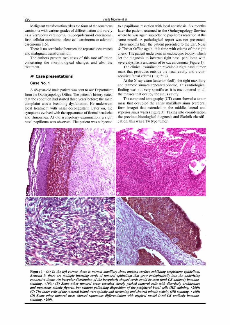
to a papilloma resection with local anesthesia. Six months later the patient returned to the Otolaryngology Service where he was again subjected to papilloma resection at the same nostril. A pathological report was not presented. Three months later the patient presented to the Ear, Nose & Throat Office again, this time with edema of the right cheek. The patient underwent an endoscopic biopsy, which set the diagnosis to inverted right nasal papilloma with severe dysplasia and areas of in situ carcinoma (Figure 1).
The clinical examination revealed a right nasal tumor mass that protrudes outside the nasal cavity and a con-secutive facial edema (Figure 2).
At the X-ray exam (anterior skull), the right maxillary and ethmoid sinuses appeared opaque. This radiological finding was not very specific as it is encountered in all the masses that occupy the sinus cavity.
The computed tomography (CT) exam showed a tumor mass that occupied the entire maxillary sinus (cerebral form image) that extended to the middle, lateral and superior sinus walls (Figure 3). Taking into consideration the previous histological diagnosis and Skolnik classifi-cation, this was a T4 type tumor.
Figure 1 – (A) In the left corner, there is normal maxillary sinus mucosa surface exhibiting respiratory epithelium. Beneath it, there are multiple inverting cords of tumoral epithelium that grow endophytically into the underlying connective tissue. An irregular distribution of the irregularly shaped cords could be seen (anti-CK antibody immuno-staining, ×100); (B) Some other tumoral areas revealed closely packed tumoral cells with disorderly architecture and numerous mitotic figures, but without palisading disposition of the peripheral basal cells (HE staining, ×200); (C) The inner cells of the tumoral island were spindle and streaming and showed mitotic activity (HE staining, ×400); (D) Some other tumoral nests showed squamous differentiation with atypical nuclei (Anti-CK antibody immuno-staining, ×200).

Clinical and histological aspects of malignant inverted sinonasal papilloma. Two case reports
291
Figure 1 (continued) – (E) In solid tumoral areas, there were numerous Ki67-positive nuclei (Anti-Ki67 antibody immunostaining, ×400); (F) The stroma was made of loose fibrous tissue and contained numerous thin walled blood vessels (Anti-Vim antibody immunostaining, ×100).
Figure 2 – Preoperative photograph showing
right facial tumor mass extending in the right
nostril.
Figure 3 – CT exam: tumor mass in the nasal and
maxillary sinus cavity with extension
towards the ethmoid sinus.
The treatment was decided to consist in surgical resection. A lateral rhinotomy was performed with a superior labial incision. The surgical conduct included hemi resection of the right maxilla, external carotid artery tie and ethmoid sinus surgery.
The histological exam was malignant inverted sino-nasal papilloma.
No complications were noted after surgery. The patient was advised to undertake another surgical
procedure using a free ilium flap in order to reconstruct the tissues lost in the surgical excision. As the patient decline another surgical procedure, a recommendation for prosthetic rehabilitation was issued.
The patient was instructed to go to the Dental Prosthetic Department, where a removable appliance was manufac-tured to aid in the healing process.
After the surgical treatment was performed, the patient underwent radiotherapy (45 Gy).
No local relapses were noted at the last recall (four years recall).
Case No. 2
A 56-year-old male patient was sent to our Department from the Otolaryngology Office. The patient’s history mentioned that his condition had started two years before with non-specific symptoms of obstructive breathing dysfunctions followed by headaches and rhinorrhea. The otolaryngology specialist set the diagnosis for left nasal papilloma. He was subjected to local surgical resection. The histological exam was conclusive for inverted papilloma. One year later, the tumor relapsed, this time the breathing disorder was more severe. He was again subjected to an endoscopic resection as a papilloma mass was noted. The histological exam was definitive for inverted papilloma with areas of in situ carcinoma. Within six months, the tumor relapsed again. This time, the patient presented with ocular symptoms (chemosis, double sight) and left facial edema (Figure 4).
The CT scan showed a tumor mass that occupied the entire left maxillary sinus and extended into the medial, lateral and superior sinus walls to the orbit and base of the skull (Figure 5).
The surgical treatment consisted in tying off the external carotid artery, ethmoid sinus surgery, left hemi resection of the maxilla, removal of the involved inferior wall of the orbit with immediate reconstruction using a titanium plate (Figure 6).
The histological exam was malignant inverted sino-nasal papilloma (Figure 7).
The patient underwent radiotherapy (50 Gy). The patient had no relapse in the five-year follow-up.
Figure 4 – Preoperative photograph showing a left facial tumor that modifies the genian,
nasal and orbital regions.

Vasile Nicolae et al.
292
Figure 5 – CT exam: tumor mass that
involves the nasal cavity, the left maxillary and
ethmoid sinus and extends to the floor
of the orbit.
Figure 6 – Intra-operative photo:
immediate reconstruction of the orbital floor with a
titanium plate.
Figure 7 – (A) The tumoral inverted growth revealed an area with high-grade dysplasia, along with transition to an in situ carcinoma (HE staining, ×200); (B) Some other tumoral areas were more solid and revealed disorderly maturation, spindling, squamous differentiation (in the upper field), mitosis, and central necrosis (HE staining, ×200); (C) Island of epithelial tumoral cells with squamous differentiation and cytological atypia (Anti-CK antibody immunostaining, ×200); (D) Solid tumoral area with disorderly architecture and numerous Ki67-positive nuclei (Anti-Ki67 antibody immuno-staining, ×400).
Discussion
The inverted papilloma frequently involves the male gender [16]. The average age ranges from 50 to 80 years, the most affected people being those in their fifties [17]. The pediatric patient is seldom affected, the literature reporting only rare cases [18]. The cases presented by the authors are both adult males in their fifth decade of life.
Inverted papilloma is unilateral and derives from the oral mucosa or from the sinus mucosa, usually in the maxillary sinuses. There are few reports regarding bilateral inverted papilloma [19]. The cases presented were both dealing with unilateral inverted papillomas.
At an early stage, the symptoms are non-specific: obstructive breathing problems, headaches, loss of smell, rhinorrhea, recurrent nasal papilloma. The patients usually present to a maxillofacial surgery office in the late stages when there is eye involvement, facial edema, and neuro-logical signs on the path of the II, IV, and VI cranial nerves [20]. The cases observed had similar symptoms at presentations: facial edema, eye impairment and respiratory dysfunctions.
At the endoscopic exam, the tumor is noticed as an irregular proliferative mass, blackberry like shape, light grey in color [21].
The histopathological examination shows the thickened multilayered epithelium. The epithelium is non-keratinized

Clinical and histological aspects of malignant inverted sinonasal papilloma. Two case reports
293
and has an inverted growth towards the stroma. The normal respiratory epithelium is replaced by this squamous type. Also, transitional epithelium can be observed. Surface keratinization can also be noted. The stroma can have different patterns dense or fibrous, loose or myxoid. Inflammatory cells usually migrate at the border of these lesions, mainly polymorphonuclear neutrophils and rarely eosinophils [22]. Malignization is characterized by the presence of numerous mitosis, nucleus atypia and different stages of dysplasia [23].
The classic X-ray exam is not explanative but the CT scan shows the amount of the extension towards the bony walls, other sinuses (ethmoid), other anatomic regions (orbit). In the authors’ cases, the CT showed extensive bone destruction evolving the medial and the superior maxillary sinus wall and also the orbit rim towards the base of the skull.
One of the most important factors for malignant transformation is thought to be smoking, but also recurrent viral sino-nasal infections and local inflammation process [24]. There are a few hypotheses suggesting that pollution plays an important role in malignization [25]. The presence of HPV is considered by some authors as coincidental [26] but many reports suggest it as a direct factor for the malignant process [27–29]. Immunohistochemistry is an accurate method for viral determination in histological samples but it is rather expensive for most of the patients in the authors’ geographic area. The patients did not afford the cost of this exam thus there are no HPV determination.
Malignancy in inverted sino-nasal papiloma is mainly related to squamous cell carcinoma and adenocarcinoma [30]. It can occur in the present lesion or it can occur as a recurrence in a previous site that had underwent surgical excision [31].
The early diagnosis is hard to predict. There are a few markers, which have an increase value in this pathology, but further research is still needed: squamous carcinoma antigen [32], C/EBP alpha and beta proteins [33], and EGFR and TGD alpha growth factors [34].
The classification is according to the American Joint Committee on Cancer (AJCC): T1 for tumor mass that does not extend over the nostril, T2 involves two areas of the nasal cavity, T3 affects the maxillary sinus and T4 extends beyond the maxillary sinus [9].
The treatment involves surgical resection. When dealing with T1–T3 tumors, the surgical conduct is done by endoscopic methods [35, 36]. In the case of T4 tumors with multiple relapses, the classic surgery is preferred for the resection of the entire tumor mass [37]. Radiotherapy is used in conjunction with surgical treatment and alone only in surgical surpassed cases [38].
Surgical resection was preferred in the cases highlighted above as the operating team was dealing with an inverted papilloma that had previous relapses and had signs of carcinoma transformation. This radical procedure esta-blished a thorough surgical excision with free oncological resections margins.
The surgical treatment usually leaves the patient with a large bony defect. The rehabilitation should take into consideration the patient’s age, associated pathology and also patient’s wishes. Reconstruction can imply prosthetic devices or surgical procedures [39]. The prosthetic device have the advantages of the reduce cost, the ease of
fabrication, the removable character eases the clinician in detecting recurrences [40]. However, a surgical procedure, although more invasive and with greater costs rehabilitates the bone structures as well as the soft tissue lost. A free autologous composite flap gives the patient the possibility to reconstruct his dentition with the aid of dental implants [41]. With the help of the Department of Prosthodontics, the patients were fitted with special dental prosthesis, which were able to restore their oral functions.
The relapse rate is sometimes over 50% [10]. These patients must return to follow up examinations for a long period of time (at least five years) with complete clinical and X-ray examinations [CT, magnetic resonance imaging (MRI)].
Conclusions
Both cases reported above fit in the risk category for malignant inverted papilloma: male gender, around 50 years old, and multiple relapses. The treatment of choice due to the tumor extent was classic surgery with radical tumor resection. At follow-up, no local recurrences were noted.
Conflict of interests The authors declare that they have no conflict of
interests.
References [1] Lingen M. Head and neck. Tumors of the nose, sinuses and
nasopharynx. In: Kumar VK, Abbas AK, Fausto N, Aster JC (eds). Robbins and Cotran pathologic basis of disease. 8th edition, Saunders–Elsevier, Philadelphia, 2010, 750–752.
[2] Hyams VJ. Papillomas of the nasal cavity and paranasal sinuses. A clinicopathologic study of 315 cases. Ann Otol Rhinol Laryngol, 1971, 80(2):192–206.
[3] Hobbs CG, Sterne JA, Bailey M, Heyderman RS, Birchall MA, Thomas SJ. Human papillomavirus and head and neck cancer: a systematic review and meta-analysis. Clin Otolaryngol, 2006, 31(4):259–266.
[4] Snyder RN, Perzin KH. Papillomatosis of nasal cavity and paranasal sinuses (inverted papilloma, squamous papilloma). A clinicopathologic study. Cancer, 1972, 30(3):668–690.
[5] Christensen WN, Smith RR. Schneiderian papillomas: a clinico-pathologic study of 67 cases. Hum Pathol, 1986, 17(4):393–400.
[6] Furuta Y, Shinohara T, Sano K, Nagashima K, Inoue K, Tanaka K, Inuyama Y. Molecular pathologic study of human papillomavirus infection in inverted papilloma and squamous cell carcinoma of the nasal cavities and paranasal sinuses. Laryngoscope, 1991, 101(1 Pt 1):79–85.
[7] Myers EN, Fernau JL, Johnson JT, Tabet JC, Barnes EL. Management of inverted papilloma. Laryngoscope, 1990, 100(5):481–490.
[8] Finkelstein SD, Tiffee JC, Bakker A, Swalsky P, Barnes L. Malignant transformation in sinonasal papillomas is closely associated with aberrant p53 expression. Mol Diagn, 1998, 3(1):37–41.
[9] Sousa AM, Vicenti AB, Speck Filho J, Cahali MB. Retros-pective analysis of 26 cases of inverted nasal papillomas. Braz J Otorhinolaryngol, 2012, 78(1):26–30.
[10] Kim K, Kim D, Koo Y, Kim CH, Choi EC, Lee JG, Yoon JH. Sinonasal carcinoma associated with inverted papilloma: a report of 16 cases. J Craniomaxillofac Surg, 2012, 40(4): e125–e129.
[11] Vorasubin N, Vira D, Suh JD, Bhuta S, Wang MB. Schneiderian papillomas: comparative review of exophytic, oncocytic, and inverted types. Am J Rhinol Allergy, 2013, 27(4):287–292.
[12] Mirza S, Bradley PJ, Acharya A, Stacey M, Jones NS. Sino-nasal inverted papillomas: recurrence, and synchronous and metachronous malignancy. J Laryngol Otol, 2007, 121(9):857–864.

Vasile Nicolae et al.
294
[13] Barnes L. Schneiderian papillomas and nonsalivary glandular neoplasms of the head and neck. Mod Pathol, 2002, 15(3): 279–297.
[14] Lesperance MM, Esclamado RM. Squamous cell carcinoma arising in inverted papilloma. Laryngoscope, 1995, 105(2): 178–183.
[15] von Buchwald C, Bradley PJ. Risks of malignancy in inverted papilloma of the nose and paranasal sinuses. Curr Opin Otolaryngol Head Neck Surg, 2007, 15(2):95–98.
[16] Peter B, Grossenbacher R. Invertierte Papillome der Nase und deren Nebenhöhlen. Laryngo-Rhino-Otologie, 1997, 76(1): 14–18.
[17] Dolgin SR, Zaveri VD, Casiano RR, Maniglia AJ. Different options for treatment of inverting papilloma of the nose and paranasal sinuses: a report of 41 cases. Laryngoscope, 1992, 102(3):231–236.
[18] D’Angelo AJ Jr, Marlowe A, Marlowe FI, McFarland M. Inverted papilloma of the nose and paranasal sinuses in children. Ear Nose Throat J, 1992, 71(6):264–266.
[19] Kodama S, Kawano T, Suzuki M. Bilateral endoscopic medial maxillectomy for bilateral inverted papilloma. Case Rep Oto-laryngol, 2012, 2012:215847.
[20] Ganzer U, Donath K, Schmelzle R. Geschwulste der inneren nase, der Nasenebenhohlen, des Ober und Unterkiefers. In: Naumann HH, Helms J, Herbenhold C, Kastenbauer E (eds). Oto-Rhino-Laryngologie in Klinik und Praxis. Band 2, Thieme Verlag, Stuttgart–New York, 1992, 312–359.
[21] Eggesbø HB. Imaging of sinonasal tumours. Cancer Imaging, 2012, 12:136–152.
[22] Wang M, Liu Y, Zhou G. [Oncocytic papilloma in nasal cavity and paranasal sinuses: 3 of cases]. Lin Chung Er Bi Yan Hou Tou Jing Wai Ke Za Zhi, 2015, 29(3):279–281.
[23] Batsakis JG, Suarez P. Schneiderian papillomas and carci-nomas: a review. Adv Anat Pathol, 2001, 8(2):53–64.
[24] Skolnik EM, Loewy A, Friedman JE. Inverted papilloma of the nasal cavity. Arch Otolaryngol, 1966, 84(1):61–67.
[25] Barbieri PG, Tomenzoli D, Morassi L, Festa R, Fernicola C. [Sino-nasal inverted papillomas and occupational etiology]. G Ital Med Lav Ergon, 2005, 27(4):422–426.
[26] Jenko K, Kocjan B, Zidar N, Poljak M, Strojan P, Zargi M, Blatnik O, Gale N. In inverted papillomas HPV more likely represents incidental colonization than an etiological factor. Virchows Arch, 2011, 459(5):529–538.
[27] Hasegawa M, Deng Z, Maeda H, Yamashita Y, Matayoshi S, Kiyuna A, Agena S, Uehara T, Suzuki M. Human papilloma-virus load and physical status in sinonasal inverted papilloma and squamous cell carcinoma. Rhinology, 2012, 50(1):87–94.
[28] Zhong Z, Yan A, Jiang F, Wei H, Jiang X. [The study on the relationship between human papillomavirus infection and
pathogenesis of nasal inverted papilloma and its malignant transformation]. Lin Chung Er Bi Yan Hou Tou Jing Wai Ke Za Zhi, 2010, 24(5):209–211.
[29] Hoffmann M, Klose N, Gottschlich S, Görögh T, Fazel A, Lohrey C, Rittgen W, Ambrosch P, Schwarz E, Kahn T. Detection of human papillomavirus DNA in benign and malig-nant sinonasal neoplasms. Cancer Lett, 2006, 239(1):64–70.
[30] Katori H, Nozawa A, Tsukuda M. Histopathological parameters of recurrence and malignant transformation in sinonasal inverted papilloma. Acta Otolaryngol, 2006, 126(2):214–218.
[31] Choi JW, Kim SG, Kim YM, Yoon YH, Kim AY, Rha KS. Clinical and histologic features of inverted papilloma-associated malignancy. Eur Arch Otorhinolaryngol, 2012, 269(11):2349–2354.
[32] Matoušek P, Zeleník K, Safarčík K, Cábalová L, Komínek P. Squamous cell carcinoma antigen as a marker of sinonasal inverted papilloma. Eur Arch Otorhinolaryngol, 2014, 271(3): 535–538.
[33] Shabana EH, Depondt J, Hourseau M, Walker F, Berdal A. Production and significance of CCAAT enhancer binding proteins alpha and beta in sinonasal inverted papilloma. Histol Histopathol, 2013, 28(1):53–60.
[34] Katori H, Nozawa A, Tsukuda M. Markers of malignant trans-formation of sinonasal inverted papilloma. Eur J Surg Oncol, 2005, 31(8):905–911.
[35] Hong SL, Kim BH, Lee JH, Cho KS, Roh HJ. Smoking and malignancy in sinonasal inverted papilloma. Laryngoscope, 2013, 123(5):1087–1091.
[36] Pasquini E, Sciarretta V, Farneti G, Modugno GC, Ceroni AR. Inverted papilloma: report of 89 cases. Am J Otolaryngol, 2004, 25(3):178–185.
[37] Endo R, Ishitoya J, Kawano T, Yamada M, Sakuma Y, Shiono O, Komatsu M, Mitake D, Yamashita Y, Tsukuda M. [A study of surgical procedures for inverted papilloma in the nasal cavity and paranasal sinuses]. Nihon Jibiinkoka Gakkai Kaiho, 2008, 111(8):581–587.
[38] Sandison A. Common head and neck cases in our consultation referrals: diagnostic dilemmas in inverted papilloma. Head Neck Pathol, 2009, 3(3):260–262.
[39] DaBreo EL, Chalian VA, Lingeman R, Reisbick MH. Prosthetic and surgical management of osteogenic sarcoma of the maxilla. J Prosthet Dent, 1990, 63(3):316–320.
[40] Brown KE. Clinical considerations improving obturator treatment. J Prosthet Dent, 1970, 24(4):461–466.
[41] Abdel Fattah H, Zaghloul A. Pre-prosthetic surgical alterations in maxillectomy to enhance the prosthetic prognoses as part of rehabilitation of oral cancer patient. J Egypt Natl Canc Inst, 2010, 22(4):251–263.
Corresponding author Sorin Ibric-Cioranu, PhD Student, Faculty of Medicine, “Lucian Blaga” University of Sibiu, 2A Lucian Blaga Street, 550169 Sibiu, Romania; Phone +4021–223 55 32, +40748–365 320, e-mail: [email protected] Received: September 3, 2015
Accepted: April 25, 2016