Mechanical characterisation of porcine rectus sheath under 34 ...
Upload
kory-armstrongCategory
view
228download
2
CLINICAL ANATOMY OF CLINICAL ANATOMY OF ANTERIOR ABDOMINAL WALL ANTERIOR ABDOMINAL WALL
& RECTUS SHEATH& RECTUS SHEATH
By: Dr. Mujahid KhanBy: Dr. Mujahid Khan


Structure of Abdominal CavityStructure of Abdominal Cavity
Superiorly it is formed by diaphragm which Superiorly it is formed by diaphragm which separates the abdominal cavity from the separates the abdominal cavity from the thoracic cavitythoracic cavity
Inferiorly the abdominal cavity is Inferiorly the abdominal cavity is continuous with the pelvic cavity through continuous with the pelvic cavity through the pelvic inletthe pelvic inlet

Structure of Abdominal WallStructure of Abdominal Wall
Anteriorly:Anteriorly:
The abdominal wall is formed above by The abdominal wall is formed above by lower part of the thoracic cagelower part of the thoracic cage
Below by the rectus abdominis, external Below by the rectus abdominis, external oblique, internal oblique, and transversus oblique, internal oblique, and transversus abdominis muscles and fasciaeabdominis muscles and fasciae

Structure of Ant. Abdominal WallStructure of Ant. Abdominal Wall
It is made up of skin, superficial fascia, It is made up of skin, superficial fascia, deep fascia, muscles, extraperitoneal deep fascia, muscles, extraperitoneal fascia and parietal peritoneumfascia and parietal peritoneum
The abdominal walls are lined by a fascial The abdominal walls are lined by a fascial envelope and the parietal peritoneumenvelope and the parietal peritoneum

SkinSkin
Natural lines of cleavage in the skin are Natural lines of cleavage in the skin are constant and run almost horizontally constant and run almost horizontally around the trunkaround the trunk
An incision along a cleavage line will heal An incision along a cleavage line will heal as a narrow scar, while one that crosses as a narrow scar, while one that crosses the lines will heal as a wide scarthe lines will heal as a wide scar
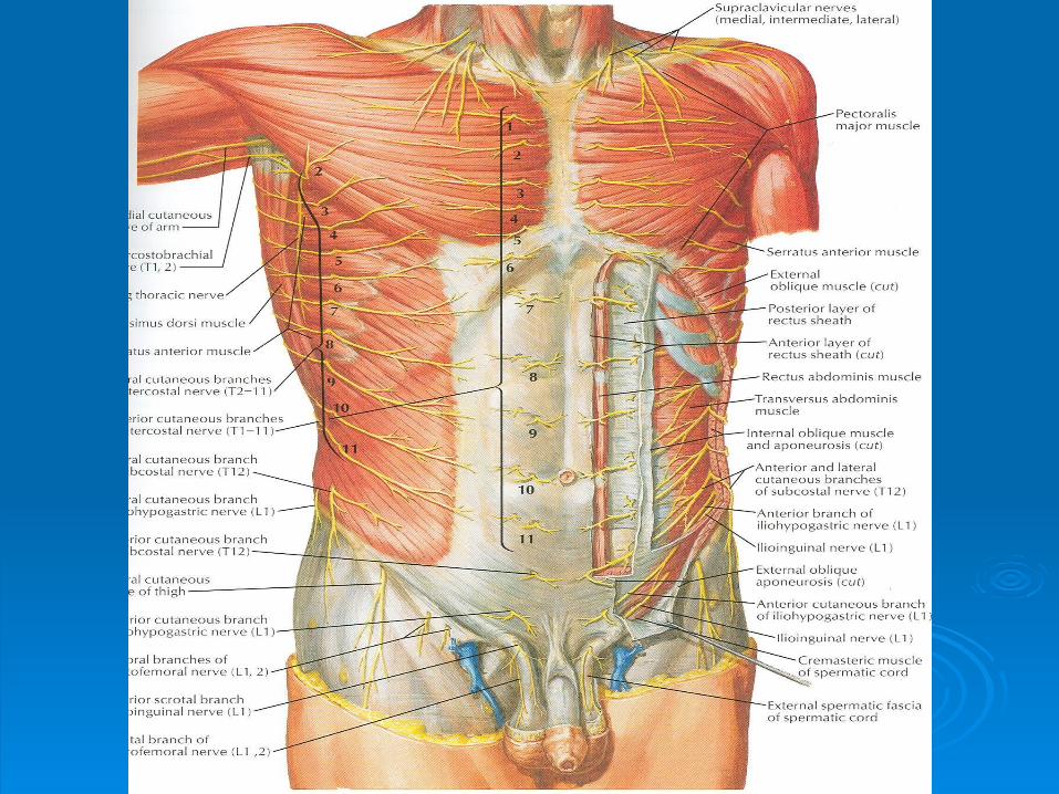
Cutaneous Nerve SupplyCutaneous Nerve Supply
Is derived from the anterior rami of the Is derived from the anterior rami of the lower six thoracic and first lumbar nerveslower six thoracic and first lumbar nerves
Thoracic nerves are the lower five Thoracic nerves are the lower five intercostal and the subcostal nervesintercostal and the subcostal nerves
First lumbar nerve is represented by the First lumbar nerve is represented by the iliohypogastric and ilioinguinal nerves iliohypogastric and ilioinguinal nerves

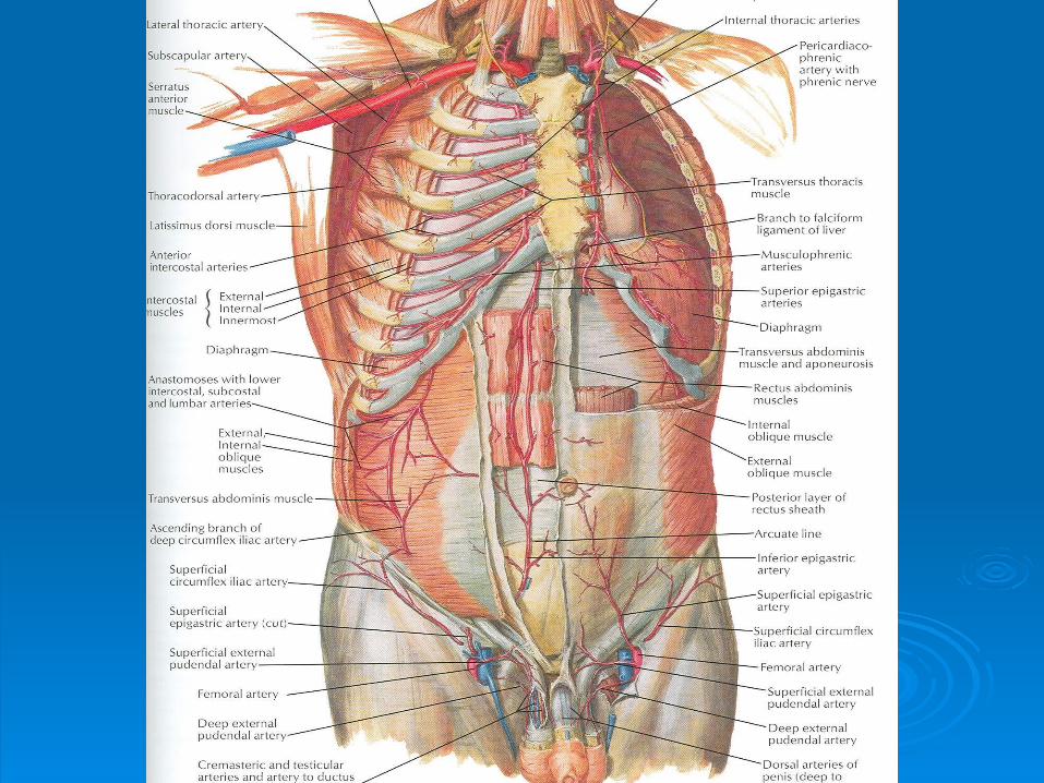
Blood SupplyBlood Supply
Skin near the midline is supplied by Skin near the midline is supplied by branches of the superior epigastric artery branches of the superior epigastric artery (br. of int. thoracic artery) and the inferior (br. of int. thoracic artery) and the inferior epigastric artery ( br. of external iliac epigastric artery ( br. of external iliac artery)artery)
Skin of the flanks is supplied by branches Skin of the flanks is supplied by branches from the intercostal, lumbar, and deep from the intercostal, lumbar, and deep circumflex arteriescircumflex arteries

Superficial FasciaSuperficial Fascia
Fatty layer or fascia of camper is Fatty layer or fascia of camper is continuous with the superficial fat over the continuous with the superficial fat over the rest of the body and may be extremely rest of the body and may be extremely thick in obese patientsthick in obese patients
The membranous layer or scarpa’s fascia The membranous layer or scarpa’s fascia is thin and fades out laterally and aboveis thin and fades out laterally and above
Becomes continuous with the superficial Becomes continuous with the superficial fascia of the back and the thoraxfascia of the back and the thorax

Superficial FasciaSuperficial Fascia
Inferiorly the membranous layer passes onto the Inferiorly the membranous layer passes onto the front of the thigh, where it fuses with the deep front of the thigh, where it fuses with the deep fasciafascia
In the midline inferiorly forms a tubular sheath In the midline inferiorly forms a tubular sheath for the penis or clitorisfor the penis or clitoris
Below in the perineum, enters the wall of the Below in the perineum, enters the wall of the scrotum or labia majorascrotum or labia majora
From there it passes to be attached on each From there it passes to be attached on each side to the margins of pubic arch, here it is side to the margins of pubic arch, here it is called Colle’s fasciacalled Colle’s fascia
Superficial FasciaSuperficial Fascia
Posteriorly it fuses with the perineal body Posteriorly it fuses with the perineal body and the margin of the perineal membraneand the margin of the perineal membrane
The fatty layer is represented as a smooth The fatty layer is represented as a smooth muscle in the scrotum, the dartos musclemuscle in the scrotum, the dartos muscle
The membranous layer persists as a The membranous layer persists as a separate layerseparate layer

Deep FasciaDeep Fascia
Deep fascia in the anterior abdominal wall Deep fascia in the anterior abdominal wall is merely a thin layer of connective tissue is merely a thin layer of connective tissue covering the musclescovering the muscles
It lies immediately deep to the It lies immediately deep to the membranous layer of the superficial fasciamembranous layer of the superficial fascia
Muscles Muscles
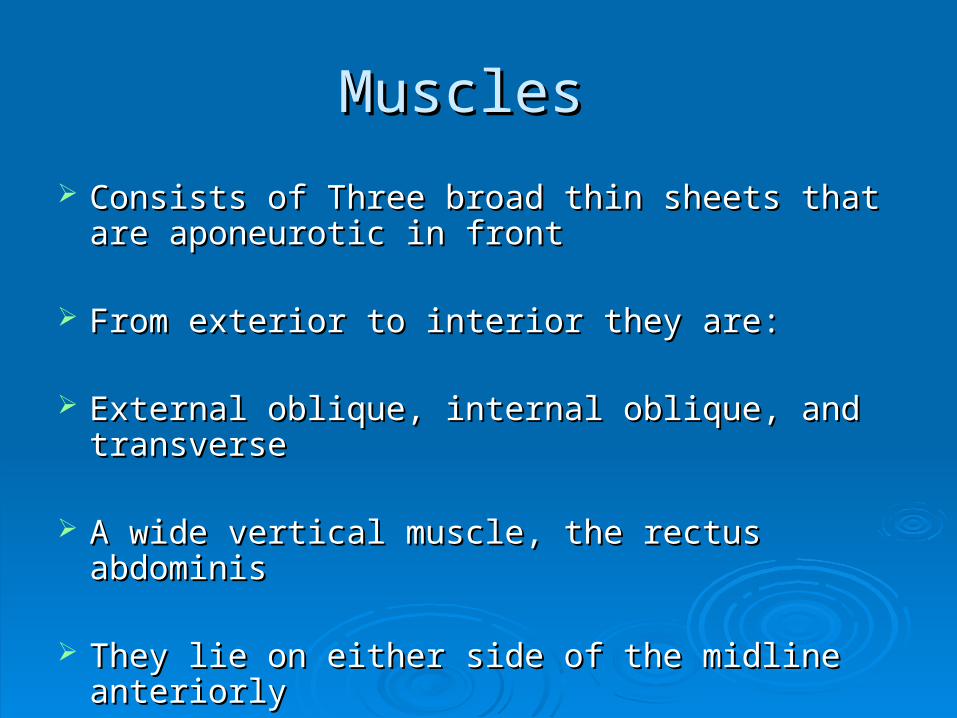
Consists of Three broad thin sheets that are Consists of Three broad thin sheets that are aponeurotic in frontaponeurotic in front
From exterior to interior they are:From exterior to interior they are:
External oblique, internal oblique, and External oblique, internal oblique, and transversetransverse
A wide vertical muscle, the rectus abdominisA wide vertical muscle, the rectus abdominis
They lie on either side of the midline anteriorlyThey lie on either side of the midline anteriorly

MusclesMuscles
As the aponeurosis of three sheets pass As the aponeurosis of three sheets pass forward, they enclose the rectus forward, they enclose the rectus abdominis to form the rectus sheathabdominis to form the rectus sheath
The cremaster muscle which is derived The cremaster muscle which is derived from the lower fibers of internal oblique, from the lower fibers of internal oblique, passes inferiorly as a covering of the passes inferiorly as a covering of the spermatic cord and enters scrotumspermatic cord and enters scrotum

External Oblique MuscleExternal Oblique Muscle
Is a broad, thin, muscular sheetIs a broad, thin, muscular sheet
Origin: Lower 8 ribsOrigin: Lower 8 ribs
Insertion: Xiphoid process, linea alba, pubic tubercle, Insertion: Xiphoid process, linea alba, pubic tubercle, iliac crestiliac crest
Nerve Supply: Lower 6 thoracic nerves, iliohypogastric & Nerve Supply: Lower 6 thoracic nerves, iliohypogastric & ilioinguinal nervesilioinguinal nerves
Action: Supports abdominal contents, assist in forced Action: Supports abdominal contents, assist in forced expiration, micturition, defecation, parturition, vomiting expiration, micturition, defecation, parturition, vomiting
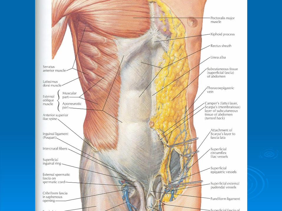

External Oblique MuscleExternal Oblique Muscle
A triangular shaped defect in the external A triangular shaped defect in the external oblique aponeurosis lies immediately oblique aponeurosis lies immediately above and medial to the pubic tubercle, above and medial to the pubic tubercle, known as superficial inguinal ringknown as superficial inguinal ring
Between the anterosuperior iliac spine and Between the anterosuperior iliac spine and the pubic tubercle, the lower border of the the pubic tubercle, the lower border of the aponeurosis is folded backward on itself, aponeurosis is folded backward on itself, forming the inguinal ligamentforming the inguinal ligament

Internal Oblique MuscleInternal Oblique Muscle
Origin: Lumbar fascia, iliac crest, lateral two-Origin: Lumbar fascia, iliac crest, lateral two-thirds of inguinal ligamentthirds of inguinal ligament
Insertion: Lower three ribs and costal cartilages, Insertion: Lower three ribs and costal cartilages, xiphoid process, linea alba, symphysis pubisxiphoid process, linea alba, symphysis pubis
Nerve Supply: Lower six thoracic nerves, Nerve Supply: Lower six thoracic nerves, iliohypogastric & ilioinguinal nervesiliohypogastric & ilioinguinal nerves
Action: Supports abdominal contents, assist in Action: Supports abdominal contents, assist in forced expiration, micturition, defecation, forced expiration, micturition, defecation, parturition, vomiting parturition, vomiting

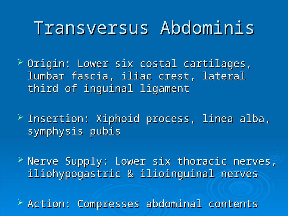
Transversus AbdominisTransversus Abdominis
Origin: Lower six costal cartilages, lumbar fascia, Origin: Lower six costal cartilages, lumbar fascia, iliac crest, lateral third of inguinal ligamentiliac crest, lateral third of inguinal ligament
Insertion: Xiphoid process, linea alba, symphysis Insertion: Xiphoid process, linea alba, symphysis pubispubis
Nerve Supply: Lower six thoracic nerves, Nerve Supply: Lower six thoracic nerves, iliohypogastric & ilioinguinal nervesiliohypogastric & ilioinguinal nerves
Action: Compresses abdominal contentsAction: Compresses abdominal contents

Rectus AbdominisRectus Abdominis
Origin: Symphysis pubis and pubic crestOrigin: Symphysis pubis and pubic crest
Insertion: 5Insertion: 5thth, 6, 6thth and 7 and 7thth costal cartilages costal cartilages and xiphoid processand xiphoid process
Nerve Supply: Lower six thoracic nervesNerve Supply: Lower six thoracic nerves
Action: Compresses abdominal contents, Action: Compresses abdominal contents, flexes vertebral column, accessory muscle flexes vertebral column, accessory muscle of expirationof expiration


Lymph DrainageLymph Drainage
Lymph drainage of the skin of the anterior Lymph drainage of the skin of the anterior abdominal wall above the umbilicus is upward to abdominal wall above the umbilicus is upward to the anterior axillary (pectoral group of nodes)the anterior axillary (pectoral group of nodes)
Below the level of umbilicus drains downward Below the level of umbilicus drains downward and laterally to the superficial inguinal nodesand laterally to the superficial inguinal nodes
Swelling in the groin is may be due to enlarged Swelling in the groin is may be due to enlarged superficial inguinal nodesuperficial inguinal node
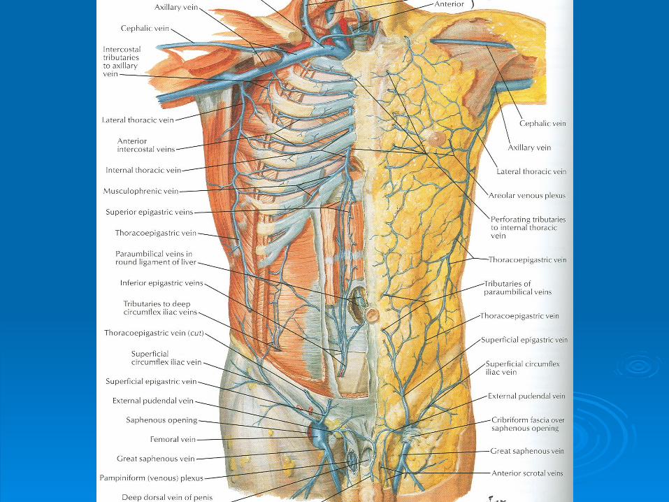
Venous DrainageVenous Drainage Venous blood is collected into a network of veins Venous blood is collected into a network of veins
that radiate from the umbilicusthat radiate from the umbilicus
The network is drained above into the axillary The network is drained above into the axillary vein via the lateral thoracic veinvein via the lateral thoracic vein
Below into the femoral vein via the superficial Below into the femoral vein via the superficial epigastric and the great saphenous veinsepigastric and the great saphenous veins
Few small veins, the paraumbilical veins form a Few small veins, the paraumbilical veins form a clinically important portal-system venous clinically important portal-system venous anastomosisanastomosis
Caput MedusaeCaput Medusae
The superficial veins around the umbilicus The superficial veins around the umbilicus and the paraumbilical veins connecting and the paraumbilical veins connecting them to the portal vein may become them to the portal vein may become grossly distended in case of portal vein grossly distended in case of portal vein obstructionobstruction
The distended subcutaneous veins radiate The distended subcutaneous veins radiate out from the umbilicus, producing in out from the umbilicus, producing in severe cases the clinical picture called severe cases the clinical picture called Caput Medusae Caput Medusae


NervesNerves
Nerves of the anterior abdominal wall supply the Nerves of the anterior abdominal wall supply the skin, muscles and the parietal peritoneumskin, muscles and the parietal peritoneum
They are derived from the anterior rami of lower They are derived from the anterior rami of lower six thoracic and the first lumbar nervessix thoracic and the first lumbar nerves
Inflammation of parietal peritoneum causes pain Inflammation of parietal peritoneum causes pain in the overlying skin and also a reflex increase in in the overlying skin and also a reflex increase in tone of the abdominal musculature in the same tone of the abdominal musculature in the same area area

Rectus SheathRectus Sheath
Is a long fibrous sheathIs a long fibrous sheath
Encloses the rectus abdominis and pyramidalis Encloses the rectus abdominis and pyramidalis muscle (if present)muscle (if present)
Contains the anterior rami of lower six thoracic Contains the anterior rami of lower six thoracic nerves and the superior and inferior epigastric nerves and the superior and inferior epigastric vessels and lymph vesselsvessels and lymph vessels
Formed mainly by aponeurosis of three lateral Formed mainly by aponeurosis of three lateral abdominal musclesabdominal muscles
Rectus SheathRectus Sheath
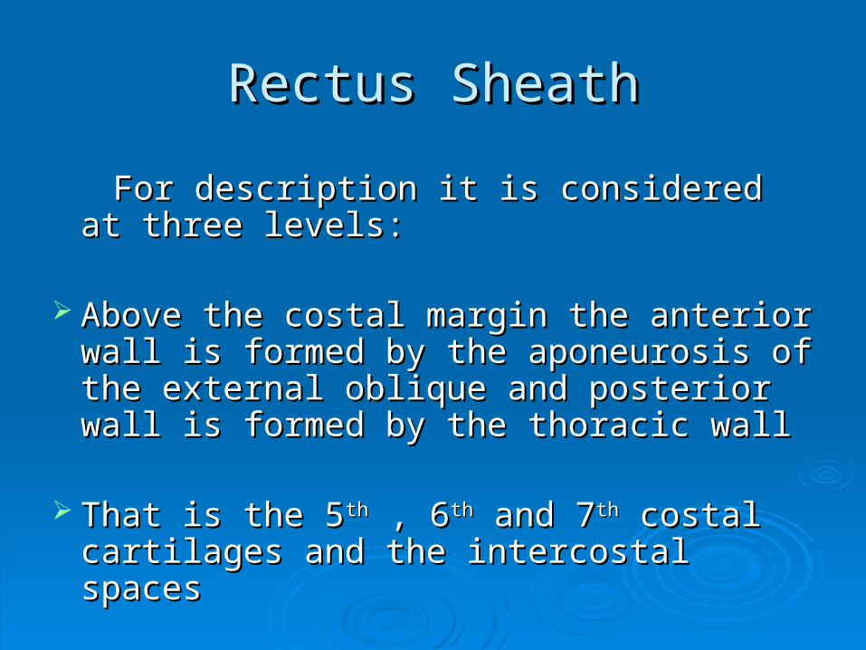
For description it is considered at three For description it is considered at three levels:levels:
Above the costal margin the anterior wall Above the costal margin the anterior wall is formed by the aponeurosis of the is formed by the aponeurosis of the external oblique and posterior wall is external oblique and posterior wall is formed by the thoracic wallformed by the thoracic wall
That is the 5That is the 5thth , 6 , 6thth and 7 and 7thth costal cartilages costal cartilages and the intercostal spacesand the intercostal spaces
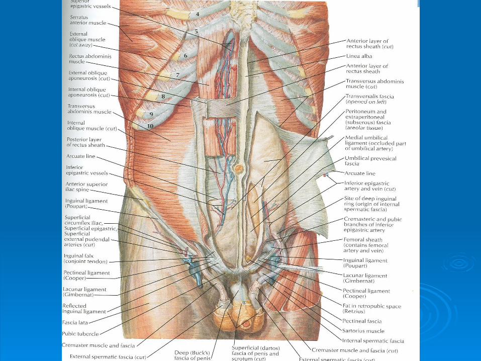
Rectus SheathRectus Sheath
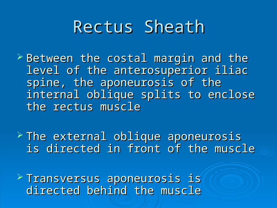
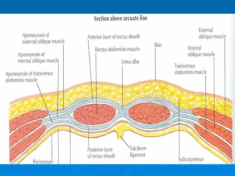
Between the costal margin and the level of Between the costal margin and the level of the anterosuperior iliac spine, the the anterosuperior iliac spine, the aponeurosis of the internal oblique splits to aponeurosis of the internal oblique splits to enclose the rectus muscleenclose the rectus muscle
The external oblique aponeurosis is The external oblique aponeurosis is directed in front of the muscledirected in front of the muscle
Transversus aponeurosis is directed Transversus aponeurosis is directed behind the musclebehind the muscle

Rectus SheathRectus Sheath
Between the level of the anterosuperior Between the level of the anterosuperior iliac spine and the pubis, the aponeurosis iliac spine and the pubis, the aponeurosis of all three muscles form the anterior wallof all three muscles form the anterior wall
The posterior wall is absentThe posterior wall is absent
The rectus muscle lies in contact with the The rectus muscle lies in contact with the fascia transversalisfascia transversalis


Rectus SheathRectus Sheath
The posterior wall of the rectus sheath is The posterior wall of the rectus sheath is not attached to the rectus abdominis not attached to the rectus abdominis musclemuscle
The anterior wall is firmly attached to it by The anterior wall is firmly attached to it by the muscle’s tendinous intersections the muscle’s tendinous intersections

Linea AlbaLinea Alba
The rectus sheath is separated from its The rectus sheath is separated from its fellow on the opposite side by a fibrous fellow on the opposite side by a fibrous band called the linea albaband called the linea alba
Extends from the xiphoid process to the Extends from the xiphoid process to the symphysis pubis symphysis pubis