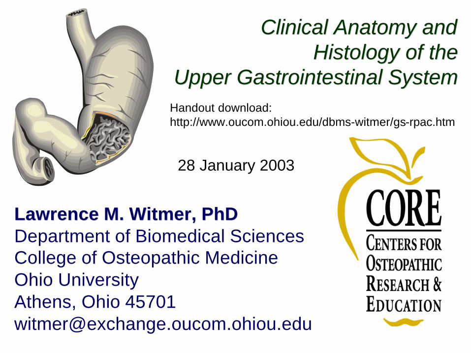
Clinical Anatomy and Histology of the Upper Gastrointestinal System
22
Clinical Anatomy and Clinical Anatomy and Histology of the Histology of the Upper Gastrointestinal System Upper Gastrointestinal System Lawrence M. Witmer, PhD Lawrence M. Witmer, PhD Department of Biomedical Sciences College of Osteopathic Medicine Ohio University Athens, Ohio 45701 [email protected] Handout download: http://www.oucom.ohiou.edu/dbms-witmer/gs-rpac.htm 28 January 2003
Transcript of Clinical Anatomy and Histology of the Upper Gastrointestinal System
Clinical Anatomy andClinical Anatomy andHistology of theHistology of the
Upper Gastrointestinal SystemUpper Gastrointestinal System
Lawrence M. Witmer, PhDLawrence M. Witmer, PhDDepartment of Biomedical SciencesCollege of Osteopathic MedicineOhio UniversityAthens, Ohio [email protected]
Handout download:http://www.oucom.ohiou.edu/dbms-witmer/gs-rpac.htm
28 January 2003
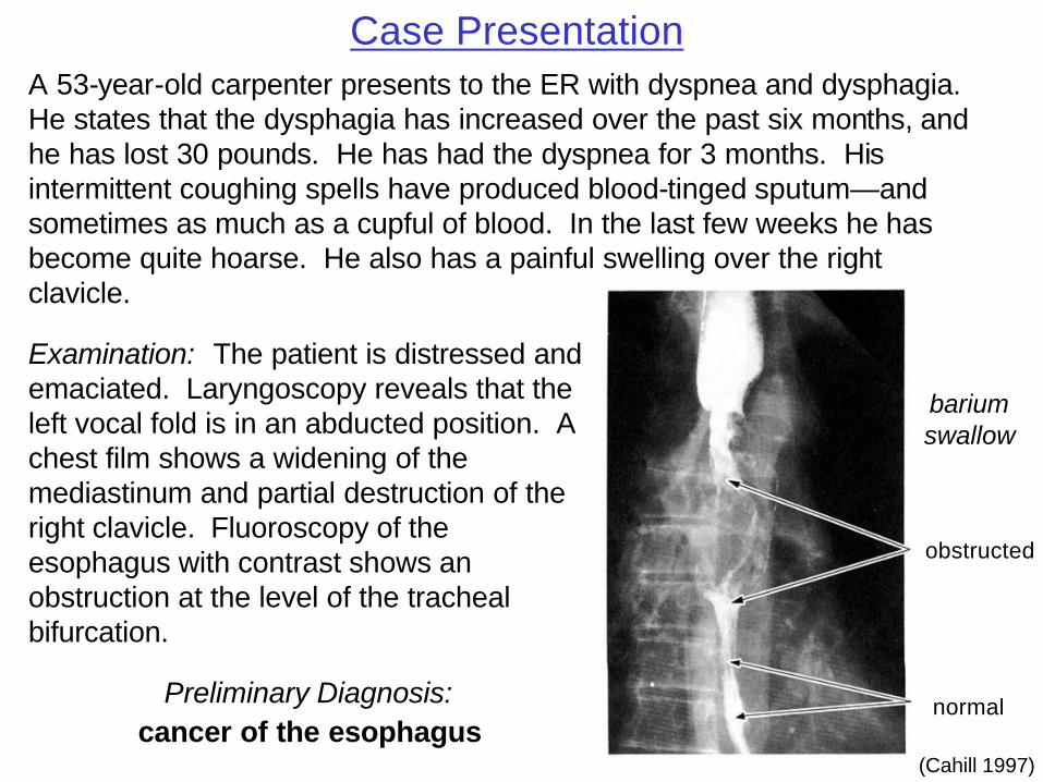
Preliminary Diagnosis:cancer of the esophagus
Case PresentationA 53-year-old carpenter presents to the ER with dyspnea and dysphagia. He states that the dysphagia has increased over the past six months, and he has lost 30 pounds. He has had the dyspnea for 3 months. His intermittent coughing spells have produced blood-tinged sputum—and sometimes as much as a cupful of blood. In the last few weeks he has become quite hoarse. He also has a painful swelling over the right clavicle.
Examination: The patient is distressed and emaciated. Laryngoscopy reveals that the left vocal fold is in an abducted position. A chest film shows a widening of the mediastinum and partial destruction of the right clavicle. Fluoroscopy of the esophagus with contrast shows an obstruction at the level of the tracheal bifurcation.
normal
obstructed
bariumswallow
(Cahill 1997)
Questions
1. Where is the cancer most likely located? Where are other common sites of esophageal cancer?
2. What may account for the dyspnea and bloody sputum?
3. Why are esophageal cancers more likely to spread to neighboring organs than are other parts of the GI tract?
4. What may account for the hoarseness and the abducted left vocal fold? Why is the right vocal fold unaffected?
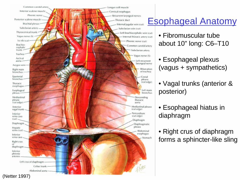
Esophageal Anatomy• Fibromuscular tube about 10” long: C6–T10
• Esophageal plexus (vagus + sympathetics)
• Vagal trunks (anterior & posterior)
• Esophageal hiatus in diaphragm
• Right crus of diaphragm forms a sphincter-like sling
(Netter 1997)

(Netter 1997)
Esophageal Constrictions
• Superiorly: level of cricoid cartilage, juncture with pharynx
• Middle: crossed by aorta and left main bronchus
• Inferiorly: diaphragmatic sphincter

(Netter 1997)
EsophagealHistology
• Inner circular & outer longitudinal muscle coats
• All the muscle fibers are actually helical: outer loose helix, inner tight helix
• Not all smooth muscle• Inferior third smooth• Superior third skeletal• Middle third mixed
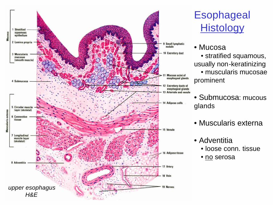
upper esophagusH&E
• Mucosa• stratified squamous,
usually non-keratinizing• muscularis mucosae
prominent
• Submucosa: mucous glands
• Muscularis externa
• Adventitia• loose conn. tissue• no serosa
EsophagealHistology
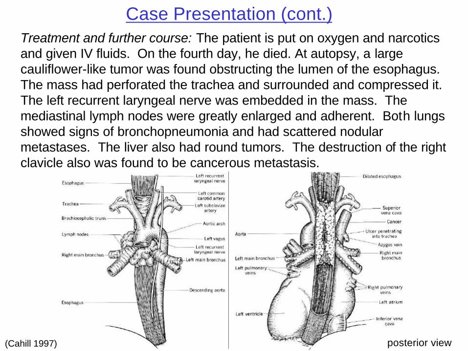
Case Presentation (cont.)Treatment and further course: The patient is put on oxygen and narcotics and given IV fluids. On the fourth day, he died. At autopsy, a large cauliflower-like tumor was found obstructing the lumen of the esophagus. The mass had perforated the trachea and surrounded and compressed it. The left recurrent laryngeal nerve was embedded in the mass. The mediastinal lymph nodes were greatly enlarged and adherent. Both lungs showed signs of bronchopneumonia and had scattered nodular metastases. The liver also had round tumors. The destruction of the right clavicle also was found to be cancerous metastasis.
(Cahill 1997) posterior view
Questions (cont.)
5. What other organs besides the trachea might be invaded by an esophageal mass?
6. Other than direct spread of the primary mass, by what route might the cancer have reached the lungs and pleura?
7. What is the most likely route of metastasis that would explain the liver involvement in this case?
8. What is the most likely route of metastasis that would explain the clavicular involvement in this case?
(Bannister 1995)
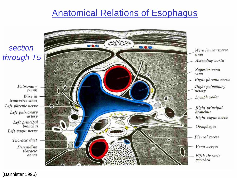
Anatomical Relations of Esophagus
sectionthrough T5

Anatomical Relations of Esophagus
(Bannister 1995)
sectionthrough T6

(Netter 1997)
EsophagealVeins
• Distribution of tumor emboli: link between lymphatic & arterial systems
• Two routes• Esophageal vv. drain into
SVC via azygous & hemiazygous
• Esophageal vv. drain into portal v. via branches of left gastric v. (a “portal-caval anastomosis”)
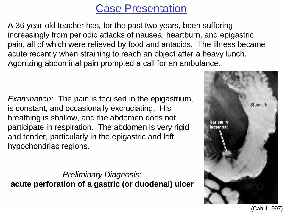
Case PresentationA 36-year-old teacher has, for the past two years, been suffering increasingly from periodic attacks of nausea, heartburn, and epigastric pain, all of which were relieved by food and antacids. The illness became acute recently when straining to reach an object after a heavy lunch. Agonizing abdominal pain prompted a call for an ambulance.
(Cahill 1997)
Examination: The pain is focused in the epigastrium, is constant, and occasionally excruciating. His breathing is shallow, and the abdomen does not participate in respiration. The abdomen is very rigid and tender, particularly in the epigastric and left hypochondriac regions.
Preliminary Diagnosis:acute perforation of a gastric (or duodenal) ulcer

Questions
1. What causes the rigidity of the abdominal wall and the costal nature of the patient’s respiration?
2. What is the anatomical basis for the difference in pain sensation before and after the perforation event?
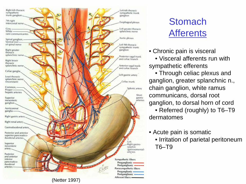
StomachAfferents
• Chronic pain is visceral• Visceral afferents run with
sympathetic efferents• Through celiac plexus and
ganglion, greater splanchnic n., chain ganglion, white ramus communicans, dorsal root ganglion, to dorsal horn of cord
• Referred (roughly) to T6–T9 dermatomes
• Acute pain is somatic• Irritation of parietal peritoneum T6–T9
(Netter 1997)
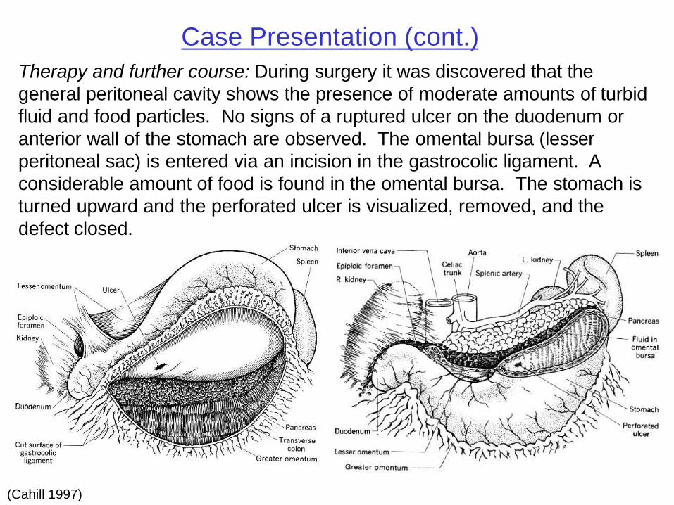
Case Presentation (cont.)Therapy and further course: During surgery it was discovered that the general peritoneal cavity shows the presence of moderate amounts of turbid fluid and food particles. No signs of a ruptured ulcer on the duodenum or anterior wall of the stomach are observed. The omental bursa (lesser peritoneal sac) is entered via an incision in the gastrocolic ligament. A considerable amount of food is found in the omental bursa. The stomach is turned upward and the perforated ulcer is visualized, removed, and the defect closed.
(Cahill 1997)

Questions
3. If the ulceration was into the lesser peritoneal sac, why wasthere extraneous material in the greater sac?
4. Which organ is most apt to be invaded if an ulcer on the posterior wall of the stomach slowly perforates through the gastric wall?
5. Which major artery would be in danger of severe hemorrhage in case of Question 4?
6. Which vessels are in danger during incision of the gastrocolic ligament?

StomachAnatomy
• Regions of stomach• Cardiac• Fundus• Corpus• Pyloric: antrum, canal,
sphincter
• Lesser & greater omenta
• Winslow’s foramen: communication of lesser & greater sacs
(Netter 1997)

Stomach Histology• Zones: cardiac, fundic, pyloric• Glands in all zones have mucous cells and enteroendocrine cells• Fundic glands
• Parietal (oxyntic) cells: HCl, Intrinsic factor• Chief (zymogenic) cells: pepsinogen, rennin, lipases• Mucous neck cells• Enteroendocrine cells: gastrin, cholecystokinin, secretin, serotonin,
glucagon
(Netter 1997)
(Netter 1997)
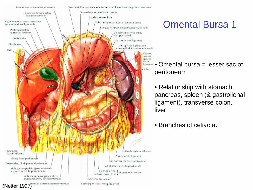
Omental Bursa 1
• Omental bursa = lesser sac of peritoneum
• Relationship with stomach, pancreas, spleen (& gastrolienal ligament), transverse colon, liver
• Branches of celiac a.
(Netter 1997)
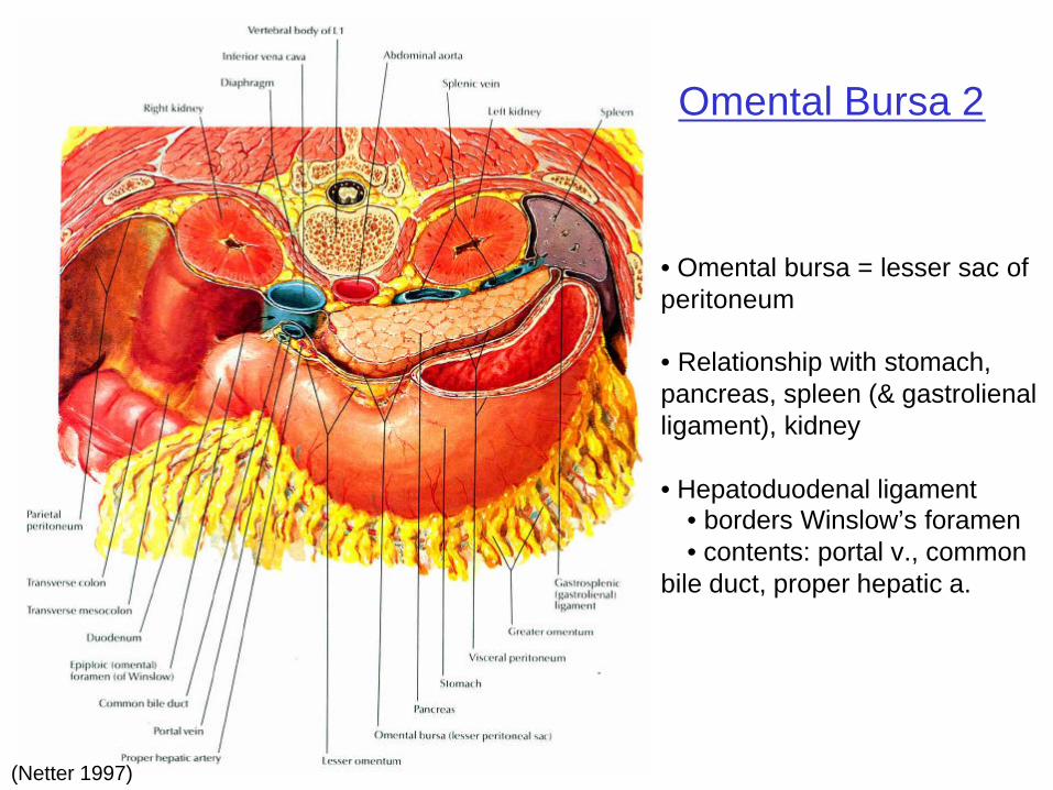
Omental Bursa 2
• Omental bursa = lesser sac of peritoneum
• Relationship with stomach, pancreas, spleen (& gastrolienal ligament), kidney
• Hepatoduodenal ligament• borders Winslow’s foramen• contents: portal v., common
bile duct, proper hepatic a.
References
Bannister, L. H. 1995. Gray’s Anatomy, 38th Edition. Churchill Livingstone, New York.
Cahill, D. R. 1997. Lachman’s Case Studies in Anatomy. Oxford Univ. Press, New York.
Netter, F. H. 1997. Atlas of Human Anatomy. CIBA-Geigy, Summit.