CliniC Consult Pulmonology Brothers Jaypeepostgraduatebooks.jaypeeapps.com/pdf/Internal...
19
Transcript of CliniC Consult Pulmonology Brothers Jaypeepostgraduatebooks.jaypeeapps.com/pdf/Internal...


CliniC Consult
Author
Digambar Behera MD DNB FCCP FAMS FICP FNCCP FICS FICP FAPSR
Senior Professor and HeadDepartment of Pulmonary Medicine
WHO Collaborating Centre for Research andCapacity Building in Chronic Respiratory Diseases
Postgraduate Institute of Medical Education and ResearchChandigarh, India
Former Director LRS Institute of Tuberculosis and Respiratory Diseases (Now known as National Institute of
Tuberculosis and Respiratory Diseases)Chairman, National Task Force for involvement of Medical Colleges in RNTCP, Government of India;
Chairman, National Operational Research Committee, RNTCP, Government of India
New Delhi | London | Panama
The Health Sciences Publisher
Tuberculosis
Pulmonology
Prelims.indd 3 24-03-2017 10:43:04
Jayp
ee B
rothe
rs

Headquarters
Jaypee Brothers Medical Publishers (P) Ltd4838/24, Ansari Road, DaryaganjNew Delhi 110 002, IndiaPhone: +91-11-43574357Fax: +91-11-43574314Email: [email protected]
Overseas OfficesJ.P. Medical Ltd Jaypee-Highlights Medical Publishers Inc83 Victoria Street, London City of Knowledge, Bld. 237, ClaytonSW1H 0HW (UK) Panama City, PanamaPhone: +44 20 3170 8910 Phone: +1 507-301-0496Fax: +44 (0)20 3008 6180 Fax: +1 507-301-0499Email: [email protected] Email: [email protected]
Jaypee Brothers Medical Publishers (P) Ltd Jaypee Brothers Medical Publishers (P) Ltd17/1-B Babar Road, Block-B, Shaymali Bhotahity, Kathmandu, NepalMohammadpur, Dhaka-1207 Phone: +977-9741283608Bangladesh Email: [email protected]: +08801912003485Email: [email protected]
Website: www.jaypeebrothers.comWebsite: www.jaypeedigital.com
© 2017, Jaypee Brothers Medical Publishers
The views and opinions expressed in this book are solely those of the original contributor(s)/author(s) and do not necessarily represent those of editor(s) of the book.
All rights reserved. No part of this publication may be reproduced, stored or transmitted in any form or by any means, electronic, mechanical, photocopying, recording or otherwise, without the prior permission in writing of the publishers.
All brand names and product names used in this book are trade names, service marks, trademarks or registered trademarks of their respective owners. The publisher is not associated with any product or vendor mentioned in this book.
Medical knowledge and practice change constantly. This book is designed to provide accurate, authoritative information about the subject matter in question. However, readers are advised to check the most current information available on procedures included and check information from the manufacturer of each product to be administered, to verify the recommended dose, formula, method and duration of administration, adverse effects and contraindications. It is the responsibility of the practitioner to take all appropriate safety precautions. Neither the publisher nor the author(s)/editor(s) assume any liability for any injury and/or damage to persons or property arising from or related to use of material in this book.
This book is sold on the understanding that the publisher is not engaged in providing professional medical services. If such advice or services are required, the services of a competent medical professional should be sought.
Every effort has been made where necessary to contact holders of copyright to obtain permission to reproduce copyright material. If any have been inadvertently overlooked, the publisher will be pleased to make the necessary arrangements at the first opportunity.
Inquiries for bulk sales may be solicited at: [email protected]
Clinic Consult Pulmonology: Tuberculosis
First Edition: 2017
ISBN: 978-93-86322-01-2
Printed at
Jaypee Brothers Medical Publishers (P) Ltd
Prelims.indd 4 24-03-2017 10:43:04
Jayp
ee B
rothe
rs

Preface
This handbook will be helpful to the practicing physicians dealing with management of tuberculosis. An attempt has been made to give precise information on the history, epidemiology, etiology, pathophysiology, clinical manifestations, and diagnosis of tuberculosis. Pertinent information on management issues are given that will help the physicians to make a quick decision for a proper prescription. Drug resistance and extrapulmonary tuberculosis are not discussed in detail as they will be taken up in subsequent and separate publications. Various illustrations and pictures incorporated in the book are from my personal collection and some are provided by Dr Amanjit Bal and Dr Mandeep Garg of my Institute. I am thankful to the Central Tuberculosis Division for allowing me to use and reproduce some of their data and recommendations in the Chapter on Revised National Tuberculosis Control Program. Last, but not the least, I express my sincere thanks to Jaypee Brothers Medical Publishers (P) Ltd. for taking up the task of publishing this book.
Digambar Behera
Prelims.indd 5 24-03-2017 10:43:04
Jayp
ee B
rothe
rs

Contents
ChAPTeR 1
history of Tuberculosis 1
ChAPTeR 2
epidemiology 9
ChAPTeR 3
The Organism 46
ChAPTeR 4
Pathogenesis of Tuberculosis 59
ChAPTeR 5
Pathology of Tuberculosis 82
ChAPTeR 6
Clinical Presentation of Tuberculosis 94
ChAPTeR 7
Radiology of Tuberculosis 107
ChAPTeR 8
Laboratory Diagnosis of Tuberculosis 122
Prelims.indd 7 24-03-2017 10:43:04
Jayp
ee B
rothe
rs

Clinic Consult Pulmonology: Tuberculosis
viii
ChAPTeR 9
Antituberculosis Drugs 150
ChAPTeR 10
Revised National Tuberculosis Control Program of India 183
Prelims.indd 8 24-03-2017 10:43:04
Jayp
ee B
rothe
rs

Plate 6
Figure 5.5 Fibrocaseous tuberculosis with cavitation (post-primary tuberculosis). (Chapter 5)
Figure 5.4 Granuloma of tuberculosis and sarcoidosis. (Chapter 5)B
A
Plates_24.03.2014.indd 14 24-03-2017 12:57:31
Jayp
ee B
rothe
rs

PRIMARY TUBERCULOSISThe bacilli will be deposited in the alveoli and a small patch of caseous bronchopneumonia develops; this encapsulates later. This is the “primary focus” or “Ghon’s focus”. The regional lymph node is soon involved and together they are called the “primary complex” (parenchymal lesion + lymph node). The bacilli reach the lymph node within less than an hour of reaching the lung and often the blood. Attraction of neutrophils and macrophages to the site of infection leads on to cascade of events. The primary infection can develop at any lung zone, but is common in the lower part of the upper lobe or in the upper part of the lower lobe, chiefly in the right; seldom found in the apex. The lesion is situated closer to pleura. Other lymph nodes become involved by lymphatic spread both upwards toward the neck and downwards to the abdomen. The first implant can occur anywhere in the lung, and the cavitary lesion is often located in the apical region of the lungs. Even if the primary implant can occur anywhere in the lungs, for the progression from infection to disease, the tubercle bacilli must gain access to the vulnerable regions in the apex of the lungs.
In areas of the world where there is low risk of infection with tubercle bacilli, low incidence of vaccination or
Pathology of Tuberculosis
CHAPTER 5
CHAPTERS 1 to 10.indd 82 24-03-2017 12:56:18
Jayp
ee B
rothe
rs
Pathology of Tuberculosis
83
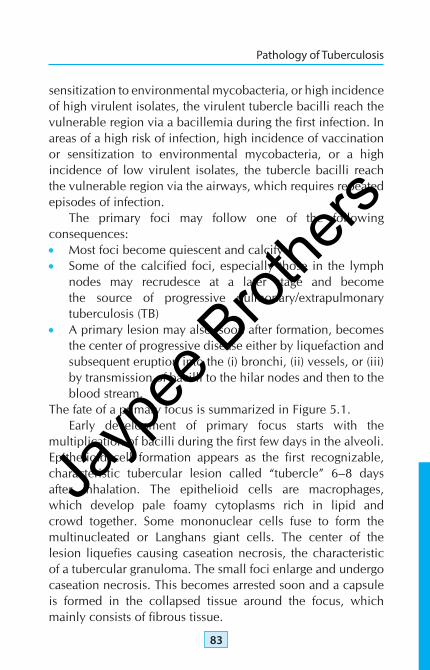
sensitization to environmental mycobacteria, or high incidence of high virulent isolates, the virulent tubercle bacilli reach the vulnerable region via a bacillemia during the first infection. In areas of a high risk of infection, high incidence of vaccination or sensitization to environmental mycobacteria, or a high incidence of low virulent isolates, the tubercle bacilli reach the vulnerable region via the airways, which requires repeated episodes of infection.
The primary foci may follow one of the following consequences:• Most foci become quiescent and calcify• Some of the calcified foci, especially those in the lymph
nodes may recrudesce at a later stage and become the source of progressive pulmonary/extrapulmonary tuberculosis (TB)
• A primary lesion may also, soon after formation, becomes the center of progressive disease either by liquefaction and subsequent eruption into the (i) bronchi, (ii) vessels, or (iii) by transmission of bacilli to the hilar nodes and then to the blood stream.
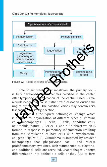
The fate of a primary focus is summarized in Figure 5.1.Early development of primary focus starts with the
multiplication of bacilli during the first few days in the alveoli. Epithelioid cell formation appears as the first recognizable, characteristic tubercular lesion called “tubercle” 6–8 days after inhalation. The epithelioid cells are macrophages, which develop pale foamy cytoplasms rich in lipid and crowd together. Some mononuclear cells fuse to form the multinucleated or Langhans giant cells. The center of the lesion liquefies causing caseation necrosis, the characteristic of a tubercular granuloma. The small foci enlarge and undergo caseation necrosis. This becomes arrested soon and a capsule is formed in the collapsed tissue around the focus, which mainly consists of fibrous tissue.
CHAPTERS 1 to 10.indd 83 24-03-2017 12:56:18
Jayp
ee B
rothe
rs
Clinic Consult Pulmonology: Tuberculosis
84
Three to six weeks after inhalation, the primary focus is fully developed and sometimes calcified in the center. After lymphocytic demarcation of the central caseous area, recrudescence may cause further fresh caseation outside the ring of lymphocytes. The calcified lesions may contain acid-fast bacilli on histologic section.
Granuloma is the typical pathological change which is a structural organization of different types of immune cells, macrophages, T cells, B cells, dendritic cells, neutrophils, natural killer cells, and a fibroblast which is formed in response to pulmonary inflammation resulting from the stimulation of host cells with mycobacterial antigens (Figure 5.2). Granuloma is initiated by resident macrophages that phagocytose bacilli and release proinflammatory cytokines, such as tumor necrosis factor-α, and additional cells are recruited. Macrophages undergo differentiation into epithelioid cells or they fuse to form
Figure 5.1 Possible course of a primary focus.
CHAPTERS 1 to 10.indd 84 24-03-2017 12:56:18
Jayp
ee B
rothe
rs

Pathology of Tuberculosis
85
multinucleated giant cells within the granuloma. Various cells mentioned above that take part in the reaction, are surrounded by a rim of lymphocytes including CD4 T cells of adaptive immune response. The bactericidal capacity of macrophages is enhanced by the release of interferon-γ. At a later stage, a tight cover of fibroblasts encloses the granuloma. The adapted cell mediated immune response and proper formation of granuloma determine the outcome of M. tuberculosis infection (Figure 5.3). Sarcoidosis is a condition where one can find granulomas. However in this condition, the granulomas are noncaseating, compact and naked and at times one may get inclusion bodies; whereas those due to tuberculosis are caseating with necrosis, they are ill-formed with intense inflammation. The two differentiating granuloas are seen in Figure 5.4.
REACTIVATED OR POST-PRIMARY TUBERCULOSIS
The primary lesion sometimes progresses and the pathologic changes will then be similar to those seen in reactivation TB. The main difference between the primary infection and
Figure 5.2 Granuloma and Langhan's giant cells. (For color version, see Plate 4)
CHAPTERS 1 to 10.indd 85 24-03-2017 12:56:19
Jayp
ee B
rothe
rs

Clinic Consult Pulmonology: Tuberculosis
86
reactivation is that, while in former the involvement of the regional lymph node is an essential component of pathology, in the latter, regional lymph node is not necessarily involved. Due to an early bacteremia following the formation of a primary lesion in the hilar glands, the bacilli are carried by the lymphatics to the right heart and then again into the lungs. These foci are called “Simon’s foci.” These are essentially the Post-primary lesions commonly seen in chronic disseminated and extrapulmonary TB, both on radiology and at autopsy. They also occur in a large number of primarily infected
A
B
Figure 5.3 Tuberculous granuloma. (For color version, see Plate 5)
CHAPTERS 1 to 10.indd 86 24-03-2017 12:56:19
Jayp
ee B
rothe
rs

Pathology of Tuberculosis
87
individuals as accidental findings. In case of primary intestinal TB, small calcified nodules may sometimes be seen in liver, which originate in a direct hematogenous transmission of the bacilli to the liver from the intestine via the portal vein.
Reactivated pulmonary TB is often seen in the upper lung zones and is limited in extent most frequently to the posterior segment of the upper lobe or apex of the lower lobe. The high alveolar partial pressure of oxygen in the upper zones relative to the other lung zones due to high ventilation-perfusion
Figure 5.4 Granuloma of tuberculosis and sarcoidosis. (For color version, see Plate 6)
B
A
CHAPTERS 1 to 10.indd 87 24-03-2017 12:56:19
Jayp
ee B
rothe
rs

Clinic Consult Pulmonology: Tuberculosis
88
predisposes to reactivation at these sites. Softening and liquefaction of the caseous material, which may discharge, into a bronchus with resultant cavity formation, follow proliferation of tubercle bacilli in the caseous center (Figure 5.5).
While 104 bacilli per gram are found in caseous tissue, up to 109 organisms may be harbored within a single cavity. Fibrous tissue forms around the periphery of such tuberculous lesions but is usually incapable of limiting extension of the tuberculous process. Hemorrhages may result from extension of the caseous process into vessels within the cavity walls.
Spread of caseous and liquefied material through the bronchial tree may disseminate the infection to other lung zones with or without the development of vigorous inflammatory exudates or tuberculous pneumonia. This type of reaction is sometimes due to a hypersensitivity reaction to tubercular protein released by the dead bacilli, and is called “epituberculosis.”
Rupture of a caseous pulmonary focus into a blood vessel may result in miliary TB with the formation of multiple
Figure 5.5 Fibrocaseous tuberculosis with cavitation (post-primary tuberculosis). (For color version, see Plate 6)
CHAPTERS 1 to 10.indd 88 24-03-2017 12:56:19
Jayp
ee B
rothe
rs

Pathology of Tuberculosis
89
0.5–2 mm tuberculous foci in the lung and in other organs of the body. Encroachment on the bronchi of pulmonary or lymph node caseous material may give rise to tuberculous bronchitis. Rupture of caseous glands into trachea or major bronchi causes collapse of lung or even sudden death by suffocation particularly in young children. Vasculitis secondary to TB affecting the affected vessels like pulmonary or cerebral is not an uncommon finding.Post-primary lesions can be divided into four main types:1. Nodular type
• Small• Large
2. Fibrocaseous type• With cavity• Without cavity
3. Miliary lesions4. Mixed nodular and fibrocaseous lesions.
All these lesions represent active tuberculous pathology meaning necrosis of varying extent with scanty to profuse epithelioid and giant cell reaction in the surrounding area.
Healed tuberculous lesions may also present as small nodules but are almost always located in the subpleural region which are completely fibrosed or show mixtures of calcification, ossification, and carbon pigment deposition. The frequency of active tuberculous disease was about 17% in autopsy series. Nodular lesions comprise a little more than half of all active cases in the lungs, the fibrocaseous variety about 45%, and the miliary almost 2%. The small nodule is 1 cm or less in diameter, has a relatively thick fibrocollagenous capsule which isolates the central area of necrosis from the almost normal appearing lung outside. The population of epithelioid and giant cells in these lesions is generally small and scattered. In contrast, large nodules which are greater than 1 cm in diameter but still
CHAPTERS 1 to 10.indd 89 24-03-2017 12:56:19
Jayp
ee B
rothe
rs
Clinic Consult Pulmonology: Tuberculosis
90
relatively well delineated from the neighboring lung parenchyma by a thin, irregular, and relatively inconspicuous capsule, shows much more prominent epithelioid cells and giant cell reaction surrounding a large area of central necrosis. Satellite small nodules may be present in the periphery of these large nodules. Special stains on these small nodules reveal preservation of the alveolar framework in the central necrotic area indicating that the basic process is one of the lobular pneumonia which has gone on to necrosis. The larger nodules can be found in close association with fibrocaseous lesion indicating a transition from the former to the latter.
The fibrocaseous and military lesions are self-explanatory. The frequency of demonstration of tubercle bacilli in the tissue section varies from 7, 29, and 75% in the small, large, and fibrocaseous type nodules, respectively. The corresponding figures in the microbiologic studies are 9, 36, and 77%, respectively. There is a good correlation between pathology and microbiology. Small nodules, which appear insignificant and inconspicuous, act as reservoirs, without producing any clinical symptom. It is potentially dangerous and under altered conditions like lowered host immunity, can break through the barrier producing more extensive tuberculous disease.
The evolution of disease will depend largely on the immune status of the individual at the time of invasion. In a nonimmune individual, an initial lobular pneumonia would most frequently lead on to necrosis, which in turn would produce the characteristic primary complex (Figure 5.6).
Rarely, the lobular pneumonia undergoes complete resolution to produce a scar. A primary complex would generally heal with fibrosis and calcification because of the development of immunity and will produce a healed nodule. Very rarely, when the immune competence is inadequate, progressive primary disease would lead to a larger complex, florid TB, extrapulmonary spread, or disseminated military TB.
CHAPTERS 1 to 10.indd 90 24-03-2017 12:56:19
Jayp
ee B
rothe
rs

Pathology of Tuberculosis
91
Figu
re 5
.6 N
atur
al h
isto
ry o
f clin
ical
tube
rcul
osis
.
CHAPTERS 1 to 10.indd 91 24-03-2017 12:56:20
Jayp
ee B
rothe
rs

Clinic Consult Pulmonology: Tuberculosis
92
In a partially immune individual, the initial lobular pneumonia would most frequently lead on to either a nodular form or a fibrocaseous type of TB. Very rarely, complete resolution would occur. The nodular lesion is usually the small nodular type, which may completely heal and like the primary complex, may lead to a healed nodule. Some of the nodules, large or small, may progress to form fibrocaseous TB. A primary complex can convert into an active nodule. Fibrocaseous form of the disease frequently cavitates but a small proportion may continue as such. These fibrocaseous lesions are caused by low immunity status of the individual and might lead to extensive form of the disease in the lungs, to extrapulmonary sites, and disseminated miliary TB. Figure 5.6 represents the natural history of pulmonary TB.
The progression of the granulomatous response in mice infected with M. tuberculosis follows five distinct immuno-pathologic stages:1. Mild scattered2. Moderate3. Moderate granulomatous4. Moderately coalescing5. Extensive.
�� Following infection with M. tuberculosis, the course of tuberculosis is variable. Either it will lead to a stable and healed lesion or can remain quiscent for a varying period of time till it will progress to disease depending upon the host immunity; or it may be a progressive disease. The disease either called as a primary or post-primary lesion. Demonstration of caseating granuloma is the hall mark of tubercular pathology; however it needs to be differentiated from other granulomatous lesions like sarcoidosis. Granuloma of both these diseases, of course, are different.
kEy mEssAgE
CHAPTERS 1 to 10.indd 92 24-03-2017 12:56:20
Jayp
ee B
rothe
rs

Pathology of Tuberculosis
93
SUGESTED READINGS 1. Co DO, Hogan LH, Kim SI, Sandor M. Mycobacterial granulomas:
keys to a long-lasting host-pathogen relationship. Clin Immunol. 2004;113:130-6.
2. Cooper AM. The protective immune response to Mycobacterium tuberculosis. Curr Opin Immunol. 1995;7:512-6.
3. Cutler RR, Baithun SI, Doran HM, Wilson P. Association between the histological diagnosis of tuberculosis and microbiological findings. Tubercle Lung Dis. 1994;75:75-9.
4. Epstein WI. Granulomatous hypersensitivity. Prog Allergy. 1967;11:36-88.
5. Gideon HP, Flynn JL. Latent tuberculosis: what the host “sees”? Immunol Res. 2011;50:202-12.
6. Lillebaek T, Dirksen A, Baess I, Strunge B, Omsen VO, Andersen AB. Molecular evidence of endogenous reactivation of Mycobacterium tuberculosis after 33 years of latent infection. J Infect Dis. 2002;185:401-4.
7. Lin PL, Flynn JL. Understanding latent tuberculosis: a moving target. J Immunol. 2010;185:15-22.
8. Nayak NC. Nature and evolution of pulmonary tuberculosis. J AIIMS. 1976;1:190-4.
9. Rhoades ER, Frank AA, Orme IM. Progression of chronic pulmonary tuberculosis in mice aerogenically infected with virulent Mycobacterium tuberculosis. Tubercle Lung Dis. 1997;78:57-66.
10. Saunders BM, Cooper AM. Restraining mycobacteria: role of granulomas in mycobacterial infections. Immunol Cell Biol. 2000;78:334-41.
11. Seal RM. The pathology of tuberculosis. Br J Hosp Med. 1971;5:783. 12. Vynnycky E, Fine PE. Lifetime risks, incubation period, and serial
interval of tuberculosis. Am J Epidemiol. 2000;152:247-63.
CHAPTERS 1 to 10.indd 93 24-03-2017 12:56:20
Jayp
ee B
rothe
rs