CLIN ICAL EFFICIENCY OF 2% CHLORHEXIDINE GEL IN … · CLIN ICAL EFFICIENCY OF 2% CHLORHEXIDINE GEL...
78
CLINICAL EFFICIENCY OF 2% CHLORHEXIDINE GEL IN REDUCING INTRACANAL BACTERIA Ching Shan Wang, DDS A thesis submitted to the faculty of the University of North Carolina at Chapel Hill in partial fulfillment of the requirements for the degree of Master of Science in the School of Dentistry (Endodontics). Chapel Hill 2007 Approved by: Fabricio B. Teixeira, DDS, PhD Martin Trope, DMD, MS Roland R. Arnold. PhD Eric Rivera, DDS, MS
Transcript of CLIN ICAL EFFICIENCY OF 2% CHLORHEXIDINE GEL IN … · CLIN ICAL EFFICIENCY OF 2% CHLORHEXIDINE GEL...

CLINICAL EFFICIENCY OF 2% CHLORHEXIDINE GEL IN REDUCINGINTRACANAL BACTERIA
Ching Shan Wang, DDS
A thesis submitted to the faculty of the University of North Carolina at Chapel Hill in partialfulfillment of the requirements for the degree of Master of Science in the School of Dentistry(Endodontics).
Chapel Hill2007
Approved by:
Fabricio B. Teixeira, DDS, PhD
Martin Trope, DMD, MS
Roland R. Arnold. PhD
Eric Rivera, DDS, MS

ii
©2007Ching Shan Wang
ALL RIGHTS RESERVED
iii
ABSTRACT
CHING S. WANG: Clinical Efficiency of 2% Chlorhexidine Gel in Reducing IntracanalBacteria.
(Under the direction of Dr. Fabricio B. Teixeira)
This study evaluated the clinical efficacy of 2% chlorhexidine (CHX) gel on
intracanal bacteria reduction during root canal instrumentation. The additional antibacterial
effect of an intracanal dressing (Ca(OH)2 mixed with 2% CHX gel) was also assessed.
Forty-three patients with apical periodontitis were recruited. Teeth were instrumented using
rotary instruments and 2% CHX gel as the disinfectant solution. Bacterial samples were
taken upon access (S1), after instrumentation (S2), and following 2 weeks of intracanal
dressing (S3). Anaerobic culture was performed. Of the samples cultured positively at S1,
10.3% (4/39) and 8.3% (4/36) sampled bacteria at S2 and S3, respectively. A significant
difference in the percentage of positive culture between S1 and S2 (p<0.001), but not
between S2 and S3 (p=0.692), was found. These results suggest that 2% CHX gel is an
effective root canal disinfectant and additional intracanal dressing did not significantly
improve the bacteria reduction on the sampled root canals.
iv
ACKNOWLEDGMENT
I would like to take this opportunity to recognize the following:
Dr. Fabricio B. Teixeira, my mentor, who always made time in his busy schedule to listenand help.
Dr. Martin Trope and Eric Rivera for their leadership, enthusiasm, and guidance over the pastthree years in my education and research.
Dr. Rolald Arnold for his insightful ideas and his vast knowledge of everything.
Eric Simmons for his help in the microbiology lab.
Jose Luis, Jason, Derek, Peter, and Anna, my co-residents. I will miss you next year.
Shu-Tang and Shu-Li Wang, my parents, who have always been there for me with love andencouragement.
Jeanne Wong, my girlfriend, who gave unconditional love and support for last 3 years. I loveher very much.

v
TABLE OF CONTENTS
LIST OF TABLES……………………………………………………………………….......vii
LIST OF FIGURES…………………………………………………………………………viii
LIST OF ABBREVIATIONS………………………………………………………………...ix
Chapter
1. INTRODUCTION…………………………………………………………………….1
a. Apical Periodontitis…………………………………………………………...1
b. Endodontic Therapy…………………………………………………………...4
c. Sodium hypochlorite…………………………………………………………10
d. Smear layer removing agent…………………………………………………13
e. Calcium hydroxide…………………………………………………………...15
f. Chlorhexidine………………………………………………………………...16
2. PURPOSE…………………………………………………………………................20
3. MATERIALS AND METHODS………………………………………………….....21
a. In Vitro Evaluation of 2% CHX Gel Neutralizer…………………………….21
b. Subject Recruitment and Qualification………………………........................23
c. Treatment Group Assignment………………………………………………..24
d. Bacterial Sampling…………………………………………………………...24
e. Data Analysis………………………………………………………………...28
vi
4. RESULTS……………………………………………………………………………29
a. In Vitro Evaluation of 2% CHX Gel Neutralizer…………………………….29
b. In Vivo Evaluation of the Clinical Efficacy of 2% CHX Gel………………..29
5. DISCUSSION………………………………………………………………………..31
6. CONCLUSION……………………………………………………………………....38
7. SUMMARY……………………………………………………………………….…39
APPENDIX I: Informed consent example…………………………………………………...41
REFERENCES……………………………………………………………………………....57
vii
LIST OF TABLES
Table
1. Description of samples………………………………………………….........46
2. Apical preparation size corresponding to canal and tooth type……………...46
3. CFU counts for each sample………………………………………………....47
4. Percentage of samples with negative culture………………………………...48

viii
LIST OF FIGURES
Figure
1. Initial, Pre-Instrumentation Sample (S1)………………………………..…...49
2. Post-Instrumentation Sample (S2)………………………………………..….50
3. Post-Dressing Sample (S3)………………………………………………..…51
4. The Sample Distribution at S1…………………………………………….....52
. 5. The Sample Distribution at S2…….…………………...………………...…..53
6. The Sample Distribution at S3……………………………………………….54
7. Percentage of samples with positive culture…………………………………55
8. Comparison of percentage of canals with positive culture at different
culturing points from selected studies………………………………………..56

ix
ABBREVIATIONS
Ca(OH)2 Calcium Hydroxide
CHX Chlorhexidine
CFU Colony Forming Unit
EDTA Disodium ethylendiamine-tetraacetate
NaOCl Sodium Hypochlorite
NiTi Nickel-Titanium
SS Stainless-Steel
RPM Revolutions Per Minute
LDTM Liquid Dental Transport Medium
CHAPTER 1
INTRODUCTION
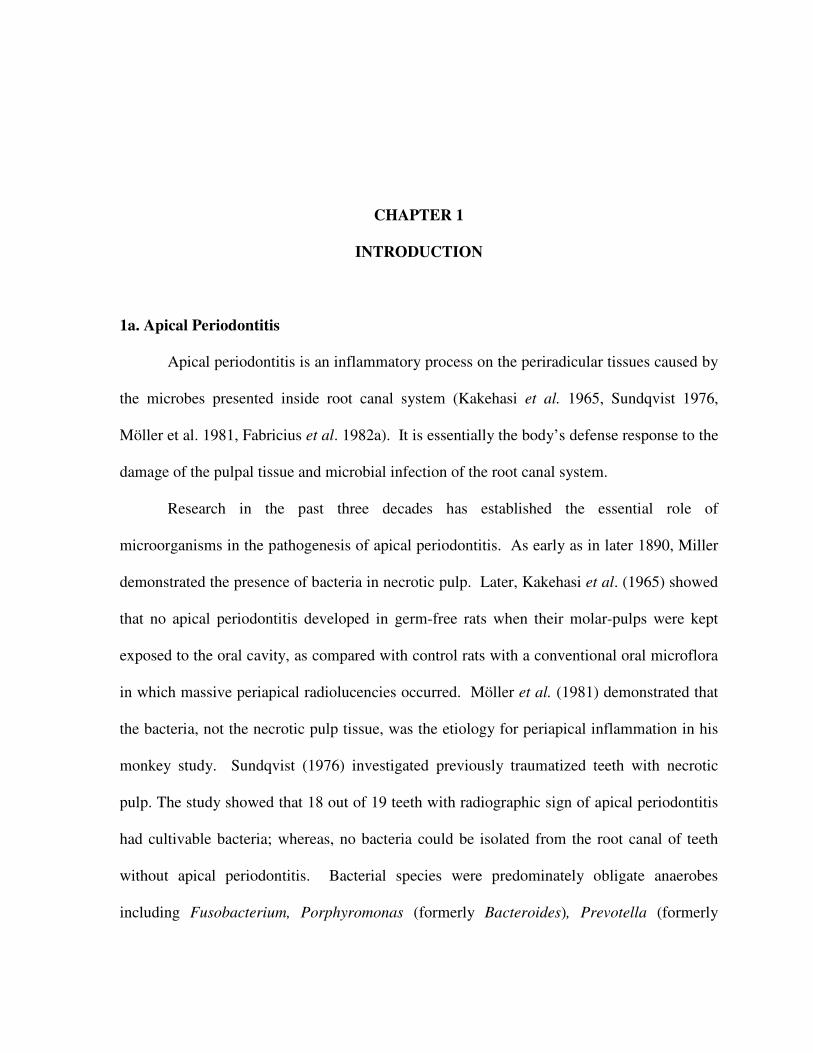
1a. Apical Periodontitis
Apical periodontitis is an inflammatory process on the periradicular tissues caused by
the microbes presented inside root canal system (Kakehasi et al. 1965, Sundqvist 1976,
Möller et al. 1981, Fabricius et al. 1982a). It is essentially the body’s defense response to the
damage of the pulpal tissue and microbial infection of the root canal system.
Research in the past three decades has established the essential role of
microorganisms in the pathogenesis of apical periodontitis. As early as in later 1890, Miller
demonstrated the presence of bacteria in necrotic pulp. Later, Kakehasi et al. (1965) showed
that no apical periodontitis developed in germ-free rats when their molar-pulps were kept
exposed to the oral cavity, as compared with control rats with a conventional oral microflora
in which massive periapical radiolucencies occurred. Möller et al. (1981) demonstrated that
the bacteria, not the necrotic pulp tissue, was the etiology for periapical inflammation in his
monkey study. Sundqvist (1976) investigated previously traumatized teeth with necrotic
pulp. The study showed that 18 out of 19 teeth with radiographic sign of apical periodontitis
had cultivable bacteria; whereas, no bacteria could be isolated from the root canal of teeth
without apical periodontitis. Bacterial species were predominately obligate anaerobes
including Fusobacterium, Porphyromonas (formerly Bacteroides), Prevotella (formerly
2
Bacteroides), Eubacterium, Peptococcus, Peptostreptococcus and Camylobacter.
Baumgartner and Falkler (1991) demonstrated that teeth with carious pulpal exposures and
periapical lesions were predominately colonized by anaerobic bacteria in the apical 5mm.
The endodontic environment provides a selective environment for establishment of a
mixed root canal flora. It is predominated by Gram-negative anaerobic bacteria, due to the
lack of oxygen and the availability of host tissues and tissue fluids as the primary nutrient
source (Sundqvist et al. 1992). In a monkey study, Fabricius et al. (1982a) inoculated
indigenous oral flora into root canal system and sealed the canals for a period of 1080 days.
In the early stage, the root canal flora was predominately colonized by facultative bacteria.
Shortly, strict anaerobes displaced the facultative bacteria. After 1080 days, 98% of bacteria
isolated from the root canal system were strict anaerobes. In another study, Fabricius et al.
(1982b) showed the mixed infections had the greatest capacity to induce apical periodontitis.
He studied the ability of 11 bacterial strains, in various combinations, to induce periapical
reaction in a monkey model. Other than Enterococcus, all other strains cannot survive as
pure culture in the root canal system. The facultative anaerobe identified in most samples
can only induce weak periapical reaction, while the combination of facultative and strict
anaerobes can induce profound periapical inflammation. Based on those studies, it appeared
there is selective pressure by the endodontic environment that favors the survival of strict
anaerobes. In addition, certain combinations of bacteria are required to induce apical
periodontitis, suggesting positive, mutually supportive, as well as negative interactions
between bacterial species.
Bacteria can potentially penetrate dentinal tubules and protect themselves from the
mechanical and chemical destruction during the root canal therapy. Ando and Hoshino

3
(1990) demonstrated the presence of bacteria 500 to 2000µm in dentinal tubules in carious
human teeth by light microscopy. Love et al. (1996) showed bacteria invaded and penetrated
deeper in cervical and mid root than the apical root. In some case, bacteria can potentially
penetrate nearly to the cementum layer of the roots in teeth with apical periodontitis (Peters
et al. 2001). Matsuo et al. (2003) also demonstrated that 70% of tubules had bacteria
penetrated up to cementum using immunohistochemistry methods.
Bacterial species found in the dentinal tubules are similar to those found in the main
root canal system. Ando and Hoshino (1990) showed the predominate bacteria found in the
dentinal tubules was Lactobacillus, Streptococcus, Propionibacterium, and
Peptostreptococcus. Matsuo et al. (2003) showed the presence of Fusobacterium,
Eubacterium, Lactobacillus and Peptostreptococcus in the dentinal tubules.
Due to the anatomic restriction, bacteria in the tubules are difficult to remove by
instrumentation and irrigation. Even with intracanal medications, viable bacteria were found
in infected human dentin tubules after the treatment (Weiger et al. 2002). Although the
clinical significance of bacteria in dentinal tubules is still not clear, studies tend to agree that
the remaining bacteria in the tubules after the root canal treatment can contribute to failure of
endodontic therapy (Love and Jenkinson 2002, Oguntebi et al. 1994, Peters et al. 2001).
In addition to bacteria, fungi and viruses were also detected in the infected root canal
system. In teeth with apical periodontitis, the presence of fungi has been demonstrated in
dentinal tubules (Sen et al. 1995), failing root canal treatment (Molander et al. 1998), and in
periapical tissues (Tronstad et al. 1987). Prevalence of fungi in infected root canal system
ranges from 0.5% to 26% in untreated root canals (Baumgartner et al. 2000) and 3.7% to
33% in cases of failed root canal treatment (Molander et al. 1998, Waltimo et al. 1997).
4
Most commonly isolated fungus is Candida albicans (Waltimo et al. 1997). At present, the
role of fungi in the pathogenesis of endodontic diseases is still not clear. Similarly, the
presence of virus in the infected root canal system has only shown in case reports (Glick et
al.1989) and their role in the development of apical lesion is not known.
Taken together, studies strongly support the primary role of bacteria in the
development of apical periodontitis and the infected root canal flora is polymicrobial and
predominately anaerobic.
1b. Endodontic Therapy
The goal of endodontic treatment is to prevent and eliminate apical periodontitis.
This objective is accomplished through disinfection of the root canal space and adequate root
canal filling to prevent recontamination. The endodontic treatment protocol should be based
on sound biologic rationale for more effective clinical management of the disease.
Long-term endodontic success for the teeth with apical periodontitis ranges from 75%
to 90% (Stringberg et al. 1956, Kerekes and Tronstand 1979, Byström et al. 1987, Sjogren et
al. 1990, Trope et al. 1999, Weiger et al. 2000, Peters & Wesslink 2002) with functional
success rate of 95% (Byström et al. 1987, Weiger et al. 2000, Peters & Wesslink 2002).
Endodontic success (healed) is defined as the absence of clinical symptoms and radiographic
resolution of periapical lesion (Stringberg et al. 1956).
In a retrospective study with 6-month to 5-year follow-up, 478 teeth endodontically
treated by undergraduate students were evaluated (Kerekes and Tronstand 1979). Overall
success rate (both vital and non-vital cases) was 83% at first year, then, increased to 91% at
3-5 year follow-up. In cases of apical periodontitis, the success rate was 85%. Other

5
retrospective studies also demonstrated similar success rate for the teeth with apical
periodontitis. Stringberg et al. (1956) showed 80% healed at 6-month to 10-year follow-up.
Sjögren et al. (1990) had 86% success rate with 8-10 year follow-up period. Toronto study
(Farzaneh et al. 2004) showed 79% success rate at 4-6 year follow-up.
Prospective studies also showed comparable success rate. Sjögren et al. (1997)
examined the 5-year outcome study on 53 single-rooted anterior teeth with radiographic
periapical lesion. Overall success rate was 83%. In another prospective study, 79
endodontically treated teeth with apical periodontitis were followed up for 2 to 5 years
(Byström et al. 1987). Eighty five percent (85%) of the cases were healed completely and
9% of the cases were healing, with functional success rate of 94%. In agreement with this
finding, other studies also showed about 95% functional success rate for endodontically
treated teeth (Weiger et al. 2000, Peters & Wesslnik 2002).
In contrast to the university-based study, cross-sectional studies showed much lower
success rate. Buckely and Spangberg (1995) assessed the prevalence of radiographically
detectable periapical disease and its relationship to the quality of endodontic care in a North
American population. The authors examined 208 randomly chosen full-mouth x-ray (FMX)
series with 5,272 teeth taken at the initial screening. They found 31.3% of root canal filled
teeth had periapical disease and only in about 40% of cases was technically satisfactory root
canal treatment achieved. Similarly, Ray and Trope (1995) evaluated 1,010 endodontically
treated teeth from FMX series with minimal of 1 year follow-up. Overall, they found 61% of
the teeth had no radiographic sign of apical periodontitis. Lower success rate observed in
cross-sectional studies is likely due to the poor quality of endodontic care performed in
private practice setting.
6
Long term success of endodontic therapy depends on many factors including tooth
vitality, level of root-canal disinfection, length and quality of root canal filling, and coronal
restoration (Stoll et al. 2005, Schaeffer et al. 2005, Caplan & Weintraub 1997, Sorensen et
al. 1984, 1985, Vire et al. 1991, Aquilino & Caplan 2002, Ray & Trope 1995).
Outcome studies of root canal treatment consistently showed higher long-term
success rate for the teeth without apical periodontitis than the teeth with apical periodontitis
(Sjögren et al. 1990, Kerekes & Tronstad 1979, Stringberg et al. 1956, Stoll et al. 2005).
Teeth with vital pulp have long-term success rates of 95-97%, while teeth with apical
periodontitis range from 75% to 90% (Stringberg et al. 1956, Kerekes and Tronstand 1979,
Byström et al. 1987, Sjögren et al 1990, Trope et al. 1999, Weiger et al. 2000, Peters &
Wesslink 2002). The observed difference in the outcome between these two groups is due to
the ability of operators to disinfect root canal system. In case of vital pulp, root canal space
is not infected with microorganism but filled with inflamed pulp tissue (Seltzer and Bender
1963, Mumford et al. 1967, Dummer et al. 1980). Hence, it is more likely to achieve a root
canal space free of bacteria assuming the treatment is performed under strict aseptic protocol.
On the other hand, teeth with apical periodontitis are usually associated with necrotic pulp
infected with obligate and facultative bacteria (Sundqvist 1976, Möller et al. 1981, Fabricius
et al. 1981). In such case, complete elimination of bacteria from the root canal space is
limited due to the complexity of root canal anatomy and resistant bacteria.
Sjögren et al. (1997) investigated the role of infection on the prognosis of endodontic
therapy by following-up teeth that had their canals cleaned and obturated during a single
appointment. In this study, the root canals of 55 single rooted teeth with apical periodontitis
were thoroughly instrumented and irrigated with NaOCl solution. Periapical healing was

7
followed up for 5 years. Complete healing occurred in 94% of cases that yielded negative
culture. Where the samples were positive prior to root filling, the success rate of treatment
was just 68%. Byström et al. (1987) also demonstrated 95% healing when the root canal
filled after the negative culture. Other studies also demonstrated similar results (Zeldow &
Ingle 1963, Engström et al. 1964). Hence, the presence of bacteria prior to root canal filling
is a negative predictor for the long term success of root canal treatment.
Root canal disinfection can be accomplished by mechanical and chemical means.
Mechanical microbial control phase involves root canal instrumentation using hand and/or
rotary instruments. In contrast, chemical microbial control phase involves antimicrobial
agents during and after instrumentation.
Mechanical instrumentation can reduce intra-canal bacteria significantly but can not
predictably eradicate them. Byström and Sundqvist (1981) evaluated the efficacy of the
mechanical instrumentation in bacterial reduction during root canal treatment. Fifteen single-
rooted teeth with apical periodontitis were treated in five visits with no inter-appointment
dressing. Root canals were instrumented with hand files and with physiological saline as
irrigant. Mechanical instrumentation with saline reduced the number of bacteria
significantly, by 100-1000 fold. However, despite being treated 5 times, bacteria can not be
predictably eliminated from the root canal system. They also showed that 0.5% NaOCl was
more effective than physiological saline when used as irrigant. Other studies also support
this finding (Siqueira et al. 2000, Byström & Sundqvist 1985, Dalton et al. 1998, Shuping et
al. 2000)
On average, 40% - 60% of root canals have no cultivable bacteria after mechanical
root canal instrumentation with NaOCl solution (Dalton et al. 1999, Shuping et al. 2001,

8
Card et al. 2002, McGurkin-Smith et al. 2005, Kvist et al. 2004). Shuping et al. (2000)
evaluated the extent of bacterial reduction with NiTi rotary instrumentation and 1.25%
NaOCl irrigation in 42 patients with apical periodontitis. Bacterial samples were taken
before and after the instrumentation, and they were cultured anaerobically for 7 days. The
authors found 62% of canals were rendered bacteria-free after instrumentation with 1.5%
NaOCl. McGurkin-Smith et al. (2005) obtained 40% of canals sampled bacteria free after
instrumentation and a strict irrigation protocol using 5.25% NaOCl and EDTA.
Another method to improve root canal disinfection is to enlarge the apical preparation
(Dalton et al. 1999, Siqueira et al. 1999, Shuping et al. 2000, Card et al. 2002, Baugh &
Wallace 2005). Dalton et al. (1999) demonstrated that regardless of technique (hand file or
rotary file), uniform reduction of bacteria occurred with progressive filing. Siqueira et al.
(1999) showed the same result from in vitro study. In a clinical study, Card (2002) was able
to demonstrate 100% of canine and premolar, and 89% of molars rendered bacteria free after
preparing apical size to #60 and #80 using LightSpeed instruments. In additional to
mechanical reduction of bacteria, apical enlargement also allows better access for the irrigant
to reach the apical region and disinfect the bacterial contents (Shuping et al. 2000, Salzgeber
et al. 1977, Ram et al. 1977, Chow et al. 1983, Siqueira et al. 1999). Studies have shown
that canals need to be enlarged to at least #35 file for adequate irrigation to reach apical third.
Ram et al. (1977) concluded that canals need to be enlarged to a #40 file size so that
maximum irrigation is in contact with the apical debris. Chow et al. (1983) also showed the
canal system had to be instrumented to at least #40 file for proper irrigation. Shuping et al.
(2000) and Siqueira et al. (1999) later confirmed the findings that NaOCl requires a certain
size canal to become beneficial in bacterial reduction.
9
Nonetheless, eradication of endodontic infection by instrumentation and NaOCl
solution alone is limited (Byström& Sundqvist 1981, 1985, Sjögren et al. 1991, Dalton et
al.1999, Shuping et al. 2000, Card et al. 2002). To obtain a more predictable microbial
control, placement of intracanal medicaments such as calcium hydroxide (Ca(OH)2) is
recommended (Ørstavik et al. 1991, Shuping et al. 2000, Sjogren et al. 1991). Intracanal
medicaments are used to: (a) eliminate bacteria in the root canal; (b) prevent bacterial
proliferation between appointments; and (c) act as a physicochemical barrier, preventing
root-canal reinfection and nutrient supply to the remaining bacteria (Byström & Sundqvist
1985). Calcium hydroxide is one of most widely used intracanal medications in endodontics
today and remains the best medicament available to reduce residual microbial flora (Law et
al. 2004). It is a strong alkaline substance, which has a pH of approximately 12.5. In an
aqueous solution, calcium hydroxide dissociates into calcium and hydroxyl ions that will lead
to a lowered oxygen tension and an increase in the pH in the inflamed periapical tissues
(Siqueira et al. 1999). With a high pH, calcium hydroxide has an excellent broad
antimicrobial effect.
To support this, studies consistently demonstrated that Ca(OH)2 can help to further
eliminate surviving bacteria in the root canals. Sjögren et al. (14) showed that bacteria were
not found in teeth with apical periodontitis dressed with Ca(OH)2 for 1 wk. Ørstavik et al.
(1991) demonstrated that extensive apical reaming and 1 week dressing with Ca(OH)2
significantly reduced bacterial growth from root canal and apical dentin samples. Shuping et
al. (2000) showed that placement of Ca(OH)2 for at least 1 week rendered 92.5% of canals
bacteria free in teeth with apical periodontitis. Law et al. (2004) reviewed the literature
evaluating the antibacterial effectiveness of Ca(OH)2 used in the management of apical
10
periodontitis and found 27% of canals showed detectable bacteria growth after medication.
However, Ca(OH)2 has been shown to be ineffective when used as a short-term dressing (< 1
d) (Sjögren et al. 1991, Safavi et al. 1990) and requires at least 1 week to properly exert its
antibacterial action. Thus, a minimum of two-visit root canal treatment is strongly
recommended for adequate intracanal disinfection in cases of apical periodontitis (Trope et
al. 1991).
1c. Sodium Hypochlorite (NaOCl)
Sodium hypochlorite was first recommended as an antiseptic solution by Henry
Dakin (1915) to irrigate open wounds during World War I. Later, Coolidge (1919)
introduced NaOCl to endodontics and it has remained the most used irrigant.
NaOCl has several characteristics that make this solution a desirable endodontic
irrigant, including its antimicrobial (Byström & Sundqvist, 1983, 1985, Siqueira et al. 2000,
Zehnder et al. 2002), tissue dissolving (Thé et al. 1980, Moorer & Wesselink1982, Zehnder
et al. 2002) and lubricating properties.
NaOCl has a broad-spectrum antimicrobial activity (Byström & Sundqvist 1983). It
can kill vegetative bacteria, spore-forming bacteria, fungi, protozoa, and viruses (Rutala &
Weber 1997). Effectiveness of NaOCl as intracanal antimicrobial agent has been well
documented.
There have been conflicting results concerning the antimicrobial efficacy of different
NaOCl concentrations. In general, in vitro studies (Siqueira et al. 1998, Yesilsoy et al. 1995)
showed that 5.25% NaOCl is more effective than lower concentration solution in eliminating
bacteria. In contrast, clinical studies found no difference in the antimicrobial effect between
0.5% and 5% NaOCl (Byström & Sundqvist 1985, Cvek et al. 1976). This disparity between

11
in vitro and clinical studies is likely attributable to the differences in methodology. In vitro
studies for testing antimicrobial properties of a solution are often carried out in test tubes or
agar plates, which do not take into consideration of root canal complexity. To eliminate
bacterial cells within the root canal system, irrigants must be able to reach them. Many areas
in the root canal system have the potential to harbor microorganisms, thus making them
inaccessible to the effects of chemomechanical preparation. In case of using weaker NaOCl
solution, many authors recommended frequent replenishing of the irrigants in order to
compensate for the quick neutralization of NaOCl (Byström & Sundqvist 1985, Siqueira et
al. 2000).
The tissue dissolving ability of NaOCl has been well investigated. Grossman and
Meiman (1941) showed that chlorinated soda (solution containing NaOCl and NaCl) was an
effective solvent of pulp tissue in an in vitro study. It was found to dissolve the pulps of
freshly extracted teeth in less than 24 hours. Since then, numerous studies have been
published on the tissue dissolution property of NaOCl when used as endodontic irrigant
(Moorer & Wesselink 1982, Thé et al. 1979, Zender et al. 2002). There are several factors
thought to be important for the efficacy of NaOCl irrigant in dissolving pulp tissue. In an in
vitro study, Moorer & Wesselink (1982) investigated the effect of fluid flow, pH and
available surface area of tissue on the tissue dissolving capability of NaOCl. The authors
concluded that the tissue-dissolving power of NaOCl solution appeared to be strongly
depended on (1) the amount of organic matter in the hypochlorite/tissue system (2) the
frequency and intensity of mechanical agitation (fluid flow) and (3) the available surface area
of free or enclosed tissue. For hypochlorite solution to be effective, it should act quickly and
be in an excess in relation to the amount of organic material that was to be digested. In
12
addition, the authors suggested that it was the amount of NaOCl that was important for the
tissue dissolution rather than the concentration. Hence, they recommend the continuous and
intensive mechanical debridement of the canal system and repeated refreshment of the
NaOCl. These authors also showed the importance of mechanical agitation on tissue
dissolving ability of NaOCl, particularly ultrasonic agitation shown to be very effective.
Although higher concentrations of NaOCl show more effective tissue dissolving
properties on a fixed surface area (Zehnder et al. 2002, Moorer & Wesselink 1982), it is not
true when there are only small amounts of tissue (Moorer & Wesselink 1982). Baumgartner
and Cuenin (1992) showed that NaOCl in concentration of 1%, 2.5%, and 5.25% completely
removed pulpal remnants and predentin from uninstrumented dentinal walls. Thus it appears
that low concentration of NaOCl retains adequate tissue dissolving and antibacterial
properties.
Cytotoxicity of NaOCl is a major disadvantage for its clinical use. Pashley et al.
(1985) had tested the cytotoxicity of various dilutions of sodium hypochlorite on 3
independent biological models (hemolysis of RBC, rabbit eye experiments, and intradermal
injections). All dilution caused hemolysis, moderate to severe irritation to rabbit eyes, and
skin ulcertation. It was found that higher concentration NaOCl causes more cytotoxic effects
on the vital tissue. The authors concluded that 5% NaOCl must be used judiciously and with
great caution to prevent it from reaching the periapex where it could elicit severe
inflammatory reactions. Spangberg et al. (1973) suggested that 5.25% NaOCl was
considerably stronger than necessary to eliminate bacterial strains commonly found in
infected root canals. At this concentration, the author believed NaOCl was considered too
cytotoxic to be routinely used in endodontic therapy.

13
Taken together, low concentration of NaOCl is sufficient to dissolve pulp tissue and
eliminate bacteria in the root canal system. Clinical use of full-strength NaOCl should be
avoided due to its strong cytotoxicity.
1d. Smear Layer Removing Agents
Current methods of root canal instrumentation produce a smear layer on the surface
of canal walls (McComb & Smith 1975, Goldman et al. 1988, Yamada et al. 1983,
Torabinejad et al. 2002). Under Scanning Electronic Microscopy (SEM), this layer has
amorphous, irregular, and granular appearance that can penetrate into and occlude the
dentinal tubules (McComb & Smith 1975). It contains both inorganic and organic substances
that include dentine, remnant of pulp tissue, odontoblastic processes, and microorganisms
(Torabinejad et al. 2002).
In infected root canals, bacteria, predominantly Gram-negative anaerobes, can be
identified from the root canal system and in the dentinal tubules (Ando & Hoshino 1990, Sen
et al. 1995, Love & Jenkinson 2002). Viable bacteria were isolated from the infected
dentinal tubules of root canal system in extracted human teeth (Peters et al. 2001).
Histological and SEM studies showed bacteria can penetrate into the dentinal tubules at
various distances from the surface of root canal wall (Ando & Hoshino 1990, Sen et al. 1995,
Peters et al. 2001). In some case, bacteria can potentially penetrate to the cementum layer
(Peters et al. 2001). Under in vitro conditions, several investigators have successfully
infected the dentinal tubules with microorganisms (Haapasalo & Ørstavik 1987, Siqueira et
al. 1996, Perez et al. 1993). Hence the available evidence strongly suggests that viable
bacteria can invade the dentinal tubule during root canal infection process.

14
The presence of smear layer after root canal instrumentation raises some concerns.
Several investigators have found that the smear layer itself may be infected and may protect
bacteria already in the dentinal tubules (McComb & Smith 1975, Haapasalo & Ørstavik
1987). It has been shown that the smear layer is not a complete barrier to bacteria and it
delays but does not abolish the action of endodontic disinfectants (Ørstavik & Haapasalo
1990). Clinically, whether bacteria in the dentinal tubule can be predictably eliminated by
the disinfectants, in the presence of smear layer, is questionable. The remaining viable
bacteria in the dentinal tubule may be responsible for the persistent endodontic infection after
root canal therapy (Stuart et al. 2006). Hence, it is prudent to remove the initially created
smear layer in infected root canals and to allow penetration of endodontic disinfectants into
the dentinal tubule more effectively (Torabinejad et al 2002).
The smear layer of root canal walls also act as a physical barrier interfering with
adhesion and penetration of sealers into dentinal tubules (Abramovich & Goldberg 1976,
Tidmarsh 1978, White et al. 1984). Studies have shown that root canal sealers can penetrate
into dentinal tubules in the absence of smear layer; whereas, no penetration was observed
with the smear layer intact (Gutiérrez et al. 1990, Pallarés et al. 1995, Kouvas et al. 1998).
This characteristic is particularly important for some root canal filling materials such as
Resilon (Shipper et al. 2004) and many other resin-based sealers. Removal of smear layer
prior to root canal filling has also shown to reduce microleakage (Pashley & Depew 1986,
Taylor et al. 1997, Timpawat et al. 2001).
EDTA (Ethylenediaminetetracetic acid) is a chelating agent, commonly used for
removal of smear layer from the instrumented root canal walls (McComb & Smith 1975,
Yamada et al. 1983, Baumgartner & Mader 1987). Since smear layer is composed of both

15
organic and inorganic materials, the combination of EDTA and sodium hypochlorite
(NaOCl) is often recommended (Goldman et al. 1988, Yamada et al. 1983, Baumgartner &
Mader 1987). In addition to EDTA, citric acid can also be used (Baumgartner et al. 1984).
Recently, new solutions have been proposed with enhanced antimicrobial properties in
addition to their smear layer removal ability such as BiopureTM MTAD (Torabinejad 2003a,
Torabinejad 2003b) and SmearClearTM. Although their abilities to remove smear layer are
well demonstrated, antimicrobial efficiency of BiopureTM MTAD has been questioned (Kho
and Baumgartner 2006, Baumgartner et al. 2007)
1e. Calcium hydroxide (Ca(OH)2)
Calcium hydroxide was introduced in endodontic therapy by Hermann in 1920 and
often recommended for disinfection of the root canal space in teeth with apical periodontitis.
The effectiveness of interappointment Ca(OH)2 has been well investigated. Byström
et al. (1985) reported that Ca(OH)2 was an effective intracanal medicament rendering 34 out
of 35 canals bacteria free after 4 weeks. Sjögren et al. (1991) who demonstrated that a 7-day
dressing with Ca(OH)2 eliminated all bacteria in the root canal but it is ineffective when used
for short-term. Shuping et al. (2000) also demonstrated the effectiveness of Ca(OH)2 as root
canal disinfectant when used for > 1 week.
Even though Ca(OH)2 is a good antimicrobial agent, it is ineffective against some of
microorganisms such as Enterococcus faecalis (Sjögren et al. 1991, Haapasalo and Ørstavik
1987, Heling et al. 1992), which is found in the case of persistent root canal infection.
Byström et al. (1985) showed the most resistant strain to Ca(OH)2 belonged to the genus
Enterococcus, which can survive at pH 11.5, but not at pH 12.5. In addition, some studies

16
have questioned the effectiveness of Ca(OH)2 to disinfect the canal and reported a residual
flora in the canal after one or more weeks with Ca(OH)2 (Reit et al.1999, Peters et al. 2002,
Kvist et al. 2004, Waltimo et al. 2005). Hence, it is necessary to explore new antimicrobial
agents that are effective in eliminating intracanal microorganisms including facultative
microorganisms such as E. faecalis.
1f. Chlorhexidine (CHX)
Despite the fact that NaOCl eradicates bacteria effectively, it is caustic if accidentally
expressed into the periapical area or adjacent structures (Hülsmann and Hahn 2000). In
addition, it has strong bleaching effect and foul odor. Chlorhexidine (CHX) gluconate has
been suggested as an alternative irrigating solution that could replace NaOCl.
CHX is one of the most widely used biocides in antiseptic products in general. It is a
cationic bisguanide that seems to act by adsorbing onto the cell wall of microorganism and
cause leakage of intracellular components. At low concentration, it has bacteriostatic effect.
While at high concentration, it is bactericidal due to precipitation and/or coagulation of
intracellular constituents (Mcdonnell and Russell 1999). Its optimal antimicrobial activity is
at pH 5.5-7.0 (Russell and Day 1993).
CHX gluconate has been in use for a long time in dentistry. It has a broad spectrum
antimicrobial activity, targeting both Gram positive and Gram negative bacteria (Delany et
al. 1982, Ringel et al. 1982, Jeansonne & White 1994). In general, in vitro studies suggested
that CHX and NaOCl have comparable antibacterial effect when used in similar
concentration (Jeansonne & White 1994, Estrela et al. 2003, Vianna et al. 2004). Jeansonne
and White (1994) compared the antimicrobial activity of 2.0% CHX gluconate with that of
17
5.25% NaOCl in an in vitro root canal system. Microbiological samples were taken before
and after instrumentation with each endodontic irrigant. The number of positive cultures and
colony-forming units after instrumentation were lower in CHX-treated than NaOCl-treated
teeth, but the differences were not statistically significant. Estrela et al. (2003) tested the
minimum inhibitory concentration and antimicrobial effectiveness by the direct exposure of
endodontic irrigants for microbes commonly isolated from the infected root canal system and
found that 2% CHX and 5.25% NaOCl had similar antimicrobial activity against those
organisms. Vianna et al. (2004) showed the time required for 1.0% and 2.0% CHX liquid to
eliminate Staphylococcus aureus, Candida albicans, E. faecalis, Prevotella endodontalis,
Porphyromonas gingivalis, and Prevotella intermedia, was the same as required for 5.25%
NaOCl.
In addition to its antimicrobial property, CHX has substantivity in root canal dentin
(Basrani et al. 2002, Rosenthal et al. 2004, Dametto et al. 2005). Rosenthal et al. (2004)
investigated the substantivity of CHX in bovine roots. Roots with standardized length were
instrumented with 1% sodium hypochlorite and EDTA followed by placing in 2% CHX
solution for 10 minutes prior to obturation. After a certain period, all specimens were halved
and canal wall dentin was ground out with Peeso reamers followed by mixing the dentin
debris with E. faecalis in a test tube. It was shown that CHX can be retained in root canal
dentin in antimicrobial effective amounts for up to 12 weeks. Other studies also
demonstrated the substantivity of CHX in rood canal dentin (Basrani et al. 2002, Dametto et
al. 2005).
The relatively low toxicity of CHX is an additional advantage. Filho et al. (2002)
evaluated the inflammatory response to irrigation solutions (0.5% NaOCl and 2% CHX)

18
injected into the peritoneal cavity of mice. They demonstrated 0.5% NaOCl solution induced
inflammatory response while 2% CHX solution did not induce a significant inflammatory
response. In a recent study, Dammaschke et al. (2005) assessed the effects of 2% CHX gel
and calcium hydroxide on apical periodontitis in Wistar rats. The investigators showed that
CHX gel used as intra-canal medicament led to good periapical regeneration, suggesting that
this may be an alternative to calcium hydroxide root canal dressing.
Despite the advantages of CHX, its activity is pH dependent and is greatly reduced in
the presence of organic matter (Russell and Day 1993). Unlike sodium hypochlorite, it lacks
tissue dissolving properties (Naenni et al. 2004).
2% CHX in a gel formulation (Endogel, Itapetininga, SP, Brazil) was proposed as an
alternative to 5.25% NaOCl solution for root canal disinfection (Ferraz et al. 2001). This
lubricant is composed of a gel base (1% natrosol) which is a nonionic, highly efficient, inert,
water-soluble agent, and 2% CHX gluconate at pH 7.0. Ferraz et al. (2001) assessed the
CHX gel as an endodontic irrigant in vitro. The results indicated that the CHX gel produced
a cleaner root canal surface and had an antimicrobial activity comparable with that obtained
with the other solutions (NaOCl and CHX liquid) tested. It was concluded that chlorhexidine
gluconate in gel form has potential for use as an endodontic lubricant. Dametto et al. (2005)
assessed in vitro the antimicrobial activity of 2% CHX gel against teeth infected with E.
faecalis, comparing it to other endodontic irrigants (2% CHX liquid and 5.25% NaOCl). The
results showed that the antimicrobial abilities of 2% CHX gluconate, gel and liquid form,
were more effective than 5.25% NaOCl in suppressing CFU of E faecalis for 7 days after the
biomechanical preparation. In another study, Gomes et al. (2003) showed that 2% CHX gel
alone was more effective against E. faecalis than calcium hydroxide as an intracanal dressing

19
in extracted bovine teeth infected with E. faecalis. It appeared that CHX in a gel form
required a much longer time to kill E. faecalis than the corresponding concentration in a
liquid (Vianna et al. 2004).
There are limited clinical studies available in the literature regarding the effectiveness
of 2% CHX in disinfecting root canal system. One study showed that the antibacterial
activity of 2% CHX gluconate solution is superior to that of 5.25% NaOCl in infected root
canal (Ercan et al. 2004). However, there was no statistical difference between two
endodontic irrigants, probably due to the small sample numbers. Another study evaluated the
addition of a 2% CHX rinse to a conventional treatment protocol in the enhancement of the
rate of the successful disinfection of the root canal system (Zamany et al. 2004). Cultivable
bacteria were recovered in 7 out of 12 cases treated with a conventional protocol
(instrumentation with 5.25% NaOCl) while only 1 out of 12 cases treated with a conventional
protocol and final rinse with 2% CHX solution had bacterial growth.

CHAPTER 2
PURPOSE
Despite its promising results from in vitro studies, clinical studies addressing
effectiveness of 2% CHX gel have been limited. The objective of this study was to evaluate
the effectiveness of 2% CHX gel to eradicate endodontic infection in patients with apical
periodontitis. The additional antibacterial effect of Ca(OH)2 mixed with 2% CHX gel as a
paste for an intracanal dressing was also assessed.

CHAPTER 3
MATERIALS AND METHODS
3a. In Vitro Evaluation of 2% CHX Gel Neutralizer
An effective method of inactivating 2% CHX solution by 3% Tween 80 and 0.3% L-
α-lecithin was previously shown in the literature (Zamany and Spångberg 2002). However,
whether 3% Tween 80 and 0.3% L-α-lecithin can effectively neutralize 2% CHX gel
(Endogel, Itapetininga, SP, Brazil) has not been evaluated. To address this issue, the
following in vitro study was carried out based on the protocol described by Zamany and
Spångberg (2002).
Bacterial Cell Suspensions
E. faecalis was cultivated on blood agar medium for 24 hours. Colonies were
harvested from the surface of the agar plate and suspended in a 0.43% solution of sodium
chloride, centrifuged at about 400g for 10 minutes, washed twice, and resuspended. They
were centrifuged and finally resuspended in 0.43% solution of sodium chloride. A stock cell
suspension with a density of 2 x 10⁴ viable cells per mL was prepared. The cell density was
determined by measuring light absorbance with a spectrophotometer (Ultrospec LKB
Biochrom, Rochester, NY) at a wavelength of 580 nm according to MacFarland’s scale
(Remel, Lenexa, Kan). Utility cell suspensions were prepared by diluting the stock cell
22
suspension in 0.43% solution of sodium chloride. Each cell suspension was used within 30
minutes of preparation.
Test Procedure
Three percent (3%) Tween 80 plus 0.3% L-α-phosphatidylcholine (L-α-lecithin) was
prepared by dissolving 0.75g of L-α-lecithin (Sigma Chemical) in 3 mL of Tween 80, and
adjusting the final volume by adding 97 mL of 0.43% sterile saline solution.
1 mL of 2% CHX gel (Endogel, Itapetininga, SP, Brazil) was tested against 9 mL of
3% Tween 80 plus 0.3% L-α-lecithin at room temperature (25°C) for 5 min in a sterile test
tube containing glass beads (diameter, 3 mm). 0.1 mL of bacterial cell suspension,
containing 2 x 10⁴ viable cells was added to this mixture. At 10 and 60 minutes, 3 glass
beads and 0.1 mL aliquots were withdrawn and spread over the surface of two blood agar
plates by means of the glass beads, which were incubated at 37°C for 72 hours. The numbers
of colony-forming units (CFU) on the blood agar plates were recorded. The experiment was
repeated and the mean CFUs from two experiments were calculated and recorded. To
observe the storage life of the neutralizing agent, the same experiment was carried out
weekly for a period of 3 months.
To test the effectiveness of 2% CHX gel, 0.1 mL of bacterial cell suspension was
tested against the mixture of 9mL of 0.43% sterile saline and 1 mL of 2% CHX gel, which
was equilibrated for 5 min.
To confirm that 3% Tween 80 plus 0.3% L-α-lecithin did not have antimicrobial
effect, 0.1 mL of bacterial cell suspension was tested against the mixture of 9mL of 0.43%
sterile saline and 1 mL of the neutralizing agent which was equilibrated for 5 min.
23
For the positive control, 0.1 ml of bacterial cell suspension was added to the mixture
of 10 mL of 0.43% sterile saline.
3b. Subject Recruitment and Qualification
Approval for the project was obtained from the University of North Carolina School
of Dentistry Committee on Investigation Involving Human Subjects. Patients presenting to
the University of North Carolina School of Dentistry graduate endodontic clinic for
evaluation and treatment of infected pulps with apical periodontitis as verified
radiographically were considered for this study. The primary investigator conducted all
clinical and sampling procedures. Patient selection and treatment began in January 2006 and
ended in December 2006. The nature of study, complications and associated risks were fully
explained to the patients or patients’ guardians and consents were obtained prior to the
treatment (Appendix 1). Each patient was assigned a reference number that only the primary
investigator knew. References to individual treatment were by reference number to maintain
confidentiality.
All teeth types were included in the study. The mesio-buccal roots of the mandibular
first and second molars, the distal roots of maxillary first and second molars, the buccal roots
of maxillary first premolars, and single rooted teeth were sampled and included in the study.
Inclusion criteria were:
• Radiographic evidence of a periapical radiolucent lesion associated with tooth.
• Necrotic pulp as indicated by thermal or electric pulp testing.
• No history of previous endodontic treatment of the tooth
• Enough crown structure for adequate isolation

24
Exclusion criteria were:
• Teeth with unfavorable conditions for rubber-dam application.
• Vital pulp tissue observed during the treatment.
• Immature teeth with open apices
3c. Treatment Group Assignment
Qualified subjects were accepted into the study in a non-random consecutive sample
and treated with ProFile 0.04 taper nickel-titanium files (Dentsply/Tulsa Dental, Tulsa,
Oklahoma) and 2% CHX gel as disinfectant. Upon the completion of root canal
instrumentation at the end of first appointment, all teeth were dressed with Ca(OH)2 mixed
with 2% CHX gel for at least 2 weeks. In this study, forty-three patients diagnosed with
apical periodontitis were included as the test subjects and four patients with irreversible
pulpitis as the negative control subjects.
3d. Bacteria Sampling
Each tooth was isolated with rubber dam and disinfected with 30% hydrogen
peroxide until no further bubbling of the peroxide occurred. If difficulty occurred in
attaining a bubble-free status, Oraseal Putty (Ultradent Products Inc., South Jordan, UT) was
placed around the neck of the tooth and the process repeated. All surfaces were then coated
with tincture of iodine and allowed to dry. Gross caries removal and initial access form were
accomplished with sterile high speed and low speed burs. The rubber dam and surrounding
tooth structure were disinfected with iodine tincture before completing the access with
another sterile bur. After access was achieved, cases were randomly selected for sterility
25
testing of the operating field. The tooth surface was swabbed with a 5% sodium thiosulfate
solution to inactivate the iodine tincture so that residual iodine would not influence
bacteriologic sampling (Pocock 1983). To assess the efficacy of the disinfection procedure, a
sterile cotton pellet was moistened in 5% sodium thiosulfate solution and used to swab the
access cavity. The swab was then transferred to a vial containing 1 ml of liquid dental
transport medium (LDTM) (Anaerobe Systems, Morgan Hill, CA) and sampled for bacterial
growth. Sterile saline was used to flush debris within the chamber. A description of the 3
samples (S1, S2, S3) taken in the study is found in Table 1.
Initial Sample (S1)
Bacteria samples were collected from the mesiobuccal canals of mandibular molars,
distobuccal canals of maxillary molars, buccal canals of upper premolars, and single-rooted
teeth. In multi-rooted teeth, sterile orifice openers (Dentsply/Tulsa Dental) were used to
open the orifices of canals not used in this study, and those canal orifices were sealed with
Cavit (ESPE, Norristown, PA) until all samples collected. A new sterile orifice opener
(Dentsply/Tulsa Dental) was used to initiate the access into the canals of interest. Sterile
saline was again used to flush any debris from the chamber. The chamber was dried with
sterile cotton pellets and/or paper points before placement of Liquid Dental Transport Media
(LDTM) (Anaerobic Systems, Morgan Hill, CA) into the canals of interest with a sterile
tuberculin syringe. The canals were instrumented to size ISO #15 with sterile stainless steel
K-file (Kerr, Romulus, MI) to within 1 mm of the estimated working length. The LDTM
remaining in the canal was soaked up and transferred to the LDTM vial with sterile xx-fine
paper points (Mynol, Block Drug Corp., Jersey City, NJ) placed as close to working length as
possible. This constituted the initial sample (S1). All samples were reached the laboratory
within 24 h. The description of initial sampling is shown in Figure 1.
26
Instrumentation
Working length was established to the root terminus using an apex locator (Root ZX®,
J. Morita, Irvine, CA) and confirmed radiographically, 0.5-1.0mm short of radiographic
apex. The canals and pulp chambers were filled with 2% CHX gel using 30 gauge Maxi-
Probe irrigation needles (Dentsply Int. Inc., York, PA) before instrumentation. Sterile ProFile
0.04 taper nickel-titanium files (Dentsply/Tulsa Dental, Tulsa, Oklahoma) were used to
instrument the canals by a crown-down technique to standardized apical sizes. Rotary
instrumentation was used with an Aseptico ITR Electric Torque Control Motor
(Dentsply/Tulsa Dental, Tulsa, Oklahoma) rotating at 300 rpm. The mesiobuccal canals of
mandibular molars, distobuccal canals of maxillary molars, and buccal canals of upper
premolars were instrumented to apical size #40.04. All single-rooted teeth were
instrumented to apical size #60.04. The apical preparation size based on the tooth type is
shown in Table 2. The canals were irrigated with saline using a 30 gauge Maxi-Probe
irrigation needles (Dentsply International, PA) followed by replenishing with 2% CHX gel
between files. After the instrumentation, the canal spaces were filled with 2% CHX gel for 2
min followed by final rinse with 10mL of physiological saline.
Post-instrumentation Sample (S2)
The canals were flushed with 2ml of 3% Tween 80 plus 0.3% L-α-lecithin (Sigma
Corp, St. Louis, MO) (Zamany and Spångberg 2002) to neutralize 2% CHX gel. The canals
were then flushed with 2ml sterile saline and dried with sterile paper points. Using a new set
of sterile instruments, the canals were filled with LDTM and the final file sizes were placed
to the working length. Files were pumped five times with minimal reaming motion. The
entire canal content were absorbed with sterile paper points and transferred to the LDTM
sample vial. This constituted the post-instrumentation sample (S2). All samples reached the

27
laboratory within 24 h. Once samples were collected from the canals of interest, Cavit
(ESPE, Norristown, PA) was removed from the remaining canals followed by
instrumentation with 2% CHX gel. The description of post-instrumentation sampling is
shown in Figure 2.
Intracanal medicament
The canals were again filled with 2% CHX gel for 2 min, then saline and dried with
paper points. A mixture of Ca(OH)2 and 2% CHX gel was placed into all canals with a
Lentulo spiral filler (Caulk, Milford, Delaware) and the access cavity was sealed with IRM
(Dentsply Int. Inc., York, PA). The intracanal medication of the Ca(OH)2 mixed with 2%
CHX gel was placed for a minimum of two weeks.
Post-Dressing (S3)
At the second appointment, under rubber dam isolation the tooth was accessed with
the strict aseptic protocol described above. Intracanal medicament was passively removed
with a K-file and sterile saline irrigation. The remaining canals were again sealed with Cavit
(ESPE, Norristown, PA) till the sample collected from the canal of interest. Neutralization of
the Ca(OH)2/2% CHX gel dressing was accomplished with 2 ml 0.5% citric acid followed
by 2ml 3% Tween 80/0.3% L-α-lecithin introduced into each canal with a sterile tuberculin
syringe with 30 gauge Maxi-Probe irrigation needle (Dentsply Int. Inc., York, PA). The
canals were irrigated again with sterile saline and dried. As described above, LDTM was
introduced and collected, constituting the final sample (S3). The description of post-dressing
sampling is shown in Figure 3.
The laboratory procedures were performed at the University of North Carolina Dental
Microbiology Laboratory (a CLIA certified laboratory). The vials with the paper point
samples were agitated with a vortex with a setting of four before aliquot disbursement.

28
Sample dilutions of 10 and 100-fold were prepared under anaerobic conditions using sterile
glassware. Petri dishes with anaerobic sheep blood agar supplemented with heme and
vitamin K were quantitatively inoculated by spiral plating of undiluted sample, as well as
each of the two dilutions. A model D spiral plater (Microbiology International) delivered
49µl of sample to each agar plate. The Model D spiral plater delivered a 2.3 log dilution of
the sample across each plate. Plates were incubated at 37°C for 7 days in an anaerobic
chamber containing 10% hydrogen, 85% nitrogen, and 5% CO2. The bacteria growth was
measured by direct counting of colonies and grid specific calculations. The spiral plater
deposited a known volume of the sample to areas of the plate or grid. Once the colonies
were counted in each grid, a dilution factor (determined by manufacturer) was used to
translate the grid calculations to the original bacterial count in the sample.
3e. Data Analysis
The number of positive culture was also recorded at S1, S2, and S3. Fisher’s exact
tests were used to detect the differences in the number of positive culture between S1 and S2,
S1 and S3, or S2 and S3. The level of significance was set at 0.05 for all analyses.

CHAPTER 4
RESULTS
4a. In Vitro Evaluation of 2% CHX Gel Neutralizer
3% Tween 80 plus 0.3% L-α-lecithin was found to be an effective inactivating agent,
ensuring full recovery of E. faecalis in the presence of 2% CHX gel. The duration of contact
time (10 min or 60 min) had no effect on the recovery of the organisms when 3% Tween
80/0.3% L-α-lecithin tested with and without 2% CHX gel. No test organisms were
recovered when E. faecalis tested against 2% CHX gel (negative control). 3% Tween
80/0.3% L-α-lecithin yielded full recovery of E. faecalis (positive control).
The same experimented repeated weekly for 12 weeks with the same neutralizing
agent prepared initially. At the 12th week, full recovery of E. faecalis was still obtained when
3% Tween 80 plus 0.3% L-α-lecithin mixed with 2% CHX gel.
4b. In Vivo Evaluation of the Clinical Efficacy of 2% CHX Gel
Forty-three test subjects and four negative control subjects were included in the study.
Teeth were instrumented to standardized sizes, #40 or #60, depending on the tooth type
(Table 2). The bacterial counts (CFU/ml) for each sample are listed in Table 3. The sample
distribution based on the bacterial counts at S1, S2, and S3 are described in Figure 4-6.
Percentage of root canals with negative culture in the S2 and S3 samples are summarized in

30
Table 4 and Figure 7. All four negative control subjects showed no bacteria growth at S1,
S2, and S3.
Bacteria were initially present in 39 of the 43 test teeth. Four test teeth with no initial
bacterial growth were excluded from analysis. Of the teeth with positive culture at S1, 18
teeth were prepared to #60 and 21 teeth were prepared to #40.
Of the teeth that were culture positive at S1, 35 out of 39 (89.7%) were negative for
culture at S2. For the size #40 and #60 groups, 19/21 (90.5%) and 16/18 (88.9%) had
negative culture, respectively. There was no statistical difference in the numbers of negative
culture between these two groups at S2 (p= 0.871).
The average number of days of calcium hydroxide therapy was 19 days with a range
of 14 to 29 days. Three samples in S3 were lost during culturing. Of the remaining samples,
33/36 (91.7%) were free of bacteria at S3. For the size #40 and #60 groups, 18/19 (94.7%)
and 15/17 (88.2%) had negative culture, respectively. There was no statistical difference in
the numbers of negative culture between these two groups at S3 (p= 0.481). All four samples
that were culture positive at S2 were free of bacteria at S3, but three samples that were
culture negative at S2 had recoverable CFU at S3.
The Fisher’s Exact test showed a significant difference in the numbers of positive
culture between S1 and S2 (p<0.001) and between S1 and S3 (p<0.001). There was no
statistical difference in the numbers of negative culture between S2 and S3 (p=0.692).
CHAPTER 5
DISCUSSION
Effectiveness of 2% CHX solution or gel as antimicrobial agent was well investigated
in vitro. It appeared that CHX and NaOCl have similar antimicrobial activity against the
common organisms isolated from the root canal system (Estrela et al. 2003, Vianna et al.
2004). There are several advantages for the clinical use of CHX as root canal disinfectant
over NaOCl, including low toxicity (Filho et al. 2002), substantivity (Rosenthal et al. 2004),
and lack of bleaching. Despite its promising results from in vitro studies, there are not
enough clinical studies published on literature so far. In this study, we evaluated the bacteria
reduction activity of 2% CHX gel as root canal disinfectant in patients with apical
periodontitis.
An effective method of inactivating 2% CHX solution was shown previously by
Zamany and Spångberg (2002). The authors used L-α-lecithin, Tween 80, and sodium
thiosulfate in different proportions to prepare 6 potential inactivating solutions. Inactivating
agents and 2% CHX solution were tested against E. faecalis on blood agar plate. They
showed that the combination of 3% Tween-80 and 0.3% L-α-lecithin was the most effective
inactivating agent. However whether 3% Tween 80 plus 0.3% L-α-lecithin can effectively
neutralize 2% CHX gel (Endogel, Itapetininga, SP, Brazil) had not been evaluated yet. To
address this, an in vitro experiment was carried out based on the protocol described by

32
Zamany and Spångberg (2002). In addition, we repeated the same experiment weekly for 12
weeks using the same neutralizing agent prepared. Our data confirmed that 3% Tween 80
plus 0.3% L-α-lecithin is a good neutralizing agent for 2% CHX gel and it is effective at least
for 12 weeks.
Although numerous in vitro studies evaluated the antimicrobial properties of 2%
CHX gel, there is only one clinical study available in the literature. Vianna et al. (2006)
evaluated the microbial reduction after chemo-mechanical preparation of human root canals
containing necrotic pulp tissue. Thirty-two single rooted teeth with necrotic pulp and apical
periodontitis (from 32 patients) were selected for this study. One group (n=16) was irrigated
with 2.5% NaOCl during the root canal instrumentation while the other group (n=16) was
irrigated with 2% CHX gel. Bacterial load was assessed by the use of real-time quantitative-
polymerase chain reaction (RTQ-PCR) and the traditional culturing techniques. RTQ-PCR
showed that the bacterial reduction was greater in the NaOCl-group than in the CHX-group.
According to culture technique 75% of cases were free of bacteria after chemo-mechanical
preparation in the NaOCl-group, while 50% of cases were bacteria free in the CHX-group.
The authors concluded that 2.5% NaOCl was a more effective root canal irrigant than 2%
CHX gel.
In contrast, our results showed that 2% CHX gel was an effective root canal
disinfectant and close to 90% of cases were void of recoverable bacteria after the chemo-
mechanical preparation. This disparity is probably due to the differences in clinical
procedures of the two studies. In our study, all single rooted teeth were prepared to a
standardized size of #60/.04 using rotary NiTi instruments while Vianna et al. (2006)
prepared to the apical size of #35-45 using K-files followed by step back instrumentation.

33
Previous studies have shown that uniform reduction of bacteria occurred with progressive
filing (Dalton et al. 1999, Siqueira et al. 1999, Shuping et al. 2000, Card et al. 2000). Higher
load of bacterial reduction is expected for a larger apical preparation. Thus, favorable results
observed in our study could be due to the combination of larger apical preparation and
chemical disinfection property of 2% CHX gel.
In addition, the irrigation methods used for irrigation were different between these
studies. Vianna et al. (2006) stated that the use of each instrument was followed by irrigation
with a syringe containing 1ml of 2% CHX gel and immediately after with 4ml of saline. On
the other hand, we filled the root canal with 2% CHX gel and instrumented with a rotary
NiTi file followed by rinse with physiologic saline. This procedure was repeated for each
file until the final preparation. In addition, the root canals were soaked with 2% CHX gel for
2min after the instrumentation followed by copious rinsing with physiologic saline. The
advantages of our approach are to allow 2% CHX gel to have sufficient contact time and
physical contact with the root canal walls. Unlike 2% CHX solution, the gel form requires a
longer contact time for effective disinfection. In vitro studies showed 2% CHX solution and
5.25% NaOCl had similar antimicrobial activities and required about the same amount of
time to eliminate microorganisms (Estrela et al. 2003, Vianna et al. 2004). However, 2%
CHX gel requires much longer time to eliminate microorganisms compared to 2% CHX
solution and 5.25% NaOCl (Vianna et al. 2004).
In addition to the contact time, adequate delivery of 2% CHX gel to the root canal
system is an important factor, particularly in curved roots. Unlike the solution, the gel form
does not have a good flowing property. It requires additional attention to deliver the gel to
the root canal system. This was accomplished in our study by several ways. First, root canal

34
preparations were performed with rotary NiTi instruments in the canals filled with 2% CHX
gel. By this way, the gel was physically delivered throughout the root canal system as the
rotary files contacting the walls. Secondly, larger apical preparation would allow adequate
lubrication of the apical third (Shuping et al. 2000, Ram et al. 1977, Chow et al. 1983,
Siqueira et al. 1999). Third, we used 30 gauge Maxi-Probe irrigation needle (Dentsply Int.
Inc., York, PA) to deliver 2% CHX gel. Because of its small diameter, the needle was able
to be placed close to the working length and allowed the proximity to the anatomical
foramen.
On average, 40% - 60% of root canals have no cultivable bacteria after chemo-
mechanical root canal preparation with NaOCl solution (Dalton et al. 1999, Shuping et al.
2001, Card et al. 2002, McGurkin-Smith et al. 2005, Kvist et al. 2004). Shuping et al.
(2000) evaluated the extent of bacterial reduction with NiTi rotary instrumentation and 1.5%
NaOCl irrigation in 42 patients with apical periodontitis. Bacterial samples were taken
before and after the instrumentation, and they were cultured anaerobically for 7 days. The
authors found 62% of canals were rendered bacteria-free after instrumentation with 1.5%
NaOCl. Using similar clinical protocol, McGurkin-Smith et al. (2005) obtained 40% of
canals sampled bacteria free after instrumentation and a strict irrigation protocol using 5.25%
NaOCl and EDTA. It seems that increasing the NaOCl concentration from 1.5% to 5.25%
did not increase the percentage of canals sampled bacteria free. On the other hand, our
results showed that 89.7 % of canals prepared with 2% CHX gel cultured bacteria free.
There was no difference between the canals prepared to #40.04 and #60.04 in the percentage
of bacteria reduction (Figure 7). It appears that 2% CHX gel is a more effective disinfectant
than 1.5-5.25% NaOCl when it is used as in this study (Figure 8). Nonetheless, direct
comparison among these studies is not possible even though they were done from the same

35
institution following similar clinical procedures and treatment philosophy. Currently, we are
investigating the clinical efficacy of 2% CHX gel vs. 5.25% NaOCl as well.
To obtain a more predictable microbial control, placement of intracanal medicament
of Ca(OH)2 is often recommended (Byström et al. 1985, Sjögren et al. 1991, Shuping et al.
2002). Studies consistently demonstrated that Ca(OH)2 can help to further eliminate
surviving bacteria in the root canals (Sjögren et al. 1991, Ørstavik et al. 1991, Shuping et al.
2002). However, a minimum of two-visit root canal treatment is required when using
Ca(OH)2 intracanal dressing because it is ineffective when used as short-term medicament
(Sjögren et al. 1991). Although Ca(OH)2 is a good antimicrobial agent, it is ineffective
against some of the microorganisms such as E. faecalis (Sjögren et al. 1991, Haapasalo and
Ørstavik 1987, Heling et al. 1992), which is found in the case of persistent root canal
infection.
To improve the antimicrobial efficacy of Ca(OH)2 against E. faecalis, the
combination of Ca(OH)2 and 2% CHX gel was used in this study. Gomes et al. (2006)
demonstrated that Ca(OH)2 mixed with 2% CHX gel had better antimicrobial activity than
Ca(OH)2 manipulated with sterile water in an in vitro study. In addition, Siren et al. (2004)
showed Ca(OH)2 was unable to kill E. faecalis in the dentine, Ca(OH)2 combined with CHX
effectively disinfected the dentine. Other studies also support that mixture of Ca(OH)2 and
CHX solution is more effective in eliminate E. faecalis than Ca(OH)2 alone (Ercan et al.
2006, Zerrella et al. 2005, Podbielski et al. 2003, Evan et al. 2003).
In our study, placement of Ca(OH)2 /2% CHX gel intracanal dressing for at least 2
weeks rendered 91.7 % of canals bacteria free in teeth with apical periodontitis. This value is
consistent with the previous reports (Figure 8). Shuping et al. (2000) had 92.5% of canals
bacteria free when placing for Ca(OH)2 alone at least 1 week. McGurkin-Smith et al. (2005)

36
had 86% of canals bacteria free after 2 weeks of Ca(OH)2 alone. Interestingly, unlike those
studies, we did not find significant improvement of the root canal disinfection with additional
intracanal dressing (Figure 8). This does not imply that Ca(OH)2 /2% CHX gel intracanal
dressing is ineffective. Rather it demonstrated that 2% CHX gel is an effective root canal
disinfectant.
The purpose of two visit root canal treatment using intracanal dressing (such as
Ca(OH)2) is to predictably control the root canal infection (Trope et al. 1991). This is
important because the negative culture prior to the root canal filling is related to long term
root canal treatment success (Sjögren et al. 1997, Byström et al.1987). However, some
studies questioned the effectiveness of calcium hydroxide to disinfect the canals, and
reported a residual flora in the canals after more than one week of the intracanal dressing.
(Reit et al.1999, Peters et al. 2002, Kvist et al. 2004, Waltimo et al. 2005). In some cases,
residual bacteria in the canal grew in number even in the presence of calcium hydroxide
(Peters et al. 2002, Waltimo et al. 2005). We had a similar finding in our study. Three
samples showed no bacteria growth after root canal preparation with 2% CHX gel but the
bacteria were detected after two weeks of Ca(OH)2 /2% CHX gel intracanal dressing.
Ideally, one-visit root canal treatment is desirable if predictable root canal disinfection can be
achieved. Our results showed that 2% CHX gel is an effective root canal disinfectant and
additional intracanal dressing did not significantly improve the disinfection. Hence, we can
speculate that one-visit root canal treatment with 2% CHX gel can be performed without
compromising its long term success. Nonetheless, further investigations are required to
support this hypothesis.

CHAPTER 6
CONCLUSION
There was a statistically significant difference in bacterial reduction between the
initial sample (S1) and the post-instrumentation sample (S2) when 2% CHX gel used as root
canal disinfectant. Instrumentation with 2% CHX gel was effective in bacterial reduction
and resulted in 89.7 % of canals becoming free of bacteria. The addition of the mixture of
Ca(OH)2 and 2% CHX gel paste as an intracanal medicament for at least two weeks
produced 91.7 % of canals void of bacteria. However, there is no statistically significant
decrease in the numbers of canal free of bacteria between the post-instrumentation sample
(S2) and post intracanal medicament sample (S3).

CHAPTER 7
SUMMARY
In this study, we assessed the ability of Ni-Ti rotary instrumentation with 2% CHX
gel in the reduction of intracanal bacteria. In addition, the effect of at least 2 weeks of
Ca(OH)2/2% CHX gel dressing on intracanal bacteria was also evaluated.
Forty-three patients with clinical and radiographic signs of apical periodontitis were
accepted into the study. All patients received instrumentation with 0.04 NiTi rotary
instruments and 2% CHX gel, followed by at least 2 weeks of Ca(OH)2/2% CHX gel therapy.
Four additional vital teeth with a diagnosis of irreversible pulpitis and a normal periapex
were treated in the same manner as the study teeth and served as negative controls. The
mesiobuccal canals of mandibular molars, distobuccal canals of maxillary molars, and buccal
canals of upper premolars were instrumented to apical size #40.04. All single-rooted teeth
were instrumented to apical size #60.04. Samples were collected from those canals.
All teeth were sampled prior to (S1), after instrumentation (S2), and after 2 weeks of
Ca(OH)2/2% CHX gel dressing (S3). The teeth were sampled using the pumping maximum
removal method with Liquid Dental Transport Media (LDTM) as the sampling media. All
samples were diluted, plated on sheep agar, and incubated anaerobically at 37°C for seven
days. The plates were then observed, the CFUs counted, and positive culture counted.
40
Fisher’s exact tests were used to detect the differences in the number of positive
culture between S1 and S2, S1 and S3, or S2 and S3. The level of significance was set at
0.05 for all analyses.
The relationship between bacteria and apical periodontitis was shown from the initial
samples of the study subjects. Four samples showed no bacteria growth at S1. Of the
samples with positive growth (n=39), 10.3% (4/39) sampled bacteria at S2. At S3, three
samples were lost during culture and 8.3% (3/36) of the remaining samples cultured
positively. All the cases cultured positively at S2 showed no growth at S3. However, three
samples cultured negatively at S2 showed bacteria growth at S3. The Fisher’s exact test
showed a significant difference in the percentage of positive culture between S1 and S2
(p<0.0001) and between S1 and S3 (p<0.0001). No significant difference was found
between S2 and S3 (p=0.692).
These results suggest that 2% CHX gel is an effective root canal disinfectant and can
be used alternative to NaOCl solution.

41
Appendix I
Consent Form Example
University of North Carolina-Chapel HillConsent to Participate in a Research StudyAdult SubjectsBiomedical Form________________________________________________________________________
IRB Study #___05-DENT-769____Consent Form Version Date:___1-18-2006___
Title of Study: Efficacy of 2% Chlorhexidine Gel in Disinfecting the Root-Canal System: aclinical study
Principal Investigator: Ching Shan Wang, DDSUNC-Chapel Hill Department: EndodonticsUNC-Chapel Hill Phone number: (919)966-2709Email Address: [email protected] Advisor: Fabricio B. Teixeira, DDS, MS, Ph.D
Martin Trope, DDS, MSRolald Arnold, DDS, Ph.D.
Funding Source: AAE foundation
Study Contact telephone number: (919)966-2709Study Contact email: [email protected]_________________________________________________________________
What are some general things you should know about research studies?
You are being asked to take part in a research study. To join the study is voluntary.You may refuse to join, or you may withdraw your consent to be in the study, for any reason.
Research studies are designed to obtain new knowledge that may help other people in thefuture. You may not receive any direct benefit from being in the research study. There alsomay be risks to being in research studies.
Deciding not to be in the study or leaving the study before it is done will not affect yourrelationship with the researcher, your health care provider, or the University of NorthCarolina-Chapel Hill. If you are a patient with an illness, you do not have to be in theresearch study in order to receive health care.
42
Details about this study are discussed below. It is important that you understand thisinformation so that you can make an informed choice about being in this research study.You will be given a copy of this consent form. You should ask the researchers named above,or staff members who may assist them, any questions you have about this study at any time.
What is the purpose of this study?The purpose of this study is to test how effective 2% chlorhexidine gel can disinfect a rootcanal. To reduce the bacteria in a root canal is important, because it can significantly affectthe long term outcome of root canal treatment. Chlorhexidine, an antiseptic belonging tofamily of biguanides, is used extensively in the medical and surgical environment. Indentistry, it is often used as mouth rinse solution.
Recently, 2% chlorhexidine gel was introduced as root canal disinfectant. Research studiesnot using patients have shown that 2% chlorhexidine gel is a very effective antimicrobialagent for disinfecting root canal, especially it can kill certain bacteria resisting to theconventional disinfectant. In addition, chlorhexidine does cause obvious toxic effects whenused intraorally. Unlike some disinfectant, it does not have bad taste and stain cloth easily.Therefore, these properties make 2% chlorhexidine gel a suitable alternative to theconventional root canal disinfectant.
In this study, we want to evaluate the efficacy of 2% chlorhexidine gel clinically.
You are being asked to be in the study because one of your teeth has been diagnosed asnecrotic pulp with apical periodontitis, indicating the need for a root canal treatment.
Are there any reasons you should not be in this study?Pregnant women, decisionally impaired patients and patients younger than 14 years old willbe excluded from the study. Medically compromised patients with conditions that arecontraindicated to the dental treatment will also be excluded from the study. These includesevere hypertension, uncontrollable diabetes, recent history of stroke, etc
How many people will take part in this study?If you decide to be in this study, you will be one of approximately 40 people in this researchstudy.
How long will your part in this study last?You are not expected to spend any more time than 2 regular visits for your root canaltreatment.
What will happen if you take part in the study?You will have a routine root canal treatment (RCT) that follows the UNC endodonticprotocol. The exception is that you might be treated with either sodium hypochlorite or 2%chlorhexidine gel as the disinfectant during the treatment. Sodium hypochlorite is mostcommonly used disinfectant in RCT and this solution will be used as control in this study.You will be randomly assigned to be treated with either 2% chlorhexidine gel or sodiumhypochlorite.

43
During the treatment, we will take bacterial samples from the root canals of your treatingtooth. Total of 3 samples at 3 different time period will be taken: 1) before cleaning rootcanal 2) after cleaning root canal, and 3) right before root canal filling. Bacterial sampleswill be sent to a microbiologic lab as soon as possible for analysis. The number of bacterialcount will be analyzed to examine the effectiveness of 2% chlorohexidine gel in disinfectingroot canals compared to that of sodium hypochlorite.
What are the possible benefits from being in this study?Research is designed to benefit society by gaining new knowledge.A potential benefit to you from being in this study is that you might have better healingprocess after the treatment when using 2% chlorhexidine gel as compared to that of sodiumhypochlorite.In addition, you might have less unpleasant experience from foul taste and toxic effects whenusing 2% chlorhexidine gel as the disinfectant. Although there are possible benefits frombeing in this study, this cannot be guaranteed.
What are the possible risks or discomforts involved with being in this study?It is expected that the procedures incur minimal risk. You should be aware that the treatmentin this study has the same risks and possible complications as all root canal procedures,including modest pain following treatment and failure for the tooth to completely healdespite treatment. Anytime a root canal treatment is undertaken, modest pain may beanticipated. You will receive a prescription for medication or reduce discomfort if needed,though this is usually unnecessary.
Chlorhexidine is a relatively nontoxic material. Research studies have shown that 2%chlorhexidine solution when used as a periodontal irrigant did not cause obvious toxic effectson gingival tissue, suggesting its safety for intraoral use. In very rare occasion, chlorhexidinecan cause acute allergic reaction when applied to skin or mucosa. However, there has beenno report of allergic reaction to chlorhexidine when used as the root canal disinfectant.
In addition, there may be uncommon or previously unknown risks that might occur. Youshould report any problems to the researchers.
What are the risks to a pregnancy or to a nursing child?Pregnant women will be excluded from this study. We do not know the effect of the studydrug on nursing children.
If you choose not to be in the study, what other treatment options do you have?You do not have to be in this research study in order to receive treatment. The otherprocedures or treatments that are available include routine root canal treatment.
What if we learn about new findings or information during the study?
44
You will be given any new information gained during the course of the study that mightaffect your willingness to continue your participation.
How will your privacy be protected?Only the treating dentist will know about your identity throughout the study. Specific IDnumbers will be given to patients and bacterial samples. Only ID numbers will be used tocommunicate with the laboratory. At the end of data collection, the patient list from thisstudy will be destroyed by paper shredder.
No subjects will be identified in any report or publication about this study. Although everyeffort will be made to keep research records private, there may be times when federal or statelaw requires the disclosure of such records, including personal information. This is veryunlikely, but if disclosure is ever required, UNC-Chapel Hill will take steps allowable by lawto protect the privacy of personal information. In some cases, your information in thisresearch study could be reviewed by representatives of the University, research sponsors, orgovernment agencies for purposes such as quality control or safety.
A copy of this consent form will go in to your medical record. This will allow the doctorscaring for you to know what study medications or tests you may be receiving as a part of thestudy and know how to take care of you if you have other health problems or needs duringthe study.
What will happen if you are injured by this research?All research involves a chance that something bad might happen to you. This may includethe risk of personal injury. In spite of all safety measures, you might develop a reaction orinjury from being in this study. If such problems occur, the researchers will help you getmedical care, but any costs for the medical care will be billed to you and/or your insurancecompany. The University of North Carolina at Chapel Hill has not set aside funds to pay youfor any such reactions or injuries, or for the related medical care. However, by signing thisform, you do not give up any of your legal rights.
What if you want to stop before your part in the study is complete?You can withdraw from this study at any time, without penalty. The investigators also havethe right to stop your participation at any time. This could be because you have had anunexpected reaction, or have failed to follow instructions, or because the entire study hasbeen stopped.
Will you receive anything for being in this study?You will not receive anything for taking part in this study.
Will it cost you anything to be in this study?It will not cost you anything in addition to what you will be billed for your routine root canaltreatment to be in this study. All tests, visits or procedures other than what is done for thisstudy will be related to dental care that is part of the usual care for your condition and wouldbe suggested even if you decided not to be in the research study.

45
What if you are a UNC student?You may choose not to be in the study or to stop being in the study before it is over at anytime. This will not affect your class standing or grades at UNC-Chapel Hill. You will not beoffered or receive any special consideration if you take part in this research.
What if you are a UNC employee?Taking part in this research is not a part of your University duties, and refusing will not affectyour job. You will not be offered or receive any special job-related consideration if you takepart in this research.
Who is sponsoring this study?The study is carried out by the Department of Endodontics, UNC School of Dentistry.Currently it is not sponsored by any organization. Investigators have no financial affiliationwith the products used in this study.
What if you have questions about this study?You have the right to ask, and have answered, any questions you may have about thisresearch. If you have questions, or if a research-related injury occurs, you should contact theresearchers listed on the first page of this form.
What if you have questions about your rights as a research subject?All research on human volunteers is reviewed by a committee that works to protect yourrights and welfare. If you have questions or concerns about your rights as a research subjectyou may contact, anonymously if you wish, the Institutional Review Board at 919-966-3113or by email to [email protected].
- - - - - - - - - - - - - - - - - - - - - - - - - - - - - - - - - - - - - - - - - - - - - - - - - - - - - - - - - - - - - -Subject’s Agreement:
I have read the information provided above. I have asked all the questions I have at this time.I voluntarily agree to participate in this research study.
_________________________________________ _________________Signature of Research Subject Date
_________________________________________Printed Name of Research Subject
_________________________________________ _________________Signature of Person Obtaining Consent Date
_________________________________________Printed Name of Person Obtaining Consent
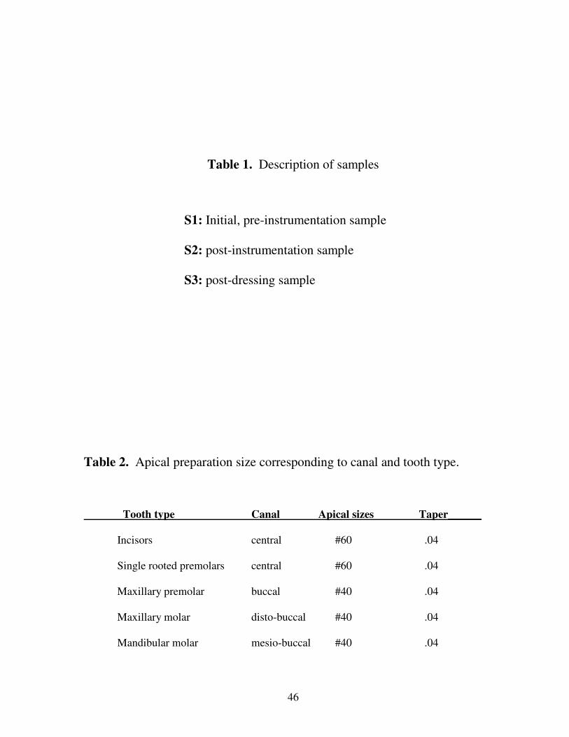
46
Table 1. Description of samples
S1: Initial, pre-instrumentation sample
S2: post-instrumentation sample
S3: post-dressing sample
Table 2. Apical preparation size corresponding to canal and tooth type.
Tooth type Canal Apical sizes Taper______
Incisors central #60 .04
Single rooted premolars central #60 .04
Maxillary premolar buccal #40 .04
Maxillary molar disto-buccal #40 .04
Mandibular molar mesio-buccal #40 .04
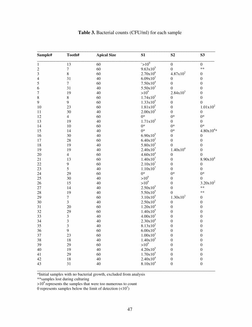
47
Table 3. Bacterial counts (CFU/ml) for each sample
________________________________________________________________________Sample# Tooth# Apical Size S1 S2 S3______________________________________________________________________________________1 13 60 `>109 0 02 7 60 9.63x10⁵ 0 **3 8 60 2.70x10⁶ 4.87x10² 04 31 40 6.09x10² 0 05 7 60 7.50x10⁴ 0 06 31 40 5.50x10³ 0 07 19 40 >109 2.84x10³ 08 8 60 1.74x10⁵ 0 09 9 60 1.33x10⁵ 0 010 23 60 1.81x10⁵ 0 1.01x10²
11 30 40 2.00x10⁵ 0 012 4 60 0* 0* 0* 13 19 40 1.71x10⁵ 0 014 10 60 0* 0* 0*15 14 40 0* 0* 4.80x104*16 30 40 6.90x10⁵ 0 017 28 60 6.40x10² 0 018 19 40 5.80x10⁵ 0 019 19 40 2.40x10⁷ 1.40x10⁴ 020 4 60 4.60x10⁴ 0 021 13 60 1.40x10⁷ 0 8.90x10⁴
22 9 60 2.10x10⁷ 0 023 5 40 1.10x10⁷ 0 024 29 60 0* 0* 0*25 30 40 >109 0 026 15 40 >109 0 3.20x10²
27 14 40 2.50x10⁵ 0 ** 28 19 40 5.50x10⁵ 0 ** 29 7 60 3.10x10⁵ 1.30x10³ 030 3 40 2.50x10⁵ 0 031 20 60 1.20x10⁴ 0 032 29 60 1.40x10⁵ 0 033 3 40 4.00x10³ 0 034 3 40 2.30x10⁴ 0 035 3 40 8.13x10² 0 036 9 60 6.00x10⁵ 0 037 23 60 1.00x10³ 0 038 18 40 1.40x10⁵ 0 039 29 60 >109 0 040 19 40 4.20x10⁵ 0 041 29 60 1.70x10⁵ 0 042 18 40 2.40x10⁴ 0 043 31 40 8.10x10⁴ 0 0________________________________________________________________________*Initial samples with no bacterial growth, excluded from analysis**samples lost during culturing>109 represents the samples that were too numerous to count0 represents samples below the limit of detection (<102)

48
Table 4. Percentage of samples with negative culture
_______________________________________________________________________Groups Sample Size S2 S3_______________________________________________________________________
Size #60 18 16/18 (88.9%) 15/17 (88.2%)
Size #40 21 19/21 (90.5%) 18/19 (94.7%)________________________________________________________________________Total 39 35/39 (89.7%) 33/36 (91.7%)________________________________________________________________________

49
Figure 1: Initial, Pre-Instrumentation Sample (S1)
•Rubber dam isolation•Oraseal•Disinfection of operatoryfield•Caries removal
•Access prep•Saline irrigation
•Transfer medium•Patency with #15 file•5x filing motion
•Collect bacterialsample 3x paperpoint per canal
•Anaerobic culture for 1 week
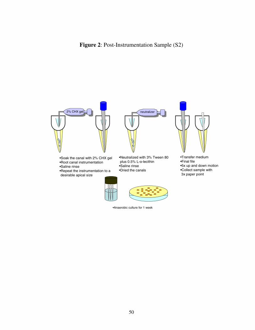
50
Figure 2: Post-Instrumentation Sample (S2)
2% CHX gel
•Soak the canal with 2% CHX gel•Root canal instrumentation•Saline rinse•Repeat the instrumentation to adesirable apical size
•Neutralized with 3% Tween 80plus 0.5% L-α-lecithin
•Saline rinse•Dried the canals
•Transfer medium•Final file•5x up and down motion•Collect sample with3x paper point
•Anaerobic culture for 1 week
neutralizer

51
Figure 3: Post-Dressing Sample (S3)
•>2 wk of mixture ofCa(OH)2 + 2% CHXgel dressing
Citric acid neutralizer
•Removed the intracanal dressing with saline•Neutralized with 0.5% citric acid and3% Tween 80 plus 0.5% L-α-lecithin•Saline rinse•Dried the canal
•Anaerobic culture for 1 week
•Transfer medium•Final file•5x up and down motion•Collect sample with3x paper point
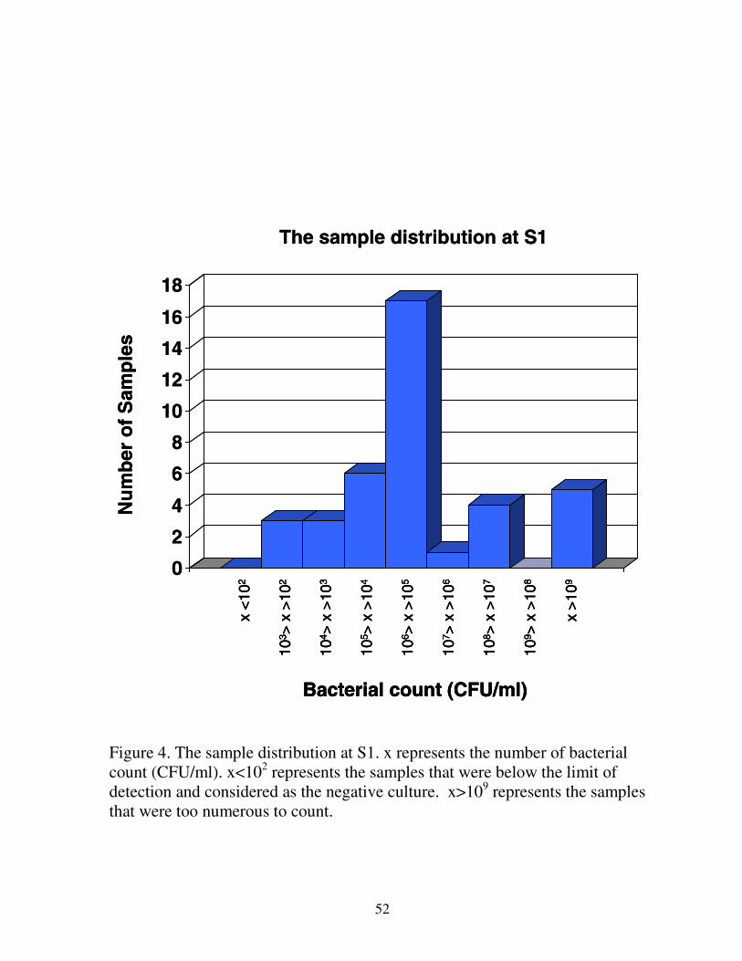
52
0
2
4
6
8
10
12
14
16
18
Nu
mb
ero
fS
amp
les
The sample distribution at S1x
<10
2
104 >
x>
103
105 >
x>
104
106 >
x>
105
107 >
x>
106
108 >
x>
107
109 >
x>
108
x>
109
103 >
x>
102
Bacterial count (CFU/ml)
0
2
4
6
8
10
12
14
16
18
Nu
mb
ero
fS
amp
les
The sample distribution at S1x
<10
2
104 >
x>
103
105 >
x>
104
106 >
x>
105
107 >
x>
106
108 >
x>
107
109 >
x>
108
x>
109
103 >
x>
102
Bacterial count (CFU/ml)
x<
102
104 >
x>
103
105 >
x>
104
106 >
x>
105
107 >
x>
106
108 >
x>
107
109 >
x>
108
x>
109
103 >
x>
102
Bacterial count (CFU/ml)
Figure 4. The sample distribution at S1. x represents the number of bacterialcount (CFU/ml). x<102 represents the samples that were below the limit ofdetection and considered as the negative culture. x>109 represents the samplesthat were too numerous to count.

53
0
5
10
15
20
25
30
35x
<10
2
104 >
x>
103
105 >
x>
104
106 >
x>
105
107 >
x>
106
108 >
x>
107
109 >
x>
108
x>
109
103 >
x>
102
Bacterial count (CFU/ml)
Nu
mb
ero
fS
amp
les
The sample distribution at S2
0
5
10
15
20
25
30
35x
<10
2
104 >
x>
103
105 >
x>
104
106 >
x>
105
107 >
x>
106
108 >
x>
107
109 >
x>
108
x>
109
103 >
x>
102
Bacterial count (CFU/ml)
x<
102
104 >
x>
103
105 >
x>
104
106 >
x>
105
107 >
x>
106
108 >
x>
107
109 >
x>
108
x>
109
103 >
x>
102
Bacterial count (CFU/ml)
Nu
mb
ero
fS
amp
les
The sample distribution at S2
Figure 5. The sample distribution at S2. x represents the number of bacterialcount (CFU/ml). x<102 represents the samples that were below the limit ofdetection and considered as the negative culture. x>109 represents the samplesthat were too numerous to count
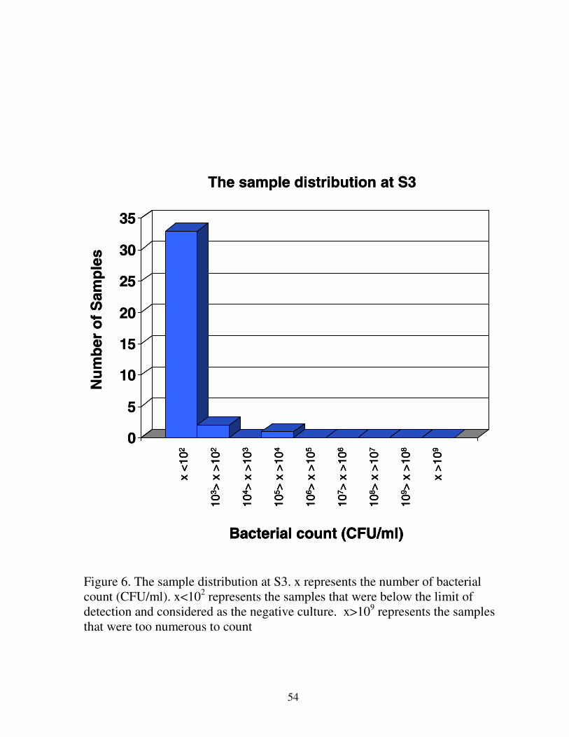
54
0
5
10
15
20
25
30
35
x<
102
104 >
x>1
03
105 >
x>1
04
106 >
x>1
05
107 >
x>1
06
108 >
x>1
07
109 >
x>1
08
x>
109
103 >
x>1
02
Bacterial count (CFU/ml)
Nu
mb
ero
fS
amp
les
The sample distribution at S3
0
5
10
15
20
25
30
35
x<
102
104 >
x>1
03
105 >
x>1
04
106 >
x>1
05
107 >
x>1
06
108 >
x>1
07
109 >
x>1
08
x>
109
103 >
x>1
02
Bacterial count (CFU/ml)
x<
102
104 >
x>1
03
105 >
x>1
04
106 >
x>1
05
107 >
x>1
06
108 >
x>1
07
109 >
x>1
08
x>
109
103 >
x>1
02
Bacterial count (CFU/ml)
Nu
mb
ero
fS
amp
les
The sample distribution at S3
Figure 6. The sample distribution at S3. x represents the number of bacterialcount (CFU/ml). x<102 represents the samples that were below the limit ofdetection and considered as the negative culture. x>109 represents the samplesthat were too numerous to count

55
Figure 7. Percentage of samples with positive culture
0%
20%
40%
60%
80%
100%
size #40 size #60 TotalS1 (initial sample)S2 (post-instrumentation sample)S3 (post-dressing sample)
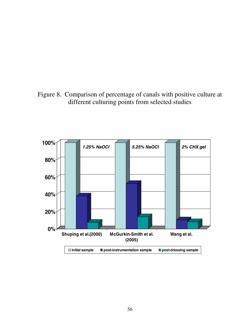
56
Figure 8. Comparison of percentage of canals with positive culture atdifferent culturing points from selected studies
0%
20%
40%
60%
80%
100%
Shuping et al.(2000) McGurkin-Smith et al.(2005)
Wang et al.
initial sample post-instrumentation sample post-dressing sample
2% CHX gel5.25% NaOCl1.25% NaOCl

57
REFERENCES
Abramovich A, Goldberg F. The relationship of the root canal sealer to the dentine wall. Anin vitro study using scanning electron microscope. J Br Endod Soc 1976; 9: 81-6.
Allard U, Nord CE, Sjoberg L, Stromberg T. Experimental infections with Staphylococcusaureus, Streptococcus sanguis, Pseudomonas aeruginosa, and Bacteroides fragilis in thejaws of dogs. Oral Surg Oral Med Oral Pathol. 1979; 48:454-62.
Ando N, Hoshino E. Predominant obligate anaerobes invading the deep layers or root canaldentine. Int Endod J. 1990; 23:20-7.
Aquilino SA, Caplan DJ. Relationship between crown placement and the survival ofendodontically treated teeth. J Prosthet Dent. 2002; 87:256-63.
Barnes GW, Langeland K. Antibody formation in primates following introduction ofantigens into the root canal. J Dent Res. 1966; 45:1111-4
Basrani B, Santos JM, Tjaderhane L, Grad H, Gorduysus O, Huang J, Lawrence HP,
Friedman S. Substantive antimicrobial activity in chlorhexidine-treated human root dentin.Oral Surg Oral Med Oral Pathol Oral Radiol Endod 2002; 94:240-5
Barthel CR, Zimmer S, Trope M. Relationship of radiologic and histologic signs of
inflammation in human root-filled teeth. J Endod 2004; 30:75-9 Baugh D, Wallace J. The role of apical instrumentation in root canal treatment: a review of
the literature. J Endod 2005; 31:333-40
Baumgartner JC, Brown CM, Mader CL, Peters DD, Shulman JD. A scanning electronmicroscopic evaluation of root canal debridement using saline, sodium hypochlorite, andcitric acid. J Endod 1984; 10:525-531
Baumgartner JC, Falkler WA. Reactivity of IgG from explant cultures of periapical lesionswith implicated microorganisms. J Endod 1991; 17:207-12.
Baumgartner JC, Cuenin PR. Efficacy of several concentrations of sodium hypochlorite forroot canal irrigation. J Endod. 1992; 18:605-12.
Baumgartner JC, Watts CM, Xia T. Occurrence of Candida albicans in infections ofendodontic origin. J Endod. 2000; 26:695-8.
Baumgartner JC, Johal S, Marshall JG. Comparison of the antimicrobial efficacy of 1.3%NaOCl/BioPure MTAD to 5.25% NaOCl/15% EDTA for root canal irrigation. J Endod.2007; 33:48-51
58
Bender IB, Seltzer S. Roentgenographic and direct observation of experimental lesions inbone: I. 1961. J Endod. 2003; 29:702-6
Bender IB, Seltzer S. Roentgenographic and direct observation of experimental lesions in
bone: II. 1961. J Endod. 2003; 29:707-12
Bergenholtz G, Lindhe J. Effect of experimentally induced marginal periodontitis andperiodontal scaling on the dental pulp. J Clin Periodontol. 1978; 5:59-73.
Brynolf I. Histological and roentgenological study of periapical region of human upperincisors. Odontol. Revy 1967; 18: suppl. 11
Buckley M, Spangberg LS. The prevalence and technical quality of endodontic treatment inan American subpopulation. Oral Surg Oral Med Oral Pathol Oral Radiol Endod. 1995;79:92-100
Burke GW Jr, Knighton HT. The localization of microorganisms in inflamed dental pulps ofrats following bacteremia. J Dent Res. 1960; 39:205-14.
Byström A, Sundqvist G. Bacteriologic evaluation of the efficacy of mechanical root canalinstrumentation in endodontic therapy. Scan. J Dent. Res. 1981; 89:321-328
Byström A, Sundqvist G. The antibacterial action of sodium hypochlorite and EDTA in 60cases of endodontic therapy. Int Endod J. 1985; 18: 35-40
Byström A, Happonen RP, Sjögren U, Sundqvist G. Healing of periapical lesions of pulplessteeth after endodontic treatment with controlled asepsis. Endod Dent Traumatol 1987;3:58-63
Caplan DJ, Weintraub JA. Factors related to loss of root canal filled teeth. J Public HealthDent. 1997;57:31-9.
Card SJ, Sigurdsson A, Ørstavik D, Trope M. The effectiveness of increased apicalenlargement in reducing intracanal bacteria. J Endod. 2002; 28:779-83.
Chow TW. Mechanical effectiveness of root canal irrigation. J Endod. 1983; 9:475-9.
Chyun YS, Raisz LG. Stimulation of bone formation by prostaglandin E2. Prostaglandins.1984; 27:97-103.
Coolidge E. The diagnosis and treatment of conditions resulting from diseased dental pulps.JADA 1919; 6:337-49
Cvek M, Nord CE, Hollender L. Antimicrobial effect of root canal debridement in teeth withimmature root. A clinical and microbiologic study. Odontol Revy. 1976; 27:1-10.

59
Czarnecki RT, Schilder H. Effect of experimentally induced marginal periodontitis andperiodontal scaling on the dental pulp. J Clin Periodontol. 1978; 5:59-73.
Dahlen G. Immune response in rats against lipopolysaccharides of Fusobacterium nucleatumand Bacteroides oralis administered in the root canal. Scand J Dent Res. 1980; 88:122-9.
Dahlen G, Fabricius L, Holm SE, Moller AJ. Circulating antibodies after experimentalchronic infection in the root canal of teeth in monkeys.Scand J Dent Res. 1982; 90:338-44.
Dakin HD. On the use of certain antiseptic substances in the treatment of infected wounds.Br Med J 1915; ii:318-20
Dalton BC, Ørstavik D, Phillips C, Pettiette M, Trope M. Bacterial reduction with nickel-titanium rotary instrumentation. J Endod 1998; 24:763-7
Dametto FR, Ferraz CC, de Almeida Gomes BP, Zaia AA, Teixeira FB, de Souza-Filho FJ.
In vitro assessment of the immediate and prolonged antimicrobial action of chlorhexidinegel as an endododontic irrigant against Enterococcus faecalis. Oral Surg Oral Med OralPathol 2005; 99:768-72
Dammaschke T, Schneider U, Stratmann U, Yoo JM, Schafer E. Effect of root canaldressings on the regeneration of inflamed periapical tissue. Acta Odontol Scand. 2005;63:143-52
Delany GM, Patterson SS, Miller CH, Newton CW. The effect of chlorhexidine gluconateirrigation on the root canal flora of freshly extracted necrotic teeth.Oral Surg Oral Med Oral Pathol. 1982; 53:518-23
Delivanis PD, Snowden RB, Doyle RJ. Localization of blood-borne bacteria in instrumentedunfilled root canals. Oral Surg Oral Med Oral Pathol. 1981; 52:430-2.
Delivanis PD, Fan VS. The localization of blood-borne bacteria in instrumented unfilled andoverinstrumented canals. J Endod. 1984; 11:521-4.
Dummer PM, Hicks R, Huws D. Clinical signs and symptoms in pulp disease. Int Endod J.1980; 13:27-35.
Engström B, Frostell G. Experiences of bacteriological root canal control. Acta OdontolScand. 1964; 22:43-69.
Ercan E, Ozekinci T, Atakul F, Gul K. Antibacterial activity of 2% chlorhexidine gluconateand 5.25% sodium hypochlorite in infected root canal: in vivo study. J Endod 2004; 39: 84-6

60
Ercan E, DalliM, Dulgergil CT. In vitro assessment of the effectiveness of chlorhexidine geland calcium hydroxide paste with chlorhexidine against Enterococcus faecalis and Candidaalbicans. Oral Surg Oral Med Oral Pathol Oral Radiol Endod. 2006; 102:27-31
Espevik T, Waage A, Faxyaag A, Shalaby MR. Regulation of interleukin-2 and interleukin-6production from T-cells: involvement of interleukin-1 beta and transforming growth factor-beta.Cell Immunol. 1990; 126:47-56.
Estrela CR, Estrela C, Reis C, Bammann LL, Pecora JD. Control of microorganisms in vitroby endodontic irrigants. Braz Dent J. 2003; 14:187-92
Evans MD, Baumgartner JC, Khemaleelakul SU, Xia T. Efficacy of calcium hydroxide:chlorhexidine paste as an intracanal medication in bovine dentin. J Endod 2003; 29:338-9
Fabricius L, Dahlén G, Öhman A, Möller AJ. Predominant indigenous oral bacteria isolated
from infected root canals after varied times of closure. Scan J Dent Res 1982(a); 9:134-44
Fabricius L, Dahlén G, Holm SE, Möller AJ. Influence of combinations of oral bacteria onperiapical tissues of monkeys. Scand J Dent Res. 1982(b); 90:200-6.
Farzaneh M, Abitbol S, Friedman S. Treatment outcome in endodontics: the Toronto study.Phases I and II: Orthograde retreatment. J Endod. 2004; 30:627-33
Ferraz CC, Figueiredo de Almeida Gomes BP, Zaia AA, Teixeira FB, de Souza-Filho FJ. Invitro assessment of the antimicrobial action and the mechanical ability of chlorhexidine gelas an endodontic irrigant. J Endod. 2001; 27:452-5
Filho M, Leonardo M, Silva L, Anibal F, Faccioli L. Inflammatory responses to differentendodontic irrigating solutions. Int Endod J. 2002; 35: 735-9
Gier RE, Mitchell DF. Anachoretic effect of pulpitis. J Dent Res. 1968; 47:564-70.
Glick M, Trope M, Pliskin ME. Detection of HIV in the dental pulp of a patient with AIDS.JAm Dent Assoc. 1989; 119:649-50.
Goldman M, White RR, Moser CR, Tenca JI: A comparison of three methods of cleaning andshaping the root canal in vitro. J Endod 1988; 14: 7-12.
Gomes BPFA, Ferraz CCR, Vianna ME, Berver VB, Teixeira FB, Souza-Filho FJ. In vitroantimicrobial activity of several concentrations of sodium hypochlorite and chlorhexidinegluconate in the elimination of Enterococcus faecalis. Int Endod J 2001; 34:424-8
Gomes BP, Vianna ME, Sena NT, Zaia AA, Ferraz CC, de Souza Filho FJ. In vitro
evaluation of the antimicrobial activity of calcium hydroxide combined with chlorhexidinegel used as intracanal medicament. Oral Surg Oral Med Oral Pathol Oral Radiol Endod.2006; 102:544-50

61
Green TL, Walton RE, Taylor JK, Merrell P. Radiographic and histologic periapical findingsof root canal treated teeth in cadaver. Oral Surg Oral Med Oral Pathol Oral Radiol Endod.1997; 83:707-11.
Grossman LI, Meiman BW. Solution of pulp tissue by chemical agents. JADA 1941;28:223-5
Gutiérrez JH, Herrera VR, Berg EH, Villena F, Jofré A. The risk of intentional dissolution of
the smear layer after mechanical preparation of root canals. Oral Surg Oral Med OralPathol 1990; 70:96-108.
Haapasalo M, Ørstavik D. In vitro infection and disinfection of dentinal tubules. J Dent Res1987; 66:1375-9.
Hermann BW. Calciumhydroxid als mittel zum behandel und füllungen vonzahnwurzelkanälen. Med Diss V; Würzburg; 1920
Heling I, Steinberg D, Kenig S, Gavrilovich I, Sela MN, Friedman M. Efficacy of asustained-release device containing chlorhexidine and Ca(OH)2 in preventing secondaryinfection of dentinal tubules. International Endodontic Journal 1992; 25:20-4
Hülsmann and Hahn W. Complications during root canal irrigation--literature review and
case reports. Int Endod J. 2000; 33:186-93
Jeansonne MJ, White RR. A comparision of 2.0% chlorhexidine gluconate and 5.25%sodium hypochlorite as antimicrobial endodontic irrigants. J Endod 1994; 20:276-8
Kakehashi S., Stanley HR, Fitzgerald RJ. The effects of surgical exposures of dental pulps in
germ-free and conventional laboratory rats. Oral Surg Oral Med Oral Pathol 1965; 20:340-349
Kerekes K, Tronstad L. Long-term results of endodontic treatment performed with astandardized technique. J Endod 1979; 5: 83-90
Kho P, Baumgartner JC. A comparison of the antimicrobial efficacy of NaOCl/BiopureMTAD versus NaOCl/EDTA against Enterococcus faecalis. J Endod. 2006;32:652-5
Kuttler Y. Microscopic investigation of root apexes. J Am Dent Assoc. 1955; 50:544-52.
Kouvas V, Liolios E, Vassiliadis L, Parissis-Messimeris S, Boutsioukis A. Influence ofsmear layer depth of penetration of three endodontic sealers: an SEM study. Endod DentTruamatol 1998; 14: 191-5

62
Kvist T, Molander A, Dahlén G, and Reit C. Microbiological evaluation of one- and two-visitendodontic treatment of teeth with apical periodontitis: a randomized, clinical trial. J Endod2004; 30: 572 -576
Law A, Messer H. An evidence-based analysis of the antibacterial effectiveness of intracanalmedicaments. J Endod 2004; 30: 689-693
Langeland K, Rodrigues H, Dowden W. Periodontal disease, bacteria, and pulpalhistopathology. Oral Surg Oral Med Oral Pathol. 1974; 37:257-70.
Love RM. Bacterial penetration of the root canal of intact incisor teeth after a simulatedtraumatic injury. Endod Dent Traumatol. 1996; 12:289-93.
Love RM, Jenkinson HF. Invasion of dentinal tubules by oral bacteria. Crit Rev Oral BiolMed 2002; 13:171-183.
Matsuo T, Nakanishi T, Ebisu S. Immunoglobulins in periapical exudates of infected rootcanals: correlations with the clinical findings of the involved teeth. Endod Dent Traumatol.1995; 11:95-9.
Matsuo T, Shirakami T, Ozaki K, Nakanishi T, Yumoto H, Ebisu S. An immunohistologicalstudy of the localization of bacteria invading root pulpal walls of teeth with periapicallesions. J Endod. 2003; 29:194-200.
McComb D, Smith DC. A preliminary scanning electron microscopic study of root canalsafter endodontic procedures. J Endod 1975; 1: 238-42.
McDonnell G, Russell AD. Antiseptics and disinfectants: activity, action, and resistance.Clin Microbiol Rev. 1999; 12:147-79
McGurkin-Smith R, Trope M, Caplan D, Sigurdsson A. Reduction of intracanal bacteriausing GT rotary Instrumentation, 5.25% NaOCl, EDTA, and Ca(OH)2. J Endod 2005; 21:359-363
Miller WD. The micro-organisms of the human mouth. Philadelphia, 1980, White Dental
Molander A, Reit C, Dahlén G, Kvist T. Microbiological status of root-filled teeth withapical periodontitis. Int Endod J. 1998; 31:1-7.
Möller AJ. Microbiological examination of root canals and periapical tissues of human teeth.Methodological studies. Odontologisk Tidskrift 1966; 74(Suppl.): 1-380
Möller AJ, Fabricius L, Dahlén G, Öhman A, Heyden G. Influence on periradicular tissuesof indigenous oral bacteria and necrotic pulp tissue in monkeys. Scan J Dent Res 1981;89:475-84

63
Moorer WR, Wesselink PR. Factors promoting the tissue dissolving capability of sodiumhypochlorite. Int Endod J. 1982; 15:187-96.
Mumford JM. Pain perception threshold on stimulating human teeth and the histologicalcondition of the pulp. Br Dent J. 1967; 123:427-33.
Naenni N, Thoma K, Zehnder M. Soft tissue dissolution capacity of currently used andpotential endodontic irrigants. J Endod. 2004; 30:785-7
Nagaoka S, Miyazaki Y, Liu HJ, Iwamoto Y, Kitano M, Kawagoe M. Bacterial invasion into
dentinal tubules of human vital and nonvital teeth. J Endod. 1995; 21:70-3.
Nair PN. Pathogenesis of apical periodontitis and the causes of endodontic failures.Crit Rev Oral Biol Med. 2004; 15:348-81.
Oguntebi BR. Dentine tubule infection and endodontic therapy implications.Int Endod J. 1994; 27:218-22.
Okiji T, Morita I, Sunada I, Murota S. Involvement of arachidonic acid metabolites inincreases in vascular permeability in experimental dental pulpal inflammation in the rat.Arch Oral Biol. 1989; 34:523-8.
Ørstavik D, Haapasalo M. Disinfection by endodontic irrigants and dressings ofexperimentally infected dentinal tubules. Endod Dent Traumatol. 1990; 6:142-9.
Ørstavik D, Kerekes K, Molven O. Effects of extensive apical reaming and calciumhydroxide dressing on bacterial infection during treatment of apical periodontitis: a pilotstudy. Int Endod J. 1991; 24: 1-7
Pallarés A, Faus V, Glickman GN. The adaptation of mechanically softened gutta-percha to
the canal walls in the presence or absence of smear layer: a scanning electron microscopicstudy. Int Endod J 1995; 28: 266-9.
Pashley EL, Birdsong NL, Bowman K, Pashley DH. Cytotoxic effects of NaOCl on vitaltissue. J Endod. 1985; 11:525-8.
Pashley DH, Depew DD. Effects of the smear layer, Copalite, and oxalate on microleakage.Oper Dent 1986;11: 95-105
Perez F, Rochd T, Lodter J-P, Calas P, Michel G. In vitro study of the penetration of threebacterial strains into root dentin. Oral Surg Oral Med Oral Pathol 1993; 76: 97-103.
Peters LB, Wesselink PR, Buijs JF, van Winkelhoff AJ. Viable bacteria in root dentinaltubules of teeth with apical periodontitis. J Endod 2001; 27: 76-81.

64
Peters LB, Wesselink PR. Periapical healing of endodontically treated teeth in one and twovisits obturated in the presence or absence of detectable microorganisms.Int Endod J. 2002; 35:660-7.
Podbielski A, Spahr A, Haller B. Additive antimicrobial activity of calcium hydroxide andchlorhexidine on common endodontic bacterial pathogens. J Endod 2003; 29:340-5
Pulver WH, Taubman MA, Smith DJ. Immune components in human dental periapicallesions. Arch Oral Biol. 1978; 23:435-43.
Rutala WA, Weber DJ. Uses of inorganic hypochlorite (bleach) in health-care facilities.Clin Microbiol Rev. 1997; 10:597-610.
Ram Z. Effectiveness of root canal irrigation. Oral Surg Oral Med Oral Pathol. 1977; 44:306-12.
Ray HA, Trope M. Periapical status of endodontically treated teeth in relation to the technicalquality of the root filling and the coronal restoration. Int Endod J. 1995; 28:12-8.
Reit C, Molander A, Dahlen G. The diagnostic accuracy of microbiologic root canalsampling and the influence of antimicrobial dressings. Endod Dent Traumatol. 1999;15:278-83
Riccuci D. Apical limit of root canal instrumentation and obturation, part 1. Literaturereview. Int Endod J. 1998; 3:384-93.
Riccuci D, Langeland K. Apical limit of root canal instrumentation and obturation, part 2. Ahistological study. Int Endod J. 1998; 3:394-409.
Ringel AM, Patterson SS, Newton CW, Miller CH, Mülhern JM. In vivo evaluation ofchlorhexidine gluconate solution and sodium hypochlorite solution as root canal irrigants.J Endod. 1982; 8:200-4
Robinson HBG, Boling LR: The anachoretic effect in pulpitis: bacteriologic studies. J Am
Dental Assoc 1941; 28:268
Rosenthal S, Spangberg L, Safavi K. Chlorhexidine substantivity in root canal dentin. OralSurg Oral Med Oral Pathol Oral Radiol Endod 2004; 98:488-92
Rubach WC, Mitchell DF. Periodontal disease, age, and pulp status. Oral Surg Oral MedOral Pathol. 1965; 19:482-93.
Russell AD, Day MJ. Antibacterial activity of chlorhexidine. J Hosp Infect. 1993; 25:229-38
Safavi KE, Spangberg LS, Langeland K. Root canal dentinal tubule disinfection. J Endod.1990; 16:207-10.

65
Salzgeber RM, Brilliant JD. An in vivo evaluation of the penetration of an irrigating solutionin root canals. J Endod. 1977; 3:394-8.
Schaeffer MA, White RR, Walton RE. Determining the optimal obturation length: a meta-analysis of literature. J Endod. 2005; 31:271-4.
Schilder H. Filling root canals in three dimensions.Dent Clin North Am. 1967: 723-44.
Schilder H. Cleaning and shaping the root canal. Dent Clin North Am. 1974; 18:269-96
Seltzer S, Bender IB, Ziontz M. The interrelationship of pulp and periodontal disease. OralSurg Oral Med Oral Pathol. 1963; 16:1474-90
Seltzer S, Bender IB, Ziontz M. The dynamics of pulp inflammation: correlations betweendiagnostic data and actual histologic findings in the pulp. Oral Surg Oral Med Oral Pathol.1963; 16:846-71
Seltzer S, Bender IB, Nazimov H, Sinai I. Pulpitis-induced interradicular periodontalchanges in experimental animals. J Periodontol. 1967; 38:124-9.
Sen BH, Pi’kin B, Demirci T. Observation of bacteria and fungi in infected root canals anddentinal tubules by SEM. Endod Dent Traumatol 1995; 11: 6-9.
Shuping GB, Ørstavik D, Sigurdsson A, Trope M. Reduction of intracanal bacteria usingnickel-titanium rotary instrumentation and various medications. J Endod 2000; 26: 751-755
Shipper G., Ørstavik D, Teixeira FB, and Trope M. An evaluation of microbial leakage inroots filled with a thermoplastic synthetic polymer-based root canal filling material(Resilon). J Endod 2004; 30: 342-347
Siqueira JF Jr, de Uzeda M, Fonseca MEF. A scanning electron microscopic evaluation of invitro dentinal tubules penetration by selected anaerobic bacteria. J Endod 1996; 22: 308-10.
Siqueira JF Jr, da Silva CH, Cerqueira M das D, Lopes HP, de Uzeda M. Effectiveness offour chemical solutions in eliminating Bacillus subtilis spores on gutta-percha cones.Endod Dent Traumatol. 1998; 14:124-6.
Siqueira JF Jr, Lopes HP. Mechanisms of antimicrobial activity of calcium hydroxide: acritical review. Int Endod J. 1999; 32:361-9.
Siqueira JF Jr, Lima KC, Magalhaes FA, Lopes HP, de Uzeda M. Mechanical reduction ofthe bacterial population in the root canal by three instrumentation techniques. J Endod.1999; 25:332-5.

66
Siqueira JF Jr., Rocas IN, Alves FR, Campos LC. Periradicular status related to the quality ofcoronal restorations and root canal fillings in a Brazilian population. Oral Surg Oral MedOral Pathol Oral Radiol Endod. 2005; 100:369-74
Siqueira JF Jr, Rocas IN, Favieri A, Lima KC. Chemomechanical reduction of the bacterialpopulation in the root canal after instrumentation and irrigation with 1%, 2.5%, and 5.25%sodium hypochlorite. J Endod. 2000; 26:331-4
Sirén EK, Haapasalo MP, Waltimo TM, Ørstavik D. In vitro antibacterial effect of calcium
hydroxide combined with chlorhexidine or iodine potassium iodide on Enterococcusfaecalis. Eur J Oral Sci. 2004; 112:326-31
Sjögren U, Hagglund B, Sundqvist G, Wing K. Factors affecting the long-term results ofendodontic treatment. J Endod 1990; 16:498-504
Sjögren U, Figdor D, Spangberg L, Sundqvist G. The antimicrobial effect of calciumhydroxide as a short-term intracanal dressing. Int Endod J. 1991; 24, 119-125
Sjögren U, Figdor D, Persson S, Sundqvist G. Influence of infection at the time of root fillingon the outcome of endodontic treatment of teeth with apical peridontitis. Int Endod J. 1997;30, 297-306
Sorensen JA, Martinoff JT. Intracoronal reinforcement and coronal coverage: a study ofendodontically treated teeth. J Prosthet Dent. 1984; 51:780-4
Sorensen JA, Martinoff JT. Endodontically treated teeth as abutments. J Prosthet Dent. 1985;
53:631-6.
Spangberg L, Engstrom B, Langeland K. Biologic effects of dental materials. 3. Toxicity andantimicrobial effect of endodontic antiseptics in vitro. Oral Surg Oral Med Oral Pathol.1973; 36:856-71.
Stashenko P, Dewhirst FE, Peros WJ, Kent RL, Ago JM. Synergistic interactions betweeninterleukin 1, tumor necrosis factor, and lymphotoxin in bone resorption. J Immunol. 1987;138:1464-8.
Stashenko P, Yu SM. T helper and T suppressor cell reversal during the development ofinduced rat periapical lesions. J Dent Res. 1989; 68:830-4
Stashenko P, Wang CY, Tani-Ishii N, Yu SM. Pathogenesis of induced rat periapical
lesions. Oral Surg Oral Med Oral Pathol. 1994; 78:494-502.
Stashenko P, Wang CY, Riley E, Wu Y, Ostroff G, Niederman R. Reduction of infection-stimulated periapical bone resorption by the biological response modifier PGG glucan. JDent Res. 1995; 74:323-30
67
Stashenko P, Teles R, D’Souza R. Periapical inflammatory responses and their modulation.Crit Rev Oral Biol Med. 1998; 9:498-521.
Stern MH, Dreizen S, Mackler BF, Selbst AG, Levy BM. Quantitative analysis of cellularcomposition of human periapical granuloma. J Endod. 1981; 7:117-22.
Stern MH, Mackler BF, Dreizen S. A quantitative method for the analysis of humanperiapical inflammation. J Endod. 1981; 7:70-4.
Stoll R, Betke K, Stachniss V. The influence of different factors on the survival of root canalfillings: a 10-year retrospective study. J Endod. 2005; 31:783-90.
Stringberg L. The dependence of the results of pulp therapy of certain factors. An analyticalstudy based on radiographic and clinical follow-up examinations. Acta Odontol Scan 1956;14 (supplement 21)
Stuart CH, Schwartz SA, Beeson TJ, Owatz CB. Enterococcus faecalis: its role in root canaltreatment failure and current concepts in retreatment. J Endod. 2006; 32:93-8
Sundqvist G. Bacteriological studies of necrotic dental pulps. Umea University
Odontological Dissertations No. 7, University of Umea, Sweden; 1976
Sundqvist G, Figdor D, Persson S. Microbiologic findings of teeth with failed endodontictreatment and the outcome of conservative re-treatment. Oral Surg. 1998; 85:86-93
Sundqvist G. Ecology of the root canal flora. J Endod 1992; 18:427-30.
Tani-Ishii N, Wang CY, Stashenko P. Immunolocalization of bone-resorptive cytokines inrat pulp and periapical lesions following surgical pulp exposure. Oral Microbiol Immunol.1995; 10:213-9.
Taylor JK, Jeansonne BG, Lemon RR. Coronal leakage: effects of smear layer, obturationtechnique, and sealer. J Endod 1997; 23: 508-12
Thé SD, Maltha JC, Plasschaert AJ. Reactions of guinea pig subcutaneous connective tissuefollowing exposure to sodium hypochlorite. Oral Surg Oral Med Oral Pathol. 1980;49:460-6.
Tidmarsh BG. Acid-cleansed and resin-sealer root canals. J Endod 1978; 4: 117-121.
Timpawat S, Vongsavan N, Messer HH. Effect of removal of the smear layer on apicalmicroleakage. J Endod 2001; 27: 351-3.
Torabinejad M, Bakland LK. An animal model for the study of immunopathogenesis ofperiapical lesions. J Endod. 1978; 4:273-7.

68
Torabinejad M, Kettering JD. Identification and relative concentration of B and Tlymphocytes in human chronic periapical lesions.J Endod. 1985; 1:122-5.
Torabinejad M, Eby WC, Naidorf IJ. Inflammatory and immunological aspects of thepathogenesis of human periapical lesions. J Endod. 1985; 11:479-88.
Torabinejad M, Kiger RD. A histologic evaluation of dental pulp tissue of a patient withperiodontal disease. Oral Surg Oral Med Oral Pathol. 1985; 59:198-200.
Torabinejad M, Handysides R, Khademi AA, Bakland L K. Clinical implications of thesmear layer in endodontics: A review. Oral Surg Oral Med Oral Pathol Oral Radiol Endod2002; 94: 658-66.
Torabinejad M, Khademi AA, Babagoli J, et al. A new solution for the removal of the smearlayer. J Endod 2003(a); 29: 170-175.
Torabinejad M, Cho Y, Khademi AA, Bakland LK, Shabahang S. The effect of variousconcentration of sodium hypochlorite on the ability of MTAD to remove the smear layer. JEndod 2003(b); 29: 233-239.
Tronstad L, Barnett F, Riso K, Slots J. Extraradicular endodontic infections. Endod DentTraumatol. 1987; 3:86-90.
Tronstad L, Asbjornsen K, Doving L, Pedersen I, Eriksen HM. Influence of coronalrestorations on the periapical health of endodontically treated teeth. Endod Dent Traumatol.2000; 16:218-21.
Trope M, Delano EO, Ørstavik D. Endodontic treatment of teeth with apical periodontitis:single vs. multivisit treatment. J Endod. 1999; 25: 345-50
Vianna ME, Gomes BP, Berber VB, Zaia AA, Ferraz CC, de Souza-Filho FJ. In vitroevaluation of the antimicrobial activity of chlorhexidine and sodium hypochlorite.Oral Surg Oral Med Oral Pathol Oral Radiol Endod. 2004; 97:79-84
Vianna ME, Horz HP, Gomes BP, Conrads G. In vivo evaluation of microbial reduction afterchemo-mechanical preparation of human root canals containing necrotic pulp tissue. IntEndod J. 2006; 39:484-492
Vire DE. Failure of endodontically treated teeth: classification and evaluation. J Endod.1991; 17:338-42.
Wahl SM. Transforming growth factor beta: the good, the bad, and the ugly.J Exp Med. 1994; 180:1587-90.
Waltimo TM, Siren EK, Torkko HL, Olsen I, Haapasalo MP. Fungi in therapy-resistantapical periodontitis. Int Endod J. 1997; 30:96-101.

69
Waltimo T, Trope M, Haapasalo M, Orstavik D. Clinical efficacy of treatment procedures inendodontic infection control and one year follow-up of periapical healing. J Endod. 2005;31:863-6
Wang CY, Stashenko P. Characterization of bone-resorbing activity in human periapical
lesions. J Endod. 1993; 19:107-11.
Wang CY, Stashenko P. The role of interleukin-1 alpha in the pathogenesis of periapicalbone destruction in a rat model system. Oral Microbiol Immunol. 1993; 8:50-6.
Wang CY, Tani-Ishii N, Stashenko P. Bone-resorptive cytokine gene expression inperiapical lesions in the rat. Oral Microbiol Immunol. 1997; 12:65-71.
Weiger R, de Lucena J, Decker HE, Lost C. Vitality status of microorganisms in infectedhuman root dentine. Int Endod J. 2002; 35:166-71.
Weiger R, Rosendahl R, Lost C. Influence of calcium hydroxide intracanal dressings on theprognosis of teeth with endodontically induced periapical lesions.Int Endod J. 2000; 33:219-26
Weine FS. Initiating endodontic therapy in posterior teeth. Part II. Maxillary molars.Compend Contin Educ Dent. 1982; 3:455-64
White R, Goldman M, Peck S-L. The influence of the smeared layer upon dentinal tubulepenetration by plastic filling materials. J Endod 1984; 10:558-62.
Yamada RS, Armas A, Goldman M, Lin PS. A scanning electron microscopic comparison ofa high volume final flush with several solutions: Part 3. J Endod 1983; 9: 137-142.
Yamasaki M, Kumazawa M, Kohsaka T, Nakamura H, Kameyama Y. Pulpal and periapicaltissue reactions after experimental pulpal exposure in rats. J Endod. 1994 ; 20:13-7.
Yesilsoy C, Whitakers E, Cleveland D, Phillips E, Trope M. Antimicrobial and toxic effectsof established and potential root canal irrigants. J Endod. 1995; 21:513-5.
Yu SM, Stashenko P. Identification of inflammatory cells in developing rat periapicallesions. J Endod. 1987; 13:535-40.
Zamany A, Spangberg LS. An effective method of inactivating chlorhexidine. Oral Surg OralMed Oral Pathol Oral Radiol Endod. 2002; 93:617-20
Zeldow BI, Ingle JI. Correlation of the positive culture to the prognosis of endodonticallytreated teeth: a clinical study. J Am Dent Assoc. 1963; 66:9-13.

70
Zehnder M, Kosicki D, Luder H, Sener B, Waltimo T. Tissue-dissolving capacity andantibacterial effect of buffered and unbuffered hypochlorite solutions. Oral Surg Oral MedOral Pathol Oral Radiol Endod. 2002; 94:756-62.
Zerella JA, Fouad AF, Spangberg LS. Effectiveness of a calcium hydroxide andchlorhexidine digluconate mixture as disinfectant during retreatment of failed endodonticcases. Oral Surg Oral Med Oral Pathol Oral Radiol Endod 2005; 100:756-61


![Effectiveness of an implementation optimisation intervention … · 2017-08-27 · mentation research is conducted following trials for clin-ical effectiveness [23], it is argued](https://static.fdocuments.in/doc/165x107/5f03e9e47e708231d40b63e2/effectiveness-of-an-implementation-optimisation-intervention-2017-08-27-mentation.jpg)