Classifying Taiwan Lianas with Radiating Plates of Xylem
9
Taiwania 60(4):151‒159, 2015 DOI: 10.6165/tai.2015.60.151 151 Classifying Taiwan Lianas with Radiating Plates of Xylem Sheng-Zehn Yang * and Po-Hao Chen National Pingtung University of Science and Technology. Corresponding author. No. 1, Shuefu Rd., Neipu, Pingtung, 912, Taiwan. *Corresponding author: [email protected] (Manuscript received 21 Janurary 2015; accepted 18 June 2015) ABSTRACT: Radiating plates of xylem are a lianas cambium variation, of which, 22 families have this feature. This study investigates 15 liana species representing nine families with radiating plates of xylem structures. The features of the transverse section and epidermis in fresh liana samples are documented, including shapes and colors of xylem and phloem, ray width and numbers, and skin morphology. Experimental results indicated that the shape of phloem fibers in Ampelopsis brevipedunculata var. hancei is gradually tapered and flame-like, which is in contrast with the other characteristics of this type, including those classified as rays. Both inner and outer cylinders of vascular bundles are found in Piper kwashoense, and the irregularly inner cylinder persists yet gradually diminishes. Red crystals are numerous in the cortex of Celastrus kusanoi. Aristolochia shimadai and A. zollingeriana develop a combination of two cambium variants, radiating plates of xylem and a lobed xylem. The shape of phloem in Stauntonia obovatifoliola is square or truncate, and its rays are numerous. Meanwhile, that of Neoalsomitra integrifolia is blunt and its rays are fewer. As for the features of a stem surface within the same family, Cyclea ochiaiana is brownish in color and has a deep vertical depression with lenticels, Pericampylus glaucus is greenish in color with a vertical shallow depression. Within the same genus, Aristolochia shimadai develops lenticels, which are not in A. zollingeriana; although the periderm developed in Clematis grata is a ring bark and tears easily, that of Clematis tamura is thick and soft. KEY WORDS: Cambial production unit, lianas, single cambium activity, xylem in plates. INTRODUCTION Lianas situated in different layers of forested areas must compete with plants for resources such as nutrients, sunlight, as well as factors such as impact trees net rate of increase, growth, diversity, reproduction, and survival (Schnitzer and Bongers, 2002; Schnitzer et al., 2002; Schnitzer and Carson, 2010). For adaptation, lianas may gradually produce unique habits or structures. Additionally, lianas do not require a self-support habit during the development of their stems; this unique habit creates variations of their stems. These variations originate from cambium, which changes the shape and structure of the stems into irregular types, called cambial variants or anomalous structures (Isnard and Silk, 2009). These irregular structures can distinguish between different families (Caballé, 1993; Angyalossy et al., 2012). Different irregular features of lianas usually derive from a single cambium and multiple cambia, which develop different types and arrangements of xylem and phloem, as well as variations of rays. By using different terms, previous studies have described the irregular structures and categorized them into different cambial variants (Angyalossy et al., 2012, 2015; Sun et al., 2014; Yang et al., 2014). The cambial variants include furrowed xylem, such as in Bignoniaceae (Angyalossy et al., 2012); lobed xylem, such as in Passifloraceae (Acevedo-Rodriguez, 2005); radiating plates of xylem, such as in Asteraceae, Aristolochiaceae, Cucurbitaceae, Lardizabalaceae, Menispermaceae, Ranunculaceae (Isnard et al., 2003; Jacques and Franceschi, 2007); included phloem, such as in Loganiaceae (Isnard and Silk, 2009); intraxylary phloem, such as in Apocynaceae (Patil et al., 2009; Yang et al., 2013). The above five types evolve from a single cambium activity. The following five types emerging from multiple cambium activities include successive cambia, such as in Mucuna macrocarpa (Sun et al., 2014); duplicated xylem cylinders, such as in Convolvulaceae (Acevedo-Rodriguez, 2005; Rajput et al., 2009); fissured/blocks xylem, such as in Bauhinia championii (Carlquist, 2001); compound vascular cylinders and divided xylem mass, such as in Sapindaceae (Tamaio et al., 2011). Beck (2011) observed that although the fascicular cambium functions normally, the interfascicular cambium produces only ray-like parenchyma cells in lianas. Consequently, these regions become large ray parenchyma cells, commonly referred to as giant rays. For instance, although cambium regions of Aristolochia genus function normally, the adjacent areas of both the inner and outer sides produce pure parenchyma (Mauseth, 1988). These irregular growth forms control the patterns of rays, in which fascicular cambium regions produce a certain percentage of xylem and phloem. Therefore, cambium activity leads to a large, radically extended region of secondary vascular tissues that are distinct from each other and alternated with giant rays, so that it becomes ultimately leading to a vascular cylinder. Metcalfe and Chalk (1985) described the above variation as xylem reduced to narrow radiating plates;
Transcript of Classifying Taiwan Lianas with Radiating Plates of Xylem
Taiwania 60(4):151‒159, 2015
DOI: 10.6165/tai.2015.60.151
151
Classifying Taiwan Lianas with Radiating Plates of Xylem Sheng-Zehn Yang* and Po-Hao Chen National Pingtung University of Science and Technology. Corresponding author. No. 1, Shuefu Rd., Neipu, Pingtung, 912, Taiwan. *Corresponding author: [email protected] (Manuscript received 21 Janurary 2015; accepted 18 June 2015) ABSTRACT: Radiating plates of xylem are a lianas cambium variation, of which, 22 families have this feature. This study investigates 15 liana species representing nine families with radiating plates of xylem structures. The features of the transverse section and epidermis in fresh liana samples are documented, including shapes and colors of xylem and phloem, ray width and numbers, and skin morphology. Experimental results indicated that the shape of phloem fibers in Ampelopsis brevipedunculata var. hancei is gradually tapered and flame-like, which is in contrast with the other characteristics of this type, including those classified as rays. Both inner and outer cylinders of vascular bundles are found in Piper kwashoense, and the irregularly inner cylinder persists yet gradually diminishes. Red crystals are numerous in the cortex of Celastrus kusanoi. Aristolochia shimadai and A. zollingeriana develop a combination of two cambium variants, radiating plates of xylem and a lobed xylem. The shape of phloem in Stauntonia obovatifoliola is square or truncate, and its rays are numerous. Meanwhile, that of Neoalsomitra integrifolia is blunt and its rays are fewer. As for the features of a stem surface within the same family, Cyclea ochiaiana is brownish in color and has a deep vertical depression with lenticels, Pericampylus glaucus is greenish in color with a vertical shallow depression. Within the same genus, Aristolochia shimadai develops lenticels, which are not in A. zollingeriana; although the periderm developed in Clematis grata is a ring bark and tears easily, that of Clematis tamura is thick and soft. KEY WORDS: Cambial production unit, lianas, single cambium activity, xylem in plates. INTRODUCTION
Lianas situated in different layers of forested areas must compete with plants for resources such as nutrients, sunlight, as well as factors such as impact trees net rate of increase, growth, diversity, reproduction, and survival (Schnitzer and Bongers, 2002; Schnitzer et al., 2002; Schnitzer and Carson, 2010). For adaptation, lianas may gradually produce unique habits or structures. Additionally, lianas do not require a self-support habit during the development of their stems; this unique habit creates variations of their stems. These variations originate from cambium, which changes the shape and structure of the stems into irregular types, called cambial variants or anomalous structures (Isnard and Silk, 2009). These irregular structures can distinguish between different families (Caballé, 1993; Angyalossy et al., 2012).
Different irregular features of lianas usually derive from a single cambium and multiple cambia, which develop different types and arrangements of xylem and phloem, as well as variations of rays. By using different terms, previous studies have described the irregular structures and categorized them into different cambial variants (Angyalossy et al., 2012, 2015; Sun et al., 2014; Yang et al., 2014). The cambial variants include furrowed xylem, such as in Bignoniaceae (Angyalossy et al., 2012); lobed xylem, such as in Passifloraceae (Acevedo-Rodriguez, 2005); radiating plates of xylem, such as in Asteraceae, Aristolochiaceae, Cucurbitaceae, Lardizabalaceae, Menispermaceae, Ranunculaceae
(Isnard et al., 2003; Jacques and Franceschi, 2007); included phloem, such as in Loganiaceae (Isnard and Silk, 2009); intraxylary phloem, such as in Apocynaceae (Patil et al., 2009; Yang et al., 2013). The above five types evolve from a single cambium activity. The following five types emerging from multiple cambium activities include successive cambia, such as in Mucuna macrocarpa (Sun et al., 2014); duplicated xylem cylinders, such as in Convolvulaceae (Acevedo-Rodriguez, 2005; Rajput et al., 2009); fissured/blocks xylem, such as in Bauhinia championii (Carlquist, 2001); compound vascular cylinders and divided xylem mass, such as in Sapindaceae (Tamaio et al., 2011).
Beck (2011) observed that although the fascicular cambium functions normally, the interfascicular cambium produces only ray-like parenchyma cells in lianas. Consequently, these regions become large ray parenchyma cells, commonly referred to as giant rays. For instance, although cambium regions of Aristolochia genus function normally, the adjacent areas of both the inner and outer sides produce pure parenchyma (Mauseth, 1988). These irregular growth forms control the patterns of rays, in which fascicular cambium regions produce a certain percentage of xylem and phloem. Therefore, cambium activity leads to a large, radically extended region of secondary vascular tissues that are distinct from each other and alternated with giant rays, so that it becomes ultimately leading to a vascular cylinder.
Metcalfe and Chalk (1985) described the above variation as xylem reduced to narrow radiating plates;
Taiwania Vol. 60, No. 4
152
Carlquist (1991, 2001) as xylem in plates; Acevedo-Rodríguez (2005) as cylindrical stems with conspicuous rays, where a cross section of stems exhibits wide parenchymatous rays inserted in the xylem tissue; Angyalossy et al. (2012) as axial vascular elements in segments, in which axial elements of xylem and phloem are divided in segments separated by large portions of xylem and phloem rays. Despite all of the explanations, above descriptions and illustrations from previous studies, we believe that naming narrow radiating plates of xylem or xylem in plates is a more meanful way of defining related reports. Some climbing species of the genera such as Clematis, Aristolochia, and Tinospora develop these irregular structures (Carlquist 2001).
By integrating the reports related to cambial variants on radiating plates of xylem (Metcalfe and Chalk, 1985; Caballé, 1993; Carlquist, 1991, 2001; Isnard et al., 2003; Acevedo-Rodríguez, 2005; Jacques and Franceschi, 2007; Angyalossy et al., 2012), twenty two families develop this type. Given the scarcity of literature on such a pattern and the diverse and complex characteristics of their transverse sections, this study elucidates the differences among species from the features of stem surfaces and cross-sections by focusing on these xylem radiating plates. Results of this study significantly contribute to efforts to identify and classify lianas.
MATERIALS AND METHODS
The materials of lianas were collected mainly from southern and eastern Taiwan from 2010 to 2014, and fifteen lianas belonging to nine families were selected as follows:
1. Aristolochia shimadai (Aristolochiaceae), collected in two locations, with a diameter size of around 10–18 mm; 2. A. zollingeriana (Aristolochiaceae), collected in one location, with a diameter size of about 6–8 mm; 3. Vernonia gratiosa (Asteraceae), collected in three locations, with a diameter size of approximately 5–10 mm; 4. Celastrus kusanoi (Celastraceae), collected in six locations, with a diameter size of around 5–25 mm; 5. Neoalsomitra integrifolia (Cucurbitaceae), collected in five locations, with a diameter size of about 5–10 mm; 6. Stauntonia obovatifoliola (Lardizabalaceae), collected in four locations, with a diameter size of approximately 10–20 mm; 7. Cyclea ochiaiana (Menispermaceae), collected in three locations, with a diameter size of about 5–15 mm; 8. Pericampylus glaucus (Menispermaceae), collected in three locations, with a diameter size of around 3–5 mm; 9. Stephania merrillii (Menispermaceae), collected in one location, with a diameter size of around 5–10 mm; 10. Tinospora dentate (Menispermaceae), collected in three locations, with a diameter size of about 3–10 mm; 11. Piper
kwashoense (Piperaceae), collected in one location, with a diameter size of around 8–30 mm; 12. Clematis crassifolia (Ranunculaceae), collected in one location, with a diameter size of about 6–10 mm; 13. C. grata (Ranunculaceae), collected in four locations, with a diameter size of approximately 12–25 mm; 14. C. tamurae (Ranunculaceae), collected in two locations, with a diameter size of about 10–15 mm; and 15. Ampelopsis brevipedunculata var. hancei (Vitaceae), collected in seven locations, with a diameter size of around 10–25 mm.
The features of liana transverse sections were observed and measured, including stem surface morphology and color, maximum diameter, periderm shape, the largest periderm thickness, cortex color, maximum cortex thickness, rays, maximum ray width, phloem color, phloem shape, xylem color, xylem lobed, the largest vessel diameter, and pith. Based on this data, keys are constructed to identify above fifteen lianas.
Lianas samples were selected with obvious secondary growth from different diameter sizes to evaluate the structural variations of stems in developmental stages. Fresh materials were cut into pieces of 4–5 cm in length, and the transverse surfaces were smoothened (Gartner and Schweingruber, 2013) for analysis via microscopic (model Stemi 2000-c, Carl Zeiss) observations and drawings of the cambium variants, and photographs of the cross sections were taken by using a Nikon D80 SLR. All of the voucher specimens used were dried by an oven (temp. 60℃) and then deposited in PPI herbarium at National Pingtung University of Science and Technology. The scientific names of plants were taken from the Flora of Taiwan Volume 3 (Boufford et al., 2003), Flora of China (Lo et al., 2008) and Lu and Wang (2014).
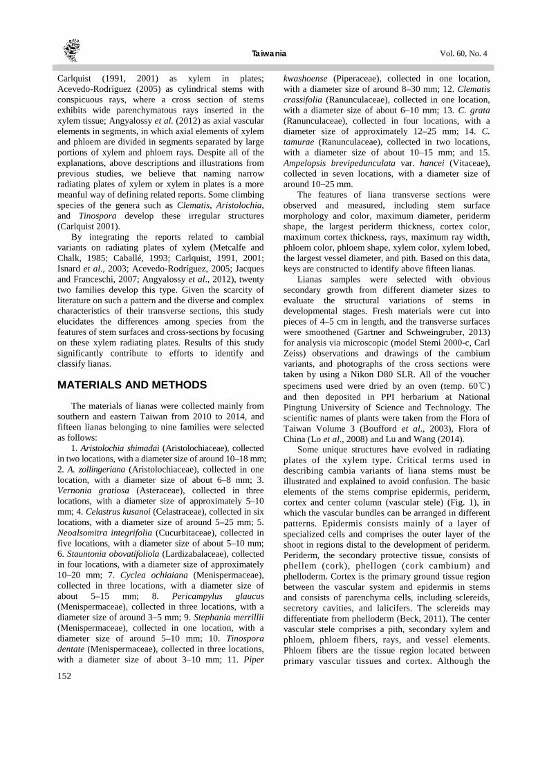
Some unique structures have evolved in radiating plates of the xylem type. Critical terms used in describing cambia variants of liana stems must be illustrated and explained to avoid confusion. The basic elements of the stems comprise epidermis, periderm, cortex and center column (vascular stele) (Fig. 1), in which the vascular bundles can be arranged in different patterns. Epidermis consists mainly of a layer of specialized cells and comprises the outer layer of the shoot in regions distal to the development of periderm. Periderm, the secondary protective tissue, consists of phellem (cork), phellogen (cork cambium) and phelloderm. Cortex is the primary ground tissue region between the vascular system and epidermis in stems and consists of parenchyma cells, including sclereids, secretory cavities, and lalicifers. The sclereids may differentiate from phelloderm (Beck, 2011). The center vascular stele comprises a pith, secondary xylem and phloem, phloem fibers, rays, and vessel elements. Phloem fibers are the tissue region located between primary vascular tissues and cortex. Although the
December 2015 Yang and Chen: Classifying Taiwan Lianas with Radiating Plates of Xylem
153
Fig. 1. Diagram showing the terms used in the descriptions with respect to radiating plates of xylem. Labels: ep = epidermis, pe = periderm, co = cortex, pf = phloem fiber, fc = fascicular cambium, ic = interfascicular cambium, p = pith, v = vessels, xy = xylem, r = ray, ph = phloem. functions of fascicular cambium normally produce xylem and phloem, the interfascicular cambium produces only ray-like parenchyma tissues.
RESULTS
Characteristics of the transverse sections and stem surfaces of fifteen lianas are described as follows:
(1) Aristolochia shimadai (Aristolochiaceae) (Fig. 2A)
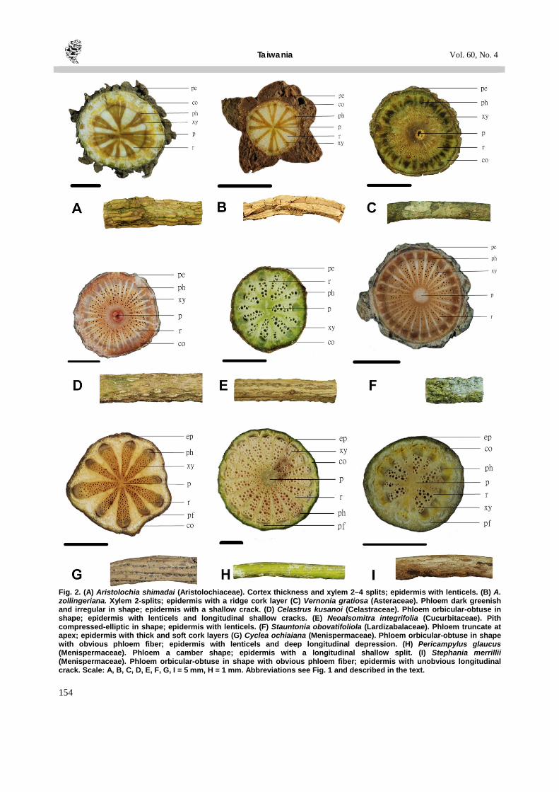
The epidermis is khaki in color with lenticels making up the cork layer. The periderm thickness ranges from 0.4–2.4 mm. The cortex is whitish in color, and its thickness ranges from 1.5–2.4 mm with yellow crystals. There are around 20 rays, in which the widest one is about 1.5 mm. The phloem has an orbicular-obtuse shape and is brownish in color. The xylem is brownish in color, with usually 2–4 splits of different sizes of the vessels distributed and maximum vessel is about 0.1 mm in diameter. The pith is invisible to the naked eye.
(2) Aristolochia zollingeriana (Aristolochiaceae) (Fig. 2B)
The epidermis is khaki in color with a ridge cork layer. The periderm has a triangular shape, and its thickness ranges from 1.6–2.8 mm. The cortex is gray-whitish in color, and its thickness ranges from 0.2–0.4 mm with yellow crystals. There are about 13 rays, in which the widest one is about 1 mm. The phloem has an orbicular-obtuse shape and is brownish in color. The xylem is brownish in color and usually 2 splits with different sizes of the vessels distributed and maximum vessel is about 0.1 mm in diameter. The pith is invisible to the naked eye.
(3) Vernonia gratiosa (Asteraceae) (Fig. 2C) The epidermis is yellow-greenish in color with a
shallow crack. The periderm ranges from 0.2–0.6 mm in thickness. The cortex is yellow-greenish in color, and its thickness ranges from 0.3–0.9 mm. There are about 37 irregular size rays, in which the widest one is about 0.3 mm. The phloem has a lengthy-obtuse irregular shape and is dark greenish in color. The xylem is yellowish in color, not split with different sizes of the vessels distributed and maximum vessel is about 0.2 mm in diameter. The pith has a circular shape, ranging from 2–2.1 mm in diameter.
(4) Celastrus kusanoi (Celastraceae) (Fig. 2D)
The epidermis is yellow-reddish in color with lenticels and longitudinal shallow cracks. The periderm ranges from 0.1–0.5 mm in thickness. The cortex is whitish in color, and its thickness ranges from 0.5–1.1 mm with red crystals. There are around 40 rays, in which the widest one is about 0.4 mm and those rays width are varied and irregular. The phloem has a truncate shape and is light-grayish in color. The xylem is light-yellowish in color, not split with different size of the vessels distributed and maximum vessel is about 0.3 mm in diameter. The pith has a circular shape, ranging from 2.1–2.4 mm in diameter.
(5) Neoalsomitra integrifolia (Cucurbitaceae) (Fig. 2E)
The epidermis is yellowish in color with lenticels. The periderm ranges from 0.1–0.2 mm in thickness, and the cortex is greenish in color, ranging from 0.3–0.9 mm in thickness. There are about 14 rays, in which the widest is about 0.9 mm. The phloem has an orbicular-obtuse shape and is greenish in color. The xylem is whitish in color, not split and with different sizes of the vessels distributed; the maximum vessel is about 0.4 mm in diameter. The pith has a compressed-elliptic shape, ranging from 0.7–2.2 mm in diameter.
(6) Stauntonia obovatifoliola (Lardizabalaceae) (Fig. 2F)
The epidermis is beige in color with thick and soft cork layers. The irregular periderm ranges from 0.2–1.9 mm in thickness, and the cortex is gray in color, ranging from 0.1–0.4 mm in thickness. There are about 25 rays, in which the widest one is about 0.3 mm. The phloem has a truncate shape, and has a coffee color. The xylem is brownish in color, not split with different sizes of the vessels distributed; the maximum vessel is about 0.2 mm in diameter. The pith has a circular shape, ranging from 1.6–1.7 mm in diameter.
(7) Cyclea ochiaiana (Menispermaceae) (Fig. 2G)
The epidermis has a coffee color with lenticels and longitudinal deep split. The periderm ranges from 0.1–0.4 mm in thickness. The cortex is cream in color,
Taiwania Vol. 60, No. 4
154
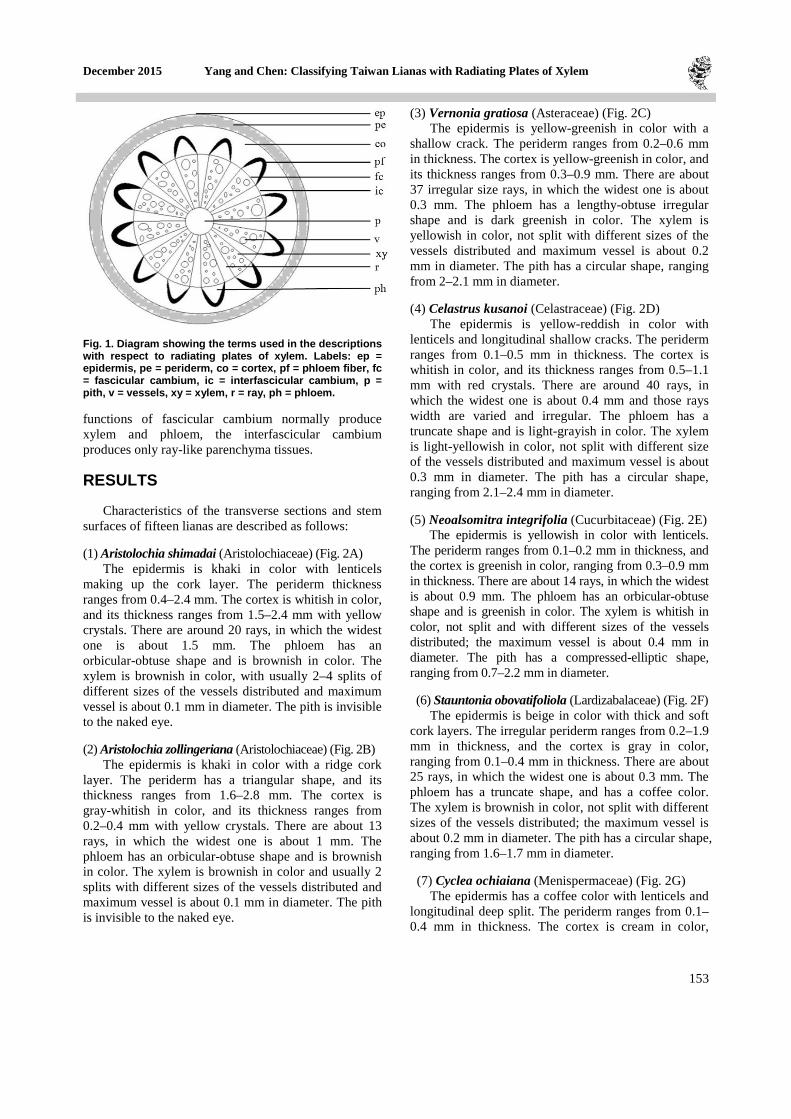
Fig. 2. (A) Aristolochia shimadai (Aristolochiaceae). Cortex thickness and xylem 2–4 splits; epidermis with lenticels. (B) A. zollingeriana. Xylem 2-splits; epidermis with a ridge cork layer (C) Vernonia gratiosa (Asteraceae). Phloem dark greenish and irregular in shape; epidermis with a shallow crack. (D) Celastrus kusanoi (Celastraceae). Phloem orbicular-obtuse in shape; epidermis with lenticels and longitudinal shallow cracks. (E) Neoalsomitra integrifolia (Cucurbitaceae). Pith compressed-elliptic in shape; epidermis with lenticels. (F) Stauntonia obovatifoliola (Lardizabalaceae). Phloem truncate at apex; epidermis with thick and soft cork layers (G) Cyclea ochiaiana (Menispermaceae). Phloem orbicular-obtuse in shape with obvious phloem fiber; epidermis with lenticels and deep longitudinal depression. (H) Pericampylus glaucus (Menispermaceae). Phloem a camber shape; epidermis with a longitudinal shallow split. (I) Stephania merrillii (Menispermaceae). Phloem orbicular-obtuse in shape with obvious phloem fiber; epidermis with unobvious longitudinal crack. Scale: A, B, C, D, E, F, G, I = 5 mm, H = 1 mm. Abbreviations see Fig. 1 and described in the text.

December 2015 Yang and Chen: Classifying Taiwan Lianas with Radiating Plates of Xylem
155
and its thickness ranges from 0.3–1.5 mm. There are about 10 rays, in which the widest one is about 1.5 mm. The phloem has an orbicular-obtuse shape and coffee color; the phloem fiber is obviously blackish in color.The xylem is yellowish in color and not split with different sizes of the vessels distributed; the maximum vessel is about 0.2 mm in diameter. The pith has a circular shape, ranging from 0.7–1.1 mm in diameter. (8) Pericampylus glaucus (Menispermaceae) (Fig. 2H) The epidermis is obviously greenish in color with a longitudinal shallow split. The periderm is less than 0.1 mm in thickness. The cortex is greenish in color and ranges from 0.1–0.2 mm in thickness. There are about 17 rays, in which the widest one is about 0.2 mm. The phloem has a camber and is grayish in color. The phloem fiber is unobvious greenish in color. The xylem is cream in color, not split with different sizes of the vessels distributed; the maximum vessel is about 0.1 mm in diameter. The pith has a circular shape, ranging from 0.6 mm in diameter. (9) Stephania merrillii (Menispermaceae) (Fig. 2I)
The epidermis has a coffee or brownish color and an unobvious longitudinal crack. The periderm is less than 0.1 mm in thickness. The cortex is cream in color and ranges from 0.3–1 mm in thickness. There are about 12 rays, in which the widest one is about 0.5 mm. The phloem has an orbicular-obtuse shape, blackish color with obvious phloem fiber, and light whitish color. The xylem is cream or light yellowish in color, not split with different sizes of the vessels distributed; the maximum of vessel is about 0.3 mm in diameter. The pith has a compressed-elliptic shape, ranging from 1–1.6 mm in diameter. (10) Tinospora dentata (Menispermaceae) (Fig. 3A)
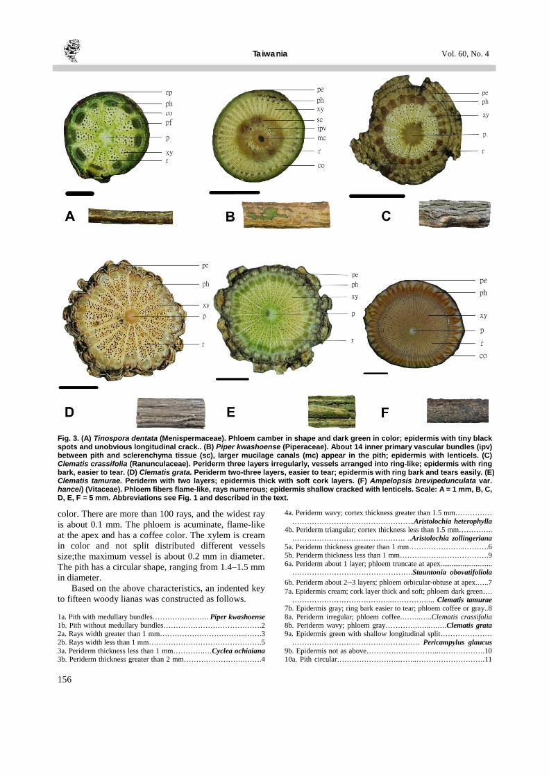
The epidermis has a coffee or light brownish color with tiny black spots and unobvious longitudinal crack. The periderm is less than 0.1 mm in thickness. The cortex is greenish in color and ranges from 0.1–0.2 mm in thickness. There are about 11 rays, in which the widest one is about 0.2 mm. The phloem is camber, darkish green or gray-blackish in color. The phloem fiber is light greenish in color and unobvious. The xylem is cream in color and not split with different size of the vessels distributed; the maximum vessel is about 0.1 mm in diameter. The pith has a compressed-elliptic shape, ranging from 0.6–0.8 mm in diameter. (11) Piper kwashoense (Piperaceae) (Fig. 3B)
The epidermis is yellow-reddish in color with lenticels. The periderm ranges from 0.1–0.3 mm in thickness. The cortex ranges from 0.4–0.6 mm in thickness and is grayish in color. There are about 40 rays, in which the widest one is about 0.1 mm. The
phloem has an orbicular-obtuse shape and is grayish in color. The xylem is cream in color and not split with different size of the vessels distributed; the maximum vessel is about 0.1 mm in diameter. The pith has a compressed-elliptic shape, ranging from 1.5–2 mm in diameter. The inner primary vascular bundles (called medullary bundles) are about 14 between pith and sclerenchyma tissues. (12) Clematis crassifolia (Ranunculaceae) (Fig. 3C)
The epidermis is grayish in color with ring bark and tears easily. The periderm is irregular with about 3 layers and a thickness ranging from 0.4–2.1 mm. the cortex is cream in color and less than 0.1 mm in thickness. There are about 20 rays, in which the widest one is about 0.3 mm. The phloem has an orbicular-obtuse shape with a coffee color. The xylem is cream in color and not split with different size of the vessels distributed; the maximum vessel is about 0.2 mm in diameter. The pith has a circular shape, ranging from 1.9–2.1 mm in diameter. (13) Clematis grata (Ranunculaceae) (Fig. 3D)
The epidermis is grayish in color with ring bark and tears easily. The periderm has about 2–3 layers and ranges from 0.3–1.7 mm in thickness. The cortex is cream in color and ranges from 0.1–0.3 mm in thickness. There are about 35 rays, in which the widest one is about 0.3 mm. The phloem has an orbicular-obtuse shape, grayish color with obvious secondary sclerenchymatous fibers. The xylem is cream in color and not split with different sizes of the vessels distributed; the maximum diameter of vessel is about 0.2 mm. The pith has a circular shape, ranging from 1.5–1.7 mm in diameter. (14) Clematis tamurae (Ranunculaceae) (Fig. 3E)
The epidermis has a cream color with thick and soft cork layers. The periderm has about two layers, and its thickness ranges from 0.9–2.9 mm. The cortex is gray in color and ranges from 0.3–0.9 mm in thickness. There are about 50 rays, in which the widest one is about 0.2 mm. The phloem has an orbicular-obtuse shape and is dark greenish in color. The xylem is cream in color and not split with different sizes of vessels distributed; the maximum vessel is about 0.2 mm in diameter. The pith has a circular shape, and is about 2 mm in diameter. (15) Ampelopsis brevipedunculata var. hancei (Vitaceae) (Fig. 3F)
The epidermis is black-brownish in color and shallow cracked with lenticels, amount of watery outpour when cutting the stem. The periderm and cortex range from 0.1–0.3 mm and 0.7–1.8 mm in thickness, respectively, in which the cortex is whitish in
Taiwania Vol. 60, No. 4
156
Fig. 3. (A) Tinospora dentata (Menispermaceae). Phloem camber in shape and dark green in color; epidermis with tiny black spots and unobvious longitudinal crack.. (B) Piper kwashoense (Piperaceae). About 14 inner primary vascular bundles (ipv) between pith and sclerenchyma tissue (sc), larger mucilage canals (mc) appear in the pith; epidermis with lenticels. (C) Clematis crassifolia (Ranunculaceae). Periderm three layers irregularly, vessels arranged into ring-like; epidermis with ring bark, easier to tear. (D) Clematis grata. Periderm two-three layers, easier to tear; epidermis with ring bark and tears easily. (E) Clematis tamurae. Periderm with two layers; epidermis thick with soft cork layers. (F) Ampelopsis brevipedunculata var. hancei) (Vitaceae). Phloem fibers flame-like, rays numerous; epidermis shallow cracked with lenticels. Scale: A = 1 mm, B, C, D, E, F = 5 mm. Abbreviations see Fig. 1 and described in the text.
color. There are more than 100 rays, and the widest ray is about 0.1 mm. The phloem is acuminate, flame-like at the apex and has a coffee color. The xylem is cream in color and not split distributed different vessels size;the maximum vessel is about 0.2 mm in diameter. The pith has a circular shape, ranging from 1.4–1.5 mm in diameter.
Based on the above characteristics, an indented key to fifteen woody lianas was constructed as follows.
1a. Pith with medullary bundles………………….. Piper kwashoense 1b. Pith without medullary bundles………………………………….2 2a. Rays width greater than 1 mm……………………………..…….3 2b. Rays width less than 1 mm……………………………………….5 3a. Periderm thickness less than 1 mm………….…Cyclea ochiaiana 3b. Periderm thickness greater than 2 mm…………………….…….4
4a. Periderm wavy; cortex thickness greater than 1.5 mm…………… …………………………………………..Aristolochia heterophylla
4b. Periderm triangular; cortex thickness less than 1.5 mm………….. ………………………………………. ..Aristolochia zollingeriana
5a. Periderm thickness greater than 1 mm…………………..……….6 5b. Periderm thickness less than 1 mm……….……..……………….9 6a. Periderm about 1 layer; phloem truncate at apex............................
………………………………………….Stauntonia obovatifoliola 6b. Periderm about 2–3 layers; phloem orbicular-obtuse at apex.…..7 7a. Epidermis cream; cork layer thick and soft; phloem dark green….
…………………………………..……………... Clematis tamurae 7b. Epidermis gray; ring bark easier to tear; phloem coffee or gray..8 8a. Periderm irregular; phloem coffee.……..…..Clematis crassifolia 8b. Periderm wavy; phloem gray………….…..….….Clematis grata 9a. Epidermis green with shallow longitudinal split…………………
……………………………………………. Pericampylus glaucus 9b. Epidermis not as above…………….…………..……………….10 10a. Pith circular…………………….……..……………………….11

December 2015 Yang and Chen: Classifying Taiwan Lianas with Radiating Plates of Xylem
157
10b. Pith compressed-elliptic……………………………………….13 11a. Number of rays greater than 100…………………………………
…………………..…….Ampelopsis brevipedunculata var. hancei 11b. Number of rays less than 50………………..………………….12 12a. Phloem truncate, gray color……….……..….. Celastrus kusanoi 12b. Phloem lengthy-obtuse, dark green color………………………...
………………………………….……………….Vernonia gratiosa 13a. Phloem camber……………………………….Tinospora dentata 13b. Phloem orbicular-obtuse………..……………………………..14 14a. Epidermis without lenticel……..….……...…Stephania merrillii 14b. Epidermis with lenticel.……..………..Neoalsomitra integrifolia DISCUSSION
The phloem anatomy is poorly known since most examples used were dry samples and incompletely showed phloem features and parenchyma (Carlquist, 2001). Based on the fresh examples offered in this study, the characteristics of xylem and phloem were observed and described, thus contributing to widen the borders of knowledge in this field.
Jacques and Franceschi (2007) referred to the radiating plates of xylem as cambial production unit (CPU), which sequentially consisted of pith, secondary xylem, fascicular cambium, secondary phloem, primary phloem, and phloem fibers. The repeated appearance of the CPU unit leads to the formation of a successive cambium. Two subtypes of pericycle, fibrous pericycle and parenchymal pericycle, were developed in Cissampelos andromorpha (Menispermaceae) (Tamaio et al., 2010). However, no endodermis is found in most stems, and there are no pericyclic fibers in stems (Evert, 2006; Carlquist, 2007, 2013). There are eleven woody or herbaceous climber species and one shrub-like species of Menispermaceae in Taiwan, but only Cocculus laurifolius has a successive cambium structure. Four species in this study have a single cambium; the remaining seven species are worthy of further exploration. The parts of CPU, including shape of CPU, phloem fiber, shape of phloem at apex (rounded, curved, arched, truncate), size, color, and texture of phloem, are important terms and available for wood or herbaceous vines identification.
Radiating plates of xylem group included Vitaceae (Carlquist, 2001) (Table 1). However, in Ampelopsis brevipedunculata var. hancei, its xylem tissue appeared to be linear (Fig. 3F). Additionally, its vascular bundles are divided by linear ray-like parenchyma cells, and the secondary phloem at apex is acuminate and flame-like. These features differ from those of Aristolochiaceae, Cucurbitaceae, Menispermaceae and Ranunculaceae. Linear rays are distributed evenly in the cross-section of a stem (Caballé, 1993), and differ from the wide rays, large rays, or giant rays (Acevedo-Rodríguez, 2005; Angyalossy et al., 2012). Therefore, those characteristics of xylem and phloem in A. brevipedunculata var. hancei should be classified into rays. Although the 21 species of Vitaceae in Taiwan are all climbers, the other species
with large rays or not in this family must be studied in the future. The ray area in radiating plates of xylem (Carlquist, 2001) is much less than the fascicular area in cambial activities, and breakup of ray areas is nonexistent. Examples of this tendency are Mikania (Asteraceae); Cocculus and Tinospora (Menispermaceae) (Pfeiffer, 1926). The characteristics of Tinospora dentata (Fig. 3A) revealed that the width of CPU is significantly larger than that of the ray, implying that it is truly classified as this pattern. About fifty species are climbers and belong to four families in Taiwan, including Asteraceae (20 spp.), Celastraceae (7 spp.), Cucurbitaceae (22 spp.) and Lardizabalaceae (5 spp.). Further information regarding the radiating plates of xylem among these families is necessary. Alternatively, Beck (2011) suggested that fascicular areas are few and separated by large ray areas, which are thin-walled parenchyma produced by interfascicular vascular cambium. Comparing this pattern with the radiating plates of xylem may be of worthwhile interest. Table 1 Twenty two families develop radiating plates of xylem (cited from Carlquist, 1991).
Family Actinidiaceae Hippocrateaceae* Apocynaceae Icacinaceae Araliaceae Lardizabalaceae Aristolochiaceae Marcgraviaceae* Asclepiadaceae Menispermaceae Asteraceae Misodendraceae* Begoniaceae Passifloraceae Cactaceae Piperaceae Celastraceae Ranuaculaceae Cucurbitaceae Schisandraceae Dilleniaceae Vitaceae
*Not found in Taiwan.
Combinations of cambial variants were found in lianas, including flattened stems with successive cambium (Angyalossy et al., 2012). The transverse sections of Aristolochia shimadai and A. zollingeriana (Aristolochiaceae) also revealed two cambial variants combinations, i.e. lobed xylem and radiating plates of xylem are found in this study. Although the periderm is extremely obvious in these two species, no periderm is developed in Aristolochia grandiflora (Wagner et al., 2014). Information is still lacking regarding the absence/presence or thin/thickness of periderm in Taiwan Aristolochia genus, making this a worthwhile area of study. In Cyclea ochiaiana (Fig. 2G), phellem is unobvious or none; three Clematis species also obviously develop phellem (Fig. 3C, D, E), explaining why the epidermis variations are useful characteristics for identifying lianas. Nineteen species are climbers in Taiwan Ranunculaceae, in which Clematis genus includes mostly woody or herbaceous vines and a few shrubs, in which their phloem comprises sclerenchymatous fibers (Isnard et al., 2003). According to that study, these
Taiwania Vol. 60, No. 4
158
fibers refer to phloem located in the apex of CPU. Therefore, accuracy in term definition and nomenclature is of priority concern in climber classification.
The stem of Piper betle develops two vascular cylinders (Beck, 2011), i.e. an inner irregular cylinder of primary vascular bundles enclosing the pith, and an outer cylinder of smaller cortical bundles to an undulating cylinder of sclerenchyma. The primary vascular bundles are usually called medullary bundles and occupy the pith. A thick cylinder of secondary tissues consists of regions of tracheary tissues capped by phloem and often with fiber (pf) (Fig. 1) separated by ray-like regions of secondary parenchyma, called medullary rays. Larger mucilage canals (mc) appear in the pith and inner cortex. The stem of Piper kwashoense obviously has both internal and external cylinders, in which inner primary vascular bundles (ipv) persist yet gradually diminish, surrounded by an external cylinder of sclerenchyma (sc) (Fig. 3B). For seven climbing species of Piperaceae in Taiwan, whether they have two vascular cylinders are unknown and warrant further research.
Within the same Aristolochia genus, the skin of Aristolochia shimadai develops lenticels (Fig. 2A), yet those of A. zollingeriana develop none (Fig. 2B). Within different genera in the same family, Cyclea ochiaiana stem surface is brown in color and covered with vertical and deeply depressed lenticels (Fig. 2G), but Pericampylus glaucus surface is greenish in color with a vertical shallow depression (Fig. 2H). Therefore, the skin features of lianas as diagnostic characteristics can be examined to determine the inter-specific or inter-generic differences. However, flowers and fruits collections are limited by different seasons and the parts of leaves have no crucial characters, explaining why the features of stem cross-section or surface offer important evidence in distinguishing between species. Therefore, integrating additional features of stem surface and cross-section plays an important role in liana identification.
The formation of radiating plates of xylem is brought by interpolation of softer tissues such as broad medullary rays, and axial parenchyma in the hard tissue of secondary xylem (Haberlandt, 1914). Wide rays composed of thin-walled cells are involved in the xylem in plates of cambial variants. Indeed, the ray cells are thin-walled and the rays alter little in ontogeny, the xylem is effectively subdivided into plates (Carlquist, 1991). The significance of such wide rays in lianas may be as a means whereby vascular segments of the wood can yield to torsion without sustaining damage to vessels and contribute to liana stem flexibility (Putz and Holbrook, 1991). Here the gross features of xylem plates were described, more data are still desirable to clarify the frequency and distribution pattern of vessels and axial parenchyma. The functional
significance of radiating plates of xylem is needed to be checked and whether this function occurs in other lianas is also worthy of investigation.
ACKNOWLEDGEMENTS
The authors would like to thank Dr. Sherwin Carlquist for some important suggestions about how to use the correct terms of cambial variations. We also thank all staff of PPI Herbarium for kindly helping with specimen collection and identification. Teddy Knoy is appreciated for his editorial assistance. LITERATURE CITED Acevedo-Rodríguez, P. 2005. Vines and climbing plants of
Puerto Rico and the Virgin Islands. The United States National Herbarium Vol. 51. Department of Botany, National Museum of Natural History, Washington, DC. 483 pp.
Angyalossy, V., G. Angeles, M. R. Pace, A. C. Lima, C. L. Dias-Leme, L.G. Lohmann and C. Madero-Vega. 2012. An overview of the anatomy, development and evolution of the vascular system of lianas. Plant Ecol. Divers. 5(2): 167–182.
Angyalossy, V., M. R. Pace, A. C. Lima. 2015. Lianas anotomy: A broad perspective on structural evolution of the vascular system. In: Schnitzer, S. A., F. Bongers, R. J. Burnham and F. E. Putz. (eds.), Ecology of Lianas, 253–287. John Wiley Blackwell, UK.
Beck, C. B. 2011. An Introduction to Plant Structure and Development: plant anatomy for the twenty-first century, 2nd ed. Cambridge University Press, UK. 441 pp.
Boufford, D. E., H. Ohashi, T.-C. Huang, C.-F. Hsieh, J.-L. Tsai, K.-C. Yang, C.-I. Peng, C.-S. Kuoh and A. Hsiao 2003. A checklist of the vascular plants of Taiwan. In: Huang, T.-C. et al. (eds.), Flora of Taiwan, 2nd ed., 6: 15–139. Editorial Committee, Dept. Bot., NTU, Taipei, Taiwan.
Caballé, G. U. Y. 1993. Liana structure, function and selection: a comparative study of xylem cylinders of tropical rainforest species in Africa and America. Bot. J. Linn. Soc. 113: 41–60.
Carlquist, S. 1985. Observation on the functional histology of vine and lianas: vessel dimorphism, tracheids, vasicentric tracheids, narrow vessels, and parenchyma. Aliso 11: 139–157.
Carlquist, S. 1991. Anatomy of vine and liana stems: a review and synthesis. In: Putz, F. E. and H. A. Mooney (eds.), The Biology of Vines, 53–71. Cambridge University Press. UK.
Carlquist, S. 1993. Wood and bark anatomy of Aristolochiaceae: systematic and habital correlation. IAWA J. 14(4): 341–357.
Carlquist, S. 2001. Comparative Wood Anatomy. 2nd ed. Spinger-Verlag Press, Berlin, Germany. 448 pp.
Carlquist, S. 2007. Successive cambia revisited: ontogeny, histology, diversity, and functional significance. J. Torrey Bot. Soc. 134(2): 301–332.
Carlquist, S. 2013. Interxylary phloem: diversity and functions. Brittonia 65(4): 477–495.
December 2015 Yang and Chen: Classifying Taiwan Lianas with Radiating Plates of Xylem
159
Chiang, S.-H. 1973. Anatomy of Plant. National Institute for Compilation and Translation, Taipei, Taiwan. 358 pp. (in Chinese)
Evert, R. F. 2006. Esau's Plant Anatomy: meristems, cells, and tissues of the plant body: their structure, function, and development. 3rd ed. John Wiley and Sons, Inc. Hoboken, New Jersey. 601 pp.
Gartner, H. and F. H. Schweingruber. 2013. Microscopic preparation techniques for plant stem analysis. Swiss Federal Research Institute WSL. Kessel Publishing House, Switzerland. 78 pp.
Haberlandt, G. 1914. Physiological Plant Anatomy. Translated from the 4th German edition by M. Drummond. Mac-Millan: London.
Isnard, S. and W. K. Silk. 2009. Moving with climbing plants from Charles Darwin's time into the 21st century. Amer. J. Bot. 96: 1205–1221.
Isnard, S., T. Speck and N. P. Rowe. 2003. Mechanical architecture and development in Clematis: implications for canalised evolution of growth forms. New Phytol. 158: 543–559.
Jacques, F. M. B. and D. de Franceschi. 2007. Menispermaceae wood anatomy and cambial variants. IAWA J. 28(2): 139–172.
Lo, H. S., T. M. Chen and G. Gilbert. 2008. Menisperaceae. In: Wu, C. Y. and Raven, P. H. (eds.), Flora of China 7: 1–31. Science Press, Beijing, China and Missouri Botanical Garden Press, Saint Louis, USA.
Lu, C.-T. and J.-C. Wang. 2014. Aristolochia yujungiana (Aristolochiaceae): a new species from Taiwan. Taiwan J. For. Sci. 29: 291–299.
Mauseth, J. D. 1988. Plant Anatomy. The Benjamin/Cummings Publishing Company, Menlo Park, California. p.369–379.
Metcalfe, C. R. and L. Chalk. 1950. Anatomy of the dicotyledons. Vol. 2. Clarendon Press, Oxford. 1500 pp.
Metcalfe, C. R. and L. Chalk. 1985. Anatomy of the dicotyledons: Vol. 2: wood structure and conclusion of the general introduction, Ed. 2. Oxford University Press, New York. 297 pp.
Patil, V. S., K. S. Rao and K. S. Rajput. 2009. Development of intraxylary phloem and internal cambium in Ipomoea hederifolia (Convolvulaceae). J. Torrey Bot. Soc. 136(4): 423–432.
Pfeiffer, H. 1926. Das abnorme dickenwachstum. Handbuch der Pflanzenanatomie, Vol. 9. Gebruder Borntraeger, Berlin. 272 pp.
Putz, F. E. and N. M. Holbrook. 1991. Biomechanical studiesof vines. In: Putz, F. E. and H. A. Mooney (eds.), The Biology of Vines, 72–97. Cambridge University Press. UK.
Rajput, K. S., V. S. Patil and K. S. Rao. 2009. Development of included phloem of Calycepteris floribunda Lamk. (Combretaceae). J. Torrey Bot. Soc. 136: 302–312.
Schnitzer, S. A. and F. Bongers. 2002. The ecology of lianas and their role in forests. Trends Ecol. Evol. 17(5): 223–230.
Schnitzer, S. A., P. B. Reich, B. Bergner and W. P. Carson. 2002. Herbivore and pathogen damage on grassland and woodland plants: a test of the herbivore uncertainty principle. Ecol. Lett. 5(4): 531–539.
Schnitzer, S. A. and W. P. Carson. 2010. Lianas suppress tree regeneration and diversity in treefall gaps. Ecol. Lett. 13(7): 849–857.
Sun, S.-F., C.-F. Chen, Y.-H. Chen and S.-Z. Yang. 2014. The anomalous structure of Fabaceae seven lianas. CRJBS 2: 1–10.
Tamaio, N., A. Joffily, J. M. A. Braga and K. S. Rajput. 2010. Stem anatomy and pattern of secondary growth in some herbaceous vine species of Menispermaceae. J. Torrey Bot. Soc. 137(2-3): 157–165.
Tamaio, N., M. F. Neves, A. F. N. Brandes and R. C. Vieira. 2011. Quantitative analyses establish the central vascular cylinder as the standard for wood-anatomy studies in lianas having compound stems (Paullinieae: Sapindaceae). Flora 206(11): 987–996.
Wagner, S. T., L. Hesse1, S. Isnard, M. S. Samain, J. Bolin, E. Maass, C. Neinhuis, N. P. Rowe and S. Wanke. 2014. Major trends in stem anatomy and growth forms in the perianth-bearing Piperales, with special focus on Aristolochia. Ann. Bot. 113(7): 1139–1154.
Yang, S.-Z., C.-F. Chen and K.-L. Lai. 2013. The anomalous structure of stems of Apocynaceae and Asclepiadaceae lianas. J. Natl. Taiwan Mus. 66(3): 57–86. (in Chinese with English summary)
Yang, S.-Z., P.-H. Chen and Y.-J. Chen. 2014. Classification on anomalous structure of lianas stem. J. Natl Taiwan Mus. 67(3): 45–74. (in Chinese with English summary)