Classification and Volumetric Analysis of Temporal Bone ...
28
University of Connecticut OpenCommons@UConn Master's eses University of Connecticut Graduate School 7-9-2013 Classification and Volumetric Analysis of Temporal Bone Pneumatization Using Cone Beam Computed Tomography Aniket B. Jadhav UCHC School of Dental Medicine, [email protected] is work is brought to you for free and open access by the University of Connecticut Graduate School at OpenCommons@UConn. It has been accepted for inclusion in Master's eses by an authorized administrator of OpenCommons@UConn. For more information, please contact [email protected]. Recommended Citation Jadhav, Aniket B., "Classification and Volumetric Analysis of Temporal Bone Pneumatization Using Cone Beam Computed Tomography" (2013). Master's eses. 460. hps://opencommons.uconn.edu/gs_theses/460
Transcript of Classification and Volumetric Analysis of Temporal Bone ...

University of ConnecticutOpenCommons@UConn
Master's Theses University of Connecticut Graduate School
7-9-2013
Classification and Volumetric Analysis of TemporalBone Pneumatization Using Cone BeamComputed TomographyAniket B. JadhavUCHC School of Dental Medicine, [email protected]
This work is brought to you for free and open access by the University of Connecticut Graduate School at OpenCommons@UConn. It has beenaccepted for inclusion in Master's Theses by an authorized administrator of OpenCommons@UConn. For more information, please [email protected].
Recommended CitationJadhav, Aniket B., "Classification and Volumetric Analysis of Temporal Bone Pneumatization Using Cone Beam ComputedTomography" (2013). Master's Theses. 460.https://opencommons.uconn.edu/gs_theses/460

i
Classification and Volumetric Analysis of Temporal Bone Pneumatization Using Cone
Beam Computed Tomography
Aniket Jadhav
B.D.S., MGV’s KBH Dental College and Hospital- India 2008.
A Thesis
Submitted in Partial Fulfillment of the
Requirements for the Degree of
Master of Dental Science
At the
University of Connecticut
2013

ii
APPROVAL PAGE
Masters of Dental Science Thesis
Classification and Volumetric Analysis of Temporal Bone Pneumatization
Using Cone Beam Computed Tomography
Presented by
Aniket Jadhav, B.D.S.
Major Advisor________________________________________________________________
Dr. Alan G. Lurie
Associate Advisor_____________________________________________________________
Dr. Douglas Fellows
Associate Advisor_____________________________________________________________
Dr. Arthur R. Hand
Associate Advisor_____________________________________________________________
Dr. Aditya Tadinada
University of Connecticut
2013

iii
Acknowledgment
I take this opportunity to express my gratitude to the people who have been instrumental in the
successful completion of this project.
I would like to show my greatest appreciation to Dr. Alan Lurie. I can’t say thank you enough for
his tremendous support and help. Without his encouragement and guidance this project would
not have materialized. The guidance and support received from all the members who contributed
and who are contributing to this project, was vital for the success of the project. I am grateful for
their constant support and help I would like to thank my committee members, Dr. Douglas
Fellows, Dr. Arthur Hand and Dr. Aditya Tadinada for their encouragement and insightful
comments. Furthermore I would also like to acknowledge with much appreciation the crucial
role of Dr. Kandasamy Rengasamy who gave me permission to use all required data for this
project. Finally I would like to thank my parents for supporting my education at this premier
institute and for their continued encouragement.

iv
TABLE OF CONTENTS
Title Page i
Approval Page ii
Acknowledgment iii

v
Table of Contents iv
1. Introduction 1
2. Objective and Hypothesis 3
3. Materials and Methods 4
4. Data and Statistical Analysis 10
5. Results 11
6. Discussion 14
7. Conclusion 18
8. Future Directions 19
9. References 20

1
1. Introduction:
The advent of Cone Beam Computed Tomography (CBCT) over the past decade, with its
applications in dentistry in general and Oral and Maxillofacial Radiology (OMFR) in particular,
has led to a variety of maxillofacial applications, including evaluation of portions of the skull
base. During routine OMF radiology practice, portions of the skull base that include the temporal
bone are visualized. The temporal bone often presents a varied pneumatization pattern that has a
specific clinical significance especially important for planning any surgical procedures in this
area.
Each temporal bone consists of four components: the squamous, petromastoid and tympanic
parts and the styloid processes [10]. A proposed function of aeration includes resonance, acting
as a reservoir of air for the middle ear to compensate for altered function of the Eustachian tube
(ET), thereby preventing negative pressure, and avoiding changes in middle ear mucosa which
may progress to otitis media [18, 29].
Pneumatization is the process whereby epithelium infiltrates the developing bone and forms
epithelial lined air cell cavities [14]. The squamous, petromastoid and tympanic parts are the
most frequently pneumatized parts [5, 1, 32], but pneumatization may be extend to the articular
eminence [32, 7] of the zygomatic process. The environmental theory of Wittmaack suggests that
middle ear mucosa is a pre-requisite for normal pneumatization while Diamant suggests that
pneumatization is genetically dependent. The pneumatization process usually begins prenatally,
during the 22nd-24th gestational weeks. At around 28 weeks of gestation, the petrous apex
begins to pneumatize [20]. The pattern of pneumatization of the temporal bone is usually
completed by the age of 10 years in females and 15 years in males [9].
Numerous studies have reported on the variety of pneumatization patterns of the temporal bone
and their classifications using computed tomography (CT). Although CT has several advantages
over CBCT, especially for depicting soft tissues, comparable evaluation of the osseous

2
components of the skull base and sinonasal anatomy are possible at a lower radiation dose and at
a lower cost with CBCT.
In routine OMF radiology practice, a bulk of the temporal bone is routinely visualized on TMJ
and orthodontic studies; thus, it is important for dentists in general and oral radiologists in
particular to know normal anatomical variations and pneumatization patterns to identify any
pathological changes and make appropriate referrals to specialists in this region like
otorhinolaryngologists and neuroradiologists.

3
2. Objective and Hypothesis
Objective: To classify and characterize the pneumatization patterns of the temporal bone using
CBCT data sets.
Specific Aim: To perform volumetric analysis and classify different repeated patterns of
temporal bone pneumatization in adults using CBCT data sets.
Hypothesis: Cone Beam Computed Tomography can be used for qualitative and quantitative
assessment of pneumatization patterns of the temporal bone in adults.

4
3. Materials and Methods:
Data Collection:
Data for the study were gathered after an approval from the Institutional Review Board (IRB) of
The UCONN Health Center was obtained for the study. (IRB # 13-120-1.)
One hundred and fifty five temporal bones were analyzed from 78 patients who were
asymptomatic for any oro-nasal pathology.
The inclusion and exclusion criteria for this retrospective analysis of CBCT scans were as
follows (Table 1).
1. Patients above the age of 18 and not over the age of 70 years.
2. All the scans with a field of view (FOV) more than eight inches in diameter.
The inclusion criteria for the selected scans consist of complete visualization of external auditory
canal, middle ear, Perilabyrinthine area and articular eminence. Cases with presence of soft
tissue, fluid levels, bone sclerosis and destruction and any radiographic evidence of surgery were
excluded (Table 1).
Table 1: Inclusion and Exclusion Criteria
Inclusion Criteria Exclusion Criteria
Complete visualization of:
External ear
Middle ear
Perilabyrinthine region
Petrous apex
Articular eminence
Soft tissue and/or fluid
levels
Sclerosis
Destruction of the bony
margins
Radiographic evidence of
surgical procedure

5
Analysis of the Scans:
There are three primary regions where pneumatization in the temporal bone can be seen:
mastoid, perilabyrinthine and the petrous apex. There are numerous studies which looked at
mastoid pneumatization due to their large air cells based on Multi-slice CT (MSCT) and
conventional radiography. In this study we limited the assessment of the pneumatization pattern
to the middle ear, perilabyrinthine and petrous apex regions because the majority of large field of
view scans of this region show only a part of the mastoid air cells. The mastoid air cells are not
included en toto in the large volume CBCT scanning protocols because it is usually not the
specific area of interest. Both sides of the skull base were assessed for quantitative and
qualitative analysis using CB works 3.0 (Hitachi Medical System America, Inc.) DICOM viewer.
To see the greatest area of pneumatization, a correction of the Z-axis was done in such way that
the malleoincudal complex appeared as an ice-cream cone shaped structure on axial images.
Establishing reference structures for simple classification of temporal bone
pneumatization:
This study followed the method of selection of the reference structures from a study conducted
by Han et al. [12]. Temporal bone pneumatization is classified into the three groups using
labyrinthine and petrous segment of the internal carotid canal as reference structures.
Labyrinthine is used as a reference structure to classify the pneumatization of the temporal
bone around the inner ear structures using the following grouping:
Group 1: No evidence of pneumatization in the region of inner ear (Fig 1A).
Group 2: Pneumatization present either medial or lateral to the superior semicircular
canal on axial section (Fig 1B).
Group 3: Peri- labyrinthine pneumatization (Fig 1C).

6
Fig 1 A B C
Petrous segment of Internal Carotid Canal is used for assessing the pneumatization of the
petrous apex.
Group 1: No pneumatization of petrous apex (Fig 2A).
Group 2: (Mild pneumatization of petrous apex); there are irregularly evident small
numbers of air cells on either side (medial or lateral) of the carotid canal (Fig 2B).
Group 3: (Complete Pneumatization) of petrous apex; pneumatization is present
surrounding the carotid canal (Fig 2C).
Fig 2 A B C

7
Direct communication of peritubal cells with ET was also assessed to assess possible
correlation with degree of pneumatization:
Presence of peritubal cells was assessed to see its correlation with degree of pneumatization of
the temporal bone and further incidence of direct communication (Fig. 3) of peritubal cells with
the bony portion of the eustachian tube was also analyzed.
A B C
Fig 3: Showing direct communication of peritubal cell with bony portion of ET.
Calculation of Volume:
Pneumatization in the temporal bone is mainly present in the mastoids, perilabyrinthine region,
middle ear/tympanic cavity and petrous part of the temporal bone. Since CBCT utilizes isotropic
voxels with thin slices (0.2mm-0.4mm), it can precisely locate and measure small pneumatized
spaces like the Perilabyrinthine region, peritubal region and petrous apex. This is the first type of
study in the English literature looking at pneumatization in the middle ear, perilabyrinthine and
petrous apex region only using a CBCT data set. Measurements of pneumatization of the
temporal bone included the spaces of:
Middle ear cavity including eustachian tube;
Perilabyrinthine region;
Petrous apex.

8
The acquired data were stored in DICOM format (Digital imaging and communication in
medicine) and imported into the CB Works 3.0 (Hitachi Medical System America Inc).
The slice thicknesses and slice distances varied from 0.2mm to 0.4mm. Images were segmented
using lower and upper window levels of -1024 and -290 pixel intensity values to mark the
pneumatized spaces. The windowing function in computed tomography enables the operator to
narrow the shades of gray. The human eye is capable of distinguishing 32 shades of gray while
CT data has a range of 4,096 shades of gray per pixel. In this study we selected the window
threshold of -1024 and -290 pixel intensity to accurately mark the air cavities. In the thresholding
process, the window width and level settings map the measured attenuation of each voxel to a
corresponding gray scale value to appropriately mark the tissue of interest.
All other regions such as pneumatization in the articular eminence, part of sphenoid sinus and the
external auditory canal were excluded using the region of interest tool (Fig. 4). In Fig. 4A, the
initial process of thresholding shows marking of all air cavities in the craniofacial area which
includes paranasal sinuses, nasal cavity, air cells in the skull base/pneumatized bone in the skull
base and airway. As with MSCT data, CBCT data sets can be easily used to perform thresholding
and segmentation of bone and air cavities.
Once all the air cavities are marked, the region of interest tool is applied. Since CBCT utilizes
isotropic voxels, multiplanar views (axial, sagittal and coronal) which precisely mark the area of
interest can be used to limit or extend the area of volume measurement accordingly (Fig. 4B).
Once air spaces are selected, the software algorithm calculates the volume of area of interest
(Fig. 4C).

9
Window thresholding to mark
the pneumatized spaces
Marking region of Interest
Other regions such as articular eminence,
sphenoid sinus and external auditory canal
were excluded.
Selected air spaces

10
4. Data and Statistical Analysis:
Data analysis to evaluate the pattern of pneumatization was done by one OMF radiology faculty
member (AT) and one third year OMF radiology resident (AJ). Differences in the volumes of
temporal bone pneumatization according to the degree of pneumatization between the groups for
each reference structure were investigated using one way analysis of variance (ANOVA) and a
post hoc test. Student’s t-Test was performed to assess possible differences in volume between
right and left temporal bones in the same subject. Correlation of direct communication of
peritubal cells to ET was analyzed using Pearson’s correlation. All statistical analyses were done
using SPSS version 21 [17]; p values less than 0.05 were considered statistically significant.

11
5. Results:
This study population consisted of 78 patients; 27 males and 51 females with age ranges from 18
to 70 years. A total of 155 temporal bones were analyzed to classify the pattern of
pneumatization; 78 on the right side and 77 on the left side. The mean volume of temporal bone
pneumatization was 1337+/-9mm3. The difference between right and left sides was not
statistically significant (p= 0.930).
Classification of temporal bone pneumatization and its corresponding volumetric analysis
Evaluation of temporal bone using labyrinthine as a reference structure:
Cases were classified into three groups. Group 1 consisted of 62 cases, with a mean volume of
822 mm³ and a volume range of 450 mm³ to 1325 mm³. Since group 1 indicates no
pneumatization of the temporal bone, the calculated value represents the volume of the middle
ear cavity. Group 2 consisted of 41 cases with a mean volume of 1269 mm³ and a volume range
of 760 mm³ to 2185 mm³. Group 3 consisted of 52 cases with a mean volume of 2008 mm³ and a
volume range of 1010 mm³ to 3050 mm³.
Overlapping of volumetric measurements between three groups in few cases were observed in
this study due to the large size of the air cells. Despite this, cases were segregated according to
their extension and not based on their volumetric measurements.
Using ANOVA, the three groups showed statistically significant differences (p<0.0001) in their
mean volumes (Fig. 5A). A post hoc test was performed to see the source of significant
differences in the groups. Significant differences existed between all groups (p<0.0001) [Table
2].
Evaluation of temporal bone using carotid canal as a reference structure
Cases were again classified into three groups. Group 1 consisted of 51 cases with a mean volume
of 781 mm³ and a volume range of 443 mm³ to 1325 mm³. Since group 1 indicates no
pneumatization of the temporal bone, the calculated value represents the volume of the middle
ear cavity. Group 2 consisted of 57 cases with a mean volume of 1268 mm³ and a volume range
12
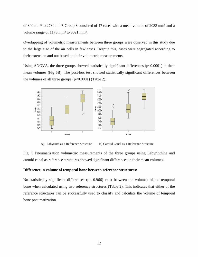
of 840 mm³ to 2780 mm³. Group 3 consisted of 47 cases with a mean volume of 2033 mm³ and a
volume range of 1178 mm³ to 3021 mm³.
Overlapping of volumetric measurements between three groups were observed in this study due
to the large size of the air cells in few cases. Despite this, cases were segregated according to
their extension and not based on their volumetric measurements.
Using ANOVA, the three groups showed statistically significant differences (p<0.0001) in their
mean volumes (Fig 5B). The post-hoc test showed statistically significant differences between
the volumes of all three groups (p<0.0001) (Table 2).
A) Labyrinth as a Reference Structure B) Carotid Canal as a Reference Structure
Fig: 5 Pneumatization volumetric measurements of the three groups using Labyrinthine and
carotid canal as reference structures showed significant differences in their mean volumes.
Difference in volume of temporal bone between reference structures:
No statistically significant differences (p= 0.966) exist between the volumes of the temporal
bone when calculated using two reference structures (Table 2). This indicates that either of the
reference structures can be successfully used to classify and calculate the volume of temporal
bone pneumatization.

13
Table 2: Mean volume of temporal bone.
Labyrinthine as reference Carotid canal as a
reference
ANOVA Post Hoc
Group 1 822mm3
SD: 184.61mm3
781 mm3
SD: 177.2 mm3
P<0.0001
P<0.0001 Group 2 1269mm3
SD: 294.5mm3
1268mm3
SD: 391.9mm3
Group 3 2008mm3
SD: 516.3mm3
2033mm3
SD: 477.3mm3
t -Test P=0.966
Presence of peritubal cells and its correlation with degree of pneumatization:
Of the 155 temporal bones, 99 temporal bones (63.9%) showed the presence of peritubal cells
while 56 (36.1%) had no evidence of peritubal cells. A statistically significant correlation (p<
0.0001) was found between degree of pneumatization and presence of peritubal cells. The
presence of peritubal cells was further categorized on the basis of direct communication with the
bony part of the eustachian tube. Direct communication of peritubal cells at the lateral aspect of
the bony segment of the eustachian tube occurred in 41.3% of the temporal bones.
Communication on the medial aspect was seen in 12.3 % of the temporal bones, while 10.3%
showed communication at the anterior third of the bony segment of the eustachian tube.

14
6. Discussion:
Cone Beam CT technology, also known as volumetric tomography, was developed in the early
1970s and was then utilized in vascular imaging until the early 1980s. This initial prototype was
based on a C-arm instrument. In 1995, two Italian inventors introduced CBCT to dentistry and
the New Tom DVT 9000 was launched in European markets in 1999 [28]. Apart from dentistry,
this technology has gained popularity in the field of otolaryngology, especially for imaging of
paranasal sinuses and the temporal bone.
CBCT is based on a rotating gantry with a fixed source of x-rays and a detector. During rotation
multiple planar projection images are acquired in an arc of more than 180 degrees. CBCT
technology utilizes a cone shaped beam vs. a fan shaped beam in computed tomography.
Orthogonal planar images are secondarily reconstructed from multiple basis projection while in
computed tomography, primary reconstruction produces axial images and then secondary
reconstruction generates orthogonal slices.
To date, numerous studies have been published on uses of CBCT technology for imaging of the
temporal bone, specifically middle and inner ear structures [16, 21, 22, 25, and 34]. CBCT is not
only being used in pre-surgical evaluation of the temporal bone, it is also now being tested intra-
operatively for surgery of the skull base and the temporal bone. Multiple commercial CBCT
systems are available, from limited fields of view to large fields of view. Most of the new
generation CBCT machines give the operator flexibility to choose from various FOV’s targeted
to the area of interest.
Pneumatization in the temporal bone is divided into five major regions; middle ear, mastoid,
petrous apex, perilabyrinthine and accessory. In this study we examined pneumatization in the
middle ear, perilabyrinthine and the petrous apex regions only. In maxillofacial CBCT imaging,
these are the primary regions that usually appear on large field of view scans, as opposed to
dedicated temporal bone studies which include the entire temporal bone. Pneumatic spaces in the
tympanic cavity arise independently of the mastoid antrum, while perilabyrinthine and petrosal
apex cells originate from extensions of the mastoid air cells. The supralabyrinthine region is
pneumatized by the posterior superior and subarcuate tracts while the infralabyrinthine region is

15
pneumatized by hypotympanum, posteromedial and peritubal cells. The petrous apex is
pneumatized by cells from the perilabyrinthine region and peritubal tracts [33].
There are numerous studies of pneumatization of the temporal bone and its volumetric analysis
using conventional radiography and computed tomography. A study conducted by Han et al.
[12] showed a mean volume of 15.28+/-5.34 cm3 using computed tomography. Stieglitz [29]
reported a mean petrous bone air cell volume of 10.97 ml based on MSCT imaging. In our study
the mean volume of temporal bone, was 1337mm3 which is relatively less than these two studies
because we did not include the mastoid air cell system in the classification system and its
volumetric analysis.
In this study we used two reference structures and classified the pneumatization into three
groups. By grouping pneumatization, we were able to show the repeated patterns of extension of
air cells in the temporal bone. In regard to reference structures, labyrinthine and carotid canal are
easy to locate anatomical landmarks on CBCT scans. The numbers of cases in each group are
different for both the reference structures but the calculated mean volume for each group is not
statistically different (Table 2). In this study we showed that either of the reference structures can
be used to classify and perform volume analysis of the pneumatization pattern in the temporal
bone.
CBCT utilizes isotropic voxels with slice thickness of less than 0.4mm and has higher spatial
resolution than MSCT. This makes CBCT technology superior to MSCT when looking at bone
and the air cavities where high contrast resolution is not indicated. In the present study we
calculated the volume of temporal bone air cells including the middle ear cavity on CBCT data.
Group 1 which represents no pneumatization of temporal bone showed a mean volume of 822+/-
50mm3. This value indicates the volume of the middle ear cavity. Petrous apex cells and cells
around labyrinthine were easily located when performing window thresholding and hence
labyrinthine was used as a reference structure. In this study we found that the perilabyrinthine
cells in group 3 were often extended to the posterior wall of the internal auditory canal. These air
cells are of major concern in vestibular schwannoma surgery as they are known as the origin of
CSF fistulae. [31]

16
Apical cells are anatomically located anterior-medial to the internal auditory canal (IAC) and
posterior medial to the carotid canal, again consistently present in group 3. These cells
significantly contributed to the total volume of the temporal bone in the present study. When the
carotid canal was used as the reference structure, group 3 cases (33%) showed extensive
pneumatization surrounding the carotid canal and the petrous apex region. During sub-temporal
transpetrosal transtentorial approach surgery, the petrous apex region medial to the IAC is
resected and it often becomes a route for CSF rhinorrhea. The pneumatized petrous apex is
frequently responsible for CSF leakage after skull base surgery. Stieglitz et al. showed that a
10% increase in petrous bone pneumatization increased the risk of CSF fistulae by 26% [31].
The eustachian tube (ET) has important physiologic functions including pressure regulation and
protection of the middle ear from infectious pathogens from the nasopharynx [19, 29]. ET
dysfunction can be caused by a variety of mechanisms, including upper respiratory tract
infections, sinusitis and adenoidal hypertrophy. The presence of peritubal cells and their
communication with the ET were highly correlated with the degree of pneumatization in the
present study. The relationship of peritubal cells to the bony segment of the eustachian tube plays
a major role in development of CSF rhinorrhea after skull base surgery.
Temporal bones in our study showed direct communication of peritubal cells with the ET in
63.9% of the cases. The most common communication was at the posterior lateral part of the ET
towards the middle ear cavity. ET is known as a common pathway for development of CSF
rhinorrhea during translabyrinthine, suboccipital and middle cranial fossa surgery for acoustic
neuromas. In a study conducted by Jen et al., it was stated that direct communication of peritubal
cells with the osseous portion of ET plays an important role in the development of persistent CSF
rhinorrhea after cerebellopontine angle surgery [19].
CBCT clearly showed the presence of air cells in the temporal bone due to high spatial
resolution. Although there are many significant uses of CBCT in the imaging of the temporal
bone, the low radiation dose which increases noise and gives poor density resolution of soft
tissues, may restrict utilizing this imaging modality rather than MSCT and significantly affect its
use for pre- and post-surgical evaluation of skull base surgeries. If the indication for using CBCT
is correct it can be used to reduce radiation and as an inexpensive technique. Hodez et al. [16]
showed that CBCT images of a well pneumatized middle ear and temporal bone are comparable

17
to high resolution MSCT for visualizing ossicular erosions, traumatic lesions, osseous
labyrinthine wall dehiscence and bony dysplasias.

18
7. Conclusion:
This study showed that CBCT can be effectively used for imaging temporal bone air cavities.
The mean volume of air cells in perilabyrinthine, middle ear, carotid and petrous regions was
1339 mm3. The petrous apex, labyrinthine and petrous segment of carotid canal are critical
structures which can be located easily on CBCT scans and used as references to assess the
pneumatization patterns. As an OMF radiologist it is important to define the normal appearances
and patterns of the temporal bone pneumatization, and to identify potential pathologic changes.
In a variety of temporal bone diseases and otitis media, pneumatization can be affected
significantly; this study showed that analysis of air cells and their volume measurements can be
done effectively utilizing CBCT.
As CBCT is being used widely, and may be approaching standard of care across the field of
dentistry, especially for TMJ and orthodontic evaluation, it is important to identify early changes
in the temporal bone, especially in the regions of inner ear and the central skull base, to make
appropriate referrals to neuroradiologist and ENT specialists to avoid long term functional
disability.

19
8. Future Directions:
Chronic inflammation of the middle ear especially in children can suppress pneumatization of
the temporal bone. It also causes bony erosion and disturbances in the ossicular chain. A
comparative study of diseased bones vs. normal bones needs to be done to correctly identify
changes in the pneumatization pattern and differences in the degree of pneumatization.

20
9. References
1. Allam, A. F. (1969). Pneumatization of the temporal bone. Annals of Otology, Rhinology
and Laryngology, 78(1), 49-64.
2. Apuhan, T., Yildirim, Y. S., & Özaslan, H. (2011). Is there any developmental
relationship between mastoid pneumatization and adenoid tissue volume? International
Journal of Pediatric Otorhinolaryngology, 75(3), 415-419.
3. Austin, D. F. (1977). On the function of the mastoid. Otolaryngologic Clinics of North
America, 10(3), 541-547.
4. Balzeau, A., & Radovčić, J. (2008). Variation and modalities of growth and development
of the temporal bone pneumatization in neandertals. Journal of Human Evolution, 54(5),
546-567.
5. Bast, T. H., & Forester, H. B. (1939). Origin and distribution of air cells in the temporal
bone. Arch Otolaryngol, 30, 183-205.
6. Çakli, H., Cingi, C., Ay, Y., Oghan, F., Ozer, T., & Kaya, E. (2012). Use of cone beam
computed tomography in otolaryngologic treatments. European Archives of Oto-Rhino-
Laryngology, 269(3), 711-720.
7. Carter, L. C., Haller, A. D., Calamel, A. D., & Pfaffenbach, A. C. (1999). Zygomatic air
cell defect (ZACD). prevalence and characteristics in a dental clinic outpatient
population. Dentomaxillofacial Radiology, 28(2), 116-122.
8. Dahmani-Causse, M., Marx, M., Deguine, O., Fraysse, B., Lepage, B., & Escudé, B.
(2011). Morphologic examination of the temporal bone by cone beam computed
tomography: Comparison with multislice helical computed tomography. European
Annals of Otorhinolaryngology, Head and Neck Diseases, 128(5), 230-235.
9. Diamant, M. (1940). Otitis and pneumatization of the mastoid bone. Acta Otolaryngol,
41(SUPPL.), 1-149.
10. Gray’s Anatomy 40th edition (p-615)
11. Habesoglu, T. E., Habesoglu, M., Toros, S. Z., Deveci, I., Surmeli, M., Sheidaei, S., et al.
(2010). How does childhood otitis media change the radiological findings of the temporal
bone? Acta Oto-Laryngologica, 130(11), 1225-1229.
12. Han, S. -., Song, M. H., Kim, J., Lee, W. -., & Lee, H. -. (2007). Classification of
temporal bone pneumatization based on sigmoid sinus using computed tomography.
Clinical Radiology, 62(11), 1110-1118.
13. Hentona, H., Ohkubo, J., Tsutsumi, T., Tanaka, H., & Komatsuzaki, A. (1994).
Pneumatization of the petrous apex. Journal of Otolaryngology of Japan, 97(3), 450-456.
14. Hill, C. A. (2011). Ontogenetic change in temporal bone pneumatization in humans.
Anatomical Record, 294(7), 1103-1115.
15. Hill, C. A., & Richtsmeier, J. T. (2008). A quantitative method for the evaluation of
three-dimensional structure of temporal bone pneumatization. Journal of Human
Evolution, 55(4), 682-690.

21
16. Hodez, C., Griffaton-Taillandier, C., & Bensimon, I. (2011). Cone-beam imaging:
Applications in ENT. European Annals of Otorhinolaryngology, Head and Neck
Diseases, 128(2), 65-78.
17. IBM Corp. Released 2012. IBM SPSS Statistics for Windows, Version 21.0. Armonk,
NY: IBM Corp.
18. Jen, A., Sanelli, P. C., Banthia, V., Victor, J. D., & Selesnick, S. H. (2004). Relationship
of petrous temporal bone pneumatization to the eustachian tube lumen. Laryngoscope,
114(4), 656-660.
19. Jen, A., Sanelli, P. C., Banthia, V., Victor, J. D., & Selesnick, S. H. (2004). Relationship
of petrous temporal bone pneumatization to the eustachian tube lumen. Laryngoscope,
114(4), 656-660.
20. Kenna, M. A. (1996). Embryology and developmental anatomy of the ear. Pediatric
Otolaryngology, 1, 113-126.
21. Ladeira, D. B. S., Barbosa, G. L. R., Nascimento, M. C. C., Cruz, A. D., Freitas, D. Q., &
Almeida, S. M. (2013). Prevalence and characteristics of pneumatization of the temporal
bone evaluated by cone beam computed tomography. International Journal of Oral and
Maxillofacial Surgery, 42(6), 771-775.
22. Majdani, O., Thews, K., Bartling, S., Leinung, M., Dalchow, C., Labadie, R., et al.
(2009). Temporal bone imaging: Comparison of flat panel volume CT and multisection
CT. American Journal of Neuroradiology, 30(7), 1419-1424.
23. Palva, T., & Palva, A. (1966). Size of the human mastoid air cell system. Acta Oto-
Laryngologica, 62(3), 237-251.
24. Palva, T., & Ramsay, H. (2002). Fate of the mesenchyme in the process of
pneumatization. Otology and Neurotology, 23(2), 192-199.
25. Redfors, Y. D., Gröndahl, H. G., Hellgren, J., Lindfors, N., Nilsson, I., & Möller, C.
(2012). Otosclerosis: Anatomy and pathology in the temporal bone assessed by multi-
slice and cone-beam CT. Otology and Neurotology, 33(6), 922-927.
26. Richards, S. D., Saeed, S. R., Laitt, R., & Ramsden, R. T. (2004). Hypercellularity of the
mastoid as a cause of spontaneous pneumocephalus. Journal of Laryngology and
Otology, 118(6), 474-476.
27. Sade, J. (1992). The correlation of middle ear aeration with mastoid pneumatization. the
mastoid as a pressure buffer. European Archives of Oto-Rhino-Laryngology, 249(6), 301-
304.
28. Scarfe, W. C., Li, Z., Aboelmaaty, W., Scott, S. A., & Farman, A. G. (2012).
Maxillofacial cone beam computed tomography: Essence, elements and steps to
interpretation. Australian Dental Journal, 57 Suppl 1, 46-60.
29. Seibert, J. W., & Danner, C. J. (2006). Eustachian tube function and the middle ear.
Otolaryngologic Clinics of North America, 39(6), 1221-1235.
30. Sherwood, R. J. (1995). The Hominid Temporal Bone: Ontogeny and Phylogenetic
Implications,

22
31. Stieglitz, L. H., Giordano, M., Gerganov, V., Raabe, A., Samii, A., Samii, M., et al.
(2010). Petrous bone pneumatization is a risk factor for cerebrospinal fluid fistula
following vestibular schwannoma surgery. Neurosurgery, 67(SUPPL. 2), ons509-ons515.
32. Tyndall, D. A., & Matteson, S. R. (1987). The zygomatic air cell defect (ZACD) on
panoramic radiographs. Oral Surgery Oral Medicine and Oral Pathology, 64(3), 373-
376.
33. Virapongse, C., Sarwar, M., & Bhimani, S. (1985). Computed tomography of temporal
bone pneumatization: 1. normal pattern and morphology. American Journal of
Neuroradiology, 6(4), 551-559.
34. Xu, J., Reh, D. D., Carey, J. P., Mahesh, M., & Siewerdsen, J. H. (2012). Technical
assessment of a cone-beam CT scanner for otolaryngology imaging: Image quality, dose,
and technique protocols. Medical Physics, 39(8), 4932-4942.