Classification and Identification ofthe Viridans Streptococci - Clinical
14
CLINICAL MICROBIOLOGY REVIEWS, JUly 1989, p. 315-328 Vol. 2, No. 3 0893-8512/89/030315-14$02.00/0 Copyright ©) 1989, American Society for Microbiology Classification and Identification of the Viridans Streptococci ALAN L. COYKENDALL Department of Oral Diagnosis, School of Dental Medicine, University of Connecticut Health Center, Farmington, Connecticuit 06032 INTRODUCTION ........................... 315 THE EARLY CONTRIBUTORS ........................... 316 MIDCENTURY DEVELOPMENTS ........................... 317 THE MOLECULAR APPROACH ........................... 317 S. MUTANS .................................. 317 History and Taxonomy ........................... 317 Identification ........................... 318 S. SANGUIS AND S. MITIS .......................... 318 A History of Confusion........................... 318 Recent Results ........................... 319 Identification ........................... 320 S. ANGINOSUS .................................. 320 History and Genetic Relationships ........................... 320 Identification ........................... 321 S. BOVIS ........................... 322 History ........................... 322 Genetic Relationships ........................... 322 Identification ........................... 323 S. SALIVARIUS .................................. 323 History ........................... 323 DNA and Identification ........................... 323 S. VESTIBULARIS .................................. 324 A New Species ........................... 324 CONCLUSION ........................... 324 For More Information ........................... 325 Final Thoughts ........................... 325 ACKNOWLEDGMENTS ........................... 325 ADDENDUM IN PROOF ........................... 325 LITERATURE CITED ........................... 325 INTRODUCTION The nonhemolytic streptococci are those whose colonies do not produce clear zones on blood agar by the complete destruction of erythrocytes; that is, they are not beta- hemolytic. Because many produce greenish discoloration on blood agar (alpha hemolysis), the group is also called the viridans streptococci, although many are completely indif- ferent to blood. They usually do not react with Lancefield grouping sera. They are human commensals and are a major component of the oral flora. Even within this loose defini- tion, exceptions abound: some types of Streptococcus ang- inosus are beta-hemolytic, and many react with Lancefield A, C, F, or G antiserum; S. bovis has some characteristics of the enterococci, including a Lancefield D reaction. The human nonhemolytic streptococci can also be defined in a practical way by the exclusion of other streptococci; the viridans group is what remains when human pathogens and commensals that are S. pyogenes, enterococci (a separate genus now) (106), pneumococci, Lancefield group B, and "large colony" group C and G are eliminated. Although these bacteria are usually docile tenants of the mouth and gut, they are capable of causing diseases and death among their hosts, especially when they gain entrance to sites that are usually sterile. Nonhemolytic streptococci account for about one-half of all cases of streptococcal endocarditis, a life-threatening disease that requires long periods of expensive treatment. S. mutans is responsible for dental caries, a condition whose treatment accounts for great losses of time and treasure. S. anginosus, in its various guises, causes abscesses in the brain, liver, and joints and may also be important in neonatal infections. The words "nonhemolytic streptococci" are nearly al- ways preceded or followed by words such as "poorly classified." In this review, it will be shown that the taxon- omy of the human nonhemolytic streptococci has improved as ever closer examination has revealed the biochemical and genetic relationships among these bacteria. Improved taxon- omy has facilitated identification and has enabled laborato- ries to correlate species with diseases (92). Perfection has not been attained, however. Although there is considerable agreement now on which of these streptococci are related to others and which are not, there is far less unanimity on the matter of appropriate names and type strains. This review traces the history of the six most common nonhemolytic species and presents their current taxonomic positions, with emphasis on the molecular or genetic rela- tionships among these bacteria. These six species encom- pass streptococci that have been given many other names. 315 Downloaded from https://journals.asm.org/journal/cmr on 06 January 2022 by 112.167.235.15.
Transcript of Classification and Identification ofthe Viridans Streptococci - Clinical
CLINICAL MICROBIOLOGY REVIEWS, JUly 1989, p. 315-328 Vol. 2, No. 30893-8512/89/030315-14$02.00/0Copyright ©) 1989, American Society for Microbiology
Classification and Identification of the Viridans StreptococciALAN L. COYKENDALL
Department of Oral Diagnosis, School of Dental Medicine, University of Connecticut Health Center,Farmington, Connecticuit 06032
INTRODUCTION........................... 315THE EARLY CONTRIBUTORS........................... 316MIDCENTURY DEVELOPMENTS........................... 317THE MOLECULAR APPROACH........................... 317S. MUTANS.................................. 317
History and Taxonomy........................... 317Identification........................... 318
S. SANGUIS AND S. MITIS.......................... 318A History of Confusion........................... 318Recent Results........................... 319Identification........................... 320
S. ANGINOSUS.................................. 320History and Genetic Relationships ........................... 320Identification........................... 321
S. BOVIS........................... 322History........................... 322Genetic Relationships........................... 322Identification........................... 323
S. SALIVARIUS.................................. 323History........................... 323DNA and Identification........................... 323
S. VESTIBULARIS.................................. 324A New Species........................... 324
CONCLUSION........................... 324For More Information........................... 325Final Thoughts........................... 325
ACKNOWLEDGMENTS........................... 325ADDENDUM IN PROOF ........................... 325LITERATURE CITED........................... 325
INTRODUCTION
The nonhemolytic streptococci are those whose coloniesdo not produce clear zones on blood agar by the completedestruction of erythrocytes; that is, they are not beta-hemolytic. Because many produce greenish discoloration onblood agar (alpha hemolysis), the group is also called theviridans streptococci, although many are completely indif-ferent to blood. They usually do not react with Lancefieldgrouping sera. They are human commensals and are a majorcomponent of the oral flora. Even within this loose defini-tion, exceptions abound: some types of Streptococcus ang-inosus are beta-hemolytic, and many react with LancefieldA, C, F, or G antiserum; S. bovis has some characteristics ofthe enterococci, including a Lancefield D reaction. Thehuman nonhemolytic streptococci can also be defined in apractical way by the exclusion of other streptococci; theviridans group is what remains when human pathogens andcommensals that are S. pyogenes, enterococci (a separategenus now) (106), pneumococci, Lancefield group B, and"large colony" group C and G are eliminated.Although these bacteria are usually docile tenants of the
mouth and gut, they are capable of causing diseases anddeath among their hosts, especially when they gain entranceto sites that are usually sterile. Nonhemolytic streptococci
account for about one-half of all cases of streptococcalendocarditis, a life-threatening disease that requires longperiods of expensive treatment. S. mutans is responsible fordental caries, a condition whose treatment accounts for greatlosses of time and treasure. S. anginosus, in its variousguises, causes abscesses in the brain, liver, and joints andmay also be important in neonatal infections.The words "nonhemolytic streptococci" are nearly al-
ways preceded or followed by words such as "poorlyclassified." In this review, it will be shown that the taxon-omy of the human nonhemolytic streptococci has improvedas ever closer examination has revealed the biochemical andgenetic relationships among these bacteria. Improved taxon-omy has facilitated identification and has enabled laborato-ries to correlate species with diseases (92). Perfection hasnot been attained, however. Although there is considerableagreement now on which of these streptococci are related toothers and which are not, there is far less unanimity on thematter of appropriate names and type strains.
This review traces the history of the six most commonnonhemolytic species and presents their current taxonomicpositions, with emphasis on the molecular or genetic rela-tionships among these bacteria. These six species encom-pass streptococci that have been given many other names.
315
Dow
nloa
ded
from
http
s://j
ourn
als.
asm
.org
/jour
nal/c
mr
on 0
6 Ja
nuar
y 20
22 b
y 11
2.16
7.23
5.15
.
316 COYKENDALL
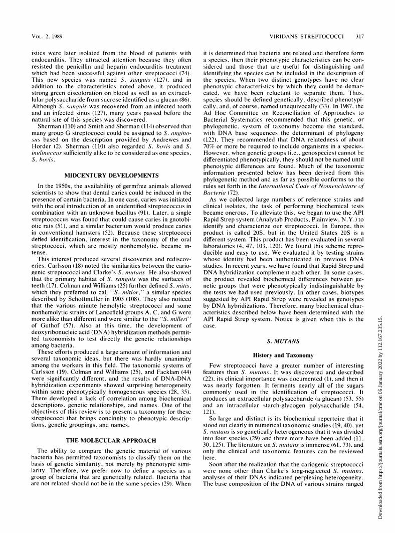
TABLE 1. The common viridans streptococci
Test'Species Comments
VP ARG ESC MAN SORB
S. anginosus Phenotypically heterogeneous; now includes S. constellatus, S. interminedius, + + + +Streptococcus MG, group F, "S. /nillenii,' and minute hemolytic strains;important in purulent infections
S. bovis Nonenterococcal group D; variants are less reactive; common blood isolate in + - + ±patients with colon cancer; animal strains may be different species
S. mitis Also called "S. mitior," S. oralis, and, when raffinose positive, S. sanguis II;includes two genetic groups
S. mnutans Actually a group of seven species which are phenotypically similar: causes + - + + +dental caries and endocarditis
S. salit'arius Rare in infections; genetically homogeneous; can resemble S. boi'is + - +S. sanguis Causes endocarditis; genetically heterogeneous; one genetic group contains the - + +
type strain of S. mnitisS. v'estibularis New species from the oral cavity + - +
" VP, Voges-Proskauer; ARG, hydrolysis of arginine; ESC, hydrolysis of esculin; MAN, fermentation of mannitol: SORB, fermentation of sorbitol.
Table 1 summarizes the species, their other names, and theirnotable attributes along with a very simple guide to theirbiochemical traits. Whenever possible, the names used arethose on the Approved List of Bacterial Names (113), andthe rationale for using these names will be explained. As willbe seen, the names and descriptions published by the earliestworkers in this field often are the most appropriate. Mostimportant, the phenotypic portraits of the species will bedrawn so that isolates can be identified and differentiatedfrom each other.The nutritionally variant streptococci are not considered
here. Although they have been considered forms of S.sanguis or S. mitis, they are genetically unrelated and format least two distinct genospecies (11a).
THE EARLY CONTRIBUTORSThree species of nonhemolytic streptococci that are still
valid were named and described in 1906 by Andrewes andHorder (2). They recognized a rarely pathogenic resident ofthe mouth and intestine which they called S. mitis. Fordecades, this species has been poorly defined because itferments or hydrolyzes few of the substrates commonly usedto describe and identify streptococci. Often, isolates thatwere not clearly members of other species were relegated toS. mitis. This species can be more accurately describedtoday but, as we shall see, the type strain does not fit thedescription and the taxonomic position of S. mitis is onceagain confused. This problem and a proposed remedy will befully discussed below.Andrewes and Horder also found a streptococcus that
fermented sucrose, lactose, and usually raffinose and was socommon in saliva that they named it S. sali'ariius (2). Thisspecies has always been well-defined, and its taxonomicposition has rarely been assailed.These two early investigators also described a hemolytic
species that they associated with disease, including scarletfever. (They were careful not to assign a definite etiologicrole to the organism.) It fermented sucrose and lactose andoften fermented raffinose. To this species they gave thename S. anginosus (2). Although it is hemolytic, we nowknow that there are many nonhemolytic variants of S.anginosus which have been described as "S. milleri" S.constellatus, S. intermedius, and Streptococcus MG. S.anginosus is now regarded as a member of the nonhemolyticgroup. This story will be told later. The observation of ahemolytic streptococcus that fermented raffinose and was
associated with disease was very perceptive, and theseobservations give validity to both the name and the impor-tance of S. anginosus today. For anyone interested in thedevelopment of taxonomy, the early application of biochem-ical tests, and the concept of a species, the treatise byAndrewes and Horder is recommended.
In 1919, Orla-Jensen (90) described S. boi'is, an organismcommon in the bovine gut. It fermented starch, inulin,raffinose, and arabinose, but not mannitol. Similar strepto-cocci that did ferment mannitol but not arabinose werenamed S. inulinaceus (67). Streptococci that resemble S.bos'is are isolated from humans and are often encountered inendocarditis, and the species is more common in the bowelsof people with colon disease. The significance of Orla-Jensen's work is that he recognized diversity among thebovine streptococci. Failure to attach significance to thisdiversity has contributed to some confusion about whatspecies S. bovis really is.
Dental caries has occupied the thoughts of scientists andphilosophers for centuries. Bacteria had been considered theagent of this disease since W. D. Miller's cogent argument of1880 (81), but there was little knowledge of which of thescores of microbial forms in the mouth were responsible forthe decay of teeth. J. K. Clarke sought bacteria that wereassociated with decayed teeth and that could produceenough acid to initiate caries. He found such an organismand named it S. mutans (22). It produced a pH of 4.2 inglucose broth and fermented glucose, lactose, raffinose,mannitol (mannite), inulin, and salicin. Clarke also suc-ceeded in producing caries in extracted teeth in vitro with hisS. nutans. His discoveries were confirmed by Maclean (79)3 years later, and in 1928 Abercrombie and Scott (1) isolatedS. mnutans from a case of endocarditis. Until the 1960s, S.muitans was mentioned as a cause of dental caries in severaltextbooks (15, 75-77, 117, 118), but little if any researchseems to have been done to explore further the landsdiscovered by Clarke.
In the 1930s and 1940s, Sherman and others refined thedefinition of several streptococci. Sherman et al. (111) re-viewed the ecology and characteristics of S. salivarius andS. mitis. They noted the lack of biochemical activity of S.mitis and the lack of properties "uniquely its own." Theysuspected that the S. mnitis "group" (as they called it)contained other entities because some members hydrolyzedarginine and esculin and fermented salicin. This observationwas perceptive, because streptococci with these character-
CLIN. MICROBIOL. REV.
Dow
nloa
ded
from
http
s://j
ourn
als.
asm
.org
/jour
nal/c
mr
on 0
6 Ja
nuar
y 20
22 b
y 11
2.16
7.23
5.15
.
VIRIDANS STREPTOCOCCI 317
istics were later isolated from the blood of patients withendocarditis. They attracted attention because they oftenresisted the penicillin and heparin endocarditis treatmentwhich had been successful against other streptococci (74).This new species was named S. sanguis (127), and inaddition to the characteristics noted above, it producedstrong green discoloration on blood as well as an extracel-lular polysaccharide from sucrose identified as a glucan (86).Although S. sainguis was recovered from an infected toothand an infected sinus (127), many years passed before thenatural site of this species was discovered.Sherman (110) and Smith and Sherman (114) observed that
many group G streptococci could be assigned to S. angino-sits based on the description provided by Andrewes andHorder (2). Sherman (110) also regarded S. boils and S.inulinaceits sufficiently alike to be considered as one species,S. boi'is.
MIDCENTURY DEVELOPMENTS
In the 1950s, the availability of germfree animals allowedscientists to show that dental caries could be induced in thepresence of certain bacteria. In one case, caries was initiatedwith the oral introduction of an unidentified streptococcus incombination with an unknown bacillus (91). Later, a singlestreptococcus was found that could cause caries in gnotobi-otic rats (51), and a similar bacterium would produce cariesin conventional hamsters (52). Because these streptococcidefied identification, interest in the taxonomy of the oralstreptococci, which are mostly nonhemolytic. became in-tense.
This interest produced several discoveries and rediscov-eries. Carlsson (18) noted the similarities between the cario-genic streptococci and Clarke's S. inuhtns. He also showedthat the primary habitat of S. sanguis was the surfaces ofteeth (17). Colman and Williams (25) further defined S. mitis,which they preferred to call "S. mitior," a similar speciesdescribed by Schottmuller in 1903 (108). They also noticedthat the various minute hemolytic streptococci and somenonhemolytic strains of Lancefield groups A. C, and G weremore alike than different and were similar to the "S. inille i"'of Guthof (57). Also at this time, the development ofdeoxyribonucleic acid (DNA) hybridization methods permit-ted taxonomists to test directly the genetic relationshipsamong bacteria.These efforts produced a large amount of information and
several taxonomic ideas, but there was hardly unanimityamong the workers in this field. The taxonomic systems ofCarlsson (19), Colman and Williams (25), and Facklam (44)were significantly different, and the results of DNA-DNAhybridization experiments showed surprising heterogeneitywithin some phenotypically homogeneous species (28, 35).There developed a lack of correlation among biochemicaldescriptions, genetic relationships, and names. One of theobjectives of this review is to present a taxonomy for thesestreptococci that brings concinnity to phenotypic descrip-tions, genetic groupings, and names.
THE MOLECULAR APPROACH
The ability to compare the genetic material of variousbacteria has permitted taxonomists to classify them on thebasis of genetic similarity, not merely by phenotypic simi-larity. Therefore, we prefer now to define a species as agroup of bacteria that are genetically related. Bacteria thatare not related should not be in the same species (29). When
it is determined that bacteria are related and therefore forma species, then their phenotypic characteristics can be con-sidered and those that are useful for distinguishing andidentifying the species can be included in the description ofthe species. When two distinct genotypes have no clearphenotypic characteristics by which they could be demar-cated, we have been reluctant to separate them. Thus,species should be defined genetically, described phenotypi-cally, and, of course, named unequivocally (33). In 1987, theAd Hoc Committee on Reconciliation of Approaches toBacterial Systematics recommended that this genetic, orphylogenetic, system of taxonomy become the standard,with DNA base sequences the determinant of phylogeny(122). They recommended that DNA relatedness of about70% or more be required to include organisms in a species.However, when genetic groups (i.e., genospecies) cannot bedifferentiated phenotypically, they should not be named untilphenotypic differences are found. Much of the taxonomicinformation presented below has been derived from thisphylogenetic method and as far as possible conforms to therules set forth in the International Code of Nomenclature ofBacteria (72).As we collected large numbers of reference strains and
clinical isolates, the task of performing biochemical testsbecame onerous. To alleviate this, we began to use the APIRapid Strep system (Analytab Products, Plainview, N.Y.) toidentify and characterize our streptococci. In Europe, thisproduct is called 20S, but in the United States 20S is adifferent system. This product has been evaluated in severallaboratories (4, 47, 103, 120). We found this scheme repro-ducible and easy to use. We evaluated it by testing strainswhose identity had been authenticated in previous DNAstudies. In recent years, we have found that Rapid Strep andDNA hybridization complement each other. In some cases,the product revealed biochemical differences between ge-netic groups that were phenotypically indistinguishable bythe tests we had used previously. In other cases, biotypessuggested by API Rapid Strep were revealed as genotypesby DNA hybridizations. Therefore, many biochemical char-acteristics described below have been determined with theAPI Rapid Strep system. Notice is given when this is thecase.
S. MUTANS
History and Taxonomy
Few streptococci have a greater number of interestingfeatures than S. inutanzs. It was discovered and described(22), its clinical importance was documented (1), and then itwas nearly forgotten. It ferments nearly all of the sugarscommonly used in the identification of streptococci. Itproduces an extracellular polysaccharide (a glucan) (53, 55)and an intracellular starch-glycogen polysaccharide (54,121).So large and distinct is its biochemical repertoire that it
stood out clearly in numerical taxonomic studies (19, 40), yetS. inhitans is so genetically heterogeneous that it was dividedinto four species (29) and three more have been added (11,30, 125). The literature on S. mutants is immense (61, 73), andonly the clinical and taxonomic features can be reviewedhere.Soon after the realization that the cariogenic streptococci
were none other than Clarke's long-neglected S. inutans,analyses of their DNAs indicated perplexing heterogeneity.The base composition of the DNA of various strains ranged
VOL. 2. 1989
Dow
nloa
ded
from
http
s://j
ourn
als.
asm
.org
/jour
nal/c
mr
on 0
6 Ja
nuar
y 20
22 b
y 11
2.16
7.23
5.15
.
318 COYKENDALL
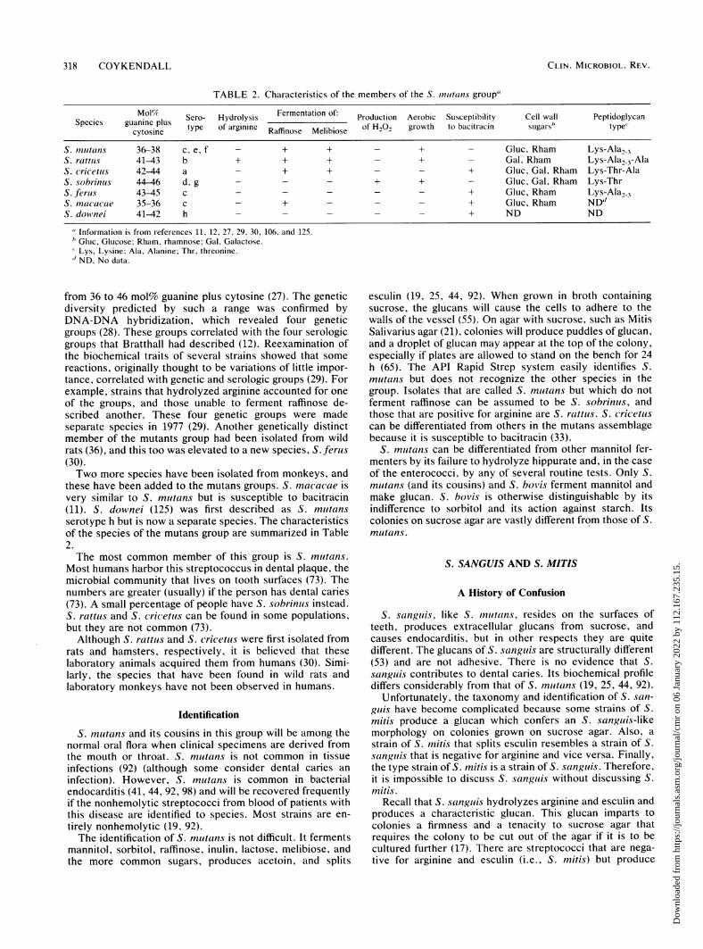
TABLE 2. Characteristics of the members of the S. inutans group'
Mol Sero- Hydrolysis Fermentation of: Production Aerobic Susceptibility Cell wall PeptidoglycanSpecies guanine plus Sero- yrolysis oH__gwht_____surbyp
cytosine type of arginine Raffinose Melibiose of H202 growth to bacitracin sugars type
S. mutans 36-38 c, e, f - + + - + - Gluc, Rham Lys-Ala2 XS. rattus 41-43 b + + + - + - Gal, Rham Lys-Ala23-AlaS. cricetus 42-44 a - + + - - + Gluc, Gal, Rham Lys-Thr-AlaS. sobrinus 44-46 d, g - - - + + - Gluc, Gal, Rham Lys-ThrS. ferus 43-45 c - - - - - + Gluc, Rham Lys-Ala2 3S. macacae 35-36 c - + - - - + Gluc, Rham ND"S. downei 41-42 h - - - - - + ND ND
" Information is from references 11, 12, 27, 29, 30, 106, and 125." Gluc, Glucose; Rham, rhamnose; Gal, Galactose.Lys, Lysine; Ala, Alanine; Thr. threonine.
"ND, No data.
from 36 to 46 mol% guanine plus cytosine (27). The geneticdiversity predicted by such a range was confirmed byDNA-DNA hybridization, which revealed four geneticgroups (28). These groups correlated with the four serologicgroups that Bratthall had described (12). Reexamination ofthe biochemical traits of several strains showed that somereactions, originally thought to be variations of little impor-tance, correlated with genetic and serologic groups (29). Forexample, strains that hydrolyzed arginine accounted for oneof the groups, and those unable to ferment raffinose de-scribed another. These four genetic groups were madeseparate species in 1977 (29). Another genetically distinctmember of the mutants group had been isolated from wildrats (36), and this too was elevated to a new species, S. ferus(30).Two more species have been isolated from monkeys, and
these have been added to the mutans groups. S. macacae isvery similar to S. mutans but is susceptible to bacitracin(11). S. downed (125) was first described as S. mutansserotype h but is now a separate species. The characteristicsof the species of the mutans group are summarized in Table2.The most common member of this group is S. mutans.
Most humans harbor this streptococcus in dental plaque, themicrobial community that lives on tooth surfaces (73). Thenumbers are greater (usually) if the person has dental caries(73). A small percentage of people have S. sobrinuts instead.S. rattus and S. cricetus can be found in some populations,but they are not common (73).
Although S. rattus and S. cricetus were first isolated fromrats and hamsters, respectively, it is believed that theselaboratory animals acquired them from humans (30). Simi-larly, the species that have been found in wild rats andlaboratory monkeys have not been observed in humans.
Identification
S. mutans and its cousins in this group will be among thenormal oral flora when clinical specimens are derived fromthe mouth or throat. S. mutans is not common in tissueinfections (92) (although some consider dental caries aninfection). However, S. mutans is common in bacterialendocarditis (41, 44, 92, 98) and will be recovered frequentlyif the nonhemolytic streptococci from blood of patients withthis disease are identified to species. Most strains are en-tirely nonhemolytic (19, 92).The identification of S. muitans is not difficult. It ferments
mannitol, sorbitol, raffinose, inulin, lactose, melibiose, andthe more common sugars, produces acetoin, and splits
esculin (19, 25, 44, 92). When grown in broth containingsucrose, the glucans will cause the cells to adhere to thewalls of the vessel (55). On agar with sucrose, such as MitisSalivarius agar (21), colonies will produce puddles of glucan,and a droplet of glucan may appear at the top of the colony,especially if plates are allowed to stand on the bench for 24h (65). The API Rapid Strep system easily identifies S.mutans but does not recognize the other species in thegroup. Isolates that are called S. mutans but which do notferment raffinose can be assumed to be S. sobrinus, andthose that are positive for arginine are S. rattus. S. cricetuscan be differentiated from others in the mutans assemblagebecause it is susceptible to bacitracin (33).
S. nutans can be differentiated from other mannitol fer-menters by its failure to hydrolyze hippurate and, in the caseof the enterococci, by any of several routine tests. Only S.mutans (and its cousins) and S. bovis ferment mannitol andmake glucan. S. bovis is otherwise distinguishable by itsindifference to sorbitol and its action against starch. Itscolonies on sucrose agar are vastly different from those of S.mutans.
S. SANGUIS AND S. MJTIS
A History of Confusion
S. sanguis, like S. mutans, resides on the surfaces ofteeth, produces extracellular glucans from sucrose, andcauses endocarditis, but in other respects they are quitedifferent. The glucans of S. sanguis are structurally different(53) and are not adhesive. There is no evidence that S.sanguis contributes to dental caries. Its biochemical profilediffers considerably from that of S. nutans (19, 25, 44, 92).
Unfortunately, the taxonomy and identification of S. san-guis have become complicated because some strains of S.mitis produce a glucan which confers an S. sanguis-likemorphology on colonies grown on sucrose agar. Also, astrain of S. mitis that splits esculin resembles a strain of S.sanguis that is negative for arginine and vice versa. Finally,the type strain of S. mitis is a strain of S. sanguis. Therefore,it is impossible to discuss S. sanguis without discussing S.Mitis.
Recall that S. sanguis hydrolyzes arginine and esculin andproduces a characteristic glucan. This glucan imparts tocolonies a firmness and a tenacity to sucrose agar thatrequires the colony to be cut out of the agar if it is to becultured further (17). There are streptococci that are nega-tive for arginine and esculin (i.e., S. mitis) but produce
CLIN. MICROBIOL. REV.
Dow
nloa
ded
from
http
s://j
ourn
als.
asm
.org
/jour
nal/c
mr
on 0
6 Ja
nuar
y 20
22 b
y 11
2.16
7.23
5.15
.
VIRIDANS STREPTOCOCCI 319
similar glucans and similar colonies. These have been calledS. sanguis. Carlsson (19), for example, recognized thebiochemical differences between these and typical S. san-guis strains but considered them S. sanguis based on otherbiochemical similarities, glucan production, and their similarecology, i.e., the tooth surface. It was Colman and Williams(25) who suggested that these isolates were glucan-producingmembers of the species they preferred to call "S. mitior."DNA studies confirmed that the streptococci that producedglucans like S. sanguis but that did not split esculin orarginine were not closely related to typical S. sanguis strains(35) but were indeed related to typical "S. mitior" (or S.mitis) strains that did not produce glucans (34). Further-more, there were two genetic groups within S. sanguis (35)and two genetic groups within "S. mitior" (34).A key to separating S. sanguis from "S. mitior" was the
discovery that "S. mitior" has ribitol, but no rhamnose, inits cell wall (24). It was also found that the peptidoglycan ofthese streptococci has direct lysine cross-links (105), basedon analysis of ATCC 10557, a strain referred to in theAmerican Type Culture Collection list as S. sanguis butwhich is biochemically and genetically a member of "S.mitior" (34, 35). Thus, "S. mitior" (or S. mitis) can beclearly described and differentiated from S. sanguis based oncell wall analysis.
In the taxonomic and identification system of Facklam(44), strains that produce the S. sanguis-like glucan andferment raffinose are called S. sanguis II. These strains areotherwise similar to S. mitis, and DNA hybridization hasshown that there is no taxonomic significance to raffinosefermentation in this species (34).
In 1980, the Approved List of Bacterial Names (113)accepted S. sanguis, with ATCC 10556 (=NCTC 7863) asthe type strain. The official description is that in Bergey'sManual of Determinative Bacteriology (8th ed.) (39). Theauthors did not accept "S. mitior" but did accept S. mitis,with NCTC 3165 chosen as the type strain (113). The officialdescription is that in Bergey's Manual (8th ed.) (39), adescription based on that by Sherman at al. (111).
Therefore, by 1980, S. sanguis was an accepted specieswith a good description, consisting of two moderately re-lated genetic groups, one of which contained the type strain.There were no clear phenotypic differences between the twogenetic groups. S. mitis was an accepted species with asomewhat equivocal description, consisting of two geneticgroups with no phenotypic tests to differentiate them. Un-fortunately, the type strain chosen for S. mitis is actually astrain of S. sanguis. It attacks arginine and esculin andferments raffinose and inulin; therefore, it fits the descriptionof S. sanguis, not that of S. mitis. Its DNA hybridizes withthat of S. sanguis strains in the genetic group that isrepresented not by the type strain, but by S. sanguis ATCC10558 (=NCTC 7865). Its DNA is not similar to that of anyS. mitis strain (A. L. Coykendall and P. Wesbecher, Abstr.10th Lancefield Conf. 1987, Cologne, Federal Republic ofGermany, p. 33).Using Rapid Strep tests, we now know that the members
of the S. sanguis genetic group that includes the type strain(ATCC 10556) and which we call genetic type 2 (group 3 inreference 35) do not produce alkaline phosphatase andusually split starch. Genetic type 1 strains, represented byATCC 10558 and the S. mitis type strain, have the oppositereactions (33). Nyvad and Kilian (89) have suggested that thegroup represented by ATCC 10558 become a separate spe-cies named S. gordonii. The group that includes the typestrain must, of course, remain S. sanguis.
This story now becomes more convoluted. In 1982, Bridgeand Sneath (14) named a new species, S. oralis. Some of thestrains of S. oralis had been described earlier as "S. mitior"(S. mitis) (34). The type strain of S. oralis (NCTC 11427) hasthe phenotypic characters of S. mitis, its peptidoglycan hasdirect lysine cross-links (107), and its DNA forms hybridswith strains that we recognize as S. mitis, such as ATCC10557 (Coykendall and Wesbecher, Abstr. 10th LancefieldConf. 1987). Although S. oralis has been reported to produceacetoin (107), we have not observed this.
This state of affairs can be resolved one of two ways, bothtaxonomically correct (72). Those strains of S. sanguiswhich are biochemically and genetically like the type strainof S. mitis could be called S. mitis, and what is now S. mitiscould be renamed S. oralis. This has been suggested (106,107). It is my opinion that this would lead to great confusion.Streptococci now called S. mitis (or "S. mitior") would becalled S. oralis. About half of what are called S. sanguiswould be renamed S. mitis, but except for DNA analysis andthe few biochemical differences noted above (starch andalkaline phosphatase), S. mitis and S. sanguis would bedifficult to differentiate. Also, the official descriptions wouldhave to be rewritten. It is unlikely that such a change wouldmeet with approval.A much simpler solution would be merely to reject the
current type strain of S. mitis and select a new type strainthat fits the description of S. mitis and whose DNA ishomologous with DNAs of other strains that are recognizedas S. mitis. The rules of taxonomy provide for this, and arequest for an opinion has been published (31). The requestproposes that the type strain of S. mitis be rejected and S.oralis be considered a later synonym of S. mitis. The currenttype strain of S. oralis would become the type strain of S.mitis.
Recent Results
Recent examination of some unusual S. sanguis strainsuncovered two additional genetic groups (Coykendall andWesbecher, Abstr. 10th Lancefield Conf. 1987). A group ofthree strains that bore tufts of fibrils on their walls (62) wasclosely related by DNA homology but less related to the twoS. sanguis genogroups described above. These strains splitarginine but gave weak esculin reactions. They did notproduce alkaline phosphatase or ferment raffinose.A fourth group was genetically quite distinct and was
represented by five strains which produced alkaline phos-phatase (four of which also fermented raffinose) and hydro-lyzed arginine. Four gave weak or negative esculin reac-tions.
Thus, the viridans streptococci that typically split arginineand esculin and do not produce acetoin form at least fourgenetic groups. We have continued to call them all S.sanguis. The groups are sufficiently disparate to be consid-ered separate species (89), but we have been reluctant topropose new species because there are so few constantbiochemical tests that would serve to discriminate amongthem. (See reference 122, cited above.) In other words, newspecies can be defined genetically but cannot be describedphenotypically. The same applies to the two genetic groupsin S. mitis.
This complicated story of S. sanguis, "S. mitior," S.sanguis II, and S. oralis has a simple conclusion. S. sanguisis a valid species with four genetic groups. Most strainshydrolyze arginine and esculin. There are some small phe-notypic differences between some of the genotypes. The
VOL. 2, 1989
Dow
nloa
ded
from
http
s://j
ourn
als.
asm
.org
/jour
nal/c
mr
on 0
6 Ja
nuar
y 20
22 b
y 11
2.16
7.23
5.15
.
320 COYKENDALL
genetic groups could become separate species, especially ifenough phenotypic differences were uncovered to facilitatetheir descriptions and identification. S. initis is a validspecies. Although "S. mitior" is a good name for thesestreptococci, it was not accepted in 1980. S. sainguis II is thesame as S. mitis. S. oralis is also a synonym for S. mitis.There are two genetic groups within S. initis, and to datethere are no phenotypic differences by which they can bediscriminated.
Identification
Typical strains of S. sanguis can be identified by tests forarginine and esculin. Other nonhemolytic species such as S.anginostus have similar reactions, but S. sanguis does notproduce acetoin (19, 25, 92). Although some isolates splitstarch, none attack glycogen. Many ferment inulin, and afew acidify sorbitol (23, 92). The API Rapid Strep system hasno difficulty with such typical strains. Both S. sanguis and S.mitis produce strong alpha hemolysis (19, 25, 44, 92).
Typical S. initis strains hydrolyze neither arginine noresculin and almost never ferment inulin. S. initis isolates areless likely to ferment trehalose and salicin compared with S.sanguis (19, 24, 44, 92). In the Facklam scheme (44), thefermentation of raffinose separates S. sanguis II (positive)from S. mitis (negative). As noted above, we found notaxonomic significance to raffinose among these streptococciand consider them all S. mitis.When an isolate of one of these two species splits arginine
but not esculin, or vice versa, identification can be difficult,especially when inulin is not fermented. Facklam (44) did notassign to S. mitis or S. sanguis II any strain that splitsesculin but allowed arginine- or esculin-negative strains intoS. sanguis. In their 1972 study (25), Colman and Williamsincluded in S. sanguis 3 (of 35) strains that were argininenegative and 15 that were esculin negative. In "S. initior,"they accepted two that split esculin and three that attackedarginine. In a later study, Colman and Ball (23) used argininehydrolysis as a strict requirement for S. sanguis. Of 174isolates, 34 did not hydrolyze esculin. Conversely, indiffer-ence to arginine was a criterion for "S. mitior" (S. mitis)and, of 189, 47 attacked esculin. Price et al. (94) tested 48strains that were S. sanguis or S. mitior and found that allbut one strain with rhamnose in the walls (and therefore notS. mitis) split arginine.Among the strains that we call S. sanguis based on DNA
hybridization with the type strain, reference strains, orstrains that fit the description of S. sanguis, only one wasarginine negative. Among S. mitis strains whose identitieswere authenticated by DNA hybridization, only two splitarginine, and two others hydrolyzed esculin. Therefore,while most strains of S. sanguis split arginine and most S.initis ("S. mitior") isolates do not, DNA studies prove thatexceptions can occur and will present difficulties for identi-fication. In these cases the fermentation of inulin and salicinwould assist the identification; S. initis rarely fermentsinulin, and salicin and trehalose are acidified by only 20 to30% of isolates (19, 25, 44, 92). The API manual (3) suggeststhat the fermentation of amygdalin by many S. sanguisstrains can help separate these from S. mnitis. The API RapidStrep system is helpful in separating these two species, but ittoo has difficulty with some strains that are arginine positiveand esculin negative or vice versa.
Both S. sanguis and S. mitis strains can produce glucans.When grown on agar with 5% sucrose, colonies will be hardand adherent, with the texture and consistency of polyeth-
ylene. The agar may be depressed and form a groove or moataround the colony (17). The production of glucans is by no
means a constant trait. Colman and Ball (23) found that 81of 174 S. sanguis isolates did not produce glucans and only37 of 189 "S. mitior" (S. mitis) isolates made these poly-mers.
S. sanguis and S. initis are normal residents in the mouthand will be found in any sample contaminated with dentalplaque or saliva. They are both important pathogens for theendocardium. Together they account for a high percentageof streptococcal endocarditis cases (10, 41, 44, 92, 98). Thereare statistical (92) and experimental (93, 52, 104) indicationsthat the production of glucans enhances attachment ofstreptococci to the endocardium, but many glucan-negativeisolates of S. Initis and S. sanguis have been recovered fromthe blood or heart valves of patients with endocarditis (A. L.Coykendall, A. Efstratiou, and G. Colman, Abstr. Annu.Meet. Am. Soc. Microbiol. 1986, B37, p. 30).
In the future, a new type strain should be selected for S.mitis. Also, efforts should be made to find phenotypicdifferences by which the four genetic groups of S. sanguisand the two genotypes of S. initis could be distinguished.This may prove useful for epidemiology and for studying theetiology of the diseases with which they are associated.
For example, S. sanguis strains that did not producealkaline phosphatase and seldom split starch (reactionswhich seem to describe genotype 2) were more common
among all S. sanguis isolates sent to a reference laboratory,but when only isolates from endocarditis cases were consid-ered, strains characteristic of genotype 1 predominated(Coykendall et al. Abstr. Annu. Meet. Am. Soc. Microbiol.1986). Finally, a rapid method to clearly distinguish S. mitisfrom S. sanguis would be a welcome development. A simpletechnique to detect ribitol in cell walls would serve thepurpose nicely.
S. ANGINOSUS
History and Genetic RelationshipsWe now regard "S. milleri," S. constellatus, S. interme-
diuis, Lancefield F streptococci, and S. anginosus as one
species whose appropriate name is S. anginosus. This spe-cies embraces streptococci that can be hemolytic or nonhe-molytic; some have Lancefield antigen A, C, F, or G; andsome ferment several sugars, while others have a limitedbiochemical repertoire.
Recall that Smith and Sherman (114) recognized that someLancefield group G streptococci fit the description of S.anginosuts, and Bergey's Manual (39) regards group F as S.anginosuts. Colman and Williams (25) realized that theminute hemolytic streptococci of Lancefield groups F and Gand nonhemolytic streptococci of Lancefield groups A, C,and G had many common traits. They also perceived simi-larities between these bacteria and "S. mnilleri," which hadbeen described and named in 1956 (57). They adopted thisname for strains that fermented lactose, sucrose, trehalose,and salicin, hydrolyzed arginine and esculin but not hippu-rate, and produced acetoin. Parker and Ball (92) used thisclassification and also included in the species all group Fstreptococci and any hemolytic strain that otherwise fit thedescription of "S. milleri." Ball and Parker (6) expanded thespecies to include isolates that fermented several othersugars such as mannitol.Around the same time, Facklam (44) noticed the similarity
between Streptococcus MG (82) and S. intermedius (63) and
CLIN. MICROBIOL. REV.
Dow
nloa
ded
from
http
s://j
ourn
als.
asm
.org
/jour
nal/c
mr
on 0
6 Ja
nuar
y 20
22 b
y 11
2.16
7.23
5.15
.
VIRIDANS STREPTOCOCCI 321
found it impossible to distinguish his reference strains of S.anginosus from S. constellates (63). Group F streptococciand hemolytic strains with no Lancefield group antigen werealso biochemically like S. constellatus. He realized thatthese bacteria could all be called "S. miller" but separatedthe taxon for epidemiologic reasons. To isolates that fer-mented lactose, the name "Streptococcus MG-intermedius"was applied. Lactose-negative strains were called "S. ang-inosus-constellatus," although S. anginosus had been de-scribed as fermenting lactose (2). In 1984, Facklam (45)amended this scheme to give the name S. anginosus to theminute beta-hemolytic streptococci of groups A, C, and Gand to eliminate hyphenated names. In the meantime, Mooreet al. (84) recognized that S. intermedius was indistinguish-able from S. anginosus and regarded it as a later synonymfor that earlier valid species.The first genetic evidence that these streptococci were
indeed alike was offered by Welborn et al. (123), who founda high degree of DNA base sequence similarity among thetype strains of S. intermedius and S. constellatus, two groupF strains, and "Streptococcus MG-intermedius." Farrowand Collins (49) and Ezaki et al. (42) also found genetichomology among various strains in this phenotypically com-plex group. However, Kilpper-Balz et al. (68) could distin-guish two genetic groups, one represented by the type strainof S. anginosus and the other represented by the type strainof S. constellatus. They found no strains homologous withthe type strain of S. intermedius.We collected 76 clinical isolates that the API Rapid Strep
system identified as "S. milleri," plus 35 type or referencestrains that others considered to be "S. milleri" group F, S.constellatus, S. intermedius, or S. anginosus (37). These 111strains represented nearly every combination of hemolysis,serologic group, and phenotype. Forty of these strains werecompared by DNA-DNA hybridization. There was consid-erable homology among nearly all strains, including the typestrains of S. anginosus and S. constellatus, although the typestrain of S. intermedius was notably less related to all otherstrains. We did not see the distinct division between S.anginosus and S. constellatus observed by Kilpper-Balz etal. (68). (The hot formamide method that they used may bemuch more stringent than our dimethyl sulfoxide technique[see the discussion in reference 37].) Strains that werebeta-hemolytic and esculin and lactose negative were lessrelated to the strains which were not hemolytic but positivefor esculin and lactose plus mannitol and raffinose. Thesetwo phenotypic types represented the extremes of a range ofbiochemical and serologic patterns and could be considereddifferent species. Yet there are so many phenotypic patternsin between that erecting distinct species borders would beimpossible. We concluded that the members of this largegroup are sufficiently alike both biochemically and geneti-cally to be called one species. The name "S. miller" wouldbe useful since it is now in common use. However, the oldestvalid species name is S. anginosus. The type strain of S.anginosus is genetically homologous with strains in thiscomplex and fits the species description. Because it is sounusual for hemolytic streptococci to ferment raffinose, thebacteria that Andrewes and Horder (2) called S. anginosutscould not have been anything else.
Identification
Despite the unusual phenotypic diversity of its members,S. anginosus has a group of characteristics which seldomvary. These traits describe the species and facilitate its
identification. The production of acetoin and alkaline phos-phatase, the hydrolysis of arginine, and the fermentation oftrehalose, with failure to split hippurate or ferment ribose,set the species apart from other streptococci (3, 6, 37). Ruoffand Ferraro (100) showed that this species could be sepa-rated from all other viridans species by just three tests:acetoin (always positive), arginine (always positive), andsorbitol (always negative). In the API system (3), failure toproduce P-glucuronidase and pyrrolidonylarylamidase forti-fies the identification. S. anginosus does not produce hydro-gen peroxide (126) or make extracellular polysaccharidesfrom sucrose (6, 37). Cultures on blood agar plates oftenproduce a sweet smell described as caramel (7, 99) or, wethink, honeysuckle. This is quite noticeable, especially whenplates sit on the bench for a day or two. The appearance ofhemolysis before the colony is visible is a recognized trait ofthe hemolytic varieties (39), and the hemolytic zones be-come much larger than the colonies.Among our 111 strains were 26 different biochemical
profiles. Yet only 3 of the 40 strains whose DNAs weretested failed to join the species based on genetic homology(37). Two failed to ferment trehalose and two producedhydrogen peroxide. Although these three strains appeared tobe S. anginosus, the API system rejected two of them. Thus,there seems to be a rigid core of characteristics in thisspecies.The Rapid Strep system recognizes three biotypes of S.
anginosus (3). Biotype 1 strains are usually hemolytic andnegative for esculin and usually lactose. Isolates of biotype 2ferment lactose and often starch, split esculin, and may ormay not be hemolytic. Strains that also ferment mannitol andraffinose form biotype 3. As one would expect, the bordersbetween the biotypes are indistinct. In our collection wefound that biotype 1 was often associated with a Lancefieldgroup, usually F. Within biotype 2, hemolysis was associ-ated with Lancefield C and nonhemolytic strains were oftengroup F. All group G isolates were hemolytic, and allhemolytic strains of biotypes 2 and 3 had a Lancefield group(37).
S. anginosuts is a normal oral, intestinal, and probablyvaginal resident (7, 56). However, its ability to produceserious purulent infections in liver, brain, joints, and otherspaces has been thoroughly documented and often reviewed(56, 99, 112). 5. anginosus should always be suspected insuch infections. Although few cases have been reported,neonatal sepsis from S. anginosius has an aggressive courseand a sad outcome (26, 115). Since neonatal viridans infec-tions seem to be increasing (58, 117) and since S. anginosushas been found often in the vagina (7, 56), this species shouldbe suspected in these cases.
S. anginosus is the most pathogenic and most phenotypi-cally variegated of the viridans streptococci, and it has themost aliases. Its pathogenic mechanisms and its role inneonatal infections deserve investigation. Production of hy-drolytic enzymes, which could contribute to virulence, is asheterogeneous as other characteristics in this species (101).In a recent review, Gossling (56) summarized what is knownand unknown about pathogenic mechanisms and also drewattention to the fact that, at least in neonatal and vaginalinfections, the streptococcal classification method usedcould make a significant difference in the conclusions. If theVoges-Proskauer test for acetoin is not used, S. anginosuscould be misidentified as S. sanguis, for example. In thestudy of infectious disease, good taxomomy leads to goodconclusions and good therapy.
VOL. 2, 1989
Dow
nloa
ded
from
http
s://j
ourn
als.
asm
.org
/jour
nal/c
mr
on 0
6 Ja
nuar
y 20
22 b
y 11
2.16
7.23
5.15
.
322 COYKENDALL
S. BOVIS
History
The discovery that a correlation exists between chronicbowel disease and S. bovis illustrates the benefits of goodtaxonomy. Facklam (43) noticed that, among Lancefieldgroup D organisms from endocarditis, many were S. bovisand urged that they be identified. There followed a series ofarticles attesting to the frequency of this nonenterococcalgroup D species in endocarditis and the need to identify it toavoid the more aggressive antibiotic treatment demanded fortrue enterococcal infections (59, 64, 73, 83, 96). Then in1977, Klein et al. noticed that two patients that had S. bovisendocarditis also had adenocarcinoma of the colon (70). Asurvey of patients with colon cancer and other bowel dis-eases (70) and healthy controls showed that 56% of thecancer patients had S. bovis in their feces compared with10% in controls and 28% in people with other inflamatorybowel diseases (70). Since then, additional studies (69, 85,97) have confirmed this association. Patients with S. bovisbacteremia are now examined for colon disease. Thus it isimportant, even imperative (102), to identify correctly S.bovis in clinical material.
History shows that the association of a streptococcus withbowel diseases is not a new idea. In the 1920s, Bargen (8, 9)thought that ulcerative colitis might be caused be a strepto-coccus. Sherman (110) reviewed this question and concludedthat the "Bargen streptococcus" was S. bovis.
It is important to remember that S. bovis was originallydescribed as a bovine bacterium that fermented arabinose,raffinose, and starch, but not mannitol (90). Another bovinebacterium, S. inulinaceus, fermented mannitol and inulin,but not arabinose (67). Sherman (110) at first agreed and thendisagreed with this separation after finding some isolates thathydrolyzed starch but did not acidify arabinose. Since then,the name S. inulinaceus has essentially disappeared. Al-though Niven et al. (87) found that "a few" S. bovis strainsproduced large mucoid colonies through the production of a
dextran (glucan) synthesized from sucrose or raffinose, mosthuman strains produce copious glucans (43, 92). Typicalstrains ferment mannitol and inulin (92), but most never
acidify sorbitol or arabinose (43, 92). There is a variantbiotype that neither ferments mannitol nor produces glu-cans. Facklam (43) called these S. bovis variant; Parker andBall (92) used the term biotype II for these and biotype I forthe more active type. Like Enterococcus spp., isolates of S.bovis have Lancefield D antigen and grow at 450C and in 40%bile, but they do not succeed at 100C or in alkaline pH (9.6),so they are referred to as nonenterococcal group D strepto-cocci (43).Animal isolates of S. bovis are more often mannitol
negative and arabinose positive, and glucan production isassociated with arabinose rather than with mannitol (38, 80),although not always (5); there are three cell wall types withinthe current species (106). When both animal and humanisolates are considered, it can be seen that S. bovis isdistinctly heterogeneous.
Genetic Relationships
The results of DNA hybridizations confirm the heteroge-neity predicted by phenotypic variety. Farrow et al. (50)found considerable genetic heterogeneity among S. bovisstrains. They found six DNA "homology groups" within a
large collection of S. bovis and S. equinus strains. The type
strain of S. bovis, NCTC 8177 (=ATCC 33317), was homol-ogous with the type strain of S. equinus, NCDO 1037(=ATCC 9812), and joined a group whose members did notferment mannitol. Members of group 4 also were mannitolnegative, but were quite unrelated genetically.
Strains in group 2 were mannitol positive and associatedwith bovine mastitis but included one human isolate. Group3 strains shared genetic homology with some S. suis strainsof group S under less than stringent hybridization condi-tions, but not under stringent conditions. These bacteria didnot produce acetoin (negative Voges-Proskauer), which isunusual for S. bovis. From the remaining two groups, twonew species were created: S. saccharolyticus and S. alac-tolyticus (50).There were no phenotypic differences which could delin-
eate the four groups except negative acetoin in group 3 andthe fact that group 4 strains (of which there were but four inthe table of biochemical reactions) never fermentedtrehalose and group 3 strains always did (50). Because the S.bovis and S. equinus type strains were homologous, Farrowet al. proposed that the earlier name, S. equinus, become thespecies name for all organisms in homology group 1. (S.equinus was described by the now familiar Andrewes andHorder in 1906 [2].)Among S. bovis from humans, Knight and Shlaes (71)
found good genetic relatedness. Five typical strains thatwere positive for mannitol, starch, and (except one) inulinand five variant strains (negative for the above) were all>60% homologous by DNA hybridization. However, thetype strain of S. bovis (ATCC 33317, NCDO 597), which isstarch and inulin positive but mannitol negative and whichwas isolated from cow dung, was not related to any humanisolate (71).We also compared the DNAs of several human isolates
(32). We too found homology among typical strains andsome, but not all, variant strains. Some variants formed aseparate genetic group which was about 40 to 60% related tothe others. The Rapid Strep system assigns typical S. bovisto a biotype I and the variants to a biotype II. Biotype II issubdivided to II/1 and 11/2 (3). Our variants that weregenetically related to the typical biotype I strains werebiotype 11/2. Those that were not closely related to biotype Iwere in biotype 11/1. When compared with biotype 11/1,biotype 11/2 strains were more likely to produce P-glucuron-idase and 3-galactosidase and to ferment trehalose and lesslikely to split starch. In the case of this genetic study,correlation with the biotypes of the API Rapid Strep re-solved what was a perplexing genetic heterogeneity, and itexemplifies the benefits of looking carefully at both biochem-ical and molecular data.
Regrettably, the type strain was not included in ourhybridizations. When it was characterized in the API RapidStrep test battery, it was identified as a member of biotypeII/1.The available data suggest that Orla-Jensen was correct.
There is S. bovis (or S. equinus), and there is anotherspecies, S. inulinaceus. Human strains now called S. bovis(biotypes I and 11/2) are perhaps S. inulinaceus. Some animalisolates and the biotype II/1 strains may be S. bovis. Theirksome array of biotypes and genotypes among animalisolates (50) and the relationship of some human strains tothe type strain yearn for clarification. Biochemical testsassociated with a numerical data base, nucleic acid hybrid-izations, and other molecular comparisons such as cell wallanalysis should be applied to a large group of human and
CLIN. MICROBIOL. REV.
Dow
nloa
ded
from
http
s://j
ourn
als.
asm
.org
/jour
nal/c
mr
on 0
6 Ja
nuar
y 20
22 b
y 11
2.16
7.23
5.15
.
VIRIDANS STREPTOCOCCI 323
animal strains to untangle the taxonomy of these strepto-cocci.
Identification
If patients who have S. boi'is bacteremia will undergo testsfor colon malignancy, then it is important to identify theorganism accurately. There are viridans streptococci thatcan mimic S. bovis, but use of adequate tests should elimi-nate these. Typical isolates of S. boi'is grow on bile-esculinand at 450C, but not in 6.5% NaCl or at 10TC. They fermentmannitol, lactose, raffinose, and salicin. To these, Parkerand Ball (92) added melibiose, trehalose, and inulin. Arabi-nose and sorbitol are almost never fermented (43, 92).Facklam (43) found that all split starch but only half fer-mented inulin and none fermented glycerol. Acetoin isproduced and arginine is rarely attacked (23, 92). Most arecompletely nonhemolytic (43, 92). In the API system, iso-lates of biotypes I and II/I acidify starch and glycogen (3).On sucrose agar, typical strains produce great amounts of
watery glucans. This promotes very large colonies which canrun together even if originally well spread. Colonies are notas firm as those of S. salivarius. When sucrose plates areincubated in the unusual inverted position, S. bovis glucanssometimes drip onto the cover.
Variant or biotype II strains neither ferment mannitol norproduce glucans; Facklam (43) further distinguished them byfailure to split starch. Otherwise they are quite similar. In theAPI system, mannitol-negative strains that acidify starch areusually identified as biotype 11/1 (the type not closely relatedto the typical sort). When neither substrate is fermented, thestrains will usually be called biotype 11/2. Other tests inRapid Strep such as trehalose (see above) are important fordiscriminating 11/1 from 11/2 isolates (3).The group D reaction, of course, helps in the identification
of S. boilis but cannot be relied upon absolutely. Parker andBall (92) used the formamide method, as well as hot acidextracts, and in some cases they resorted to concentration ofextracts by alcohol (109). In only 57% of their 67 typicalisolates was the D antigen detected; in the case of variantstrains, the proportion was 67% of 24 isolates. Ruoff et al.(102) used concentration methods on strains which did notgive a positive test and succeeded in showing a D antigen,which was confirmed by gel diffusion. Serologic tests whichare technically easier, such as latex bead agglutination, oftenfail to detect the D antigen in S. bovis (20, 46, 102).The production of glucan, fermentation of mannitol, and
tolerance of bile can make S. mutans look like S. bovis.However, S. boi's almost never ferments sorbitol, and S.inutans does not ferment starch or glycogen or give a groupD reaction (3, 19, 23, 92). The variant S. bov'is can bemistaken for S. saliv'arius. Separation of these two is de-tailed below.
S. SALIVARIUS
History
S. salivarius has endured as a species since it was de-scribed by Andrewes and Horder (2). In the early part of thecentury, it was the only well-demarcated human nonhemo-lytic species. All others were called S. mitis. Use of thespecies S. initis as a receptacle for "others" was one reasonit was, and still is, not well described. This reputation forimprecision was a motive for the adoption of the name "S.initior" espoused by Colman and Williams (25). S. salivarius
was further defined by Sherman et al. (111), and Niven et al.(88) analyzed the extracellular material that it produced fromsucrose and found that it was a levan. On sucrose agar,typical colonies are large and tall and have a soft viscoustexture. Compared with S. bovis, they are taller and firmerand do not flow across the surface. However, some strainsproduce a glucan and their colonies look like those of S.bo'iis (32, 66). S. safli-lrils produces acetoin, hydrolyzesesculin but not arginine, and usually ferments lactose, raffi-nose, trehalose, and salicin (3, 19, 23, 25, 44, 92). Manystrains ferment inulin and starch. Mannitol, sorbitol, andmelibiose are rarely, if ever, fermented. The proportion ofpositive and negative reactions varies among different stud-ies (3, 19, 23, 25, 44, 92). Many isolates produce urease (78):7 of 16 in our study (32) and 14 of 20 in another (102).
DNA and Identification
Its clear definition and its rarity in infections, includingendocarditis, have sheltered S. salivarius from inquisitivetaxonomists. Nevertheless, it can resemble the more inter-esting S. bovis because it can react with group D antiserum,grow in bile-esculin, and present a phenotypic profile resem-bling those of S. bovis variants; also, a few strains produceglucan instead of levan. Therefore, we examined 16 strainsto compare them with each other and with S. bovis (32).Although some were biochemically unusual, all shared ex-tensive DNA base sequence homology. There was no ge-netic similarity between S. salliiarius and S. bo i's.Some DNA studies have shown a close relationship be-
tween S. salivariuis and S. therinophilus (48). However,Schleifer and Kilpper-Balz (106) state that their unpublishedresults show only moderate (60%) DNA homology betweenthese two species and a much lower (30%) hybridizationunder stringent conditions, which limit the formation ofimperfect hybrid duplexes. They concluded that S. salivar-ills and S. therinophilus should remain as two species.
Although this species is rarely isolated from infections(92), its ability to mimic S. bovis obligates the clinicallaboratory to identify S. saliv'arius accurately. At least 20%of isolates will grow in 40% bile (102) and nearly all strainssplit esculin (19, 23, 92), so it is not unusual to see thisspecies on bile-esculin medium (102). The typical S. bovisisolates will ferment mannitol, but S. salih'arius will not. Theeffect of S. salivarius on starch varies. Colman and Williams(25) and Ruoff et al. (102) found no hydrolysis, but Facklam(44) found a few (7%) positive strains. In the API system (3),acid is produced from starch in about one-half of all strains,and Colman and Ball (23), who used that product, found that76% (31 of 41) of their S. salivarius isolates fermentedstarch. S. bovis of biotypes I and 11/1 usually produce acidfrom glycogen in the Rapid Strep scheme, but S. salivariusdoes not. As usual, the variants are most troublesome. S.boris isolates that ferment neither mannitol nor starch (i.e.,biotype 11/2) will be difficult to differentiate from S. salivar-ills. In our study (32), strains that were proven by DNAanalysis to be S. bovis variants were identified with someequivocation by Rapid Strep. Their biochemical patternsindicated that they were S. bovis but with a chance that theycould be S. saliv'arius. This was often the case when theP3-glucuronidase or 3-galactosidase test was negative inbiotype II/2 or when glycogen was not fermented by biotype11/1 strains. Also, in this study, we included strains of bothspecies that produced a polysaccharide opposite to the oneexpected, i.e., strains of S. bovis which produced a polymer(assumed to be levan) that gave colonies the morphology of
VOL. 2. 1989
Dow
nloa
ded
from
http
s://j
ourn
als.
asm
.org
/jour
nal/c
mr
on 0
6 Ja
nuar
y 20
22 b
y 11
2.16
7.23
5.15
.

324 COYKENDALL
TABLE 3. Characteristics of viridans streptococcus species'
Hydrolysis of: Production of: Acid from: Hemolysis
Species with Ace- -biotype or toin Escu Argi- Alkaline 13-Glu- ° : :genotype (V-P) in nine phospha- curoni- H202 c Beta Alpha None
tase dase
S. anginosus1 + - + + - - - - - - + + - - +2 + + + + - - - - - - + + V - +3 + + + + - - + - + - + + + - +
S. bovisI + + - - - ND + - + 90 + + + + +II/1 + + - - - ND - - + - 20 + + + +11/2 + + - - V ND - - + - 80P ND - - +
S. mitis - - - V - + - - V' - 30 20 V - +S. mutans + + - - - - + 90 + 90 + + - - +S. salivarius + + - V - - - - 90 V' 80d + 70 - +S. sanguis
1 - + + + - + - - V V +e + - - +2 - + + _ - + - 10 V Vc +e + + - +
S. vestibularis V + - ND ND + - - - - V + + - +
a +, Always or nearly always positive; -, negative or nearly always negative; V, variable, or about half of isolates positive. Numerals represent percent positivewhen enough information is available from literature to make an estimate. V-P, Voges-Proskauer; ND, no data. For hemolysis, + indicates only the usual type;exceptions are frequent.
b Data from API Rapid Strep. In reference 32, the percentages were 0 and 100.More often positive if glucan is produced (23, 92).
"93% in reference 32.e More often negative in strains than do not make glucan (23).
S. salivarius and vice versa. Thus, colony morphologycannot be relied upon to differentiate these two species. Seereference 32 for a comparison of phenotypic traits, colonymorphology, API identification, and DNA identification ofS. bovis and S. salivarius strains.
Although an occasional isolate may react with group Dantiserum, and more can be shown to have traces of a
D-reactive material (102), this antigen is not identical to theauthentic D antigen (102). Neither Parker and Ball (92) nor
Bratthall and Carlsson (13) found S. salivarius isolates thatreacted with group D reagents. However, at least half (92) or
more (13) joined Lancefield group K.When faced with the task of identifying an isolate that may
be either S. bovis or S. salivarius, the advice of Ruoff et al.(102) is appropriate: a broad range of tests should be used.They recommended careful serology, bile-esculin, mannitol,inulin, starch, and urease tests. They suggested use of a
commercial system for biochemical tests and advised thatfatty acid analysis would show a greater amount of eicose-noic acid in S. salivarius and could be used to supportidentification.
S. VESTIBULARIS
A New Species
As this review approached completion, a new, oral, alpha-hemolytic species was described and named S. vestibularis(124). The species includes strains that were studied byCarlsson (18, 19) but that defied assignment to any describedspecies. These strains split esculin, starch, and urea like S.salivarius, but produced hydrogen peroxide like S. mitis andS. sanguis. They synthesized neither glucans nor levans.
DNA hybridization showed that these strains were notrelated to S. anginosus, S. mitis, S. mutans, or S. sanguisbut shared some base sequences with S. salivarius and withS. thermophilus, which the authors considered a subspecies
of S. salivarius. These streptococci also contained eicose-noic acid in common with S. salivarius. Yet the extent ofDNA hybridization indicated that these strains were moreclosely related to each other than to either S. salivarius or S.thermophilus and should be considered a separate species,which was named S. vestibularis. Electrophoresis of thecellular polypeptides of strains within these species impliedthat S. vestibularis was more closely related to S. salivaniusthan to S. thermophilus (124).Because DNA relatedness of any two of these three
species was roughly 50%, while relatedness was <16% (andoften < 10%) with any one of them and the other oralstreptococci, they could be considered a trio of microbialcousins similar to the genospecies within the current S.sanguis and S. mitis.The hydrolysis of both starch and urea and the production
of hydrogen peroxide distinguish S. vestibularis. The failureto ferment raffinose and the lack of extracellular polysaccha-ride production further separate the new species from S.salivarius. The clinical importance of this species awaitsdetermination. S. vestibularis may account for some of theatypical S. salivarius strains in a large collection of clinicalisolates identified by Colman and Ball (23), so it is likely thatthe species will occasionally be encountered and identifiedby the alert microbiologist.
CONCLUSION
Table 3 presents the biochemical characteristics of thecommon viridans streptococci. S. mutans is included, butcharacteristics of the other members of the mutans groupshown in Table 2 are not repeated, nor does Table 3incorporate the reactions of the unusual S. sanguis strainsdiscussed above. The information was compiled from re-ports that tested many strains and that have been citedabove, including the work of Carlsson (19), Colman andWilliams (25), Parker and Ball (92), and Facklam (44). Our
Cl IN. MICROBIOL. REV.
Dow
nloa
ded
from
http
s://j
ourn
als.
asm
.org
/jour
nal/c
mr
on 0
6 Ja
nuar
y 20
22 b
y 11
2.16
7.23
5.15
.
VIRIDANS STREPTOCOCCI 325
own experience is included, and the API Rapid Strep infor-mation was used for alkaline phosphatase and P3-glucuroni-dase, starch, and glycogen reactions.The day will come when nucleic acid probes (119) will be
available for the identification of these streptococci. Thedevelopment of these tools depends on good taxonomy.Probes detect bacteria that are genetically related to theorganism from which the probe was developed. Thus. if aDNA probe for S. inutans was derived from a fragment ofthe genome of a streptococcus proven to cause caries inanimals, then we must know whether the strain is S. muittansor S. sobrinuis. It is essential that probes for the viridansstreptococci be developed from a foundation of moleculartaxonomy. Those who would embark on a project involvinggene probes must be cognizant of the taxonomic pitfalls.
For More Information
Considering the vast literature of the nonhemolytic strep-tococci, this review is a mere abstract. For additionalinformation, there are many detailed reviews and othertaxonomic treatises. For historical interest, Sherman's 1937review (110), Andrewes and Horder (2). and Clarke's work(22) are instructive. The detailed surveys cited just above(19, 23, 25, 44, 92) are valuable. For S. muhitns, the proceed-ings of a 1985 conference (60) and a review by Loesche (73)are good places to start. With its 600 references, an earlierreview by Hamada and Slade (61) is still a bibliographictreasure. S. anginosius ("S. ui/elein") was reviewed twice in1988 (56, 99). The genetic relationships and cell wall com-positions of the entire genus, including the now separategenera Enteroc ofccl)s and Lac(to o((cus, were compiled indetail by Schleifer and Kilpper-Bailz (106). Although I dis-agree with their use of the name S. oralis for S. mitis and S.mitis for some S. sanguis, as discussed above, their paperrepresents a valuable handbook of streptococcal moleculartaxonomy.
Final Thoughts
Science endeavors to free mankind from the anarchy andoppression of ignorance. In explaining complexity and mys-tery, it often seems to expand complexity. Thus it is withtaxonomy. Newer methods have elucidated the naturalgenetic relationships among bacteria and have made thecomplex simple, such as in S. anginosuts. Yet new strata ofcomplexity have been uncovered in the cases of S. sanguiisand the mutans group. Still we benefit, for who would wanta vaccine against dental caries based on S. ra{ttis when mostchildren have S. iuitans (73)? Who would develop a nucleicacid probe for human intestinal S. bovis by cloning a genefrom the unrelated type strain (71)'? 1 hope that in this reviewelucidation has outweighed obfuscation and that the corre-lation of genetic types and phenotypes and the application ofreasonable, valid names is more useful than theoretical, fora clearer, more logical taxonomy should serve identification.help explain disease, and guide therapy.
ACKNOWLEDGMENTS
I thank J. Scotella and M. B. Latham for preparing this paper andL1awrence Kunz and K. L. Ruoff for their encouragement. Withoutthe loyalty and dedication of K. B. Gustafson and P. M. Wesbecher.this review could not have been written.
ADDENDUM IN PROOF
At a meeting of the Streptococcus Working Group(London, 16 April 1989), the members present endorsed the
division of S. sanguis into S. sanguis and S. gordonii andsupported the rejection of the S. mnit'is type strain. Alsorecommended was the maintenance of S. orl/is, with thename S. initis reserved for the second genetic group (34) ofS. mnitis ("S. mnitior ) only (M. Kilian, manuscript in prep-aration).
LITERATURE CITED
1. Abercrombie, G. F., and W. M. Scott. 1928. A case of infectiveendocarditis due to Streptococcits inutans. Lancet ii:697-699.
2. Andrewes, F. W., and T. J. Horder. 1906. A study of thestreptococci pathogenic for man. Lancet ii:708-713.
3. API System S.A. 1985. Analytical profile index. API SystemS.A., La Balme Les Grottes, France.
4. Appelbaum, P. C., P. S. Chaurushiya, M. R. Jacobs, and A.Duffett. 1984. Evaluation of the Rapid Strep system for speciesidentification of streptococci. J. Clin. Microbiol. 19:588-591.
5. Bailey, R. W., and A. E. Oxford. 1958. A quantitative study ofthe production of dextran from sucrose by rumen strains ofStreptococcus bovis. J. Gen. Microbiol. 19:130-145.
6. Ball, L. C., and M. T. Parker. 1979. The cultural and biochem-ical characters of Streptococcus miller strains isolated fromhuman sources. J. Hyg. 82:63-78.
7. Bannatyne, R. M., and C. Randall. 1977. Ecology of 350isolates of group F streptococcus. Am. J. Clin. Pathol. 67:184-186.
8. Bargen, J. A. 1924. Experimental studies on the etiology ofchronic ulcerative colitis. J. Am. Med. Assoc. 83:332-336.
9. Bargen, J. A. 1930. Chronic ulcerative colitis. A review ofinvestigations on etiology. Arch. Intern. Med. 45:559-572.
10. Bayliss, R., C. Clarke, C. M. Oakley, W. Somerville, S. G. W.Whitfield, and S. E. Young. 1983. The microbiology andpathogenesis of infective endocarditis. Br. Heart J. 50:513-519.
11. Beighton, D., H. Hayday, R. R. B. Russell, and R. A. Whiley.1984. Streptococcus inaicacte sp. nov. from the dental plaqueof monkeys (MNacaca faliscicularis). Int. J. Syst. Bacteriol.34:332-335.
11a.Bouvet, A., F. Grimont, and P. A. D. Grimont. 1989. Strepto-cocc s defectivuis sp. nov. and Streptococcus (fjacels sp.nov.. nutritionally variant streptococci from human clinicalspecimens. Int. J. Syst. Bacteriol. 39:290-294.
12. Bratthall, D. 1970. Demonstration of five serological groups ofstreptococcal strains resembling Streptococcus unutans. Odon-tol. Revy 21:143-152.
13. Bratthall, D., and J. Carlsson. 1968. Oral streptococci andcommercial grouping sera. Odontol. Revy 19:205-209.
14. Bridge, P. D., and P. H. A. Sneath. 1982. Streptococcusgalli/aron sp. nov. and Streptococcus oralis sp. nov. Int. J.Syst. Bacteriol. 32:410-415.
15. Burnett, G. W., and H. W. Scherp. 1957. Oral microbiologyand infectious disease. The Williams & Wilkins Co.. Balti-more.
16. Burnett, G. W., and H. W. Scherp. 1962. Oral microbiologyand infectious disease. 2nd ed. The Williams & Wilkins Co..Baltimore.
17. Carlsson, J. 1965. Zooglea-forming streptococci. resemblingStreptococcus saniguis isolated from dental plaque in man.Odontol. Revy 16:348-358.
18. Carlsson, J. 1967. Presence of various types of non-haemolyticstreptococci in dental plaque and in other sites of the oralcavity in man. Odontol. Revy 18:55-74.
19. Carlsson, J. 1968. A numerical taxonomic study of human oralstreptococci. Odontol. Revy 19:137-160.
20. Chang, G. T., and P. D. Ellner. 1983. Evaluation of slideagglutination methods for identifying group D streptococci. J.Clin. Microbiol. 17:804-8X)6.
21. Chapman, G. H. 1946. The isolation and testing of fecalstreptococci. Am. J. Digest. Dis. 13:105-107.
22. Clarke, J. K. 1924. On the bacterial factor in the aetiology of
Vol. 2. 1989
Dow
nloa
ded
from
http
s://j
ourn
als.
asm
.org
/jour
nal/c
mr
on 0
6 Ja
nuar
y 20
22 b
y 11
2.16
7.23
5.15
.
326 COYKENDALL CLIN. MICaoBaoL.aREV.
dental caries. Br. J. Exp. Pathol. 5:141-147.23. Colman, G., and L. C. Ball. 1984. Identification of streptococci
in a medical laboratory. J. Apple. Bacteriol. 57:1-14.24. Colman, G., and R. E. 0. Williams. 1965. The cell walls of
streptococci. J. Gen. Microbiol. 41:375-387.25. Colman, G., and R. E. 0. Williams. 1972. Taxonomy of some
human viridans streptococci, p. 282-299. In L. W. Wannama-ker and J. M. Matsen (ed.), Streptococci and streptococcaldiseases. Academic Press, Inc., New York.
26. Cox, R. A., K. Chen, A. L. Coykendall, P. Wesbecher, andV. C. Herson. 1987. Fatal infection in neonates of 26 week'sgestation due to Streptococcus miller. J. Clin. Pathol. 40:190-193.
27. Coykendall, A. L. 1970. Base composition of deoxyribonucleicacid isolated from cariogenic streptococci. Arch. Oral Biol.15:365-368.
28. Coykendall, A. L. 1971. Genetic heterogeneity in Streptococ-cus mutans. J. Bacteriol. 106:192-196.
29. Coykendall, A. L. 1977. Proposal to elevate the subspecies ofStreptococcus mutans to species status, based on their molec-ular composition. Int. J. Syst. Bacteriol. 27:26-30.
30. Coykendall, A. L. 1983. Streptococcus sobrinus nom. rev. andStreptococcus ferus nom. rev.: habitat of these and othermutans streptococci. Int. J. Syst. Bacteriol. 33:883-885.
31. Coykendall, A. L. 1989. Rejection of the type strain of Strep-tococcus mitis (Andrewes and Horder 1906): request for anopinion. Int. J. Syst. Bacteriol. 39:207-209.
32. Coykendall, A. L., and K. B. Gustafson. 1985. Deoxyribonu-cleic acid hybridizations among strains of Streptococcus sali-varius and Streptococcus bovis. Int. J. Syst. Bacteriol. 35:274-280.
33. Coykendall, A. L., and K. B. Gustafson. 1986. Taxonomy ofStreptococcus mutans, p. 21-28. In S. Hamada, S. M.Michalek, H. Kiyono, L. Menaker, and J. R. McGhee (ed.),Molecular microbiology and immunobiology of Streptococcusmutans. Elsevier Biomedical Press, Amsterdam.
34. Coykendall, A. L., and A. J. Munzenmaier. 1978. Deoxyribo-nucleic acid base sequence studies on glucan-producing andglucan-negative strains of Streptococcus mitior. Int. J. Syst.Bacteriol. 28:511-515.
35. Coykendall, A. L., and P. A. Specht. 1975. DNA base sequencehomologies among strains of Streptococcus sanguis. J. Gen.Bacteriol. 91:92-98.
36. Coykendall, A. L., P. A. Specht, and H. H. Samol. 1974.Streptococcus mutans in a wild, sucrose-eating rat population.Infect. Immun. 10:216-219.
37. Coykendall, A. L., P. M. Wesbecher, and K. B. Gustafson.1987. "Streptococcus inilleri," Streptococcus constellatus,and Streptococcus interinedius are later synonyms of Strepto-coccus anginosus. Int. J. Syst. Bacteriol. 37:222-228.
38. Dain, J. A., A. L. Neal, and H. W. Seeley. 1956. The effect ofcarbon dioxide on polysaccharide production by Streptococ-cus bovis. J. Bacteriol. 72:209-213.
39. Deibel, R. H., and H. W. Seeley, Jr. 1974. Genus 1. Strepto-coccus Rosenbach 1884, p. 490-509. In R. E. Buchanan andN. E. Gibbons (ed.), Bergey's manual of determinative bacte-riology, 8th ed. The Williams & Wilkins Co., Baltimore.
40. Drucker, D. B., and T. H. Melville. 1969. Computer classifica-tion of streptococci mostly of oral origin. Nature (London)221:664.
41. Etienne, J., M. E. Reverdy, D. Mouren, and J. Fleurette. 1982.Etude bacteriologique de cent vingt-cinq endocardites infec-tieuses a streptocoque. Pathol. Biol. 30:707-710.
42. Ezaki, T., R. Facklam, N. Takeuchi, and E. Yabuuchi. 1986.Genetic relatedness between the type strain of Streptococcusanginosus and minute colony-forming beta-hemolytic strepto-cocci carrying different Lancefield grouping antigens. Int. J.Syst. Bacteriol. 36:345-347.
43. Facklam, R. R. 1972. Recognition of group D streptococcalspecies of human origin by biochemical and physiologicaltests. Appl. Microbiol. 23:1131-1139.
44. Facklam, R. R. 1977. Physiological differentiation of viridansstreptococci. J. Clin. Microbiol. 5:184-201.
45. Facklam, R. R. 1984. The major differences in the Americanand British Streptococcus taxonomy schemes with specialreference to Streptococcus in/leli. Eur. J. Clin. Microbiol.3:91-93.
46. Facklam, R. R., R. C. Cooksey, and E. C. Wortham. 1979.Evaluation of commercial latex agglutination reagents forgrouping streptococci. J. Clin. Microbiol. 10:641-646.
47. Facklam, R. R., D. L. Rhoden, and P. B. Smith. 1984. Evalu-ation of the Rapid Strep system for the identification of clinicalisolates of Streptococcus species. J. Clin. Microbiol. 20:894-898.
48. Farrow, J. A. E., and M. D. Collins. 1984. DNA base compo-sition, DNA-DNA homology and long-chain fatty acid studieson Streptococcus thermophilius and Streptococcus salivarius.J. Gen. Microbiol. 130:357-362.
49. Farrow, J. A. E., and M. D. Collins. 1984. Taxonomic studieson streptococci of serological groups C, G and L and possiblyrelated taxa. Syst. Appl. Microbiol. 5:483-493.
50. Farrow, J. A. E., J. Kruze, B. A. Phillips, A. J. Bramley, andM. D. Collins. 1984. Taxonomic studies on Streptococcus bovisand Streptococcus equinus: description of Streptococcus alac-tolyticus sp. nov. and Streptococcus saccharolyticus sp. nov.Syst. Appl. Microbiol. 5:467-482.
51. Fitzgerald, R. J., H. V. Jordan, and H. R. Stanley. 1960.Experimental caries and gingival pathologic changes in thegnotobiotic rat. J. Dent. Res. 39:923-935.
52. Fitzgerald, R. J., and P. H. Keyes. 1960. Demonstration of theetiologic role of streptococci in experimental caries in thehamster. J. Am. Dent. Assoc. 61:9-19.
53. Freedman, M., D. Birkhed, A. Coykendall, and D. Rizzo. 1979.Linkage analyses of extracellular glucans from Streptococcussanguis and Streptococcus initior. Infect. Immun. 23:907-909.
54. Freedman, M. L., and A. L. Coykendall. 1975. Variation ininternal polysaccharide synthesis among Streptococcus mu-tans strains. Infect. Immun. 12:475-479.
55. Gibbons, R. J., and M. Nygaard. 1968. Synthesis of insolubledextran and its significance to the formation of gelatinousdeposits by plaque-forming streptococci. Arch. Oral Biol.13:1249-1262.
56. Gossling, J. 1988. Occurrence and pathogenicity of the Strep-tococcus inilleri group. Rev. Infect. Dis. 10:257-285.
57. Guthof, 0. 1956. Ueber pathogen "vergrunende Streptokok-ken." Streptokokken-Befunde bei dentogenen Abszessen undInfiltraten in Bereich der Mundhohle. Zentralbl. Bakteriol.Parasitenkd. Infektionskr. Hyg. Abt. 1 Orig. Reihe A 166:553-564.
58. Haffar, A. A. M., P. A. Fuselier, and C. J. Baker. 1983. Speciesdistribution of non-group D alpha-hemolytic streptococci inmaternal genital and neonatal blood cultures. J. Clin. Micro-biol. 18:101-103.
59. Haldane, E. V., J. H. Haldane, G. Digout, A. West, and C. E.van Rooyen. 1974. Streptococcus bovis endocarditis. Can.Med. Assoc. J. 111:678-682.
60. Hamada, S., S. M. Michalek, H. Kiyono, L. Menaker, and J. R.McGhee (ed.). 1985. Molecular microbiology and immunobiol-ogy of Streptococcus inuttans. Elsevier Biomedical Press,Amsterdam.
61. Hamada, S., and H. D. Slade. 1980. Biology, immunology, andcariogenicity of Streptococcus iutans. Microbiol. Rev. 44:331-384.
62. Handley, P. S., P. L. Carter, J. E. Wyatt, and L. M. Herketh.1985. Surface structures (peritrichous fibrils and tufts of fibrils)found on Streptococcus sanguis strains may be related to theirability to coaggregate with other oral genera. Infect. Immun.47:217-227.
63. Holdeman, L. V., and W. E. C. Moore. 1974. New genus,Coprococcus, twelve new species, and emended descriptionsof four previously described species of bacteria from humanfeces. Int. J. Syst. Bacteriol. 24:260-277.
64. Hoppes, W. L., and P. I. Lerner. 1974. Nonenterococcal groupD endocarditis caused by Streptococcus boils. Ann. Intern.Med. 81:588-593.
65. Jordan, H. V., B. Krasse, and A. Moller. 1968. A method of
CLIN. MICROBIOL. REV.
Dow
nloa
ded
from
http
s://j
ourn
als.
asm
.org
/jour
nal/c
mr
on 0
6 Ja
nuar
y 20
22 b
y 11
2.16
7.23
5.15
.
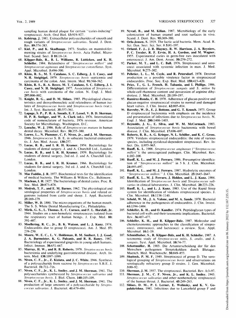
VOL.2,1989VIRIDANSSTREPTOCOCCI 327
sampling human dental plaque for certain "caries-inducing"streptococci. Arch. Oral Biol. 13:919-927.
66. Kelstrup, J. 1981. Extracellular polysaccharides of smooth andrough variants of Streptococcus salivarius. Scand. J. Dent.Res. 89:374-383.
67. Kiel, P., and K. Skadhauge. 1973. Studies on mannitol-fer-menting strains of Streptococcus boviis. Acta Pathol. Micro-biol. Scand. Sect. B 81:10-14.
68. Kilpper-Balz, R., B. L. Williams, R. Lutticken, and K. H.Schleifer. 1984. Relatedness of ''Streptococcus milleri" andStreptococcus aginosuts and Streptococcus coistellatlus. Syst.Appl. Microbiol. 5:494-500.
69. Klein, R. S., M. T. Catalano, S. C. Edberg, J. I. Casey, andN. H. Steigbigel. 1979. Streptococcus boils septicemia andcarcinoma of the colon. Ann. Intern. Med. 91:560-562.
70. Klein, R. S., R. A. Recco, M. T. Catalano, S. C. Edberg, J. I.Casey, and N. H. Steigbigel. 1977. Association of Streptococ-cus boviis with carcinoma of the colon. N. Engl. J. Med.297:800-802.
71. Knight, R. G., and D. M. Shlaes. 1985. Physiological charac-teristics and deoxyribonucleic acid relatedness of human iso-lates of Streptococcus bovis and Streptococcus boiwis (var.).Int. J. Syst. Bacteriol. 35:357-361.
72. Lapage, S. P., P. H. A. Sneath, E. F. Lessel, V. B. D. Skerman,H. P. R. Seeliger, and W. A. Clark (ed.). 1976. Internationalcode of nomenclature of bacteria, 1976 revision. AmericanSociety for Microbiology, Washington, D.C.
73. Loesche, W. J. 1986. Role of Streptococcuis unutans in humandental decay. Microbiol. Rev. 50:355-380.
74. Loewe, L., N. Plummer, C. F. Niven, Jr., and J. M. Sherman.1946. Streptococcus S. B. E. in subacute bacterial endocardi-tis. J. Am. Med. Assoc. 130:257.
75. Lucas, R. B., and I. R. H. Kramer. 1954. Bacteriology forstudents of dental surgery. J. and A. Churchill Ltd., London.
76. Lucas, R. B., and I. R. H. Kramer. 1959. Bacteriology forstudents of dental surgery, 2nd ed. J. and A. Churchill Ltd.,London.
77. Lucas, R. B., and I. R. H. Kramer. 1966. Bacteriology forstudents for dental surgery. 3rd ed. J. and A. Churchill Ltd.,London.
78. Mac Faddin, J. R. 1977. Biochemical tests for the identificationof medical bacteria. The Williams & Wilkins Co., Baltimore.
79. Maclean, I. H. 1927. The bacteriology of dental caries. Proc. R.Soc. Med. 20:873-878.
80. Medrek, T. F., and E. M. Barnes. 1962. The physiological andserological properties of Streptococcus hov'is and related or-ganisms isolated from cattle and sheep. J. Apple. Bacteriol.25:169-179.
81. Miller, W. D. 1880. The micro-organisms of the human mouth.The S. S. White Dental Manufacturing Co.. Philadelphia.
82. Mirik, G. S., L. Thomas, E. C. Curnen, and F. L. Horsfall, Jr.1944. Studies on a non-hemolytic streptococcus isolated fromthe respiratory tract of human beings. J. Exp. Med. 80:391-407.
83. Moellering, R. C., Jr., B. K. Watson, and L. J. Kunz. 1974.Endocarditis due to group D streptococci. Am. J. Med. 57:239-250.
84. Moore, W. E. C., L. V. Holdeman, R. M. Smibert, I. J. Good,J. A. Burmeister, K. G. Palcanis, and R. R. Raney. 1982.Bacteriology of experimental gingivitis in young adult humans.Infect. Immun. 38:651-667.
85. Murray, H. W., and R. B. Roberts. 1978. Streptococcus boisisbacteremia and underlying gastrointestinal disease. Arch. In-tern. Med. 138:1097-1099.
86. Niven, C. F., Jr., Z. Kiziuta, and J. C. White. 1946. Synthesisof a polysaccharide from sucrose by Streptococcus S.B.E. J.Bacteriol. 51:711-716.
87. Niven, C. F., Jr., K. L. Smiley, and J. M. Sherman. 1941. Thepolysaccharides synthesized by Streptococcus salivarius andStreptococcus boils. J. Biol. Chem. 140:105-109.
88. Niven, C. F., Jr., K. L. Smiley, and J. M. Sherman. 1941. Theproduction of large amounts of a polysaccharide by Strepto-coccus salivarius. J. Bacteriol. 41:479-484.
89. Nyvad, B., and M. Kilian. 1987. Microbiology of the earlycolonization of human enamel and root surfaces in vivo.Scand. J. Dent. Res. 95:369-380.
90. Orla-Jensen, S. 1919. The lactic acid bacteria. Mem. Acad. R.Sci. Dan. Sect. Sci. Ser. 8 5:81-197.
91. Orland, F. J., J. R. Blayney, R. W. Harrison, J. A. Reyniers,P. C. Trexler, R. F. Ervin, H. A. Gordon, and M. Wagner.1955. Experimental caries in germ-free rats inoculated withenterococci. J. Am. Dent. Assoc. 50:259-272.
92. Parker, M. T., and L. C. Ball. 1976. Streptococci and aero-cocci associated with systemic infection in man. J. Med.Microbiol. 9:275-302.
93. Pelletier, L. L., M. Coyle, and R. Petersdorf. 1978. Dextranproduction as a possible virulence factor in streptococcalendocarditis. Proc. Soc. Exp. Biol. Med. 158:415-420.
94. Price, T., G. L. French, H. Talsania, and I. Phillips. 1986.Differentiation of Streptococcus sunguis and S. initior bywhole-cell rhamnose content and possession of arginine dihy-drolase. J. Med. Microbiol. 21:189-197.
95. Ramirez-Ronda, C. H. 1978. Adherence of glucan-positive andglucan-negative streptococcal strains to normal and damagedheart valves. J. Clin. Invest. 62:805-814.
96. Ravreby, W. D., E. J. Bottone, and G. T. Keusch. 1973. GroupD streptococcal bacteremia with emphasis on the incidenceand presentation of infections due to Streptococcus bovis. N.Engl. J. Med. 280:1400-1403.
97. Reynolds, J. G., E. Silva, and W. M. McCormack. 1983.Association of Streptococcuts bovis bacteremia with boweldisease. J. Clin. Microbiol. 17:696-697.
98. Roberts, R. B., A. G. Krieger, N. L. Schiller, and K. C. Gross.1979. Viridans streptococcal endocarditis: the role of variousspecies, including pyridoxal-dependent streptococci. Rev. In-fect. Dis. 1:955-965.
99. Ruoff, K. L. 1988. Streptococcus anginosus ("Streptococcusmnilleri"): the unrecognized pathogen. Clin. Microbiol. Rev.1:102-108.
100. Ruoff, K. L., and M. J. Ferraro. 1986. Presumptive identifica-tion of ''Streptococcts mnilleri'' in 5 h. J. Clin. Microbiol.24:495-497.
101. Ruoff, K. L., and M. J. Ferraro. 1987. Hydrolytic enzymes of"Streptococcuts milleri" J. Clin. Microbiol. 25:1645-1647.
102. Ruoff, K. L., M. J. Ferraro, J. Holden, and L. J. Kunz. 1984.Identification of Streptococcuis boi'is and Streptococcuis sal-vuarius in clinical laboratories. J. Clin. Microbiol. 20:223-226.
103. Ruoff, K. L., and L. J. Kunz. 1983. Use of the Rapid Strepsystem for identification of viridans streptococcal species. J.Clin. Microbiol. 18:1138-1140.
104. Scheld, W. M., J. A. Valone, and M. A. Sande. 1978. Bacterialadherence in the pathogenesis of endocarditis. J. Clin. Invest.61: 1394-1404.
105. Schleifer, K. H., and 0. Kandler. 1974. Peptidoglycan types ofbacterial cell walls and their taxonomic implications. Bacteriol.Rev. 36:407-477.
106. Schleifer, K. H., and R. Kilpper-BAlz. 1987. Molecular andchemotaxonomic approaches to the classification of strepto-cocci. enterococci. and lactococci: a review. Syst. Appl.Microbiol. 10:1-19.
107. Schmidhuber, S., R. Kilpper-Balz, and K. H. Schleifer. 1987. Ataxonomic study of Streptococcus initis, S. oralis. and S.san guis. Syst. Appl. Microbiol. 10:74-77.
108. Schottmuller, H. 1903. Die Artunterscheidung der fur denMenschen pathogenen Streptokokken durch Blutagar.Muench. Med. Wochenschr. 50:849-853.
109. Shattock, P. M. F. 1949. Streptococci of group D. The sero-logical grouping of Streptococcus bovi's and observations onserologically refractory group D strains. J. Gen. Microbiol.3:80-92.
110. Sherman, J. M. 1937. The streptococci. Bacteriol. Rev. 1:3-97.111. Sherman, J. M., C. F. Niven, Jr., and K. L. Smiley. 1943.
Streptococcus salitvarius and other nonhemolytic streptococciof the human throat. J. Bacteriol. 45:249-263.
112. Shlaes, D. M., P. I. Lerner, E. Wolinsky, and K. V. Go-palakrishna. 1981. Infectious due to Lancefield group F and
VOL. 2, 1989
Dow
nloa
ded
from
http
s://j
ourn
als.
asm
.org
/jour
nal/c
mr
on 0
6 Ja
nuar
y 20
22 b
y 11
2.16
7.23
5.15
.
328 COYKENDALL CLIN. MICROBIOL. REV.
related streptococci (S. mi/lei. S. anginosuts). Medicine (Bal-timore) 60:197-207.
113. Skerman, V. B. D., V. McGowan, and P. H. A. Sneath. 1980.Approved list of bacterial names. Int. J. Syst. Bacteriol.30:225-420.
114. Smith, F. R., and J. M. Sherman. 1938. The hemolytic strep-tococci of human feces. J. Infect. Dis. 62:189-192.
115. Spencer, R. C., C. S. Nanayakarra, and A. J. Coup. 1982.Fulminant neonatal sepsis due to Streptococcuts ni/leri. J.Infect. 4:88-89.
116. Spigelblatt, L., J. Saintonge, R. Chicoine, and M. Laverdiere.1985. Changing pattern of neonatal streptococcal septicemia.Pediatr. Infect. Dis. 4:56-58.
117. Stones, H. H. 1948. Oral and dental diseases. E. and S.Livingstone, London.
118. Stones, H. H. 1951. Oral and dental diseases. 2nd ed. E. and S.Livingstone, London.
119. Tenover, R. C. 1988. Diagnostic deoxyribonucleic acid probesfor infectious diseases. Clin. Microbiol. Rev. 1:82-101.
120. Tillotson, G. S. 1982. An evaluation of the API-20 Strepsystem. J. Clin. Pathol. 35:468-472.
121. van Houte, J., C. De Moor, and H. Jansen. 1970. Synthesis ofiodophilic polysaccharide by human oral streptococci. Arch.Oral Biol. 15:263-266.
122. Wayne, L., G., D. J. Brenner, R. R. Colwell, P. A. D. Grimont,0. Kandler, M. 1. Krichevsky, L. H. Moore, W. E. C. Moore,R. G. E. Murray, E. Stackebrandt, M. P. Starr, and H. G.Truper. 1987. Report of the Ad Hoc Committee on Reconcil-iation of Approaches to Bacterial Systematics. Int. J. Syst.Bacteriol. 37:463-464.
123. Welborn, P. P., W. K. Hadley, E. Newbrun, and D. M. Yajko.1983. Characterization of strains of viridans streptococci bydeoxyribonucleic acid hybridization and physiologic tests. Int.J. Syst. Bacteriol. 33:293-299.
124. Whiley, R. A., and J. M. Hardie. 1988. Streptococcus iestib-u/uris sp. nov. from the human oral cavity. Int. J. Syst.Bacteriol. 38:335-339.
125. Whiley, R. A., R. R. B. Russell, J. M. Hardie, and D. Beighton.1988. Streptococcus dowei sp. nov. for strains previouslydescribed as Streptococcus inutans serotype h. Int. J. Syst.Bacteriol. 38:25-29.
126. White, J. C., and C. F. Niven, Jr. 1946. Streptococcus S.B.E.:a streptococcus associated with subacute bacterial endocardi-tis. J. Bacteriol. 51:717-722.
127. Whittenbury, R. 1964. Hydrogen peroxide formation and cat-alase activity in the lactic acid bacteria. J. Gen. Microbiol.35:13-26.
Dow
nloa
ded
from
http
s://j
ourn
als.
asm
.org
/jour
nal/c
mr
on 0
6 Ja
nuar
y 20
22 b
y 11
2.16
7.23
5.15
.