Citric acid conditioning of roots affects guided tissue ... articles/LR141... · guided tissue...
11
Journal of Periodmtal Research 1986: 21: 543-552 Citric acid conditioning of roots affects guided tissue regeneration in experimental periodontal wounds E. C. PETTERSSON AND [. AUKHIL Department of Periodonlics, University of North Carolina School of Dentistry, Chapel Hill, NC, USA The effect of citric acid conditioning of roots on the formation of a new connective tissue attachment was evaluated in the presence of a selective cell population. Fenestration wounds of standard sizes were made on the buccal aspects of mandibular premolars in 6 beagle dogs. The exposed root surfaces were curetted thoroughly and conditioned with citric acid (experimental) or distilled water (controls) for 3 minutes. The wounds were covered with Millipore filters to facilitate population of curetted root surfaces by cells from the adjacent periodontal ligament. Histologic analysis was made after 3 months of healing. The extent of new connective tissue attachment varied in both the experimental and control specimens. The percentage of surgically denuded root surface showing new cementum with inserting fibers was significantly lower in the experimental group compared with the controls. Root resorption was seen In both experimental and control specimens but involved a signifi- cantly larger percentage of denuded root surface in the experimental specimens. Ankyiosis occurred more frequently in the experimental group compared with the controls, while no difference was seen in the degree of bone regeneration. The results, indicate that new connective tissue attachment can form on denuded root surfaces by a selective cell popuiation. Citric acid conditioning of roots appears to either delay or complicate healing in the presence of a selective cell population. (Accepted for publication March 9. }986} no beneficial effects of citric acid condition- Introduction j^^ ^f j.^^^g ^^ ^j^g formation of a new Severai recent studies have evaluated the connective tissue attachment (Stahi & effects of citric acid conditioning of roots Froum 1977, Nyman, Lindhe & Karring on the formation of a new connective tissue 1981, Gottlow, Nyman & Karring i984a). attachment (Crigger et al. 1978, Selvig et al. The exact mechanism by which citric acid 198i, Nalbandian & Cote 1982). Some of conditioning promotes new attachment for- the studies have reported reestablishment mation is not ciear. Possibie explanations of connective tissue attachment on roots include "interdigitation" of coUagen fibrils following citric acid conditioning (Crigger of the dentin matrix with collagen fibriis 3t al. 1978, Cole et ai. i980, Selvig et ai. from the flap connective tissue (Ririe, Crig- 198 i). However, other studies have reported ger & Selvig 1980) and a delaying of the
Transcript of Citric acid conditioning of roots affects guided tissue ... articles/LR141... · guided tissue...

Journal of Periodmtal Research 1986: 21: 543-552
Citric acid conditioning of roots affectsguided tissue regeneration in
experimental periodontal woundsE. C. PETTERSSON AND [. AUKHIL
Department of Periodonlics, University of North Carolina School of Dentistry,Chapel Hill, NC, USA
The effect of citric acid conditioning of roots on the formation of a new connective tissueattachment was evaluated in the presence of a selective cell population. Fenestration woundsof standard sizes were made on the buccal aspects of mandibular premolars in 6 beagledogs. The exposed root surfaces were curetted thoroughly and conditioned with citric acid(experimental) or distilled water (controls) for 3 minutes. The wounds were covered withMillipore filters to facilitate population of curetted root surfaces by cells from the adjacentperiodontal ligament. Histologic analysis was made after 3 months of healing.The extent of new connective tissue attachment varied in both the experimental and controlspecimens. The percentage of surgically denuded root surface showing new cementum withinserting fibers was significantly lower in the experimental group compared with the controls.Root resorption was seen In both experimental and control specimens but involved a signifi-cantly larger percentage of denuded root surface in the experimental specimens. Ankyiosisoccurred more frequently in the experimental group compared with the controls, while nodifference was seen in the degree of bone regeneration.The results, indicate that new connective tissue attachment can form on denuded root surfacesby a selective cell popuiation. Citric acid conditioning of roots appears to either delay orcomplicate healing in the presence of a selective cell population.
(Accepted for publication March 9. }986}
no beneficial effects of citric acid condition-Introduction j ^ ^ f j. ^^g ^^ j g formation of a new
Severai recent studies have evaluated the connective tissue attachment (Stahi &effects of citric acid conditioning of roots Froum 1977, Nyman, Lindhe & Karringon the formation of a new connective tissue 1981, Gottlow, Nyman & Karring i984a).attachment (Crigger et al. 1978, Selvig et al. The exact mechanism by which citric acid198i, Nalbandian & Cote 1982). Some of conditioning promotes new attachment for-the studies have reported reestablishment mation is not ciear. Possibie explanationsof connective tissue attachment on roots include "interdigitation" of coUagen fibrilsfollowing citric acid conditioning (Crigger of the dentin matrix with collagen fibriis3t al. 1978, Cole et ai. i980, Selvig et ai. from the flap connective tissue (Ririe, Crig-198 i). However, other studies have reported ger & Selvig 1980) and a delaying of the
Laura Smith
Laura Smith
Laura Smith
Laura Smith

544 PETTERSSON AND A U K H I L
apical migration of the dento-gingival epi-thehum {Crigger et al. 1978, Poison & Proye1982).
The progenitor cells for new connectivetissue attachment are believed to be locatedin the periodontal ligament {Melcher 1976),Recent studies in animals have reported newconnective tissue attachment on denudedroots when preference was given for cellsfrom the adjacent periodontal ligament topopulate curetted root surfaces (Nyman etal. 1982a, Aukhil, Simpson & Schaberg1983, Gottlow et al. 1984b). According toKlinge et al. (1981), the extent of new at-tachment formation following citric acidconditioning of roots was related to the de-gree of coronal positioning of the muco-periosteal flaps. Thus, it is possible that thenew attachment seen after citric acid con-ditioning of roots is the result of coronalmigration of progenitor cells from thehealthy periodontal ligament. This assump-tion is supported by (i) the finding of newcementum confined to the notch region fol-lowing routine replaced flap surgery(Listgarten 1972, Frank et al. 1974) and (ii)the absence of new attachment when rootsare implanted in areas devoid of periodontalligament cells (Karring, Nyman & Lindhe1980, Nyman et al. 1980, Gottlow, Ny-man & Karring 1984a).
The present study was undertaken tostudy the effects of citric acid conditioningof roots on the formation of new connectivetissue attachment when progenitor cellsfrom the adjacent periodontal ligamentwere allowed to populate the curetted rootsurface. Nyman et al. {1982a) have de-scribed a wound model which prevents theoral epithelium and flap connective tissuesfrom growing onto the curetted root sur-face, thereby facihtating population by cellsfrom the periodontai ligament. The presentstudy used a modification of this model tostudy the effects of citric acid conditioningof roots.
Material and MethodsSix young adult female beagle dogs withchnically healthy periodontal conditionswere used in the study. Experimental proce-dures were carried out on the second andthird mandibular premolars on both sides.During a 3-wk period, the teeth were scaledand polished twice. The teeth were brushedevery 3rd d along with a topical applicationof 0.1% chiorhexidine giuconate solution.Healthy gingival conditions were estab-lished at the end of this period.
Experimental procedure: On the day ofsurgery, the dogs were anesthetized withpentobarbitai sodium injected intra-venously. The experimental areas were infil-trated with Xylocaine and epinephrine(1:50000) to reduce bleeding. Using intra-sulcular incisions, buccal mucoperiostealfiaps were reflected. Small fenestrationwounds (approximately 4 mm x 3 mm) weremade through the buccal cortical plate toexpose part of the distal roots of 2P2 andmesial roots of 3P3. The fenestrations weremade using a ^3 round bur at slow speedwith continuous saline drips. The coronalborder of the wound was 2-3 mm apical ofthe marginal bone crest. The borders of thewound were defined with a chisel and theexposed root surface was curetted tho-roughly to remove cementum and periodon-tal ligament. The exposed root surfaces wereplaned using a #3 round bur at slow speedwith cold saline drips. The wounds werethen debrided with saline.
After isolating the adjacent tissues withgauze, citric acid (pH 1) was topically ap-plied for 3 min on the exposed root surfacesof one side (experimental) using small cot-ton pellets. The contralateral side, servingas a sham-operated control, was treated byapplying cotton pellets soaked in distilledwater. The wounds were then irrigated withdistilled water. In order to prevent flap con-nective tissues from coming in contact with
Laura Smith
Laura Smith
Laura Smith

GUIDED T ISSUE REGENERATION 545
the curetted root surface, pieces of Milliporefilter (pore size 3 ftm, MiUipore Corp., Bed-ford, Mass.) were placed over the woundsas described by Nyman et ai. (1982a). Theflaps were then repositioned and sutured.Postoperative eare included instituting asoft diet and topical application of chlor-hexidine as described previously. The su-tures were removed after 1 wk and tooth-brushing with chlorhexidine applicationcontinued until the end of the experiment.The dogs were sacrificed by perfusion withbuffered neutral formalin (BNF) after 3months of healing. Following additional fix-ation in BNF for 3 d, the jaws were deealci-fied in formic acid-sodium citrate solution.Following decalcification. blocks of indivi-dual roots were trimmed, dehydrated and
MPF
embedded in paraffin. Buecolingual stepserial sections were cut at 5 p.m thicknessand stained with hematoxylin and eosin orGomori's one-step trichrome.
Histometrie analysis: Buecolingual sec-tions representing approximately the centerof the wound were selected. Histometricanalysis was performed on step-serial sec-tions at 40-50 fim intervals {average 14 sec-tions per specimen) using a calibrated gridmounted in the eyepiece of an Olympusmicroscope at 65 x . Linear measurementswere made along the wound surfaces (Fig.1). The following parameters, measured inmm and expressed as percentage of totalwound surface,, were determined:
1. The length of denuded root surface[measured as the distance between the
D
NC'9. 1. Schematic drawing illustrating the landmarks'd linear measurements used in histometric analysis. '" — • •^coronal border of wound. A = apicai border of Fig. 2. Photomicrograph of a control specimen showing'und, CC = coronal half of tooth showing new attach- new •*'- ••"—^•-" *'^""- •" *^^- = " - ^ i half3nt, AC-apicai half of tooth showing new attach-3m. B = regenerated bocxe, MPF = MiUipore filter.
lentum with inserting fibers in the apical halfof the fenestration wound, B = new bone, D = dentin,
cementum. Original magnification 65 x .
Laura Smith
Laura Smith
546 PETTERSSON AND A U K H I L
coronal (C) and apical (A) borders ofthe wound].
2. The percentage of denuded root sur-face showing new cementum with in-serting fibers [expressed separately forthe coronal (CC) and apical halves(AC) of the denuded root surface].
3. The percentage of denuded root sur-face showing root resorption [ex-pressed separately for the coronal(CR) and apical (AR) halves of thedenuded root surface].
4. Presence/absence of ankylosis.5. Regeneration of bone, expressed as
percentage of the length of denudedroot surface (distance between C andA).
For each specimen, mean values were cal-culated for the histometric parameters fromall the representative sections analyzed. Thedifferences between the experimental andcontrol specimens were compared usingStudent's / test (unpaired).
ResultsAll experimental areas had healed well ex-cept for one area where the Millipore filterwas exfoliated through the tissues after the4th wk. Only those specimens showing theMillipore filter in its original position wereselected for histometric analysis. A total of7 control and 8 experimental specimensfrom 6 beagle dogs (at least 1 experimentaland I control from each dog) were suitablefor analysis. The mean length of the de-nuded root surface was 3.12 mm (±0.11) inthe acid-treated experimental wounds and2.92 mm ( + 0.13 mm) in the controlwounds. The difference was not statisticallysignificant.
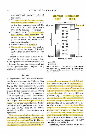
New cementum with inserting fibers wasseen in both the experimental (acid-treated)and control root surfaces (Fig. 2). The per-centage of curetted root surface showing newattachment was significantly lower in the ex-
Control Citric Acidn=7 n=8
Coronal Coronal
Apical Apical
jf. p<O-O5* * * P<OOO1
Fig. 3. Percentage of denuded root surface showingnew cementum with inserting libers in the apical andcoronal areas of the experimental (citric acid) and
perimental roots compared with the con-trols. Analyzed separately, the coronal andapical halves of the controls showed signifi-cantly higher percentages of root surfaceswith new attachment compared with the ex-perimental roots (p<0.05 and p<0,001, re-spectively) (Fig. 3). Within each group, therewere no significant differences in the amountof coronal and apical regeneration of new at-tachment (Fig. 3). At the center of the cu-retted root surface, connective tissue fiber^lying parallel to the root surface were seei-whenever no new attachment had formedThe thickness of new cementum decrease'towards the center of the curetted root sujface. Artifactual splits between new cemertum and dentin were commonly seen in th •control specimens (Fig. 4).
Resorption, mainly confined to the per -
Laura Smith
Laura Smith
Laura Smith
Laura Smith
Laura Smith
Laura Smith
Laura Smith
Laura Smith
Laura Smith
Laura Smith
Laura Smith
Laura Smith
Laura Smith

GUIDED T i S S U E REGENERATION 547
phery of the wounds, was seen in hotiigroups. However, the extent of root re-sorption was significantly greater (p 0.05)in the apical half of the experimental acid-treated roots (Figs. 5, 6).
Isolated spots of ankyiosis were a pro-nounced feature of the experimentai speci-mens (Fig. 7). Five of the 8 experimentalspecimens showed ankyiosis compared withthe 7 control specimens where ankyiosis wasnever found. Bone had regenerated to analmost equai extent in hoth the experimen-tai (99.5%, ±0.34) and control specimens(98.6%, ±i.i3).
DiscussionThe present study was designed to examinethe effects of citric acid conditioning of cu-retted roots on the formation of new attach-ment when the progenitor cells from theadjacent periodontal ligament were aiiowedto populate the wound. The present findingsindicate that new attachment can form onboth acid-conditioned and non-conditionedcuretted roots. However, citric acid con-ditioning was associated with more undesir-able features of wound heahng such as rootresorption and ankyiosis. The present find-ings are in agreement with tiiose of Nymanet al. (1982a, b), Aukhii et ai. (i983) andGottlow et al. (i984b) and suggest that faci-litating population of curetted root surfacesby cells from the periodontal ligament isadequate to regenerate new connective tis-sue attachment. The present findings alsosupport the concept that the progenitor cellsire located in the adjacent, healthy peri-idontal ligament (Meicher i976).
The formation of new attachment in both•xperimental and control specimens may be-xplained by the ceiis from the adjacentleriodontai ligament populating the cu-etted root surface. As suggested by Nymant al. (i982a), the wound model faciiitateshis by exciuding the epithelium and gin-
Fig. 4. Photomicrograph of a control specimen showingnew cementum with inserting fibers. A = artifactualsplit between new cementum (C) and dentin surface(D). B = new bone. Original magnification 130x .
givai connective tissue from contacting thecuretted root surface. Aithough Miihporefiiters may have excluded the fiap connec-tive tissues from the periodontal ligamentwound, this is no evidence that the ceiis thatfirst popuiated the curetted root surface ac-tually originated from the periodontal hga-ment. The possibility of granulation tissuearising from the cut bony margins of thefenestration wound under the Millipore fil-ter must not be overlooked. Since it is diffi-cult to identify the source of cells in thepresent wound model, the mode of healingmay to some extent explain the origin of thecells. Studies by Melcher (1970), Karring etal. (1980) and Gottiow et al. (1984a) haveshown that granulation tissue from bone
Laura Smith
Laura Smith
Laura Smith

548 PETTERSSON AND A U K H I L
Fig. 5. Percentage ol denuded root surface showingroot rasorption in the apical and coronai areas of theexpertmentai (citric acid) and controi specimens.
possesses the potential to induce root re-sorption and ankylosis. Boyko, Brunette &Melcher (1981) implanted roots to whichcultured gingival and periodonta! ligamentcells were attached in vitro and found thatonly roots bearing cultured periodontalligament cells were associated with fibroustissue resembling periodontal ligament.These Iindings, along with those by Nymanet al. (1982a, b), Aukhil et al. (1983) andGottlow et al. (1984b) suggest that new at-tachment formation is more likely a featureof periodonta] ligament cells.
The significant differences between theexperimental and control specimens interms of the extent of connective tissue at-tachment regeneration, root resorption andankylosis may be explained by several fac-tors involving cellular kinetics. It is possible
Fig. 6. Photomicrograph of an experimental (citricacid) specimen to show active root resorption, under-mining in nature, at the coronai border of the wound,B = new bone, D = dentin, F = miilipore fiiter. Originalrnagniflcation 65 .
that citric acid application in the woundused here may have damaged the adjacentperiodontal hgament cells, thereby affectingtheir capacity to migrate on curetted rootsurfaces. Delayed migration of progenitorcells from the periodontal ligament mayhave resulted in the population of curettedroot surfaces by granulation tissue frombone. Crigger, Renvert & Bogle (1983) havereported no deleterious effects of citric acidapplication on surgically exposed periodon-tal tissues. However, their different woundmodel makes it difficult to draw comparisons with the present fmdings. The wouncmodel used by Crigger et al. (1983) doenot allow a comparison of progenitor eelkinetics between the experimental and con
Laura Smith

GUIDED T ISSUE R E G E N E R A T I O N 549
Fig. 7, Photomicrograph of an experimentai (citric acid)specimen showing ankylosis and resorption in the cen-tral part of ttie wound. D = dentin, B = t\ew bone, R =root resorption. Arrows point at the area of fusionbetween new bone and dentin. Original magnification65 X.
trol wounds. In a recent study, Magnussonel al. (1985) reported root resorption andankyiosis in monkeys following citric acidconditioning of roots. Magnusson et al.11985) have suggested that "absence of peri-''dontal ligament cells may have also al-• >wed bone forming cells to contact the root'Jrface. resulting in ankylosis." It must beniphasized that root resorption was seen1 both the experimental and control speci-lens.The differences between experimentalid control specimens may also be ex-ained by possible biochemical changes ate root/soft tissue interface. Directed mi-
gration of cells in response to chemicalgradients (chemotaxis) is thought to playan important role in wound healing (Ross1968). Fibronectin, an attachment factor forfibroblasts. acctmiulates at wound sites(Mosher, Schad & Kleinman 1979) and isbelieved to enhance the migration of fibro-blasts (Gauss-Muller et al. 1980), Acid con-ditioning of roots may provide a surfacemore suitable for the establishment of fibrinlinkage (Poison & Proye 1982) and in-creased cell attachment (Boyko, Brunette &Melcher 1980, Fernyhough & Page 1983).Collagen exposed from the dentin matrix asa result of citric acid conditioning may alsobe chemotactic to fibroblasts (Postlewaithe,Seyer & Kang 1978. Rath & Reddi 1979).It is possible that such biochemical eventsat the root surface may be chemotactic toconnective tissue cells from bone and peri-odontal ligament. In the present wound mo-del, these chemotactic factors may be actingindiscriminately. Celt attachment factorslike fibronectin have been known to stimu-late the attachment of bone cells to coiiagen,and also their migration (Somerman et al.1982). This possibility can explain the fre-quent occurrence of ankylosis in acid-con-ditioned experimental wounds. Whether thesuperficially demineralized dentin matrix in-duces transformation of fibroblasts intobone-forming cells in the present model isnot clear. Decalcified dentin matrix hasbeen shown to induce transformation of fi-broblasts (Huggins & Urist 1970).
Inflammation during wound healing mayalso have contributed towards differencesin the experimental and control specimens.Multinucleated giant cells are usually pre-sent in wounds with foreign bodies (Mari-ano & Spector 1974). Millipore filters wereplaced over the fenestration wounds andsome degree of inflammation during theearly stages of wound healing may be attri-buted to their presence. However, this doesnot fully explain the differences between ex-
Laura Smith
Laura Smith
Laura Smith

550 PETTERSSON AND A U K H I L
perimenta! and control specimens becauseboth the wounds were approximately of thesame size and had MiUipore filters coveringthem. The present study describes the find-ings only in specimens where the filter wasin place. This has excluded the possibihtyof healing variation due to displacement ofMillipore filters which were acting as physi-cal barriers. The wounds were made ap-proximately at the same location on contra-lateral teeth and at least 2-3 mm of mar-ginal bone was retained above the coronalborder of the wound. This may have min-imized any variation in heahng due towound location and infiammatory influ-ences from the gingival sulcus.
The present findings are not in completeagreement with those of Crigger et al.(1978), Selvig et al. (1981) and Nalbandi-an & Cote (1982), Studies using citric acidconditioning of roots during flap surgery(Crigger et al. 1978, Cole et al. 1980, Selviget al. 1981) have reported new attachmentformation. As discussed earlier, the new at-tachment has been attributed to "splicing"of collagen fibrils from dentin and flap con-nective tissues and retarded apical mi-gration of dento-gingival epithelium. In thestudy by Nalbandian & Cote (1982), morenew attachment was seen in experimentalfenestration wounds in dogs after citric acidconditioning of roots compared to the con-trols without acid conditioning. However,no attempt was made to prevent the flapconnective tissues from growing into thewound. The new attachment they observedcan be attributed also to cells migratingfrom the adjacent periodontal ligament.Studies using citric acid (Cole et al. 1980,Crigger et al. 1978, Klinge et al, 1981, Ririeet al. 1980, Selvig et al. 1981) have demon-strated new attachment only in wound mo-dels having adjacent/apical source of peri-odontal ligament cells. Acid-conditionedroot surfaces implanted in surgically-cre-ated alveolar grooves and covered by muco-
periosteal fiaps have failed to show forma-tion of new attachment (Gottlow, Nyman &Karring 1984a, Aukhil, Greco & Torney1985).
In the present study, only the sectionsrepresenting the center of the wound wereselected. The wound mode! in the presentstudy allows healing from all borders ofthe wound towards the center. The sectionsfrom the mesial and distal corners of thewound may show extensive healing due tothe adjacent source of cells. Buccolingualsections representing the center of thewound should have minimized this error.The frequent observation of artifactualsplits between new cementum and dentinin the control specimens suggests a weakjunction between the new cementum aoddentin.
In summary, the present fmdings demon-strate that new attachment can form onboth acid-conditioned and non-acid-con-ditioned roots when progenitor cells formthe periodontal ligament are al!owed topopu!ate the curetted root surfaces. The ef-fects of citric acid conditioning of roots dur-ing periodonta! surgery on the cellu!ar kin-etics in periodontal wounds need furtherinvestigation.
AcknowledgementsThis study was supported by N.I.H. grant, DE 06766. The technical assistance ofMs. Cynthia Suggs and the secretarial as-sistance of Ms. Kathy Dodson is gratefullyacknowledged.
ReferencesAukhil, I., Greco, G,, Suggs, C, & Torney, I
1986, Root resorption potentials of grani -latioti tissue from bone and flap connecti' •:tissues. Journal of Periodontal Research ( ipress),
Aukhil. I.. Simpson. D, M. & Schaberg, T. 198 ,An experimental study of new attachment pr -
Laura Smith
Laura Smith
Laura Smith
Laura Smith

GUIDED T ISSUE REGENERATION 551cedure in beagle dogs. Journal of FeriodontalResearch 18: 643-654.
Boyko, G. A., Brunette, D. M. & Melcher. A, H,1980, Cell attachment to demineralized rootsurfaces in vitro. Journal of Periodontal Re-search 15: 297-303.
Boyko, G, A,, Melcher, A. H. & Brunette. D, M,1981. Formation of new periodontal ligamentby periodontat ligament cetis implanted in vivoafter culture in vitro. Journal of FeriodontalResearch 16: 73-88.
Coie, R., Crigger, M., Bogle, G., Egelberg. J, &Selvig, K, 1980, Connective tissue regenerationto periodontaliy diseased teeth. A histologicalstudy. Journal of Periodonta! Research 15; 1-9.
Crigger, M., Bogle, G.. Niiveus. R., EgeibergJ. & Selvig, K, 1978, The effect of citric acidapplication on the healing of experimental fur-cation defects in dogs. Journal of PeriodontalReseareh 13: 538-549,
Crigger, M., Renvert, S, & Bogle. G, 1983. Theeffect of topical citric acid application on surgi-cally exposed periodontal attachment. Journalof Feriodontal Researeh 18: 303-305.
Femyhough, W. & Page, R. 1980. Attachment,growth and synthesis by human gingival fibro-blasts on demineralized or fibronectin-treatednormal and diseased tooth roots. Journal ofPeriodontology 54: 133-140.
Frank, R., Fioro-Donno, G,, Cimasoni, G. &Matter. J. 1974, Ultrastructural study of epi-thelial and connective gingival reattachment inman. Journal of Feriodontology 45: 626.
Gauss-Muller, V., Kleinman, H. K., Martin, G,R, & Schiffman. E. 1980. Role of attachmentfactors and atlractants in fibroblast chemo-taxis. Journal of Laboratory and Cliniea! Medi-cine 96: 1071-1080,
Gottlow, J,, Nyman. S. & Karring, T. 1984a.Healing following citric acid conditioning ofroots implanted into bone and gingival connec-tive tissue. Journal of Feriodontal Research 19:214-220.
Gottlow. J,, Nyman, S.. Kardng, T. & Lindhe. J.1984b, New attachment formation as the resultof controlled tissue regeneration. Journal ofClinical Periodontologv 11: 494-503,
luggins, C, B. & Urist. M. R. 1970. Dentinmatrix transformation: Rapid induction of al-kaline phosphatase and cartilage. Science 167896-897.
-arring, X, Nyman, S. & Lindhe, J. 1980. Heal-ing following implantation of periodontitis af-tected roots into bone tissue. Journal of ClinicalFeriodontologv 7: 96-105.iinge, B., Niiveus, R., Kiger, R. & Egelberg, J,
1981. Effect of flap placement and defect sizeon healing of experimental furcation defects.Journal of Feriodontal Researeh 16: 236-248,
Listgarten, M. A. 1972. Electron microscopiostudy of the junction between surgically de-nuded root surfaces and regenerated periodon-tal tissues. Journal of Periodontal Research 7:68-70,
Magnusson, L, Claffey, N., Bogle, G., Garret,S. & Egelberg. J. 1985. Root resorption follow-ing periodontal flap procedures in monkeys.Journal of Periodontal Research 20: 79-85.
Mariano, G. & Spector, W. G, 1976, The forma-tion and properties of macrophage polykarion(inflammatory giant cells). Journal of Fathol-ogy lU: 1-19.
Melchcr, A, H, 1970. Repair of wounds in theperiodontium of the rat. Influence of periodon-tal ligament on osteogenesis. Archives of OralBiology 15: 1183-1204.
Melcher, A. H. 1976. On the repair potential ofperiodontal tissues. Journal of Feriodontology47: 256-260,
Mosher, D, F., Schad, P, E. & Kleinman, H, K.1979. Cross-linking of fibronectin lo collagenby blood coagulation factor Xllla, Journal ofClinical Investigation 64: 781-787.
Nalbandian, J. & Cote, N, 1982. Effect of citricacid on periodontal wound healing in dogs.Journal of Feriodontal Researeh 17: 552-562.
Nyman, S.. Gottlow, J., Karring, T, & Lindhe, J.1982a, The regenerative potential of the peri-odontal ligament. An experimental study in themonkey. Journal of Clinical Feriodontology 9:257-265.
Nyman, S., Karring, T., Lindhe. J. & Planten, S.1980. Healing following implantation of peri-odontitis-afTected roots into gingival connec-tive tissue. Journal of Clinical Feriodontology7:394-^1.
Nyman, S.. Lindhe, J, & Karring. T, 1981. Heal-ing following surgical treatment and root de-mineralization in monkeys with periodontaldisease. Journal of Clinical Periodontology 8:249-258.
Nyman, S,. Lindhe. J.. Karring, T. & Rylander,H. 1982b, New attachment following surgicaltreatment of human periodontal disease. Jour-nal of Clinical Feriodontology 9: 290-296.
Poison", A. M. & Proye, M. R 1982. Effect ofroot surface alterations on wound healing. ILCitric acid treatment on the denuded root.Journal of Clinical Feriodontology 9: 441-454.
Postlethwaite, A. E., Seyer, J. M. & Kang, A. H.1978. Chemotactic attraction of human flbro-biasts to type L II and III collagens and coi-

552 P E T T E R S S O N A N D A U K H I L
lagcn-derived peptides. Proceeding.^ af the Na-tional Academy of Sciences. U.S.A. 75:871-875.
Rath, N. C. & Rcddi, A. H. 1979. Collagen bonematrix is a local mitogen. Nature fLondon)278: 855-857.
Ririe, C . Crigger, M. & Selvig, K. 1980. Healingof periodontal eonneetive (issues following sur-gical wounding and application of citric acidin dogs. Journal of Periodontal Research 15:314-327.
Ross, R. 1968. Fibroblasts atid wound repair.Biological Reviews 43: 51-96.
Selvig. K. A.. Ririe. C. M., Nilveus, R. & Egel-berg, J. 1981. Fine structure of new eonneetivetissue attachment following acid treatment ofexperimental furcatiotl pockets in dogs. Journalof Periodontal Research 16: 123-129.
Somerman, M., Schiffmatl, E., Reddi, A. H. &Termine, J. 1982. Regulation of the attachmentand migration of bone cells in vitro. Journal ofPeriodontal Research 17: 527-529.
Stahl, S. S. & Froum, S. J. 1977. Human clinicaland histologic repair responses following theuse of citric acid in periodontal therapy. Jour-nal of Periodontology 48: 261.-266.
Address:
;. AukhilDepartment of PeriodonticsUNC School of DentistryChapel Hill, NC 27514U.S.A.


![3D-printed membrane for guided tissue regenerationeprints.whiterose.ac.uk/125308/3/GTR paper -o- revised 1.pdf · restoration [1-3]. While GTR mostly deals with soft tissue, guided](https://static.fdocuments.in/doc/165x107/5ed6c04ed397173fdb727a26/3d-printed-membrane-for-guided-tissue-paper-o-revised-1pdf-restoration-1-3.jpg)

