CIRRHOSIS LIVER Dr. M. A. SOFI MD; FRCP (London); FRCPEdin; FRCSEdin.
32
CIRRHOSIS LIVER Dr. M. A. SOFI MD; FRCP (London); FRCPEdin; FRCSEdin
-
Upload
logan-young -
Category
Documents
-
view
218 -
download
0
Transcript of CIRRHOSIS LIVER Dr. M. A. SOFI MD; FRCP (London); FRCPEdin; FRCSEdin.
CIRRHOSIS LIVER
Dr. M. A. SOFI MD; FRCP (London); FRCPEdin; FRCSEdin
Definition
Cirrhosis represents a late stage of progressive hepatic fibrosis characterized by distortion of the hepatic architecture and the formation of regenerative nodules.The progression of liver injury to cirrhosis may occur over weeks to years.
Epidemiology
Chronic liver disease and cirrhosis result in about 35,000 deaths each year in the United States. • Cirrhosis is the ninth
leading cause of death in the United States and is responsible for 1.2% of all US deaths. • Many patients die from the
disease in their fifth or sixth decade of life.
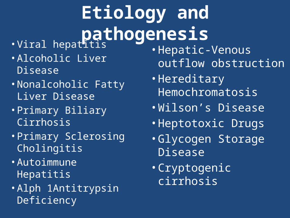
Etiology and pathogenesis
• Viral hepatitis• Alcoholic Liver
Disease • Nonalcoholic Fatty
Liver Disease• Primary Biliary
Cirrhosis• Primary Sclerosing
Cholingitis• Autoimmune Hepatitis• Alph 1Antitrypsin
Deficiency
• Hepatic-Venous outflow obstruction• Hereditary
Hemochromatosis• Wilson’s Disease• Heptotoxic Drugs• Glycogen Storage
Disease• Cryptogenic
cirrhosis
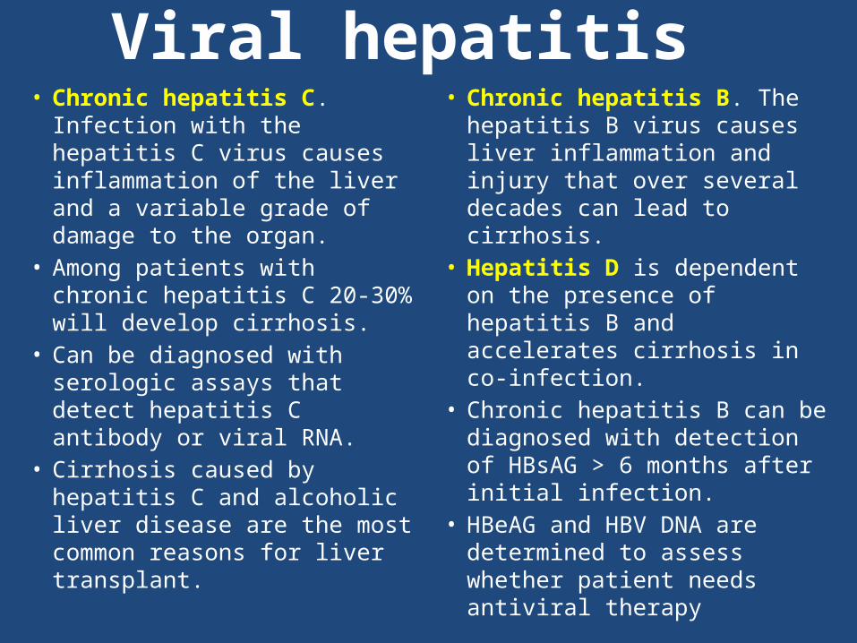
Viral hepatitis• Chronic hepatitis C.
Infection with the hepatitis C virus causes inflammation of the liver and a variable grade of damage to the organ.
• Among patients with chronic hepatitis C 20-30% will develop cirrhosis.
• Can be diagnosed with serologic assays that detect hepatitis C antibody or viral RNA.
• Cirrhosis caused by hepatitis C and alcoholic liver disease are the most common reasons for liver transplant.
• Chronic hepatitis B. The hepatitis B virus causes liver inflammation and injury that over several decades can lead to cirrhosis.
• Hepatitis D is dependent on the presence of hepatitis B and accelerates cirrhosis in co-infection.
• Chronic hepatitis B can be diagnosed with detection of HBsAG > 6 months after initial infection.
• HBeAG and HBV DNA are determined to assess whether patient needs antiviral therapy
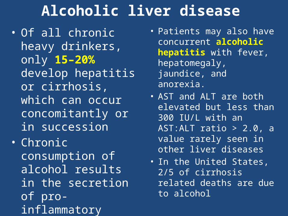
• Of all chronic heavy drinkers, only 15–20% develop hepatitis or cirrhosis, which can occur concomitantly or in succession
• Chronic consumption of alcohol results in the secretion of pro-inflammatory cytokines (TNF-alpa, IL6 andIL8], oxidative stress, peroxidation, and acetaldehyde toxicity. These factors cause inflammation, apoptosis and eventually fibrosis of liver cells.
• Patients may also have concurrent alcoholic hepatitis with fever, hepatomegaly, jaundice, and anorexia.
• AST and ALT are both elevated but less than 300 IU/L with an AST:ALT ratio > 2.0, a value rarely seen in other liver diseases
• In the United States, 2/5 of cirrhosis related deaths are due to alcohol
Alcoholic liver disease
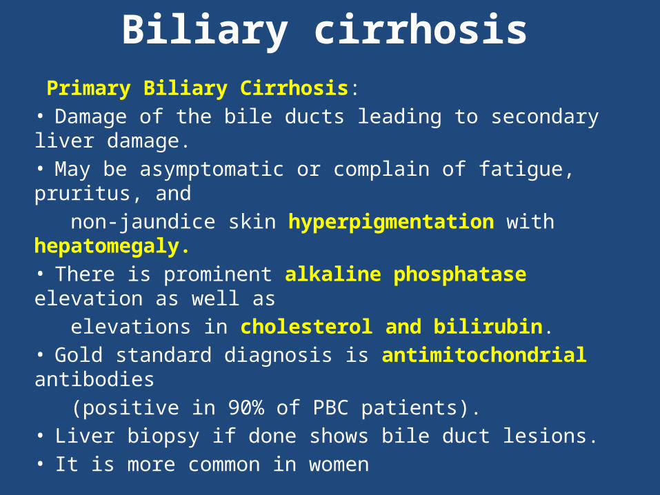
Biliary cirrhosis Primary Biliary Cirrhosis: • Damage of the bile ducts leading to secondary liver damage. • May be asymptomatic or complain of fatigue, pruritus, and non-jaundice skin hyperpigmentation with hepatomegaly. • There is prominent alkaline phosphatase elevation as well as elevations in cholesterol and bilirubin. • Gold standard diagnosis is antimitochondrial antibodies (positive in 90% of PBC patients). • Liver biopsy if done shows bile duct lesions. • It is more common in women
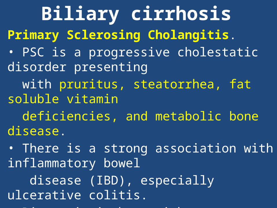
Biliary cirrhosisPrimary Sclerosing Cholangitis.• PSC is a progressive cholestatic disorder presenting with pruritus, steatorrhea, fat soluble vitamin deficiencies, and metabolic bone disease. • There is a strong association with inflammatory bowel disease (IBD), especially ulcerative colitis. • Diagnosis is best with contrast cholangiography showing diffuse, multifocal strictures and focal dilation of bile ducts, leading to a beaded appearance. Non-specific serum immunoglobulins may also be elevated
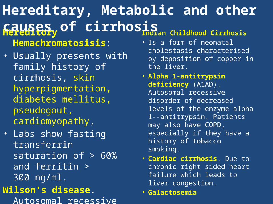
Hereditory Hemachromatosisis:
• Usually presents with family history of cirrhosis, skin hyperpigmentation, diabetes mellitus, pseudogout, cardiomyopathy,
• Labs show fasting transferrin saturation of > 60% and ferritin > 300 ng/ml.
Wilson's disease. Autosomal recessive disorder characterized by low serum ceruloplasmin and increased hepatic copper content on liver biopsy, and elevated 24-hour urine copper.
• May also have Kayser-Fleischer ring
Indian Childhood Cirrhosis • Is a form of neonatal
cholestasis characterised by deposition of copper in the liver.
• Alpha 1-antitrypsin deficiency (A1AD). Autosomal recessive disorder of decreased levels of the enzyme alpha 1--antitrypsin. Patients may also have COPD, especially if they have a history of tobacco smoking.
• Cardiac cirrhosis. Due to chronic right sided heart failure which leads to liver congestion.
• Galactosemia
Hereditary, Metabolic and other causes of cirrhosis
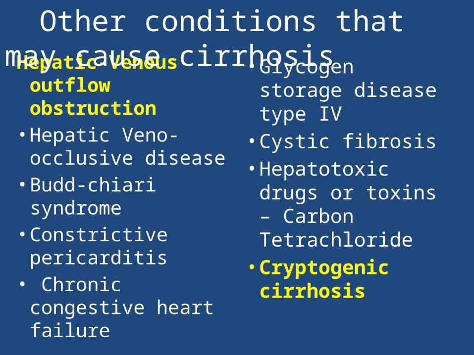
Hepatic-Venous outflow obstruction• Hepatic Veno-
occlusive disease• Budd-chiari
syndrome • Constrictive
pericarditis• Chronic
congestive heart failure
• Glycogen storage disease type IV• Cystic fibrosis• Hepatotoxic
drugs or toxins – Carbon Tetrachloride• Cryptogenic cirrhosis
Other conditions that may cause cirrhosis
11
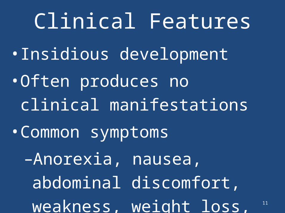
Clinical Features• Insidious development
• Often produces no clinical manifestations
• Common symptoms
–Anorexia, nausea, abdominal discomfort, weakness, weight loss, and malaise
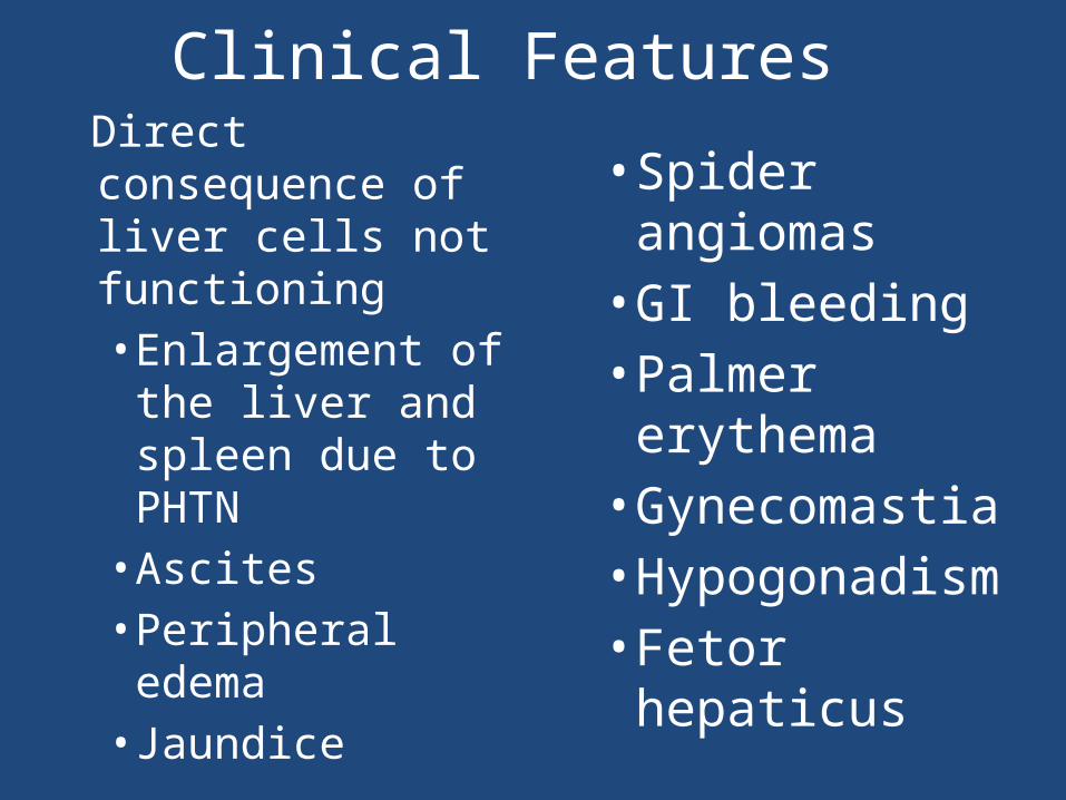
Direct consequence of liver cells not functioning• Enlargement of the liver and spleen due to PHTN• Ascites • Peripheral edema • Jaundice
• Spider angiomas• GI bleeding• Palmer erythema• Gynecomastia• Hypogonadism• Fetor hepaticus
Clinical Features
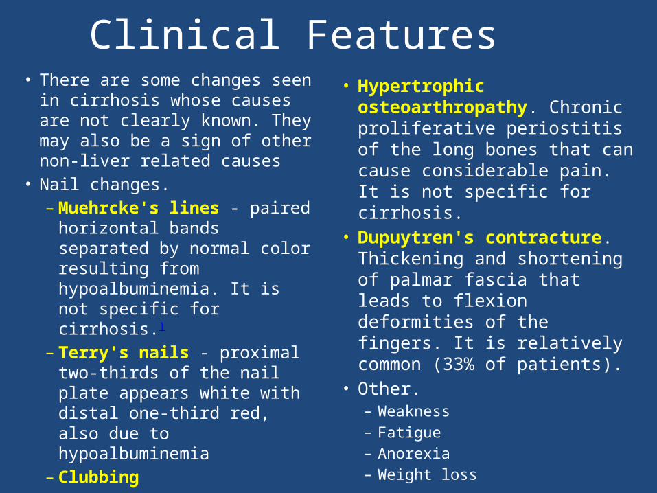
• There are some changes seen in cirrhosis whose causes are not clearly known. They may also be a sign of other non-liver related causes
• Nail changes. – Muehrcke's lines -
paired horizontal bands separated by normal color resulting from hypoalbuminemia. It is not specific for cirrhosis.]
– Terry's nails - proximal two-thirds of the nail plate appears white with distal one-third red, also due to hypoalbuminemia
– Clubbing
• Hypertrophic osteoarthropathy. Chronic proliferative periostitis of the long bones that can cause considerable pain. It is not specific for cirrhosis.
• Dupuytren's contracture. Thickening and shortening of palmar fascia that leads to flexion deformities of the fingers. It is relatively common (33% of patients).
• Other. – Weakness– Fatigue – Anorexia– Weight loss
Clinical Features
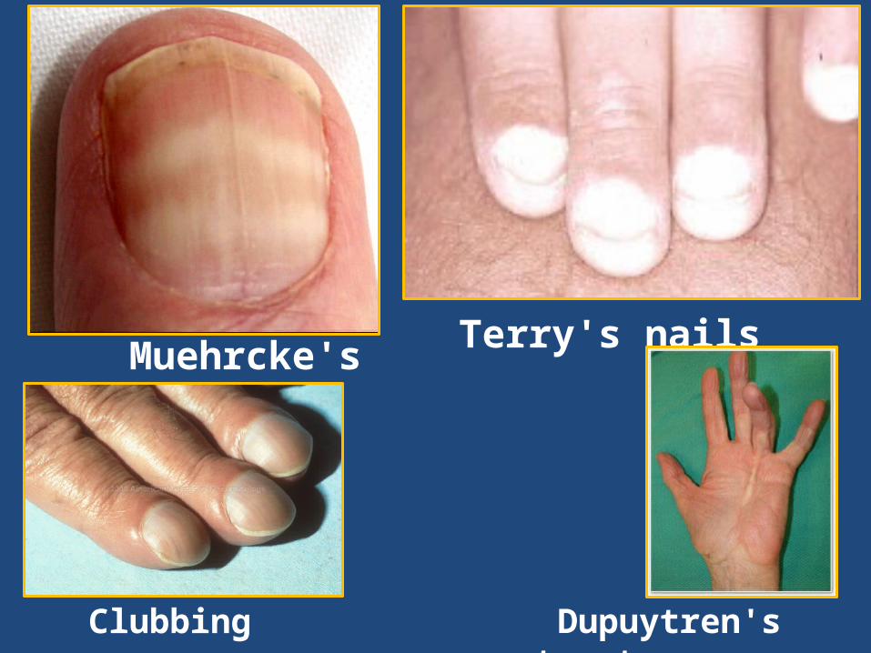
Muehrcke's lines
Terry's nails
Dupuytren's contracture
Clubbing
• Thrombocytopenia - typically multifactorial. However, this rarely results in platelet count < 50 000/mL
• Aminotransferases - AST and ALT are moderately elevated, with AST > ALT. However, normal aminotransferases do not preclude cirrhosis
• Gamma-glutamyl transferase– correlates with AP levels.
• Bilirubin- Levels normal when compensated but may elevate as cirrhosis progresses.
• Albumin - levels fall as the synthetic function of the liver declines
• Prothrombin time - increases since the liver synthesizes clotting factors.
• Globulins- increased due to shunting of bacterial antigens away from the liver to lymphoid tissue.
• Hyponatremia due to inability to excrete free water resulting from high levels of ADH and aldosterone
• Leukopenia and neutropenia - due to splenomegaly
Lab Findings
Portal HypertensionLiver cirrhosis increases resistance to blood flow and
higher pressure in the portal venous system, resulting in portal hypertension. Effects of portal hypertension include:
• Splenomegaly is found in 35% to 50% of patients.[7]
• Esophageal varices result from a process called Portacaval anastomosis). When these blood vessels become enlarged, they are called varices and are more likely to burst.
• Caput medusa are dilated periumbilical collateral veins due to portal hypertension.
• Cruveilhier-Baumgarten murmur is a venous hum heard in the epigastric region due to collateral connections forming between portal system and the periumbilical veins
Portal HypertensionComplications of portal hypertension
include:• Ascites• Hepatic encephalopathy• Variceal hemorrhage• Spontaneous bacterial peritonitis• Hepatorenal syndrome• Portal hypertensive gastropathy• Hepatic hydrothorax• Hepatopulmonary syndrome• Portopulmonary hypertension• Cirrhotic cardiomyopathy
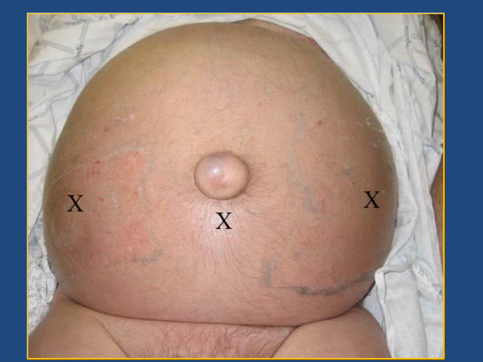
• Ascites is the accumulation of fluid within the peritoneal cavity.
• It is the most common complication of cirrhosis.
• The first step leading to fluid retention and ultimately ascites is the development of portal hypertension
• Patients without portal hypertension do not develop ascites or edema.
• Those with ascites have several circulatory, vascular, functional, and biochemical abnormalities that contribute to the pathogenesis of fluid retention
Ascites
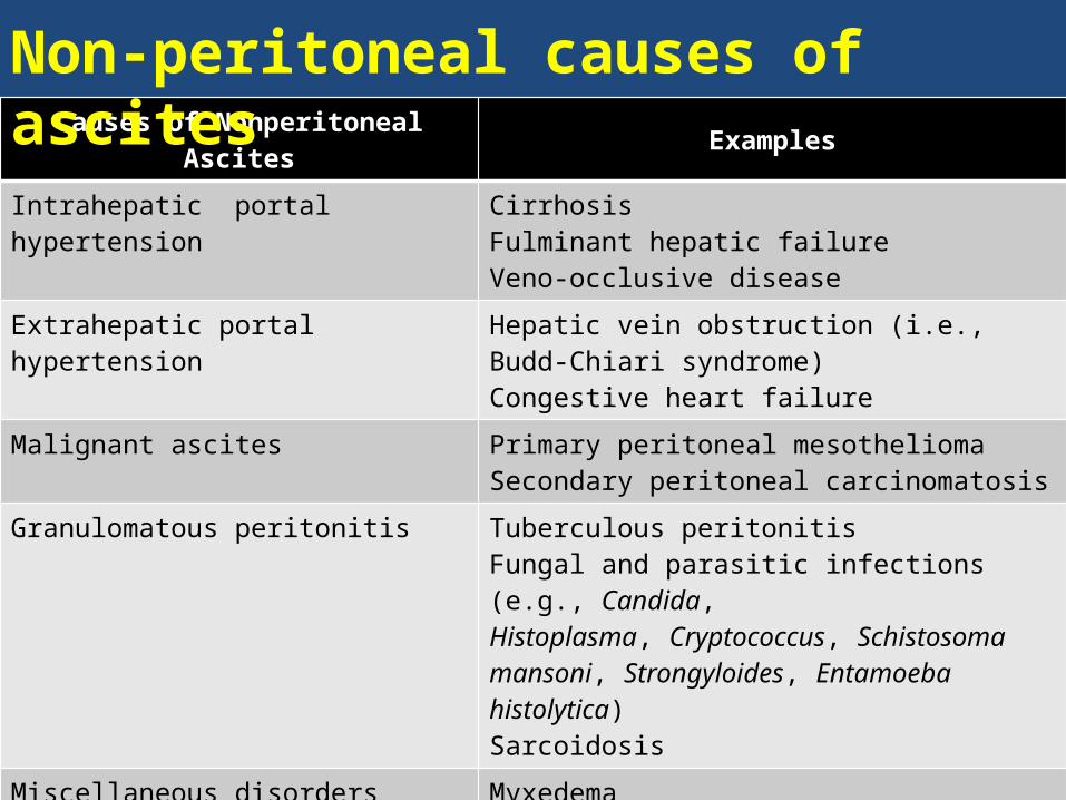
Causes of Nonperitoneal Ascites Examples
Intrahepatic portal hypertension CirrhosisFulminant hepatic failureVeno-occlusive disease
Extrahepatic portal hypertension Hepatic vein obstruction (i.e., Budd-Chiari syndrome)Congestive heart failure
Malignant ascites Primary peritoneal mesotheliomaSecondary peritoneal carcinomatosis
Granulomatous peritonitis Tuberculous peritonitisFungal and parasitic infections (e.g., Candida,Histoplasma, Cryptococcus, Schistosoma mansoni, Strongyloides, Entamoeba histolytica)Sarcoidosis
Miscellaneous disorders MyxedemaOvarian tumorsPancreatic ascitesBiliary ascites
Hypoalbuminemia Nephrotic syndromeProtein-losing enteropathy Malnutrition
Non-peritoneal causes of ascites
21
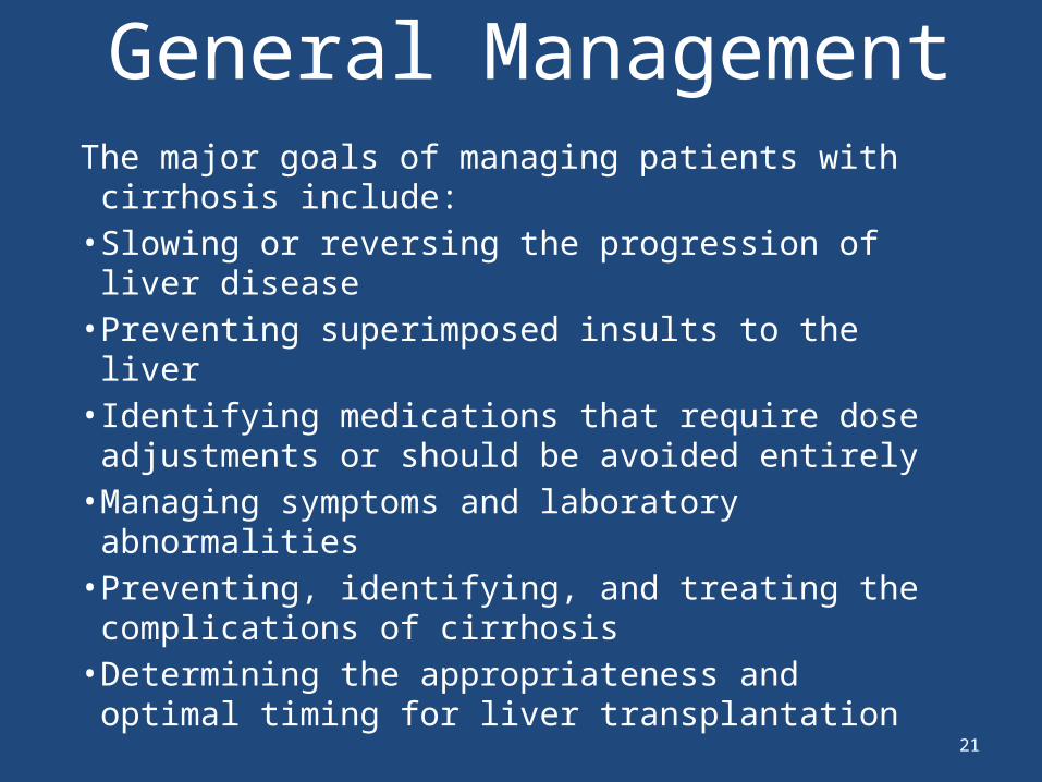
General ManagementThe major goals of managing patients with
cirrhosis include:• Slowing or reversing the progression of liver
disease• Preventing superimposed insults to the liver• Identifying medications that require dose
adjustments or should be avoided entirely • Managing symptoms and laboratory
abnormalities • Preventing, identifying, and treating the
complications of cirrhosis• Determining the appropriateness and optimal
timing for liver transplantation
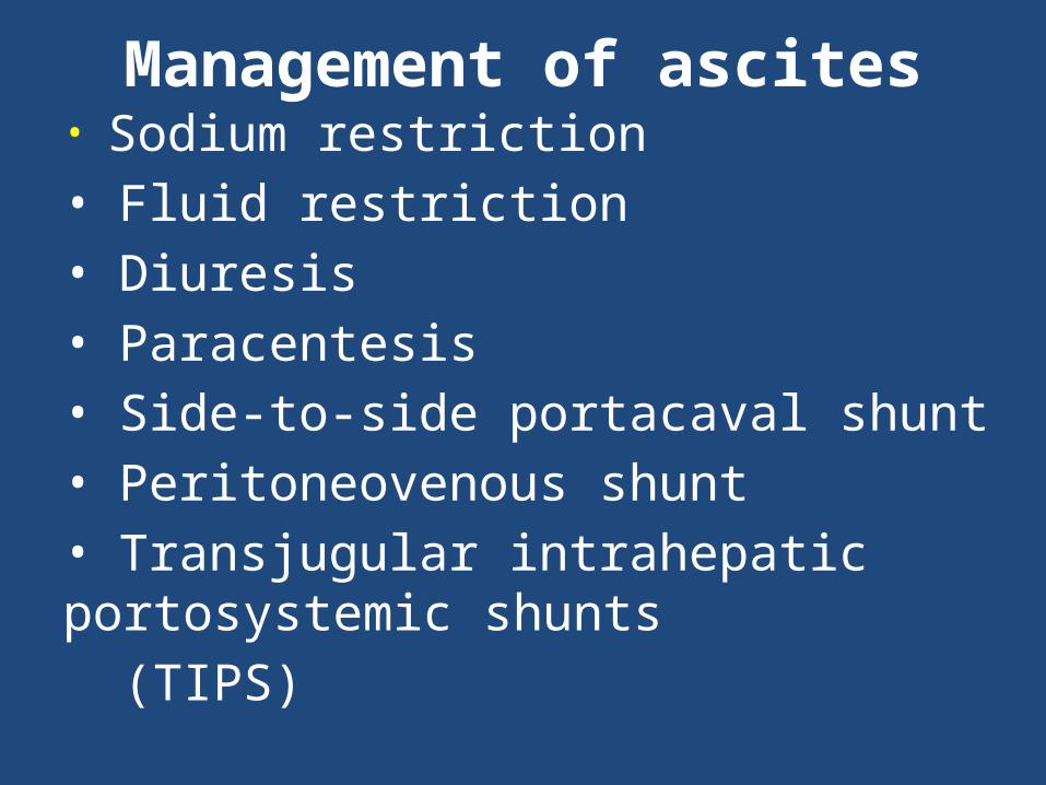
Management of ascites• Sodium restriction• Fluid restriction• Diuresis• Paracentesis• Side-to-side portacaval shunt• Peritoneovenous shunt• Transjugular intrahepatic portosystemic shunts (TIPS)
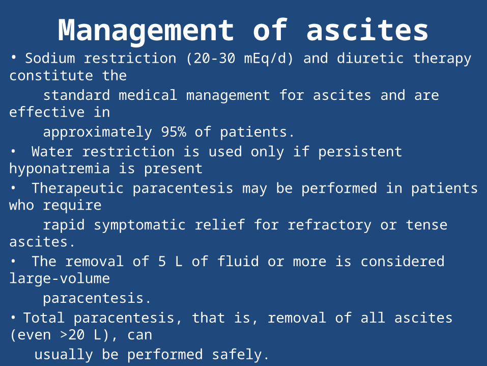
Management of ascites• Sodium restriction (20-30 mEq/d) and diuretic therapy constitute the standard medical management for ascites and are effective in approximately 95% of patients.• Water restriction is used only if persistent hyponatremia is present• Therapeutic paracentesis may be performed in patients who require rapid symptomatic relief for refractory or tense ascites.• The removal of 5 L of fluid or more is considered large-volume paracentesis. • Total paracentesis, that is, removal of all ascites (even >20 L), can usually be performed safely.
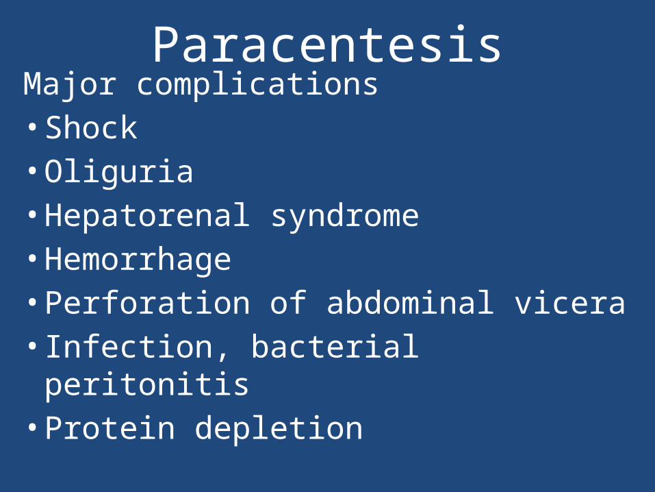
ParacentesisMajor complications• Shock• Oliguria• Hepatorenal syndrome• Hemorrhage • Perforation of abdominal vicera • Infection, bacterial peritonitis• Protein depletion
Upper gastrointestinal bleeding
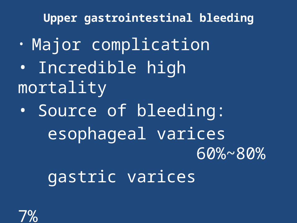
• Major complication• Incredible high mortality• Source of bleeding: esophageal varices 60%~80% gastric varices 7% congestive gastropathy 5%~20% (peptic ulcer, acute erosive gastritis etc)

Emergency endoscopy
Upper GI Bleed ? Varices
Esophageal Varices ELB/ES
Gastric Varices/ Cyanoacrylate Glue
Uncontrolled: Salvage therapyBaloon temponadeShunt - Surgical TIPS
Controlled : Secondary prophylaxisEBI eradication program+/- Beta blockadeReferral transplantation
Controlled : Secondary prophylaxisBeta blockadeConsider transplantationConsider TIPS/Surgery
• Resuscitation, multiple organ support, • correction coagulopathy /thrombocytopenia • Endotracheal intubation• Septic screen& empirical antibiotics• Medical therapy to reduce PHT
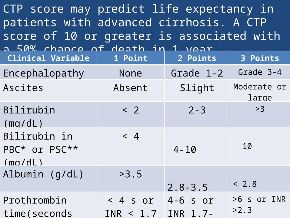
CTP score may predict life expectancy in patients with advanced cirrhosis. A CTP score of 10 or greater is associated with a 50% chance of death in 1 yearChild-Turcotte-Pugh Scoring System for CirrhosisClinical Variable 1 Point 2 Points 3 Points
Encephalopathy None Grade 1-2 Grade 3-4
Ascites Absent Slight Moderate or large
Bilirubin (mg/dL) < 2 2-3 >3
Bilirubin in PBC* or PSC** (mg/dL)
< 4 4-10 10
Albumin (g/dL) >3.5 2.8-3.5
< 2.8
Prothrombin time(seconds prolonged or INR)
< 4 s or INR < 1.7
4-6 s or INR 1.7-2.3
>6 s or INR >2.3
PBC = Primary biliary cirrhosis**PSC = Primary sclerosing cholangitisChild Class A = 5-6 points, Child Class B = 7-9 points, Child Class C = 10-15 points
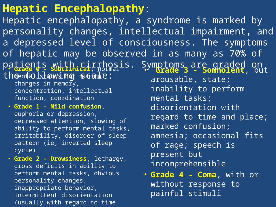
• Grade 0 - Subclinical; normal mental status but minimal changes in memory, concentration, intellectual function, coordination
• Grade 1 - Mild confusion, euphoria or depression, decreased attention, slowing of ability to perform mental tasks, irritability, disorder of sleep pattern (ie, inverted sleep cycle)
• Grade 2 - Drowsiness, lethargy, gross deficits in ability to perform mental tasks, obvious personality changes, inappropriate behavior, intermittent disorientation (usually with regard to time
• Grade 3 - Somnolent, but arousable, state; inability to perform mental tasks; disorientation with regard to time and place; marked confusion; amnesia; occasional fits of rage; speech is present but incomprehensible
• Grade 4 - Coma, with or without response to painful stimuli
Hepatic Encephalopathy:Hepatic encephalopathy, a syndrome is marked by personality changes, intellectual impairment, and a depressed level of consciousness. The symptoms of hepatic may be observed in as many as 70% of patients with cirrhosis. Symptoms are graded on the following scale:
• This syndrome represents a continuum of renal dysfunction that may be observed in patients with a combination of cirrhosis and ascites.
• Hepatorenal syndrome is caused by the vasoconstriction of large and small renal arteries and the impaired renal perfusion that results
• The syndrome is an imbalance between renal vasodilators vasoconstrictors
• Plasma levels of a number of vasoconstricting substances—including angiotensin, antidiuretic hormone, and norepinephrine—are elevated in patients with cirrhosis.
• Renal perfusion appears to be protected by vasodilators, including prostaglandins E2 and I2 and atrial natriuretic factor.
Hepatorenal syndrome
Preventing superimposed insults to the liver
• Vaccinations — Vaccination against hepatitis A and B for those who are not already immune can help prevent superimposed insults to the liver. Other vaccinations, such a yearly influenza vaccination, are also recommended
• Avoidance of hepatotoxins
• Medication adjustments
• Patients with hepatitis C and advanced fibrosis or cirrhosis who achieve a sustained virologic response (SVR) with antiviral treatment
• Abstinence from alcohol substantially improves survival in alcoholic cirrhosis
• Successful treatment of chronic viral hepatitis can improve long-term outcomes and may affect fibrosis
Slowing or reversing the progression of liver disease
Liver transplantation
Latest advance in management of cirrhosis includes liver transplantation.Indications for liver transplantation need to be carefully considered when medical and surgical treatment options have failed.
• Cirrhosis is a condition in which the liver slowly deteriorates and malfunctions due to chronic injury, preventing the liver from working as it should.
• Heavy alcohol consumption and chronic hepatitis C have been the most common causes of cirrhosis. Many people with cirrhosis have more than one cause of liver damage.
• Other causes of cirrhosis include hepatitis B, hepatitis D, and autoimmune hepatitis; diseases that damage or destroy bile ducts, inherited diseases, and nonalcoholic fatty liver disease; and drugs, toxins, and infections.
• Many people with cirrhosis have no symptoms in the early stages of the disease. As the disease progresses, symptoms may include weakness, fatigue, loss of appetite, nausea, vomiting, weight loss, abdominal pain and bloating, itching, and spiderlike blood vessels on the skin.
• As liver function deteriorates, one or more complications may develop. In some people, complications may be the first signs of the disease.
• The goals of treatment are to stop the progression of scar tissue in the liver and prevent or treat complications.
• Treatment for cirrhosis includes avoidance of alcohol and other drugs, nutrition therapy, and other therapies that treat specific complications or causes of the disease.
• Hospitalization may be necessary for cirrhosis with complications.
• A liver transplant is considered when complications of cirrhosis cannot be controlled by treatment.
Summary