Peritubular Capillary and Renal Interstitial Fluid Physical Forces
915
42Circulation and Gas Exchange
Trading Places
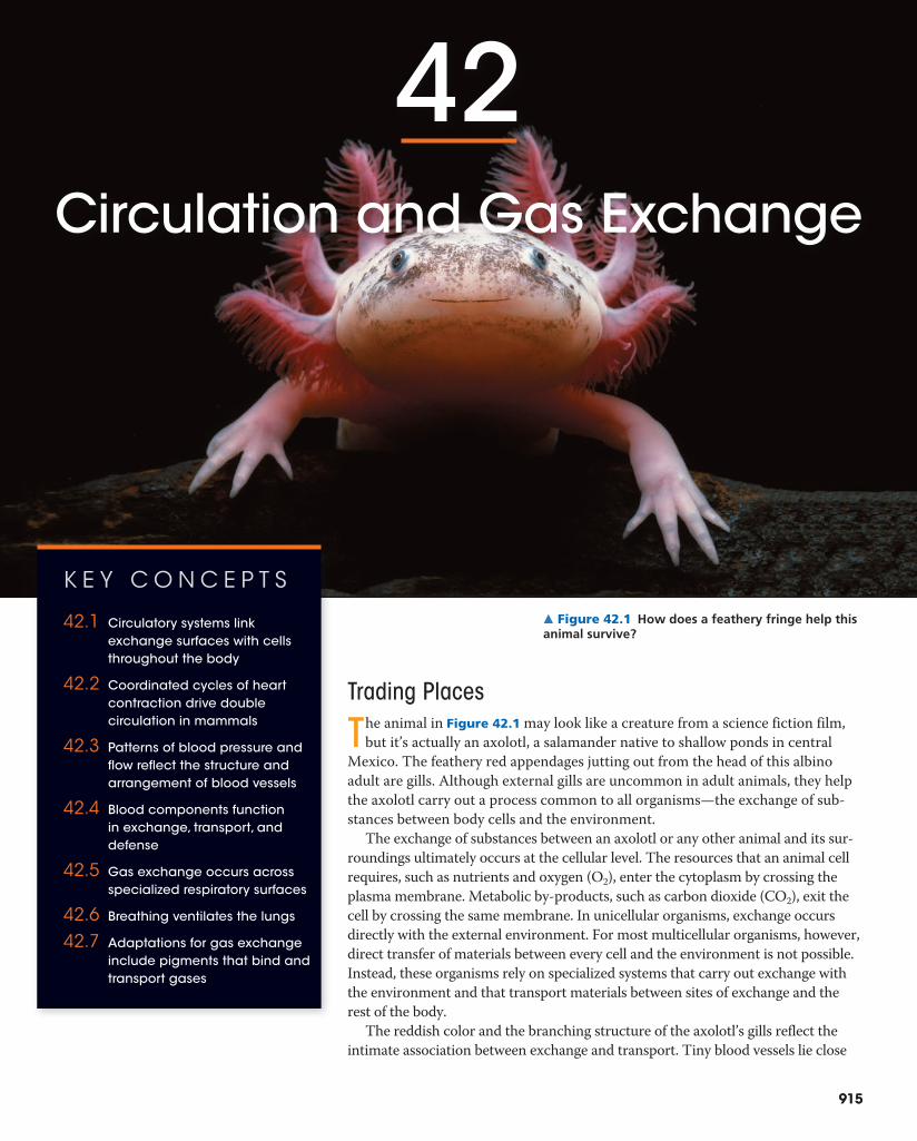
The animal in Figure 42.1 may look like a creature from a science fiction film, but it’s actually an axolotl, a salamander native to shallow ponds in central
Mexico. The feathery red appendages jutting out from the head of this albino adult are gills. Although external gills are uncommon in adult animals, they help the axolotl carry out a process common to all organisms—the exchange of sub-stances between body cells and the environment.
The exchange of substances between an axolotl or any other animal and its sur-roundings ultimately occurs at the cellular level. The resources that an animal cell requires, such as nutrients and oxygen (O2), enter the cytoplasm by crossing the plasma membrane. Metabolic by-products, such as carbon dioxide (CO2), exit the cell by crossing the same membrane. In unicellular organisms, exchange occurs directly with the external environment. For most multicellular organisms, however, direct transfer of materials between every cell and the environment is not possible. Instead, these organisms rely on specialized systems that carry out exchange with the environment and that transport materials between sites of exchange and the rest of the body.
The reddish color and the branching structure of the axolotl’s gills reflect the intimate association between exchange and transport. Tiny blood vessels lie close
▲ Figure 42.1 How does a feathery fringe help this animal survive?
915
K E Y C O N C E P T S
42.1 Circulatory systems link exchange surfaces with cells throughout the body
42.2 Coordinated cycles of heart contraction drive double circulation in mammals
42.3 Patterns of blood pressure and flow reflect the structure and arrangement of blood vessels
42.4 Blood components function in exchange, transport, and defense
42.5 Gas exchange occurs across specialized respiratory surfaces
42.6 Breathing ventilates the lungs
42.7 Adaptations for gas exchange include pigments that bind and transport gases

916 U N I T S E V E N Animal Form and Function
to the surface of each filament in the gills. Across this surface, there is a net diffusion of O2 from the surrounding water into the blood and of CO2 from the blood into the water. The short distances involved allow diffusion to be rapid. Pump-ing of the axolotl’s heart propels the oxygen-rich blood from the gill filaments to all other tissues of the body. There, more short-range exchange occurs, involving nutrients and O2 as well as CO2 and other wastes.
Because internal transport and gas exchange are function-ally related in most animals, not just axolotls, we’ll discuss circulatory and respiratory systems together in this chapter. By considering examples of these systems from a range of species, we’ll explore the common elements as well as the remarkable variation in form and organization. We’ll also highlight the roles of circulatory and respiratory systems in maintaining homeostasis.
C O N C E P T 42.1Circulatory systems link exchange surfaces with cells throughout the bodyThe molecular trade that an animal carries out with its environment—gaining O2 and nutrients while shedding CO2 and other waste products—must ultimately involve every cell in the body. Small molecules, including O2 and CO2, can move between cells and their immediate sur-roundings by diffusion (see Chapter 7). When there is a difference in concentration, diffusion can result in net movement. But such movement is very slow for distances of more than a few millimeters. That’s because the time it takes for a substance to diffuse from one place to another is proportional to the square of the distance. For example, a quantity of glucose that takes 1 second to diffuse 100 μm will take 100 seconds to diffuse 1 mm and almost 3 hours to diffuse 1 cm! This relationship between diffusion time and distance places a substantial constraint on the body plan of any animal.
Given that net movement by diffusion is rapid only over very small distances, how does each cell of an animal partici-pate in exchange? Natural selection has resulted in two basic adaptations that permit effective exchange for all of an ani-mal’s cells. One adaptation is a body plan that places many or all cells in direct contact with the environment. Each cell can thus exchange materials directly with the surrounding medium. This type of body plan is found only in certain in-vertebrates, including cnidarians and flatworms. The other adaptation, found in all other animals, is a circulatory sys-tem. Such systems move fluid between each cell’s immediate surroundings and the body tissues where exchange with the environment occurs.
PharynxMouth Gastrovascular
cavity
Mouth
Circular canal
Radial canals
1 mm
2.5 cm
(a) The moon jelly Aurelia, a cnidarian. The jelly is viewed here from its underside (oral surface). The mouth leads to an elaborate gastrovascular cavity that consists of radial canals leading to and from a circular canal. Ciliated cells lining the canals circulate fluid within the cavity.
(b) The planarian Dugesia, a flatworm. The mouth and pharynx on the ventral side lead to the highly branched gastrovascular cavity, stained dark red in this specimen (LM).
▲ Figure 42.2 Internal transport in gastrovascular cavities.
W H AT I F ? Suppose a gastrovascular cavity were open at two ends, with fluid entering one end and leaving the other. How would this affect the cavity’s functions in gas exchange and digestion?
Gastrovascular CavitiesLet’s begin by looking at some animals whose body shapes put many of their cells into contact with their environment, enabling them to live without a distinct circulatory system. In hydras, jellies, and other cnidarians, a central gastrovascular cavity functions in the distribution of substances throughout the body, as well as in digestion (see Figure 41.7). An opening at one end connects the cavity to the surrounding water. In a hydra, thin branches of the gastrovascular cavity extend into the animal’s tentacles. In jellies and some other cnidarians, the gastrovascular cavity has a much more elaborate branch-ing pattern (Figure 42.2a).

C H A P T E R 4 2 Circulation and Gas Exchange 917
and the interstitial fluid, as well as between the interstitial fluid and body cells. Annelids (including earthworms), ceph-alopods (including squids and octopuses), and all vertebrates have closed circulatory systems.
The fact that both open and closed circulatory systems are widespread among animals suggests that each system offers evolutionary advantages. The lower hydrostatic pres-sures typically associated with open circulatory systems
In animals with a gastrovascular cavity, fluid bathes both the inner and outer tissue layers, facilitating exchange of gases and cellular waste. Only the cells lining the cavity have direct access to nutrients released by digestion. However, because the body wall is a mere two cells thick, nutrients need diffuse only a short distance to reach the cells of the outer tissue layer.
Planarians and most other flatworms also survive without a circulatory system. Their combination of a gastrovascular cavity and a flat body is well suited for exchange with the en-vironment (Figure 42.2b). A flat body optimizes exchange by increasing surface area and minimizing diffusion distances.
Open and Closed Circulatory SystemsA circulatory system has three basic components: a circula-tory fluid, a set of interconnecting vessels, and a muscular pump, the heart. The heart powers circulation by using metabolic energy to elevate the circulatory fluid’s hydro-static pressure, the pressure the fluid exerts on surrounding vessels. The fluid then flows through the vessels and back to the heart.
By transporting fluid throughout the body, the circula-tory system functionally connects the aqueous environment of the body cells to the organs that exchange gases, absorb nutrients, and dispose of wastes. In mammals, for example, O2 from inhaled air diffuses across only two layers of cells in the lungs before reaching the blood. The circulatory system then carries the oxygen-rich blood to all parts of the body. As the blood courses throughout the body tissues in tiny blood vessels, O2 in the blood diffuses only a short distance before entering the fluid that directly bathes the cells.
Circulatory systems are either open or closed. In an open circulatory system, the circulatory fluid, called hemolymph, is also the interstitial fluid that bathes body cells. Arthropods, such as grasshoppers, and some molluscs, including clams, have open circulatory systems. Heart contraction pumps the hemolymph through the circulatory vessels into intercon-nected sinuses, spaces surrounding the organs (Figure 42.3a). Within the sinuses, chemical exchange occurs between the hemolymph and body cells. Relaxation of the heart draws hemolymph back in through pores, which are equipped with valves that close when the heart contracts. Body movements periodically squeeze the sinuses, helping circulate the hemo-lymph. The open circulatory system of larger crustaceans, such as lobsters and crabs, includes a more extensive system of vessels as well as an accessory pump.
In a closed circulatory system, a circulatory fluid called blood is confined to vessels and is distinct from the inter-stitial fluid (Figure 42.3b). One or more hearts pump blood into large vessels that branch into smaller ones that infiltrate the organs. Chemical exchange occurs between the blood
Dorsal vessel(main heart)
Auxiliary hearts
Ventral vessels
Hemolymph in sinusessurrounding organs
Heart
Heart
Small branch vessels in each organ
Interstitial fluid
Blood
(a) An open circulatory system
(b) A closed circulatory system
Pores
Tubular heart
In an open circulatory system, such as that of a grasshopper, hemo-lymph surrounding body tissues also acts as the circulatory fluid.
In a closed circulatory system, such as that of an earthworm, interstitial fluid surrounding body tissues is distinct from blood acting as the circulatory fluid.
▼ Figure 42.3 Open and closed circulatory systems.
918 U N I T S E V E N Animal Form and Function
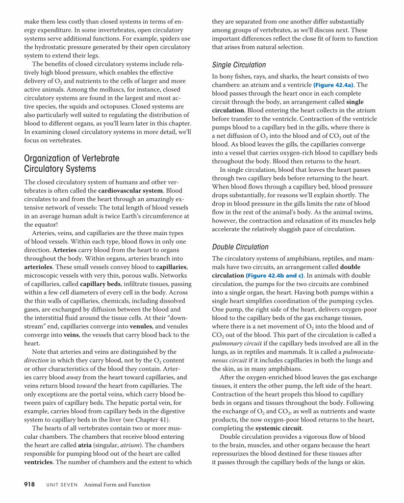
they are separated from one another differ substantially among groups of vertebrates, as we’ll discuss next. These important differences reflect the close fit of form to function that arises from natural selection.
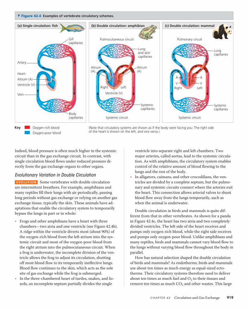
Single CirculationIn bony fishes, rays, and sharks, the heart consists of two chambers: an atrium and a ventricle (Figure 42.4a). The blood passes through the heart once in each complete circuit through the body, an arrangement called single circulation. Blood entering the heart collects in the atrium before transfer to the ventricle. Contraction of the ventricle pumps blood to a capillary bed in the gills, where there is a net diffusion of O2 into the blood and of CO2 out of the blood. As blood leaves the gills, the capillaries converge into a vessel that carries oxygen-rich blood to capillary beds throughout the body. Blood then returns to the heart.
In single circulation, blood that leaves the heart passes through two capillary beds before returning to the heart. When blood flows through a capillary bed, blood pressure drops substantially, for reasons we’ll explain shortly. The drop in blood pressure in the gills limits the rate of blood flow in the rest of the animal’s body. As the animal swims, however, the contraction and relaxation of its muscles help accelerate the relatively sluggish pace of circulation.
Double CirculationThe circulatory systems of amphibians, reptiles, and mam-mals have two circuits, an arrangement called double circulation (Figure 42.4b and c). In animals with double circulation, the pumps for the two circuits are combined into a single organ, the heart. Having both pumps within a single heart simplifies coordination of the pumping cycles. One pump, the right side of the heart, delivers oxygen-poor blood to the capillary beds of the gas exchange tissues, where there is a net movement of O2 into the blood and of CO2 out of the blood. This part of the circulation is called a pulmonary circuit if the capillary beds involved are all in the lungs, as in reptiles and mammals. It is called a pulmocuta-neous circuit if it includes capillaries in both the lungs and the skin, as in many amphibians.
After the oxygen-enriched blood leaves the gas exchange tissues, it enters the other pump, the left side of the heart. Contraction of the heart propels this blood to capillary beds in organs and tissues throughout the body. Following the exchange of O2 and CO2, as well as nutrients and waste products, the now oxygen-poor blood returns to the heart, completing the systemic circuit.
Double circulation provides a vigorous flow of blood to the brain, muscles, and other organs because the heart repressurizes the blood destined for these tissues after it passes through the capillary beds of the lungs or skin.
make them less costly than closed systems in terms of en-ergy expenditure. In some invertebrates, open circulatory systems serve additional functions. For example, spiders use the hydrostatic pressure generated by their open circulatory system to extend their legs.
The benefits of closed circulatory systems include rela-tively high blood pressure, which enables the effective delivery of O2 and nutrients to the cells of larger and more active animals. Among the molluscs, for instance, closed circulatory systems are found in the largest and most ac-tive species, the squids and octopuses. Closed systems are also particularly well suited to regulating the distribution of blood to different organs, as you’ll learn later in this chapter. In examining closed circulatory systems in more detail, we’ll focus on vertebrates.
Organization of Vertebrate Circulatory SystemsThe closed circulatory system of humans and other ver-tebrates is often called the cardiovascular system. Blood circulates to and from the heart through an amazingly ex-tensive network of vessels: The total length of blood vessels in an average human adult is twice Earth’s circumference at the equator!
Arteries, veins, and capillaries are the three main types of blood vessels. Within each type, blood flows in only one direction. Arteries carry blood from the heart to organs throughout the body. Within organs, arteries branch into arterioles. These small vessels convey blood to capillaries, microscopic vessels with very thin, porous walls. Networks of capillaries, called capillary beds, infiltrate tissues, passing within a few cell diameters of every cell in the body. Across the thin walls of capillaries, chemicals, including dissolved gases, are exchanged by diffusion between the blood and the interstitial fluid around the tissue cells. At their “down-stream” end, capillaries converge into venules, and venules converge into veins, the vessels that carry blood back to the heart.
Note that arteries and veins are distinguished by the direction in which they carry blood, not by the O2 content or other characteristics of the blood they contain. Arter-ies carry blood away from the heart toward capillaries, and veins return blood toward the heart from capillaries. The only exceptions are the portal veins, which carry blood be-tween pairs of capillary beds. The hepatic portal vein, for example, carries blood from capillary beds in the digestive system to capillary beds in the liver (see Chapter 41).
The hearts of all vertebrates contain two or more mus-cular chambers. The chambers that receive blood entering the heart are called atria (singular, atrium). The chambers responsible for pumping blood out of the heart are called ventricles. The number of chambers and the extent to which
C H A P T E R 4 2 Circulation and Gas Exchange 919
ventricle into separate right and left chambers. Two major arteries, called aortas, lead to the systemic circula-tion. As with amphibians, the circulatory system enables control of the relative amount of blood flowing to the lungs and the rest of the body.
t� In alligators, caimans, and other crocodilians, the ven-tricles are divided by a complete septum, but the pulmo-nary and systemic circuits connect where the arteries exit the heart. This connection allows arterial valves to shunt blood flow away from the lungs temporarily, such as when the animal is underwater.Double circulation in birds and mammals is quite dif-
ferent from that in other vertebrates. As shown for a panda in Figure 42.4c, the heart has two atria and two completely divided ventricles. The left side of the heart receives and pumps only oxygen-rich blood, while the right side receives and pumps only oxygen-poor blood. Unlike amphibians and many reptiles, birds and mammals cannot vary blood flow to the lungs without varying blood flow throughout the body in parallel.
How has natural selection shaped the double circulation of birds and mammals? As endotherms, birds and mammals use about ten times as much energy as equal-sized ecto-therms. Their circulatory systems therefore need to deliver about ten times as much fuel and O2 to their tissues and remove ten times as much CO2 and other wastes. This large
Indeed, blood pressure is often much higher in the systemic circuit than in the gas exchange circuit. In contrast, with single circulation blood flows under reduced pressure di-rectly from the gas exchange organs to other organs.
Evolutionary Variation in Double CirculationE VO L U T I O N Some vertebrates with double circulation
are intermittent breathers. For example, amphibians and many reptiles fill their lungs with air periodically, passing long periods without gas exchange or relying on another gas exchange tissue, typically the skin. These animals have ad-aptations that enable the circulatory system to temporarily bypass the lungs in part or in whole:t� Frogs and other amphibians have a heart with three
chambers—two atria and one ventricle (see Figure 42.4b). A ridge within the ventricle diverts most (about 90%) of the oxygen-rich blood from the left atrium into the sys-temic circuit and most of the oxygen-poor blood from the right atrium into the pulmocutaneous circuit. When a frog is underwater, the incomplete division of the ven-tricle allows the frog to adjust its circulation, shutting off most blood flow to its temporarily ineffective lungs. Blood flow continues to the skin, which acts as the sole site of gas exchange while the frog is submerged.
t� In the three-chambered heart of turtles, snakes, and liz-ards, an incomplete septum partially divides the single
Gill capillaries
Lung capillaries
Systemic capillaries
A
VLeft
A
VRight
Artery
Atrium (A)
Ventricle (V)
Vein
Body capillaries
Heart:
Oxygen-rich blood
Oxygen-poor blood
Key
Lung and skin capillaries
Systemiccapillaries
Atrium(A)
Atrium(A)
Left
Ventricle (V)
Right
Pulmocutaneous circuit
Systemic circuit
Pulmonary circuit
Systemic circuit
(a) Single circulation: fish (b) Double circulation: amphibian (c) Double circulation: mammal
(Note that circulatory systems are shown as if the body were facing you: The right side of the heart is shown on the left, and vice versa.)
▼ Figure 42.4 Examples of vertebrate circulatory schemes.
920 U N I T S E V E N Animal Form and Function
traffic of substances is made possible by the separate and in-dependently powered systemic and pulmonary circuits and by large hearts that pump the necessary volume of blood. A powerful four-chambered heart arose independently in the distinct ancestors of birds and mammals and thus reflects convergent evolution (see Chapter 34).
In the next section, we’ll restrict our focus to circulation in mammals and to the anatomy and physiology of the key circulatory organ—the heart.
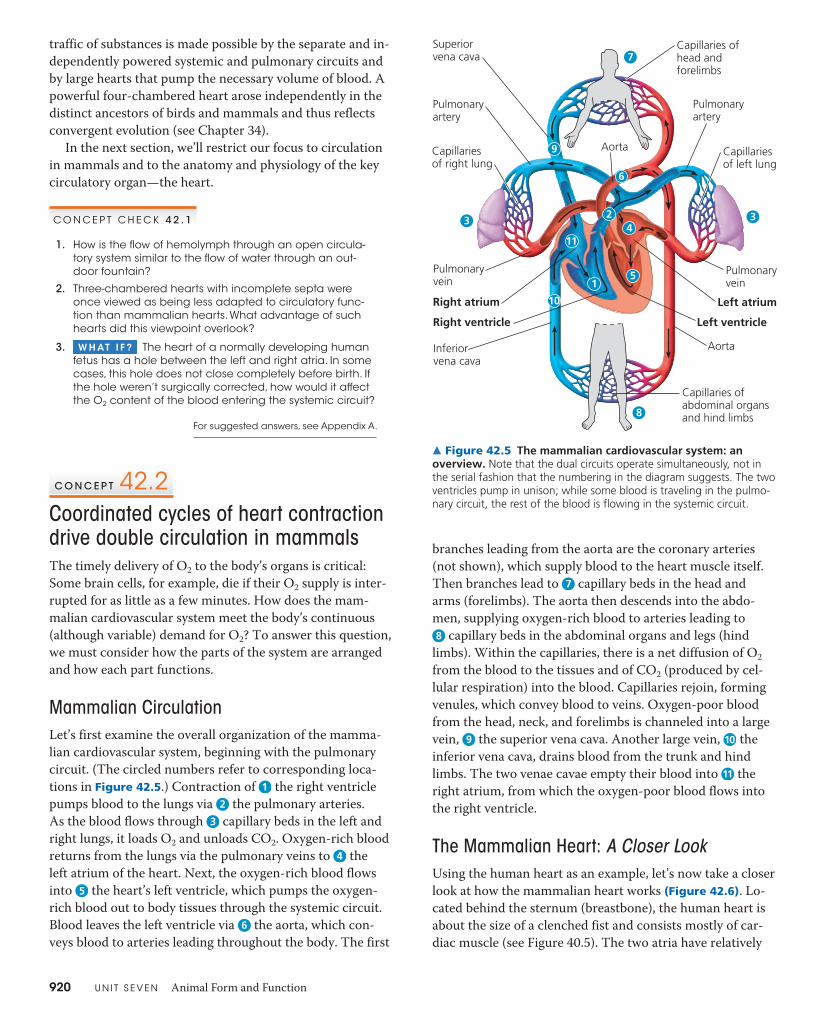
branches leading from the aorta are the coronary arteries (not shown), which supply blood to the heart muscle itself. Then branches lead to 7 capillary beds in the head and arms (forelimbs). The aorta then descends into the abdo-men, supplying oxygen-rich blood to arteries leading to 8 capillary beds in the abdominal organs and legs (hind
limbs). Within the capillaries, there is a net diffusion of O2 from the blood to the tissues and of CO2 (produced by cel-lular respiration) into the blood. Capillaries rejoin, forming venules, which convey blood to veins. Oxygen-poor blood from the head, neck, and forelimbs is channeled into a large vein, 9 the superior vena cava. Another large vein, 10 the inferior vena cava, drains blood from the trunk and hind limbs. The two venae cavae empty their blood into 11 the right atrium, from which the oxygen-poor blood flows into the right ventricle.
The Mammalian Heart: A Closer LookUsing the human heart as an example, let’s now take a closer look at how the mammalian heart works (Figure 42.6). Lo-cated behind the sternum (breastbone), the human heart is about the size of a clenched fist and consists mostly of car-diac muscle (see Figure 40.5). The two atria have relatively
C O N C E P T C H E C K 4 2 . 1
1. How is the flow of hemolymph through an open circula-tory system similar to the flow of water through an out-door fountain?
2. Three-chambered hearts with incomplete septa were once viewed as being less adapted to circulatory func-tion than mammalian hearts. What advantage of such hearts did this viewpoint overlook?
3. W H AT I F ? The heart of a normally developing human fetus has a hole between the left and right atria. In some cases, this hole does not close completely before birth. If the hole weren’t surgically corrected, how would it affect the O2 content of the blood entering the systemic circuit?
For suggested answers, see Appendix A.
C O N C E P T 42.2Coordinated cycles of heart contraction drive double circulation in mammalsThe timely delivery of O2 to the body’s organs is critical: Some brain cells, for example, die if their O2 supply is inter-rupted for as little as a few minutes. How does the mam-malian cardiovascular system meet the body’s continuous (although variable) demand for O2? To answer this question, we must consider how the parts of the system are arranged and how each part functions.
Mammalian CirculationLet’s first examine the overall organization of the mamma-lian cardiovascular system, beginning with the pulmonary circuit. (The circled numbers refer to corresponding loca-tions in Figure 42.5.) Contraction of 1 the right ventricle pumps blood to the lungs via 2 the pulmonary arteries. As the blood flows through 3 capillary beds in the left and right lungs, it loads O2 and unloads CO2. Oxygen-rich blood returns from the lungs via the pulmonary veins to 4 the left atrium of the heart. Next, the oxygen-rich blood flows into 5 the heart’s left ventricle, which pumps the oxygen-rich blood out to body tissues through the systemic circuit. Blood leaves the left ventricle via 6 the aorta, which con-veys blood to arteries leading throughout the body. The first
Superiorvena cava
Pulmonaryartery
Capillariesof right lung
Capillaries ofhead and forelimbs
Pulmonaryartery
Capillariesof left lung
Pulmonaryvein
Left atrium
Left ventricle
Aorta
Capillaries ofabdominal organsand hind limbs
Pulmonary vein
Right atrium
Inferiorvena cava
Aorta
1
10
11
3
6
34
7
8
9
5
Right ventricle
2
▲ Figure 42.5 The mammalian cardiovascular system: an overview. Note that the dual circuits operate simultaneously, not in the serial fashion that the numbering in the diagram suggests. The two ventricles pump in unison; while some blood is traveling in the pulmo-nary circuit, the rest of the blood is flowing in the systemic circuit.
C H A P T E R 4 2 Circulation and Gas Exchange 921
Made of flaps of connective tissue, the valves open when pushed from one side and close when pushed from the other. An atrioventricular (AV) valve lies between each atrium and ventricle. The AV valves are anchored by strong fibers that prevent them from turning inside out. Pressure generated by the powerful contraction of the ventricles closes the AV valves, keeping blood from flowing back into the atria. Semilunar valves are located at the two exits of the heart: where the aorta leaves the left ventricle and where the pulmonary artery leaves the right ventricle. These valves are pushed open by the pressure generated during contrac-tion of the ventricles. When the ventricles relax, blood pres-sure built up in the aorta and pulmonary artery closes the semilunar valves and prevents significant backflow.
You can follow the closing of the two sets of heart valves either with a stethoscope or by pressing your ear tightly against the chest of a friend (or a friendly dog). The sound pattern is “lub-dup, lub-dup, lub-dup.” The first heart sound
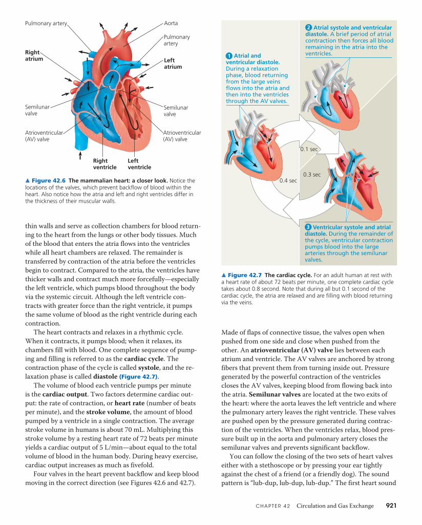
thin walls and serve as collection chambers for blood return-ing to the heart from the lungs or other body tissues. Much of the blood that enters the atria flows into the ventricles while all heart chambers are relaxed. The remainder is transferred by contraction of the atria before the ventricles begin to contract. Compared to the atria, the ventricles have thicker walls and contract much more forcefully—especially the left ventricle, which pumps blood throughout the body via the systemic circuit. Although the left ventricle con-tracts with greater force than the right ventricle, it pumps the same volume of blood as the right ventricle during each contraction.
The heart contracts and relaxes in a rhythmic cycle. When it contracts, it pumps blood; when it relaxes, its chambers fill with blood. One complete sequence of pump-ing and filling is referred to as the cardiac cycle. The contraction phase of the cycle is called systole, and the re-laxation phase is called diastole (Figure 42.7).
The volume of blood each ventricle pumps per minute is the cardiac output. Two factors determine cardiac out-put: the rate of contraction, or heart rate (number of beats per minute), and the stroke volume, the amount of blood pumped by a ventricle in a single contraction. The average stroke volume in humans is about 70 mL. Multiplying this stroke volume by a resting heart rate of 72 beats per minute yields a cardiac output of 5 L/min—about equal to the total volume of blood in the human body. During heavy exercise, cardiac output increases as much as fivefold.
Four valves in the heart prevent backflow and keep blood moving in the correct direction (see Figures 42.6 and 42.7).
Pulmonary artery
Rightatrium Left
atrium
Semilunarvalve
Atrioventricular(AV) valve
Aorta
Rightventricle
Leftventricle
Semilunarvalve
Atrioventricular(AV) valve
Pulmonaryartery
▲ Figure 42.6 The mammalian heart: a closer look. Notice the locations of the valves, which prevent backflow of blood within the heart. Also notice how the atria and left and right ventricles differ in the thickness of their muscular walls.
0.1 sec
0.3 sec0.4 sec
Atrial and ventricular diastole. During a relaxation phase, blood returning from the large veins flows into the atria and then into the ventricles through the AV valves.
Atrial systole and ventricular diastole. A brief period of atrial contraction then forces all blood remaining in the atria into the ventricles.
Ventricular systole and atrial diastole. During the remainder of the cycle, ventricular contraction pumps blood into the large arteries through the semilunar valves.
1
2
3
▲ Figure 42.7 The cardiac cycle. For an adult human at rest with a heart rate of about 72 beats per minute, one complete cardiac cycle takes about 0.8 second. Note that during all but 0.1 second of the cardiac cycle, the atria are relaxed and are filling with blood returning via the veins.

922 U N I T S E V E N Animal Form and Function
in the wall between the left and right atria. These cells form a relay point called the atrioventricular (AV) node. Here the impulses are delayed for about 0.1 second before spread-ing to the heart apex. This delay allows the atria to empty completely before the ventricles contract. Then the signals from the AV node are conducted to the heart apex and throughout the ventricular walls by specialized structures called bundle branches and Purkinje fibers.
Physiological cues alter heart tempo by regulating the pacemaker function of the SA node. Two portions of the nervous system, the sympathetic and parasympathetic divi-sions, are largely responsible for this regulation. They func-tion like the accelerator and brake in a car: For example, when you stand up and start walking, the sympathetic divi-sion speeds up your pacemaker. The resulting increase in heart rate provides the additional O2 needed by the muscles that are powering your activity. If you then sit down and relax, the parasympathetic division slows down your pace-maker, decreasing your heart rate and thus conserving en-ergy. Hormones secreted into the blood also influence the pacemaker. For instance, epinephrine, the “fight-or-flight” hormone secreted by the adrenal glands, speeds up the pacemaker. A third type of input that affects the pacemaker is body temperature. An increase of only 1°C raises the heart rate by about 10 beats per minute. This is the reason your heart beats faster when you have a fever.
Having examined the operation of the circulatory pump, we turn in the next section to the forces and structures that influence blood flow in the vessels of each circuit.
(“lub”) is created by the recoil of blood against the closed AV valves. The second sound (“dup”) is due to the vibrations caused by closing of the semilunar valves.
If blood squirts backward through a defective valve, it may produce an abnormal sound called a heart murmur. Some people are born with heart murmurs; in others, the valves may be damaged by infection (from rheumatic fever, for instance). When a valve defect is severe enough to en-danger health, surgeons may implant a mechanical replace-ment valve. However, not all heart murmurs are caused by a defect, and most valve defects do not reduce the efficiency of blood flow enough to warrant surgery.
Maintaining the Heart’s Rhythmic BeatIn vertebrates, the heartbeat originates in the heart itself. Some cardiac muscle cells are autorhythmic, meaning they can contract and relax repeatedly without any signal from the nervous system. You can see these rhythmic contrac-tions in tissue that has been removed from the heart and placed in a dish in the laboratory! Because each of these cells has its own intrinsic contraction rhythm, how are their con-tractions coordinated in the intact heart? The answer lies in a group of autorhythmic cells located in the wall of the right atrium, near where the superior vena cava enters the heart. This cluster of cells is called the sinoatrial (SA) node, or pacemaker, and it sets the rate and timing at which all cardiac muscle cells contract. (In contrast, some arthropods have pacemakers located in the nervous system, outside the heart.)
The SA node produces electrical impulses much like those produced by nerve cells. Because cardiac muscle cells are electrically coupled through gap junctions (see Figure 6.30), im-pulses from the SA node spread rap-idly within heart tissue. In addition, these impulses generate currents that are conducted to the skin via body fluids. In an electrocardiogram (ECG or, often, EKG, from the German spelling), these currents are recorded by electrodes placed on the skin. The resulting graph of current against time has a characteristic shape that repre-sents the stages in the cardiac cycle (Figure 42.8).
Impulses from the SA node first spread rapidly through the walls of the atria, causing both atria to contract in unison. During atrial contraction, the impulses originating at the SA node reach other autorhythmic cells located
Signals spreadthroughoutventricles.
1 4
SA node(pacemaker)
AVnode
ECG
Purkinjefibers
Bundlebranches Heart
apex
Signals (yellow) from SA node spread through atria.
2 Signals are delayed at AV node.
3 Bundle branches pass signals to heart apex.
▲ Figure 42.8 The control of heart rhythm. Electrical signals follow a set path through the heart in establishing the heart rhythm. The diagrams at the top trace the movement of these signals (yellow) during the cardiac cycle; specialized muscle cells involved in controlling of the rhythm are indicated in orange. Under each step, the corresponding portion of an electrocardiogram (ECG) is highlighted (yellow). In step 4, the portion of the ECG to the right of the “spike” represents electri-cal activity that reprimes the ventricles for the next round of contraction.
W H AT I F ? If your doctor gave you a copy of your ECG recording, how could you determine what your heart rate had been during the test?

C H A P T E R 4 2 Circulation and Gas Exchange 923
The walls of arteries and veins have a more complex or-ganization than those of capillaries. Both arteries and veins have two layers of tissue surrounding the endothelium. The outer layer is formed by connective tissue that contains elastic fibers, which allow the vessel to stretch and recoil, and collagen, which provides strength. The layer next to the endothelium contains smooth muscle and more elastic fibers.
While similar in organization, the walls of arteries and veins differ, reflecting distinct adaptations to the particular functions of these vessels in circulation. The walls of arteries are thick and strong, accommodating blood pumped at high pressure by the heart. They are also elastic. When the heart relaxes between contractions, the arterial walls recoil, help-ing maintain blood pressure and flow to capillaries. Signals from the nervous system and hormones circulating in the blood act on the smooth muscle in arteries and arterioles, dilating or constricting these vessels and thus controlling blood flow to different parts of the body.
C O N C E P T C H E C K 4 2 . 2
1. Explain why blood has a higher O2 concentration in the pulmonary veins than in the venae cavae, which are also veins.
2. Why is it important that the AV node delay the electrical impulse moving from the SA node and the atria to the ventricles?
3. W H AT I F ? After you exercise regularly for several months, your resting heart rate decreases, but your car-diac output at rest is unchanged. What other change in the function of your heart at rest could explain these findings?
For suggested answers, see Appendix A.
C O N C E P T 42.3Patterns of blood pressure and flow reflect the structure and arrangement of blood vesselsThe vertebrate circulatory system enables blood to deliver ox-ygen and nutrients and remove wastes throughout the body. In doing so, the circulatory system relies on a branching network of vessels much like the plumbing system that delivers fresh water to a city and removes its wastes. In fact, the same physical principles that govern the operation of plumbing systems apply to the functioning of blood vessels.
Blood Vessel Structure and FunctionBlood vessels contain a central lumen (cavity) lined with an endothelium, a single layer of flattened epithelial cells. The smooth surface of the endothe-lium minimizes resistance to the flow of blood. Surrounding the endothe-lium are layers of tissue that differ in capillaries, arteries, and veins, reflect-ing the specialized functions of these vessels.
Arteriole
Artery
Venule
Endothelium Endothelium
ValveBasal lamina
Smoothmuscle
Smoothmuscle
Connectivetissue
Connectivetissue
Vein
Capillary
Capillary
15 μ
mLM
LM
Red blood cell
100 μm
Artery
Red blood cells
Vein
▲ Figure 42.9 The structure of blood vessels.
Capillaries are the smallest blood vessels, having a diameter only slightly greater than that of a red blood cell (Figure 42.9). Capillaries also have very thin walls, which consist of just an endothelium and a surrounding extracellular layer called the basal lamina. The exchange of substances between the blood and interstitial fluid occurs only in capillaries because only there are blood vessel walls thin enough to permit this exchange.
924 U N I T S E V E N Animal Form and Function
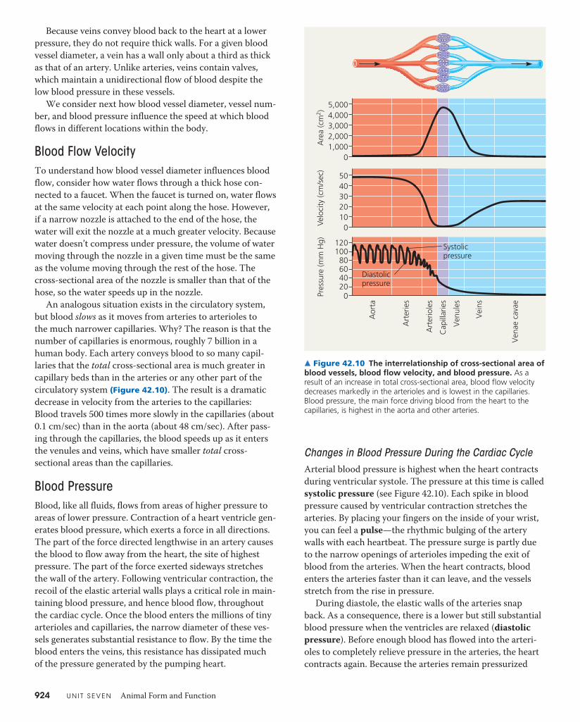
Changes in Blood Pressure During the Cardiac CycleArterial blood pressure is highest when the heart contracts during ventricular systole. The pressure at this time is called systolic pressure (see Figure 42.10). Each spike in blood pressure caused by ventricular contraction stretches the arteries. By placing your fingers on the inside of your wrist, you can feel a pulse—the rhythmic bulging of the artery walls with each heartbeat. The pressure surge is partly due to the narrow openings of arterioles impeding the exit of blood from the arteries. When the heart contracts, blood enters the arteries faster than it can leave, and the vessels stretch from the rise in pressure.
During diastole, the elastic walls of the arteries snap back. As a consequence, there is a lower but still substantial blood pressure when the ventricles are relaxed (diastolic pressure). Before enough blood has flowed into the arteri-oles to completely relieve pressure in the arteries, the heart contracts again. Because the arteries remain pressurized
Because veins convey blood back to the heart at a lower pressure, they do not require thick walls. For a given blood vessel diameter, a vein has a wall only about a third as thick as that of an artery. Unlike arteries, veins contain valves, which maintain a unidirectional flow of blood despite the low blood pressure in these vessels.
We consider next how blood vessel diameter, vessel num-ber, and blood pressure influence the speed at which blood flows in different locations within the body.
Blood Flow VelocityTo understand how blood vessel diameter influences blood flow, consider how water flows through a thick hose con-nected to a faucet. When the faucet is turned on, water flows at the same velocity at each point along the hose. However, if a narrow nozzle is attached to the end of the hose, the water will exit the nozzle at a much greater velocity. Because water doesn’t compress under pressure, the volume of water moving through the nozzle in a given time must be the same as the volume moving through the rest of the hose. The cross-sectional area of the nozzle is smaller than that of the hose, so the water speeds up in the nozzle.
An analogous situation exists in the circulatory system, but blood slows as it moves from arteries to arterioles to the much narrower capillaries. Why? The reason is that the number of capillaries is enormous, roughly 7 billion in a human body. Each artery conveys blood to so many capil-laries that the total cross-sectional area is much greater in capillary beds than in the arteries or any other part of the circulatory system (Figure 42.10). The result is a dramatic decrease in velocity from the arteries to the capillaries: Blood travels 500 times more slowly in the capillaries (about 0.1 cm/sec) than in the aorta (about 48 cm/sec). After pass-ing through the capillaries, the blood speeds up as it enters the venules and veins, which have smaller total cross- sectional areas than the capillaries.
Blood PressureBlood, like all fluids, flows from areas of higher pressure to areas of lower pressure. Contraction of a heart ventricle gen-erates blood pressure, which exerts a force in all directions. The part of the force directed lengthwise in an artery causes the blood to flow away from the heart, the site of highest pressure. The part of the force exerted sideways stretches the wall of the artery. Following ventricular contraction, the recoil of the elastic arterial walls plays a critical role in main-taining blood pressure, and hence blood flow, throughout the cardiac cycle. Once the blood enters the millions of tiny arterioles and capillaries, the narrow diameter of these ves-sels generates substantial resistance to flow. By the time the blood enters the veins, this resistance has dissipated much of the pressure generated by the pumping heart.
5,0004,0003,0002,0001,000
0
50403020100
Are
a (c
m2 )
Velo
city
(cm
/sec
)
0Pres
sure
(mm
Hg)
Aor
ta
Art
erie
s
Art
erio
les
Cap
illar
ies
Venu
les
Vein
s
Vena
e ca
vae
120100
80604020
Systolicpressure
Diastolicpressure
▲ Figure 42.10 The interrelationship of cross-sectional area of blood vessels, blood flow velocity, and blood pressure. As a result of an increase in total cross-sectional area, blood flow velocity decreases markedly in the arterioles and is lowest in the capillaries. Blood pressure, the main force driving blood from the heart to the capillaries, is highest in the aorta and other arteries.

C H A P T E R 4 2 Circulation and Gas Exchange 925
throughout the cardiac cycle (see Figure 42.10), blood con-tinuously flows into arterioles and capillaries.
Regulation of Blood PressureHomeostatic mechanisms regulate arterial blood pres-sure by altering the diameter of arterioles. As the smooth muscles in arteriole walls contract, the arterioles narrow, a process called vasoconstriction. Narrowing of the arterioles increases blood pressure upstream in the arteries. When the smooth muscles relax, the arterioles undergo vasodilation, an increase in diameter that causes blood pressure in the arteries to fall.
Researchers have identified nitric oxide (NO), a gas, as a major inducer of vasodilation and endothelin, a peptide, as the most potent inducer of vasoconstriction. Cues from the nervous and endocrine systems regulate production of NO and endothelin in blood vessels, where their activities regu-late blood pressure.
Vasoconstriction and vasodilation are often coupled to changes in cardiac output that also affect blood pressure. This coordination of regulatory mechanisms maintains adequate blood flow as the body’s demands on the circula-tory system change. During heavy exercise, for example, the arterioles in working muscles dilate, causing a greater flow of oxygen-rich blood to the muscles. By itself, this increased flow to the muscles would cause a drop in blood pressure (and therefore blood flow) in the body as a whole. However, cardiac output increases at the same time, maintaining blood pressure and supporting the nec-essary increase in blood flow.
Blood Pressure and GravityBlood pressure is generally measured for an artery in the arm at the same height as the heart (Figure 42.11). For a healthy 20-year-old human at rest, arterial blood pressure in the systemic circuit is typically about 120 millime-ters of mercury (mm Hg) at systole and 70 mm Hg at diastole, expressed as 120/70. (Arterial blood pressure in the pulmonary circuit is six to ten times lower.)
Gravity has a significant effect on blood pressure. When you are stand-ing, for example, your head is roughly 0.35 m higher than your chest, and the arterial blood pressure in your brain is about 27 mm Hg less than that near your heart. If the blood pressure in your brain is too low to provide adequate
blood flow, you will likely faint. By causing your body to col-lapse to the ground, fainting effectively places your head at the level of your heart, quickly increasing blood flow to your brain.
For animals with very long necks, the blood pressure required to overcome gravity is particularly high. A giraffe, for example, requires a systolic pressure of more than 250 mm Hg near the heart to get blood to its head. When a gi-raffe lowers its head to drink, one-way valves and sinuses, along with feedback mechanisms that reduce cardiac out-put, prevent this high pressure from damaging its brain. We can calculate that a dinosaur with a neck nearly 10 m long would have required even greater systolic pressure—nearly 760 mm Hg—to pump blood to its brain when its head was fully raised. However, calculations based on anatomy and inferred metabolic rate suggest that dinosaurs did not have a heart powerful enough to generate such high pressure. Based on this evidence as well as studies of neck bone struc-ture, some biologists have concluded that the long-necked dinosaurs fed close to the ground rather than on high foliage.
Gravity is also a consideration for blood flow in veins, especially those in the legs. When you stand or sit, gravity draws blood downward to your legs and feet and impedes its upward return to the heart. Although blood pressure in veins is relatively low, valves inside the veins help maintain
A sphygmomanometer, an inflatable cuff attached to a pressure gauge, measures blood pressure in an artery. The cuff is inflated until the pressure closes the artery, so that no blood flows past the cuff. When this occurs, the pressure exerted by the cuff exceeds the pressure in the artery.
Cuffinflatedwith air
Arteryclosed
Pressure in cuffgreater than 120 mm Hg
120
Pressure in cuffdrops below 120 mm Hg
120
Soundsaudible instethoscope
Pressure in cuff below 70 mm Hg
70
Soundsstop
1 The cuff is allowed to deflate gradually. When the pressure exerted by the cuff falls just below that in the artery, blood pulses into the forearm, generating sounds that can be heard with the stethoscope. The pressure measured at this point is the systolic pressure (120 mm Hg in this example).
2 The cuff is allowed to deflate further, just until the blood flows freely through the artery and the sounds below the cuff disappear. The pressure at this point is the diastolic pressure (70 mm Hg in this example).
3
▲ Figure 42.11 Measurement of blood pressure. Blood pressure is recorded as two numbers separated by a slash. The first number is the systolic pressure; the second is the diastolic pressure.

926 U N I T S E V E N Animal Form and Function
regulate and redirect the passage of blood into particular sets of capillaries. The signals regulating blood flow by these mechanisms include nerve impulses, hormones traveling throughout the bloodstream, and chemicals produced lo-cally. For example, the chemical histamine released by cells at a wound site causes vasodilation. The result is increased blood flow and increased access of disease-fighting white blood cells to invading microorganisms.
As you have read, the critical exchange of substances be-tween the blood and interstitial fluid takes place across the thin endothelial walls of the capillaries. Some substances are carried across the endothelium in vesicles that form on one side by endocytosis and release their contents on the oppo-site side by exocytosis. Small molecules, such as O2 and CO2, simply diffuse across the endothelial cells or, in some tissues, through microscopic pores in the capillary wall. These open-ings also provide the route for transport of small solutes such as sugars, salts, and urea, as well as for bulk flow of fluid into tissues driven by blood pressure within the capillary.
the unidirectional flow of blood within these vessels. The return of blood to the heart is further enhanced by rhythmic contractions of smooth muscles in the walls of venules and veins and by the contraction of skeletal muscles during exer-cise (Figure 42.12).
In rare instances, runners and other athletes can suffer heart failure if they stop vigorous exercise abruptly. When the leg muscles suddenly cease contracting and relaxing, less blood returns to the heart, which continues to beat rapidly. If the heart is weak or damaged, this inadequate blood flow may cause the heart to malfunction. To reduce the risk of stressing the heart excessively, athletes are encouraged to fol-low hard exercise with moderate activity, such as walking, to “cool down” until their heart rate approaches its resting level.
Capillary FunctionAt any given time, only about 5–10% of the body’s capillaries have blood flowing through them. However, each tissue has many capillaries, so every part of the body is supplied with blood at all times. Capillaries in the brain, heart, kidneys, and liver are usually filled to capacity, but at many other sites the blood supply varies over time as blood is diverted from one destination to another. For example, blood flow to the skin is regulated to help control body temperature, and blood supply to the digestive tract increases after a meal. In contrast, blood is diverted from the digestive tract and sup-plied more generously to skeletal muscles and skin during strenuous exercise.
Given that capillaries lack smooth muscle, how is blood flow in capillary beds altered? One mechanism is constric-tion or dilation of the arterioles that supply capillary beds. A second mechanism involves precapillary sphincters, rings of smooth muscle located at the entrance to capillary beds (Figure 42.13). Opening and closing these muscular rings
Valve (open)
Direction of blood flowin vein (toward heart)
Skeletal muscle
Valve (closed)
▶ Figure 42.12 Blood flow in veins. Skeletal muscle contraction squeezes and con-stricts veins. Flaps of tissue within the veins act as one-way valves that keep blood moving only toward the heart. If you sit or stand too long, the lack of muscular activity may cause your feet to swell as blood pools in your veins.
(a) Sphincters relaxed
Precapillary sphincters Thoroughfarechannel
Arteriole Venule
Arteriole Venule
Capillaries
(b) Sphincters contracted
▲ Figure 42.13 Blood flow in capillary beds. Precapillary sphincters regulate the passage of blood into capillary beds. Some blood flows directly from arterioles to venules through capillaries called thoroughfare channels, which are always open.

C H A P T E R 4 2 Circulation and Gas Exchange 927
flow in veins. Lymph ves-sels, like veins, have valves that prevent the backflow of fluid. Rhythmic contrac-tions of the vessel walls help draw fluid into the small lymphatic vessels. In addition, skeletal muscle contractions play a role in moving lymph.
Disruptions in lymph flow often result in fluid accumula-tion, or edema, in affected tissues. In some circumstances, the consequence is more severe. For example, certain species of parasitic worms that lodge in lymph vessels and thereby block lymph movement cause elephantiasis, a condition marked by extreme swelling in limbs or other body parts.
Along a lymph vessel are small, lymph-filtering organs called lymph nodes, which play an important role in the body’s defense (Figure 42.15). Inside each lymph node is a honeycomb of connective tissue with spaces filled by white blood cells, which function in defense. When the body is fighting an infection, the white blood cells multiply rapidly, and the lymph nodes become swollen and tender. This is why your doctor may check for swollen lymph nodes in your neck, armpits, or groin when you feel sick. Because lymph nodes also trap circulating cancer cells, doctors may exam-ine the lymph nodes of cancer patients to detect the spread of the disease.
In recent years, evidence has surfaced demonstrating that the lymphatic system also plays a role in harmful immune responses, such as those responsible for asthma. Because of these and other findings, the lymphatic system, largely ig-nored until the 1990s, has become a very active and promis-ing area of biomedical research.
Two opposing forces control the movement of fluid be-tween the capillaries and the surrounding tissues: Blood pres-sure tends to drive fluid out of the capillaries, and the presence of blood proteins tends to pull fluid back (Figure 42.14). Many blood proteins (and all blood cells) are too large to pass readily through the endothelium, and they remain in the capillaries. These dissolved proteins are responsible for much of the blood’s osmotic pressure (the pressure produced by the differ-ence in solute concentration across a membrane). The differ-ence in osmotic pressure between the blood and the interstitial fluid opposes fluid movement out of the capillaries. On aver-age, blood pressure is greater than the opposing forces, leading to a net loss of fluid from capillaries. The net loss is generally greatest at the arterial end of these vessels, where blood pres-sure is highest.
Fluid Return by the Lymphatic SystemThe adult human body each day loses approximately 4–8 L of fluid from capillaries to the surrounding tissues. There is also some leakage of blood proteins, even though the capil-lary wall is not very permeable to large molecules. The lost fluid and proteins return to the blood via the lymphatic sys-tem, which includes a network of tiny vessels intermingled among capillaries of the cardiovascular system, as well as larger vessels into which small vessels empty.
After entering the lymphatic system by diffusion, the fluid lost by capillaries is called lymph; its composition is about the same as that of interstitial fluid. The lymphatic system drains into large veins of the cardiovascular system at the base of the neck (see Figure 43.7). This joining of the lym-phatic and cardiovascular systems enables lipids to be trans-ferred from the small intestine to the blood (see Chapter 41).
The movement of lymph from peripheral tissues to the heart relies on much the same mechanisms that assist blood
Body cellNet fluid movement out
Bloodpressure
Osmoticpressure
INTERSTITIAL FLUID
Arterial endof capillary
Venous endof capillaryDirection of blood flow
▲ Figure 42.14 Fluid exchange between capillaries and the interstitial fluid. This diagram shows a hypothetical capillary in which blood pressure exceeds osmotic pressure throughout the entire length of the capillary. In other capillaries, blood pressure may be lower than osmotic pressure along all or part of the capillary.
Lymph nodes
▶ Figure 42.15 Human lymph nodes and vessels. In this colorized X-ray image of the groin, lymph nodes and vessels are visible next to the upper thigh bone (femur).
C O N C E P T C H E C K 4 2 . 3
1. What is the primary cause of the low velocity of blood flow in capillaries?
2. What short-term changes in cardiovascular function might best enable skeletal muscles to help an animal escape from a dangerous situation?
3. W H AT I F ? If you had additional hearts distributed throughout your body, what would be one likely advan-tage and one likely disadvantage?
For suggested answers, see Appendix A.
928 U N I T S E V E N Animal Form and Function
C O N C E P T 42.4Blood components function in exchange, transport, and defenseAs you read in Concept 42.1, the fluid transported by an open circulatory system is continuous with the fluid that surrounds all of the body cells and therefore has the same composition. In contrast, the fluid in a closed circulatory system can be much more highly specialized, as is the case for the blood of vertebrates.
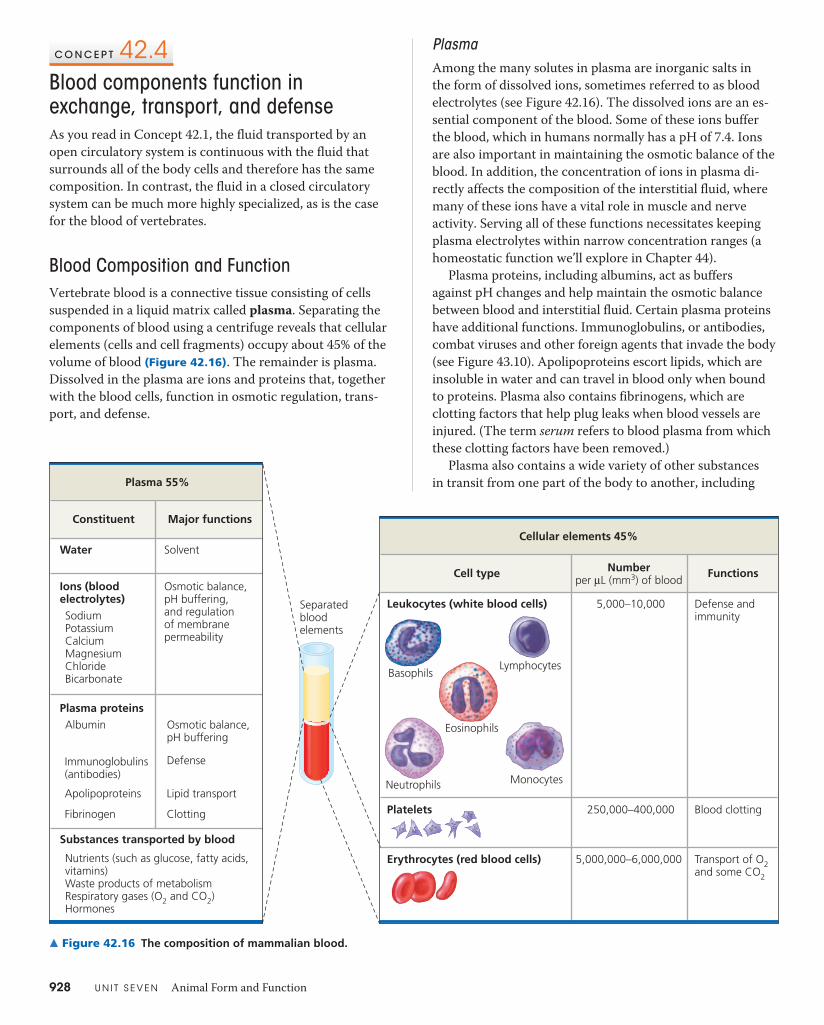
Blood Composition and FunctionVertebrate blood is a connective tissue consisting of cells suspended in a liquid matrix called plasma. Separating the components of blood using a centrifuge reveals that cellular elements (cells and cell fragments) occupy about 45% of the volume of blood (Figure 42.16). The remainder is plasma. Dissolved in the plasma are ions and proteins that, together with the blood cells, function in osmotic regulation, trans-port, and defense.
Water Solvent
BasophilsLymphocytes
Eosinophils
MonocytesNeutrophils
Ions (blood electrolytes)SodiumPotassiumCalciumMagnesiumChlorideBicarbonate
Osmotic balance,pH buffering, and regulation of membranepermeability
Separatedbloodelements
Plasma proteinsAlbumin Osmotic balance,
pH buffering Defense
Substances transported by blood
Nutrients (such as glucose, fatty acids, vitamins)Waste products of metabolismRespiratory gases (O2 and CO2)Hormones
Constituent
Plasma 55%
Leukocytes (white blood cells)
Erythrocytes (red blood cells)
Platelets
5,000–10,000
5,000,000–6,000,000
250,000–400,000
Defense andimmunity
Transport of O2and some CO2
Blood clotting
Cell type Numberper μL (mm3) of blood Functions
Cellular elements 45%Major functions
Immunoglobulins(antibodies)
Apolipoproteins
Fibrinogen Clotting
Lipid transport
▲ Figure 42.16 The composition of mammalian blood.
PlasmaAmong the many solutes in plasma are inorganic salts in the form of dissolved ions, sometimes referred to as blood electrolytes (see Figure 42.16). The dissolved ions are an es-sential component of the blood. Some of these ions buffer the blood, which in humans normally has a pH of 7.4. Ions are also important in maintaining the osmotic balance of the blood. In addition, the concentration of ions in plasma di-rectly affects the composition of the interstitial fluid, where many of these ions have a vital role in muscle and nerve activity. Serving all of these functions necessitates keeping plasma electrolytes within narrow concentration ranges (a homeostatic function we’ll explore in Chapter 44).
Plasma proteins, including albumins, act as buffers against pH changes and help maintain the osmotic balance between blood and interstitial fluid. Certain plasma proteins have additional functions. Immunoglobulins, or antibodies, combat viruses and other foreign agents that invade the body (see Figure 43.10). Apolipoproteins escort lipids, which are insoluble in water and can travel in blood only when bound to proteins. Plasma also contains fibrinogens, which are clotting factors that help plug leaks when blood vessels are injured. (The term serum refers to blood plasma from which these clotting factors have been removed.)
Plasma also contains a wide variety of other substances in transit from one part of the body to another, including
C H A P T E R 4 2 Circulation and Gas Exchange 929
that of a normal erythrocyte. The rate of erythrocyte loss outstrips their production rate. Short-term therapy in-cludes replacement of erythrocytes by blood transfusion; long-term treatments are generally aimed at inhibiting ag-gregation of HbS.
Leukocytes The blood contains five major types of white blood cells, or leukocytes. Their function is to fight infec-tions. Some are phagocytic, engulfing and digesting micro-organisms as well as debris from the body’s own dead cells. Other leukocytes, called lymphocytes, develop into B cells and T cells that mount immune responses against foreign substances (as we’ll discuss in Concepts 43.2 and 43.3). Nor-mally, 1 μL of human blood contains about 5,000–10,000 leukocytes; their numbers increase temporarily whenever the body is fighting an infection. Unlike erythrocytes, leuko-cytes are also found outside the circulatory system, patrol-ling both interstitial fluid and the lymphatic system.
Platelets Platelets are pinched-off cytoplasmic fragments of specialized bone marrow cells. They are about 2–3 μm in diameter and have no nuclei. Platelets serve both structural and molecular functions in blood clotting.
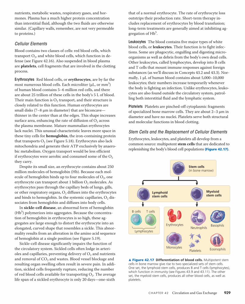
Stem Cells and the Replacement of Cellular ElementsErythrocytes, leukocytes, and platelets all develop from a common source: multipotent stem cells that are dedicated to replenishing the body’s blood cell populations (Figure 42.17).
nutrients, metabolic wastes, respiratory gases, and hor-mones. Plasma has a much higher protein concentration than interstitial fluid, although the two fluids are otherwise similar. (Capillary walls, remember, are not very permeable to proteins.)
Cellular ElementsBlood contains two classes of cells: red blood cells, which transport O2, and white blood cells, which function in de-fense (see Figure 42.16). Also suspended in blood plasma are platelets, cell fragments that are involved in the clotting process.
Erythrocytes Red blood cells, or erythrocytes, are by far the most numerous blood cells. Each microliter (μL, or mm3) of human blood contains 5–6 million red cells, and there are about 25 trillion of these cells in the body’s 5 L of blood. Their main function is O2 transport, and their structure is closely related to this function. Human erythrocytes are small disks (7–8 μm in diameter) that are biconcave— thinner in the center than at the edges. This shape increases surface area, enhancing the rate of diffusion of O2 across the plasma membrane. Mature mammalian erythrocytes lack nuclei. This unusual characteristic leaves more space in these tiny cells for hemoglobin, the iron-containing protein that transports O2 (see Figure 5.18). Erythrocytes also lack mitochondria and generate their ATP exclusively by anaero-bic metabolism. Oxygen transport would be less efficient if erythrocytes were aerobic and consumed some of the O2 they carry.
Despite its small size, an erythrocyte contains about 250 million molecules of hemoglobin (Hb). Because each mol-ecule of hemoglobin binds up to four molecules of O2, one erythrocyte can transport about 1 billion O2 molecules. As erythrocytes pass through the capillary beds of lungs, gills, or other respiratory organs, O2 diffuses into the erythrocytes and binds to hemoglobin. In the systemic capillaries, O2 dis-sociates from hemoglobin and diffuses into body cells.
In sickle-cell disease, an abnormal form of hemoglobin (HbS) polymerizes into aggregates. Because the concentra-tion of hemoglobin in erythrocytes is so high, these ag-gregates are large enough to distort the erythrocyte into an elongated, curved shape that resembles a sickle. This abnor-mality results from an alteration in the amino acid sequence of hemoglobin at a single position (see Figure 5.19).
Sickle-cell disease significantly impairs the function of the circulatory system. Sickled cells often lodge in arteri-oles and capillaries, preventing delivery of O2 and nutrients and removal of CO2 and wastes. Blood vessel blockage and resulting organ swelling often result in severe pain. In addi-tion, sickled cells frequently rupture, reducing the number of red blood cells available for transporting O2. The average life span of a sickled erythrocyte is only 20 days—one-sixth
Stem cells(in bone marrow)
Lymphoidstem cells
Myeloidstem cells
B cells
Lymphocytes
Erythrocytes BasophilsNeutrophils
MonocytesPlatelets Eosinophils
T cells
▲ Figure 42.17 Differentiation of blood cells. Multipotent stem cells in bone marrow give rise to two specialized sets of stem cells. One set, the lymphoid stem cells, produces B and T cells (lymphocytes), which function in immunity (see Figures 43.9 and 43.11). The other set, the myeloid stem cells, produces all other blood cells, as well as platelets.
930 U N I T S E V E N Animal Form and Function
The stem cells that produce blood cells are located in the red marrow inside bones, particularly the ribs, vertebrae, ster-num, and pelvis. Multipotent stem cells are so named because they have the ability to form multiple types of cells—in this case, the myeloid and lymphoid cell lineages. When a stem cell divides, one daughter cell remains a stem cell while the other takes on a specialized function.
Throughout a person’s life, stem cells replace the worn-out cellular elements of blood. Erythrocytes are the shortest-lived, circulating for only 120 days on average before being replaced. A negative-feedback mechanism, sensitive to the amount of O2 reaching the body’s tissues via the blood, con-trols erythrocyte production. If the tissues do not receive enough O2, the kidneys synthesize and secrete a hormone called erythropoietin (EPO) that stimulates the generation of more erythrocytes. If the blood is delivering more O2 than the tissues can use, the level of EPO falls and erythrocyte production slows.
Recombinant DNA technology is now used to synthesize EPO in cultured cells. Physicians use recombinant EPO to treat people with health problems such as anemia, a con-dition of lower-than-normal erythrocyte or hemoglobin levels that lowers the oxygen-carrying capacity of the blood. Some athletes inject themselves with EPO to increase their
erythrocyte levels, although this practice, a form of blood doping, has been banned by major sports organizations. In recent years, a number of well-known runners and cyclists have been found to have used EPO-related drugs and have forfeited both their records and their right to participate in future competitions.
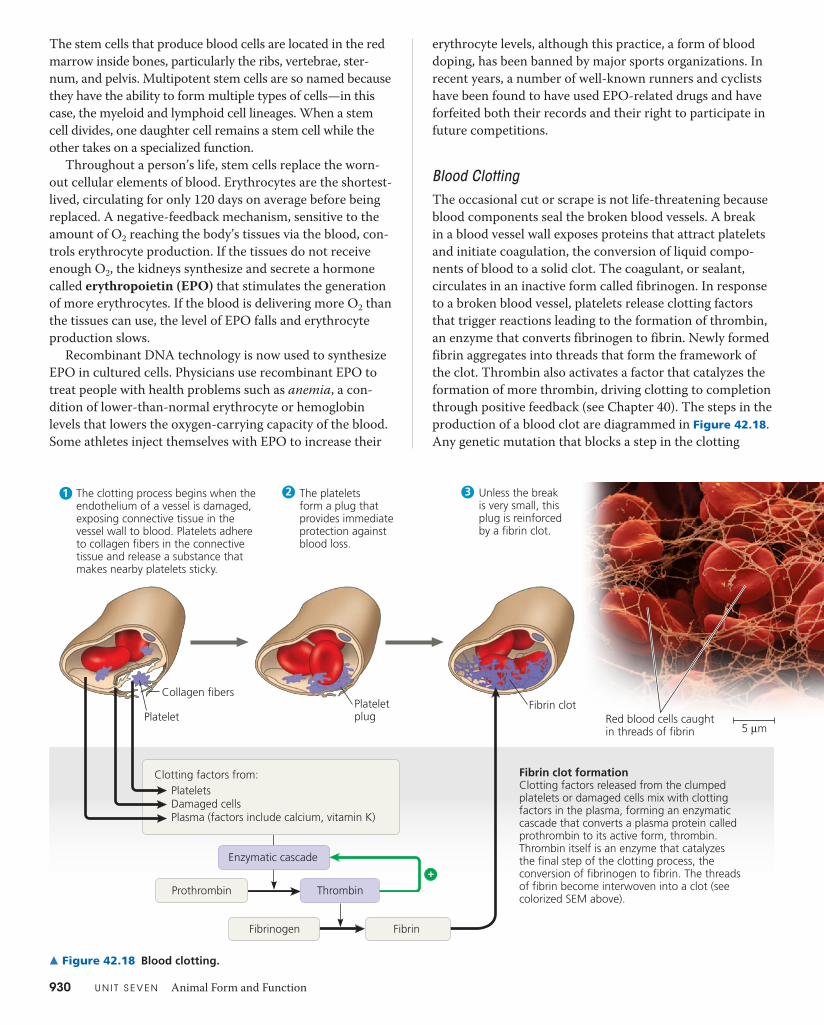
Blood ClottingThe occasional cut or scrape is not life-threatening because blood components seal the broken blood vessels. A break in a blood vessel wall exposes proteins that attract platelets and initiate coagulation, the conversion of liquid compo-nents of blood to a solid clot. The coagulant, or sealant, circulates in an inactive form called fibrinogen. In response to a broken blood vessel, platelets release clotting factors that trigger reactions leading to the formation of thrombin, an enzyme that converts fibrinogen to fibrin. Newly formed fibrin aggregates into threads that form the framework of the clot. Thrombin also activates a factor that catalyzes the formation of more thrombin, driving clotting to completion through positive feedback (see Chapter 40). The steps in the production of a blood clot are diagrammed in Figure 42.18. Any genetic mutation that blocks a step in the clotting
1 2 3
Clotting factors from:PlateletsDamaged cellsPlasma (factors include calcium, vitamin K)
Prothrombin Thrombin
Enzymatic cascade
Fibrinogen
PlateletPlateletplug Red blood cells caught
in threads of fibrin
Fibrin clotCollagen fibers
Fibrin
5 μm
The clotting process begins when the endothelium of a vessel is damaged, exposing connective tissue in the vessel wall to blood. Platelets adhere to collagen fibers in the connective tissue and release a substance that makes nearby platelets sticky.
Fibrin clot formationClotting factors released from the clumped platelets or damaged cells mix with clotting factors in the plasma, forming an enzymatic cascade that converts a plasma protein called prothrombin to its active form, thrombin. Thrombin itself is an enzyme that catalyzes the final step of the clotting process, the conversion of fibrinogen to fibrin. The threads of fibrin become interwoven into a clot (see colorized SEM above).
The platelets form a plug that provides immediate protection against blood loss.
Unless the break is very small, this plug is reinforced by a fibrin clot.
+
▲ Figure 42.18 Blood clotting.
C H A P T E R 4 2 Circulation and Gas Exchange 931
within a few minutes unless a heartbeat is restored by car-diopulmonary resuscitation (CPR) or some other emergency procedure. A stroke is the death of nervous tissue in the brain due to a lack of O2. Strokes usually result from rupture or blockage of arteries in the head. The effects of a stroke and the individual’s chance of survival depend on the extent and location of the damaged brain tissue. If a stroke results from arterial blockage by a thrombus, rapid administration of a clot-dissolving drug may help limit the damage.
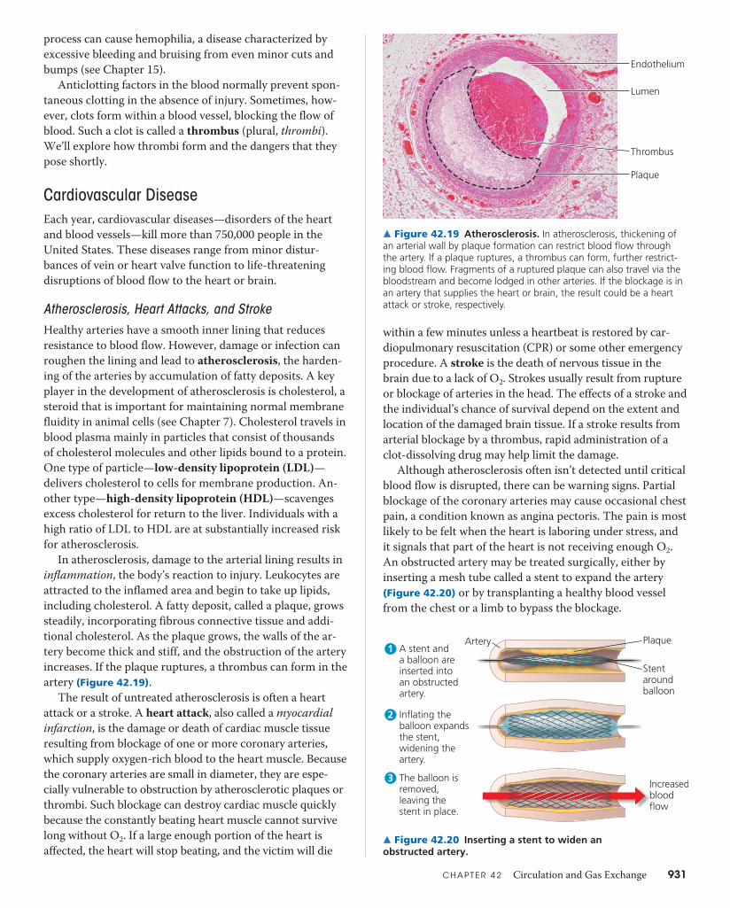
Although atherosclerosis often isn’t detected until critical blood flow is disrupted, there can be warning signs. Partial blockage of the coronary arteries may cause occasional chest pain, a condition known as angina pectoris. The pain is most likely to be felt when the heart is laboring under stress, and it signals that part of the heart is not receiving enough O2. An obstructed artery may be treated surgically, either by inserting a mesh tube called a stent to expand the artery (Figure 42.20) or by transplanting a healthy blood vessel from the chest or a limb to bypass the blockage.
process can cause hemophilia, a disease characterized by excessive bleeding and bruising from even minor cuts and bumps (see Chapter 15).
Anticlotting factors in the blood normally prevent spon-taneous clotting in the absence of injury. Sometimes, how-ever, clots form within a blood vessel, blocking the flow of blood. Such a clot is called a thrombus (plural, thrombi). We’ll explore how thrombi form and the dangers that they pose shortly.
Cardiovascular DiseaseEach year, cardiovascular diseases—disorders of the heart and blood vessels—kill more than 750,000 people in the United States. These diseases range from minor distur-bances of vein or heart valve function to life-threatening disruptions of blood flow to the heart or brain.
Atherosclerosis, Heart Attacks, and StrokeHealthy arteries have a smooth inner lining that reduces resistance to blood flow. However, damage or infection can roughen the lining and lead to atherosclerosis, the harden-ing of the arteries by accumulation of fatty deposits. A key player in the development of atherosclerosis is cholesterol, a steroid that is important for maintaining normal membrane fluidity in animal cells (see Chapter 7). Cholesterol travels in blood plasma mainly in particles that consist of thousands of cholesterol molecules and other lipids bound to a protein. One type of particle—low-density lipoprotein (LDL)— delivers cholesterol to cells for membrane production. An-other type—high-density lipoprotein (HDL)—scavenges excess cholesterol for return to the liver. Individuals with a high ratio of LDL to HDL are at substantially increased risk for atherosclerosis.
In atherosclerosis, damage to the arterial lining results in inflammation, the body’s reaction to injury. Leukocytes are attracted to the inflamed area and begin to take up lipids, including cholesterol. A fatty deposit, called a plaque, grows steadily, incorporating fibrous connective tissue and addi-tional cholesterol. As the plaque grows, the walls of the ar-tery become thick and stiff, and the obstruction of the artery increases. If the plaque ruptures, a thrombus can form in the artery (Figure 42.19).
The result of untreated atherosclerosis is often a heart attack or a stroke. A heart attack, also called a myocardial infarction, is the damage or death of cardiac muscle tissue resulting from blockage of one or more coronary arteries, which supply oxygen-rich blood to the heart muscle. Because the coronary arteries are small in diameter, they are espe-cially vulnerable to obstruction by atherosclerotic plaques or thrombi. Such blockage can destroy cardiac muscle quickly because the constantly beating heart muscle cannot survive long without O2. If a large enough portion of the heart is affected, the heart will stop beating, and the victim will die
Endothelium
Lumen
Plaque
Thrombus
▲ Figure 42.19 Atherosclerosis. In atherosclerosis, thickening of an arterial wall by plaque formation can restrict blood flow through the artery. If a plaque ruptures, a thrombus can form, further restrict-ing blood flow. Fragments of a ruptured plaque can also travel via the bloodstream and become lodged in other arteries. If the blockage is in an artery that supplies the heart or brain, the result could be a heart attack or stroke, respectively.
Artery Plaque
Stentaroundballoon
Increasedbloodflow
1 A stent and a balloon are inserted into an obstructed artery.
2 Inflating the balloon expands the stent, widening the artery.
3 The balloon is removed, leaving the stent in place.
▲ Figure 42.20 Inserting a stent to widen an obstructed artery.

932 U N I T S E V E N Animal Form and Function
Risk Factors and Treatment of Cardiovascular Disease
Although the tendency to develop particular cardiovascular diseases is inherited, it is also strongly influenced by lifestyle. For example, exercise decreases the LDL/HDL ratio, reduc-ing the risk of cardiovascular disease. In contrast, smoking and consumption of certain processed vegetable oils called trans fats (see Chapter 5) increase the LDL/HDL ratio.
There has been considerable progress in the last decade in preventing cardiovascular disease. For many individuals at high risk, treatment with drugs called statins can lower LDL levels and thereby reduce the risk of heart attacks In the Scientific Skills Exercise, you can interpret the effect of a genetic mutation on blood LDL levels.
The recognition that inflammation plays a central role in atherosclerosis and thrombus formation is also influenc-ing the treatment of cardiovascular disease. For example,
S C I E N T I F I C S K I L L S E X E R C I S E
Does Inactivating the PCSK9 Enzyme Lower LDL Levels? Researchers interested in genetic factors affecting susceptibility to car-diovascular disease examined the DNA of 15,000 individuals. They found that 3% of the individuals had a mutation that inactivates one copy of the gene for PCSK9, a liver enzyme. Because mutations that increase the activity of PCSK9 are known to increase levels of LDL cholesterol in the blood, the researchers hypothesized that inactivating mutations in this gene would lower LDL levels. In this exercise, you will interpret the results of an experiment they carried out to test this hypothesis.
How the Experiment Was Done Researchers measured LDL choles-terol levels in blood plasma from 85 individuals with one copy of the PCSK9 gene inactivated (the study group) and from 3,278 individuals with two functional copies of the gene (the control group).
Data from the Experiment The plasma LDL cholesterol levels for the control group and study group are shown in the table at the bottom.
Interpret the Data 1. Graphing often facilitates data interpretation. For this exercise,
graph the data in each row of the table as a histogram (a type of bar graph). Label the y-axis as Percent of Individuals and the x-axis as Plasma LDL cholesterol (mg/dl). Divide the x-axis into twelve equal divisions, one for each range of values (0–25, 26–50, etc). Moving along the x-axis, draw a series of twelve vertical bars, with the height of each bar indicating the percentage of samples that fall into the specified range. Note that some bars will be of zero height, such as for a plasma LDL cholesterol level in the 0–25 mg/dL (milligram/decili-ter) range. Add the percentages for the relevant bars to calculate the percentage of individuals in the study and control groups that had an LDL cholesterol level of 100 mg/dL or less. (For additional information
Making and Interpreting Histograms
about histograms, see the Scientific Skills Review in Appendix F and the Study Area in MasteringBiology.)
2. Comparing the two histograms you drew, do you find support for the researchers’ hypothesis? Explain.
3. What if instead of graphing the data you had compared the range of concentrations for plasma LDL cholesterol (low to high) in the control and study groups? How would the conclusions you could draw have differed?
4. Propose an explanation for the fact that the two histograms overlap as much as they do.
5. Consider two individuals with a plasma LDL cholesterol level of 160 mg/dL, one from the study group and one from the control group. What do you predict regarding their relative risk of develop-ing cardiovascular disease? Explain how you arrived at your predic-tion. What role did the histograms play in helping you make your prediction?
A version of this Scientific Skills Exercise can be assigned in MasteringBiology.
Data from J. C. Cohen et al., Sequence variations in PCSK9, low LDL, and protection against coronary heart disease, New England Journal of Medicine 354:1264–1272 (2006).
1MBTNB�-%-�$IPMFTUFSPM�NJMMJHSBNT�EFDJMJUFS
0–25 26–50 51–75 76–100 101–125 126–150 151–175 176–200 201–225 226–250 251-275 276–300
$POUSPM�(SPVQ 0% 1% 4% 13% 23% 23% 18% 10% 5% 2% 1% 0%
4UVEZ�(SPVQ 0% 4% 31% 23% 21% 13% 2% 1% 2% 0% 2% 0%
aspirin, which inhibits the inflammatory response, has been found to help prevent the recurrence of heart attacks and stroke. Researchers have also focused on C-reactive pro-tein (CRP), which is produced by the liver and found in the blood during episodes of acute inflammation. Like a high level of LDL cholesterol, the presence of significant amounts of CRP in blood is a useful risk indicator for cardiovascular disease.
Hypertension (high blood pressure) is yet another contributor to heart attack and stroke. According to one hypothesis, chronic high blood pressure damages the endo-thelium that lines the arteries, promoting plaque formation. The usual definition of hypertension in adults is a systolic pressure above 140 mm Hg or a diastolic pressure above 90 mm Hg. Fortunately, hypertension is simple to diagnose and can usually be controlled by dietary changes, exercise, medi-cation, or a combination of these approaches.
C H A P T E R 4 2 Circulation and Gas Exchange 933
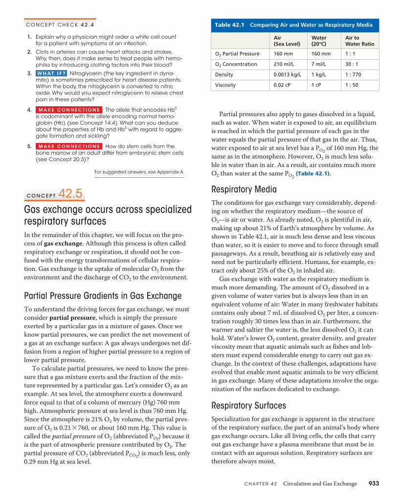
Partial pressures also apply to gases dissolved in a liquid, such as water. When water is exposed to air, an equilibrium is reached in which the partial pressure of each gas in the water equals the partial pressure of that gas in the air. Thus, water exposed to air at sea level has a PO2 of 160 mm Hg, the same as in the atmosphere. However, O2 is much less solu-ble in water than in air. As a result, air contains much more O2 than water at the same PO2 (Table 42.1).
Respiratory MediaThe conditions for gas exchange vary considerably, depend-ing on whether the respiratory medium—the source of O2—is air or water. As already noted, O2 is plentiful in air, making up about 21% of Earth’s atmosphere by volume. As shown in Table 42.1, air is much less dense and less viscous than water, so it is easier to move and to force through small passageways. As a result, breathing air is relatively easy and need not be particularly efficient. Humans, for example, ex-tract only about 25% of the O2 in inhaled air.
Gas exchange with water as the respiratory medium is much more demanding. The amount of O2 dissolved in a given volume of water varies but is always less than in an equivalent volume of air: Water in many freshwater habitats contains only about 7 mL of dissolved O2 per liter, a concen-tration roughly 30 times less than in air. Furthermore, the warmer and saltier the water is, the less dissolved O2 it can hold. Water’s lower O2 content, greater density, and greater viscosity mean that aquatic animals such as fishes and lob-sters must expend considerable energy to carry out gas ex-change. In the context of these challenges, adaptations have evolved that enable most aquatic animals to be very efficient in gas exchange. Many of these adaptations involve the orga-nization of the surfaces dedicated to exchange.
Respiratory SurfacesSpecialization for gas exchange is apparent in the structure of the respiratory surface, the part of an animal’s body where gas exchange occurs. Like all living cells, the cells that carry out gas exchange have a plasma membrane that must be in contact with an aqueous solution. Respiratory surfaces are therefore always moist.
C O N C E P T C H E C K 4 2 . 4
1. Explain why a physician might order a white cell count for a patient with symptoms of an infection.
2. Clots in arteries can cause heart attacks and strokes. Why, then, does it make sense to treat people with hemo-philia by introducing clotting factors into their blood?
3. W H AT I F ? Nitroglycerin (the key ingredient in dyna-mite) is sometimes prescribed for heart disease patients. Within the body, the nitroglycerin is converted to nitric oxide. Why would you expect nitroglycerin to relieve chest pain in these patients?
4. M A K E C O N N E C T I O N S The allele that encodes HbS is codominant with the allele encoding normal hemo-globin (Hb) (see Concept 14.4). What can you deduce about the properties of Hb and HbS with regard to aggre-gate formation and sickling?
5. M A K E C O N N E C T I O N S How do stem cells from the bone marrow of an adult differ from embryonic stem cells (see Concept 20.3)?
For suggested answers, see Appendix A.
C O N C E P T 42.5Gas exchange occurs across specialized respiratory surfacesIn the remainder of this chapter, we will focus on the pro-cess of gas exchange. Although this process is often called respiratory exchange or respiration, it should not be con-fused with the energy transformations of cellular respira-tion. Gas exchange is the uptake of molecular O2 from the environment and the discharge of CO2 to the environment.
Partial Pressure Gradients in Gas ExchangeTo understand the driving forces for gas exchange, we must consider partial pressure, which is simply the pressure exerted by a particular gas in a mixture of gases. Once we know partial pressures, we can predict the net movement of a gas at an exchange surface: A gas always undergoes net dif-fusion from a region of higher partial pressure to a region of lower partial pressure.
To calculate partial pressures, we need to know the pres-sure that a gas mixture exerts and the fraction of the mix-ture represented by a particular gas. Let’s consider O2 as an example. At sea level, the atmosphere exerts a downward force equal to that of a column of mercury (Hg) 760 mm high. Atmospheric pressure at sea level is thus 760 mm Hg. Since the atmosphere is 21% O2 by volume, the partial pres-sure of O2 is 0.21 * 760, or about 160 mm Hg. This value is called the partial pressure of O2 (abbreviated PO2) because it is the part of atmospheric pressure contributed by O2. The partial pressure of CO2 (abbreviated PCO2) is much less, only 0.29 mm Hg at sea level.
Air (Sea Level)
Water (20°C)
Air to Water Ratio
O2�1BSUJBM�1SFTTVSF ����NN ����NN �����
O2�$PODFOUSBUJPO ����NM�- ��NM�- ������
%FOTJUZ �������LH�- ��LH�- �������
7JTDPTJUZ �����D1 ��D1 ������
Table 42.1 Comparing Air and Water as Respiratory Media
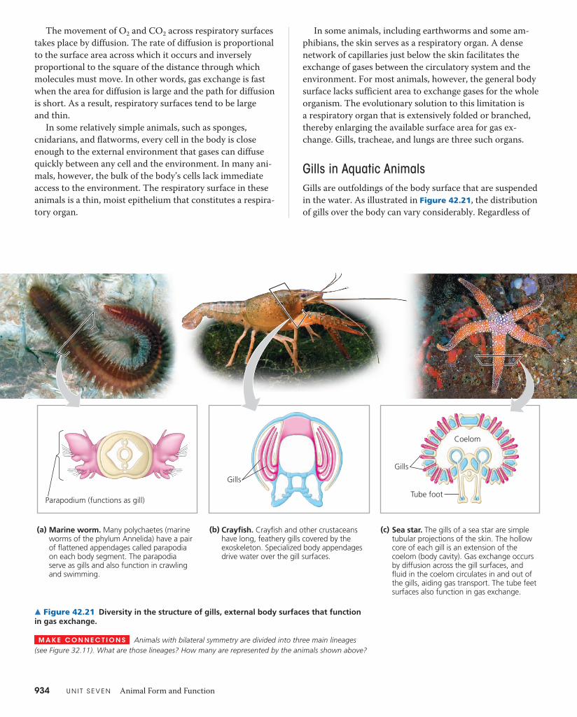
934 U N I T S E V E N Animal Form and Function
In some animals, including earthworms and some am-phibians, the skin serves as a respiratory organ. A dense network of capillaries just below the skin facilitates the exchange of gases between the circulatory system and the environment. For most animals, however, the general body surface lacks sufficient area to exchange gases for the whole organism. The evolutionary solution to this limitation is a respiratory organ that is extensively folded or branched, thereby enlarging the available surface area for gas ex-change. Gills, tracheae, and lungs are three such organs.
Gills in Aquatic AnimalsGills are outfoldings of the body surface that are suspended in the water. As illustrated in Figure 42.21, the distribution of gills over the body can vary considerably. Regardless of
The movement of O2 and CO2 across respiratory surfaces takes place by diffusion. The rate of diffusion is proportional to the surface area across which it occurs and inversely propor tional to the square of the distance through which molecules must move. In other words, gas exchange is fast when the area for diffusion is large and the path for diffusion is short. As a result, respiratory surfaces tend to be large and thin.
In some relatively simple animals, such as sponges, cnidarians, and flatworms, every cell in the body is close enough to the external environment that gases can diffuse quickly between any cell and the environment. In many ani-mals, however, the bulk of the body’s cells lack immediate access to the environment. The respiratory surface in these animals is a thin, moist epithelium that constitutes a respira-tory organ.
Parapodium (functions as gill)
(c) Sea star. The gills of a sea star are simple tubular projections of the skin. The hollow core of each gill is an extension of the coelom (body cavity). Gas exchange occurs by diffusion across the gill surfaces, and fluid in the coelom circulates in and out of the gills, aiding gas transport. The tube feet surfaces also function in gas exchange.
(b) Crayfish. Crayfish and other crustaceans have long, feathery gills covered by the exoskeleton. Specialized body appendages drive water over the gill surfaces.
(a) Marine worm. Many polychaetes (marine worms of the phylum Annelida) have a pair of flattened appendages called parapodia on each body segment. The parapodia serve as gills and also function in crawling and swimming.
Gills
Gills
Tube foot
Coelom
▲ Figure 42.21 Diversity in the structure of gills, external body surfaces that function in gas exchange.
M A K E C O N N E C T I O N S Animals with bilateral symmetry are divided into three main lineages (see Figure 32.11). What are those lineages? How many are represented by the animals shown above?
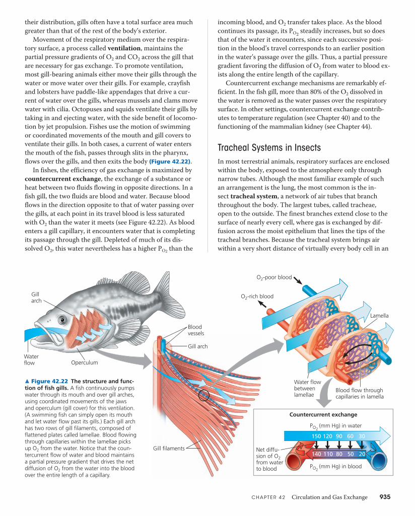
C H A P T E R 4 2 Circulation and Gas Exchange 935
incoming blood, and O2 transfer takes place. As the blood continues its passage, its PO2 steadily increases, but so does that of the water it encounters, since each successive posi-tion in the blood’s travel corresponds to an earlier position in the water’s passage over the gills. Thus, a partial pressure gradient favoring the diffusion of O2 from water to blood ex-ists along the entire length of the capillary.
Countercurrent exchange mechanisms are remarkably ef-ficient. In the fish gill, more than 80% of the O2 dissolved in the water is removed as the water passes over the respiratory surface. In other settings, countercurrent exchange contrib-utes to temperature regulation (see Chapter 40) and to the functioning of the mammalian kidney (see Chapter 44).
Tracheal Systems in InsectsIn most terrestrial animals, respiratory surfaces are enclosed within the body, exposed to the atmosphere only through narrow tubes. Although the most familiar example of such an arrangement is the lung, the most common is the in-sect tracheal system, a network of air tubes that branch throughout the body. The largest tubes, called tracheae, open to the outside. The finest branches extend close to the surface of nearly every cell, where gas is exchanged by dif-fusion across the moist epithelium that lines the tips of the tracheal branches. Because the tracheal system brings air within a very short distance of virtually every body cell in an
their distribution, gills often have a total surface area much greater than that of the rest of the body’s exterior.
Movement of the respiratory medium over the respira-tory surface, a process called ventilation, maintains the partial pressure gradients of O2 and CO2 across the gill that are necessary for gas exchange. To promote ventilation, most gill-bearing animals either move their gills through the water or move water over their gills. For example, crayfish and lobsters have paddle-like appendages that drive a cur-rent of water over the gills, whereas mussels and clams move water with cilia. Octopuses and squids ventilate their gills by taking in and ejecting water, with the side benefit of locomo-tion by jet propulsion. Fishes use the motion of swimming or coordinated movements of the mouth and gill covers to ventilate their gills. In both cases, a current of water enters the mouth of the fish, passes through slits in the pharynx, flows over the gills, and then exits the body (Figure 42.22).
In fishes, the efficiency of gas exchange is maximized by countercurrent exchange, the exchange of a substance or heat between two fluids flowing in opposite directions. In a fish gill, the two fluids are blood and water. Because blood flows in the direction opposite to that of water passing over the gills, at each point in its travel blood is less saturated with O2 than the water it meets (see Figure 42.22). As blood enters a gill capillary, it encounters water that is completing its passage through the gill. Depleted of much of its dis-solved O2, this water nevertheless has a higher PO2 than the
Water flow Operculum
Gill arch
O2-poor blood
O2-rich blood
Bloodvessels
Gill arch
Water flowbetweenlamellae
Lamella
Blood flow throughcapillaries in lamella
PO2 (mm Hg) in blood
Countercurrent exchange
Net diffu-sion of O2from waterto blood
PO2 (mm Hg) in water
150
140 110 80 50 20
120 90 60 30
Gill filaments
▲ Figure 42.22 The structure and func-tion of fish gills. A fish continuously pumps water through its mouth and over gill arches, using coordinated movements of the jaws and operculum (gill cover) for this ventilation. (A swimming fish can simply open its mouth and let water flow past its gills.) Each gill arch has two rows of gill filaments, composed of flattened plates called lamellae. Blood flowing through capillaries within the lamellae picks up O2 from the water. Notice that the coun-tercurrent flow of water and blood maintains a partial pressure gradient that drives the net diffusion of O2 from the water into the blood over the entire length of a capillary.
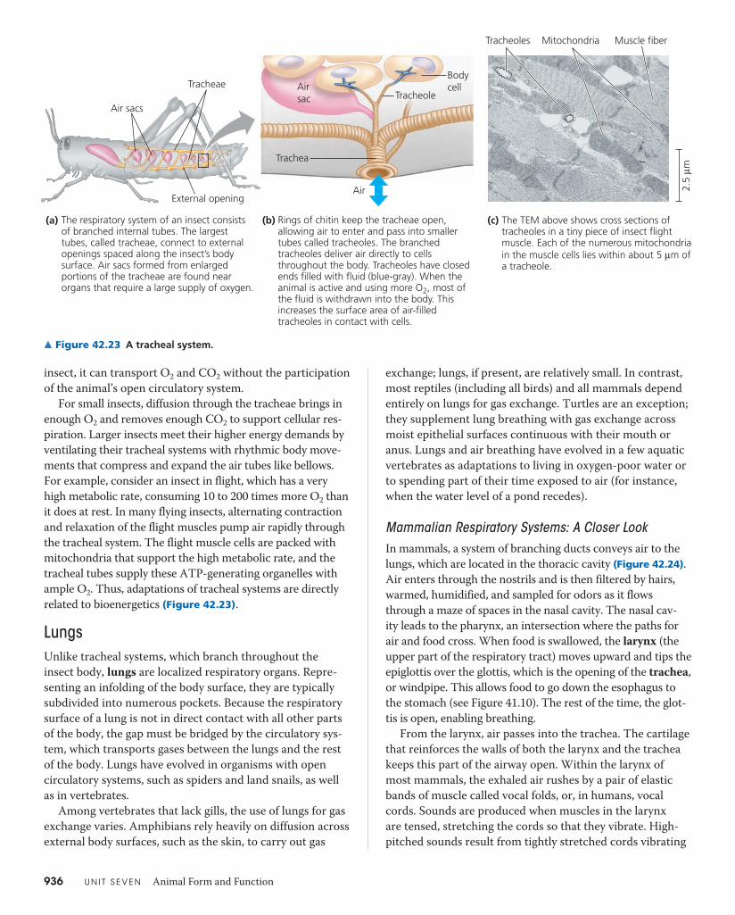
936 U N I T S E V E N Animal Form and Function
exchange; lungs, if present, are relatively small. In contrast, most reptiles (including all birds) and all mammals depend entirely on lungs for gas exchange. Turtles are an exception; they supplement lung breathing with gas exchange across moist epithelial surfaces continuous with their mouth or anus. Lungs and air breathing have evolved in a few aquatic vertebrates as adaptations to living in oxygen-poor water or to spending part of their time exposed to air (for instance, when the water level of a pond recedes).
Mammalian Respiratory Systems: A Closer LookIn mammals, a system of branching ducts conveys air to the lungs, which are located in the thoracic cavity (Figure 42.24). Air enters through the nostrils and is then filtered by hairs, warmed, humidified, and sampled for odors as it flows through a maze of spaces in the nasal cavity. The nasal cav-ity leads to the pharynx, an intersection where the paths for air and food cross. When food is swallowed, the larynx (the upper part of the respiratory tract) moves upward and tips the epiglottis over the glottis, which is the opening of the trachea, or windpipe. This allows food to go down the esophagus to the stomach (see Figure 41.10). The rest of the time, the glot-tis is open, enabling breathing.
From the larynx, air passes into the trachea. The cartilage that reinforces the walls of both the larynx and the trachea keeps this part of the airway open. Within the larynx of most mammals, the exhaled air rushes by a pair of elastic bands of muscle called vocal folds, or, in humans, vocal cords. Sounds are produced when muscles in the larynx are tensed, stretching the cords so that they vibrate. High-pitched sounds result from tightly stretched cords vibrating
insect, it can transport O2 and CO2 without the participation of the animal’s open circulatory system.
For small insects, diffusion through the tracheae brings in enough O2 and removes enough CO2 to support cellular res-piration. Larger insects meet their higher energy demands by ventilating their tracheal systems with rhythmic body move-ments that compress and expand the air tubes like bellows. For example, consider an insect in flight, which has a very high metabolic rate, consuming 10 to 200 times more O2 than it does at rest. In many flying insects, alternating contraction and relaxation of the flight muscles pump air rapidly through the tracheal system. The flight muscle cells are packed with mitochondria that support the high metabolic rate, and the tracheal tubes supply these ATP-generating organelles with ample O2. Thus, adaptations of tracheal systems are directly related to bioenergetics (Figure 42.23).
LungsUnlike tracheal systems, which branch throughout the insect body, lungs are localized respiratory organs. Repre-senting an infolding of the body surface, they are typically subdivided into numerous pockets. Because the respiratory surface of a lung is not in direct contact with all other parts of the body, the gap must be bridged by the circulatory sys-tem, which transports gases between the lungs and the rest of the body. Lungs have evolved in organisms with open circulatory systems, such as spiders and land snails, as well as in vertebrates.
Among vertebrates that lack gills, the use of lungs for gas exchange varies. Amphibians rely heavily on diffusion across external body surfaces, such as the skin, to carry out gas
External opening
Air sacs
Air sac
Bodycell
Air
Tracheae
Trachea
Tracheole
The TEM above shows cross sections of tracheoles in a tiny piece of insect flight muscle. Each of the numerous mitochondria in the muscle cells lies within about 5 μm of a tracheole.
(c)Rings of chitin keep the tracheae open, allowing air to enter and pass into smaller tubes called tracheoles. The branched tracheoles deliver air directly to cells throughout the body. Tracheoles have closed ends filled with fluid (blue-gray). When the animal is active and using more O2, most of the fluid is withdrawn into the body. This increases the surface area of air-filled tracheoles in contact with cells.
(b)The respiratory system of an insect consists of branched internal tubes. The largest tubes, called tracheae, connect to external openings spaced along the insect’s body surface. Air sacs formed from enlarged portions of the tracheae are found near organs that require a large supply of oxygen.
(a)
Tracheoles Muscle fiberMitochondria
2.5
μm
▲ Figure 42.23 A tracheal system.
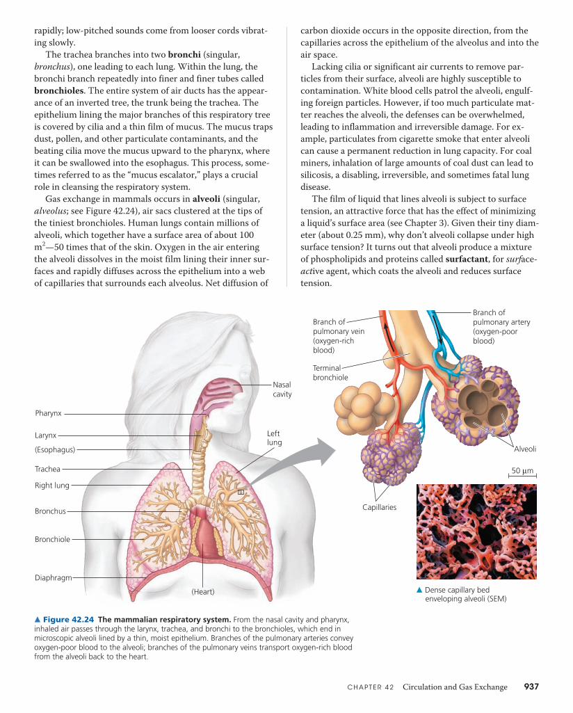
C H A P T E R 4 2 Circulation and Gas Exchange 937
carbon dioxide occurs in the opposite direction, from the capillaries across the epithelium of the alveolus and into the air space.
Lacking cilia or significant air currents to remove par-ticles from their surface, alveoli are highly susceptible to contamination. White blood cells patrol the alveoli, engulf-ing foreign particles. However, if too much particulate mat-ter reaches the alveoli, the defenses can be overwhelmed, leading to inflammation and irreversible damage. For ex-ample, particulates from cigarette smoke that enter alveoli can cause a permanent reduction in lung capacity. For coal miners, inhalation of large amounts of coal dust can lead to silicosis, a disabling, irreversible, and sometimes fatal lung disease.
The film of liquid that lines alveoli is subject to surface tension, an attractive force that has the effect of minimizing a liquid’s surface area (see Chapter 3). Given their tiny diam-eter (about 0.25 mm), why don’t alveoli collapse under high surface tension? It turns out that alveoli produce a mixture of phospholipids and proteins called surfactant, for surface-active agent, which coats the alveoli and reduces surface tension.
rapidly; low-pitched sounds come from looser cords vibrat-ing slowly.
The trachea branches into two bronchi (singular, bronchus), one leading to each lung. Within the lung, the bronchi branch repeatedly into finer and finer tubes called bronchioles. The entire system of air ducts has the appear-ance of an inverted tree, the trunk being the trachea. The epithelium lining the major branches of this respiratory tree is covered by cilia and a thin film of mucus. The mucus traps dust, pollen, and other particulate contaminants, and the beating cilia move the mucus upward to the pharynx, where it can be swallowed into the esophagus. This process, some-times referred to as the “mucus escalator,” plays a crucial role in cleansing the respiratory system.
Gas exchange in mammals occurs in alveoli (singular, alveolus; see Figure 42.24), air sacs clustered at the tips of the tiniest bronchioles. Human lungs contain millions of alveoli, which together have a surface area of about 100 m2—50 times that of the skin. Oxygen in the air entering the alveoli dissolves in the moist film lining their inner sur-faces and rapidly diffuses across the epithelium into a web of capillaries that surrounds each alveolus. Net diffusion of
Pharynx
(Esophagus)
Larynx
Trachea
Right lung
Bronchus
Bronchiole
Diaphragm
(Heart)
Nasalcavity
Leftlung
Alveoli
Branch ofpulmonary artery(oxygen-poorblood)
Branch ofpulmonary vein(oxygen-richblood)
Terminalbronchiole
Capillaries
50 μm
▲ Dense capillary bed enveloping alveoli (SEM)
▲ Figure 42.24 The mammalian respiratory system. From the nasal cavity and pharynx, inhaled air passes through the larynx, trachea, and bronchi to the bronchioles, which end in microscopic alveoli lined by a thin, moist epithelium. Branches of the pulmonary arteries convey oxygen-poor blood to the alveoli; branches of the pulmonary veins transport oxygen-rich blood from the alveoli back to the heart.
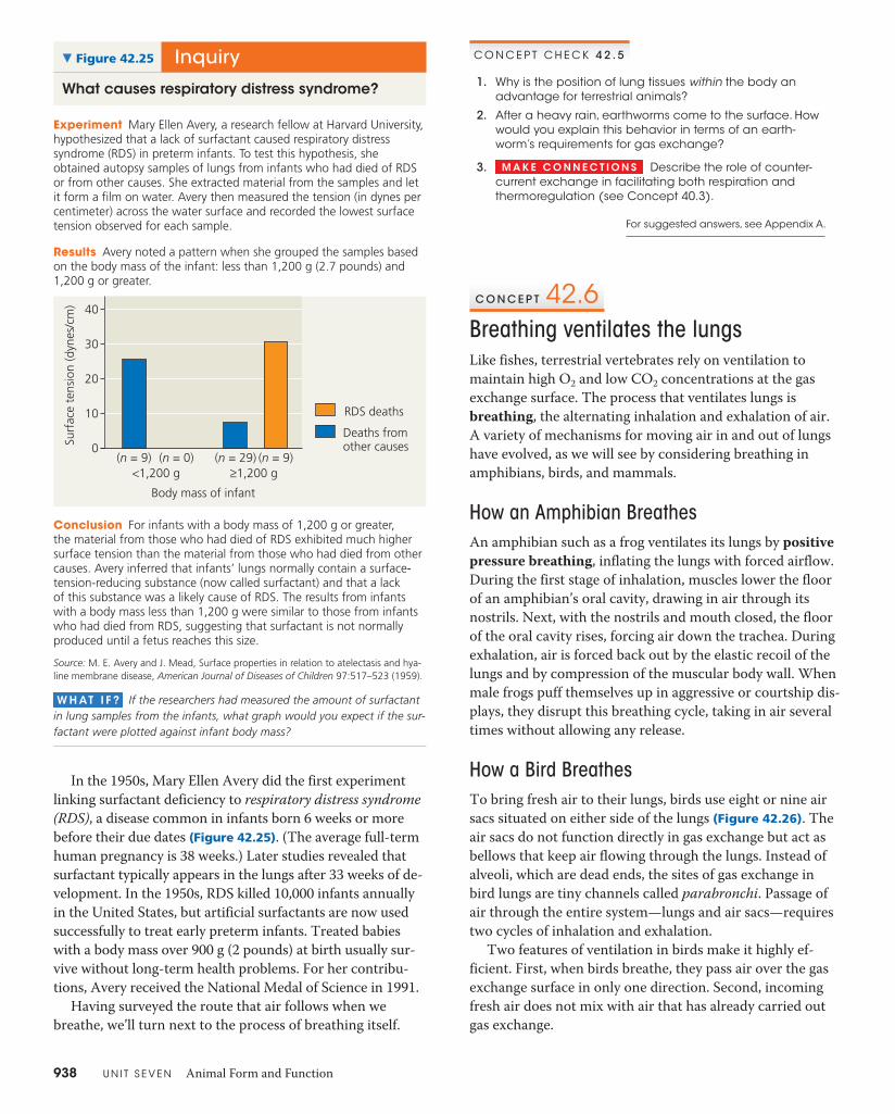
938 U N I T S E V E N Animal Form and Function
0
10
20
30
40
<1,200 g ≥1,200 g
Deaths fromother causes
RDS deaths
Body mass of infant
Surf
ace
tens
ion
(dyn
es/c
m)
(n = 9) (n = 0) (n = 9)(n = 29)
InquiryWhat causes respiratory distress syndrome?
▼ Figure 42.25
Experiment Mary Ellen Avery, a research fellow at Harvard University, hypothesized that a lack of surfactant caused respiratory distress syndrome (RDS) in preterm infants. To test this hypothesis, she obtained autopsy samples of lungs from infants who had died of RDS or from other causes. She extracted material from the samples and let it form a film on water. Avery then measured the tension (in dynes per centimeter) across the water surface and recorded the lowest surface tension observed for each sample.
Results Avery noted a pattern when she grouped the samples based on the body mass of the infant: less than 1,200 g (2.7 pounds) and 1,200 g or greater.
Conclusion For infants with a body mass of 1,200 g or greater, the material from those who had died of RDS exhibited much higher surface tension than the material from those who had died from other causes. Avery inferred that infants’ lungs normally contain a surface-tension-reducing substance (now called surfactant) and that a lack of this substance was a likely cause of RDS. The results from infants with a body mass less than 1,200 g were similar to those from infants who had died from RDS, suggesting that surfactant is not normally produced until a fetus reaches this size.
Source: M. E. Avery and J. Mead, Surface properties in relation to atelectasis and hya-line membrane disease, American Journal of Diseases of Children 97:517–523 (1959).
W H AT I F ? If the researchers had measured the amount of surfactant in lung samples from the infants, what graph would you expect if the sur-factant were plotted against infant body mass?
C O N C E P T C H E C K 4 2 . 5
1. Why is the position of lung tissues within the body an advantage for terrestrial animals?
2. After a heavy rain, earthworms come to the surface. How would you explain this behavior in terms of an earth-worm’s requirements for gas exchange?
3. M A K E C O N N E C T I O N S Describe the role of counter-current exchange in facilitating both respiration and thermoregulation (see Concept 40.3).
For suggested answers, see Appendix A.
C O N C E P T 42.6Breathing ventilates the lungsLike fishes, terrestrial vertebrates rely on ventilation to maintain high O2 and low CO2 concentrations at the gas exchange surface. The process that ventilates lungs is breathing, the alternating inhalation and exhalation of air. A variety of mechanisms for moving air in and out of lungs have evolved, as we will see by considering breathing in amphibians, birds, and mammals.
How an Amphibian BreathesAn amphibian such as a frog ventilates its lungs by positive pressure breathing, inflating the lungs with forced airflow. During the first stage of inhalation, muscles lower the floor of an amphibian’s oral cavity, drawing in air through its nostrils. Next, with the nostrils and mouth closed, the floor of the oral cavity rises, forcing air down the trachea. During exhalation, air is forced back out by the elastic recoil of the lungs and by compression of the muscular body wall. When male frogs puff themselves up in aggressive or courtship dis-plays, they disrupt this breathing cycle, taking in air several times without allowing any release.
How a Bird BreathesTo bring fresh air to their lungs, birds use eight or nine air sacs situated on either side of the lungs (Figure 42.26). The air sacs do not function directly in gas exchange but act as bellows that keep air flowing through the lungs. Instead of alveoli, which are dead ends, the sites of gas exchange in bird lungs are tiny channels called parabronchi. Passage of air through the entire system—lungs and air sacs—requires two cycles of inhalation and exhalation.
Two features of ventilation in birds make it highly ef-ficient. First, when birds breathe, they pass air over the gas exchange surface in only one direction. Second, incoming fresh air does not mix with air that has already carried out gas exchange.
In the 1950s, Mary Ellen Avery did the first experiment linking surfactant deficiency to respiratory distress syndrome (RDS), a disease common in infants born 6 weeks or more before their due dates (Figure 42.25). (The average full-term human pregnancy is 38 weeks.) Later studies revealed that surfactant typically appears in the lungs after 33 weeks of de-velopment. In the 1950s, RDS killed 10,000 infants annually in the United States, but artificial surfactants are now used successfully to treat early preterm infants. Treated babies with a body mass over 900 g (2 pounds) at birth usually sur-vive without long-term health problems. For her contribu-tions, Avery received the National Medal of Science in 1991.
Having surveyed the route that air follows when we breathe, we’ll turn next to the process of breathing itself.

C H A P T E R 4 2 Circulation and Gas Exchange 939
contracts, expanding the thoracic cavity downward. The effect of the descending diaphragm is similar to that of a plunger being drawn out of a syringe.
Within the thoracic cavity, a double membrane sur-rounds the lungs. The inner layer of this membrane adheres to the outside of the lungs, and the outer layer adheres to the wall of the thoracic cavity. A thin space filled with fluid separates the two layers. Surface tension in the fluid causes the two layers to stick together like two plates of glass sepa-rated by a film of water: The layers can slide smoothly past each other, but they cannot be pulled apart easily. Conse-quently, the volume of the thoracic cavity and the volume of the lungs change in unison.
Depending on activity level, additional muscles may be recruited to aid breathing. The rib muscles and diaphragm are sufficient to change lung volume when a mammal is at rest. During exercise, other muscles of the neck, back, and chest in-crease the volume of the thoracic cavity by raising the rib cage. In kangaroos and some other mammals, locomotion causes a rhythmic movement of organs in the abdomen, including the stomach and liver. The result is a piston-like pumping motion that pushes and pulls on the diaphragm, further increasing the volume of air moved in and out of the lungs.
The volume of air inhaled and exhaled with each breath is called tidal volume. It averages about 500 mL in resting humans. The tidal volume during maximal inhalation and exhalation is the vital capacity, which is about 3.4 L and 4.8 L for college-age women and men, respectively. The air that remains after a forced exhalation is called the residual volume. With age, the lungs lose their resilience, and resid-ual volume increases at the expense of vital capacity.
Anteriorair sacs
Posteriorair sacs
Air tubes(parabronchi)in lung
AirflowLungs
Posteriorair sacs
Anteriorair sacs
Lungs
1 mm
2
1
1
4
3
Two cycles of inhalation and exhalation are required to pass one breath through the system:
First inhalation: Air fills the posterior air sacs.First exhalation: Posterior air sacs contract, pushing air into lungs.Second inhalation: Air passes through lungs and fills anterior air sacs.Second exhalation: As anterior air sacs contract, air that entered body at first inhalation is pushed out of body.
2
3
4
▲ Figure 42.26 The avian respiratory system. This diagram traces a breath of air through the respiratory system of a bird. As shown, two cycles of inhalation and exhalation are required for the air to pass all the way through the system and out of the bird.
Rib cageexpands asrib musclescontract.
Rib cage gets smaller as rib musclesrelax.
Lung
Diaphragm
INHALATION: Diaphragm contracts (moves down).
1 EXHALATION: Diaphragm relaxes (moves up).
2
▲ Figure 42.27 Negative pressure breathing. A mammal breathes by changing the air pressure within its lungs relative to the pressure of the outside atmosphere.
W H AT I F ? The walls of alveoli contain elastic fibers that allow the alveoli to expand and contract with each breath. If the alveoli lost their elasticity, how would that affect gas exchange in the lungs?
How a Mammal BreathesUnlike amphibians and birds, mammals employ negative pressure breathing—pulling, rather than pushing, air into their lungs (Figure 42.27). Using muscle contraction to actively expand the thoracic cavity, mammals lower air pres-sure in their lungs below that of the air outside their body. Because gas flows from a region of higher pressure to a re-gion of lower pressure, air rushes through the nostrils and mouth and down the breathing tubes to the alveoli. During exhalation, the muscles controlling the thoracic cavity relax, and the volume of the cavity is reduced. The increased air pressure in the alveoli forces air up the breathing tubes and out of the body. Thus, inhalation is always active and re-quires work, whereas exhalation is usually passive.
Expanding the thoracic cavity during inhalation involves the animal’s rib muscles and the diaphragm, a sheet of skeletal muscle that forms the bottom wall of the cavity. Contracting one set of rib muscles expands the rib cage, the front wall of the thoracic cavity, by pulling the ribs upward and the sternum outward. At the same time, the diaphragm
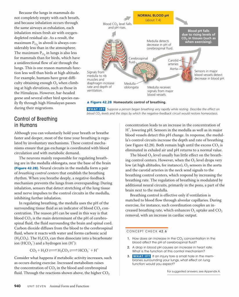
940 U N I T S E V E N Animal Form and Function
Because the lungs in mammals do not completely empty with each breath, and because inhalation occurs through the same airways as exhalation, each inhalation mixes fresh air with oxygen-depleted residual air. As a result, the maximum PO2 in alveoli is always con-siderably less than in the atmosphere. The maximum PO2 in lungs is also less for mammals than for birds, which have a unidirectional flow of air through the lungs. This is one reason mammals func-tion less well than birds at high altitude. For example, humans have great diffi-culty obtaining enough O2 when climb-ing at high elevations, such as those in the Himalayas. However, bar-headed geese and several other bird species eas-ily fly through high Himalayan passes during their migrations.
Control of Breathing in HumansAlthough you can voluntarily hold your breath or breathe faster and deeper, most of the time your breathing is regu-lated by involuntary mechanisms. These control mecha-nisms ensure that gas exchange is coordinated with blood circulation and with metabolic demand.
The neurons mainly responsible for regulating breath-ing are in the medulla oblongata, near the base of the brain (Figure 42.28). Neural circuits in the medulla form a pair of breathing control centers that establish the breathing rhythm. When you breathe deeply, a negative-feedback mechanism prevents the lungs from overexpanding: During inhalation, sensors that detect stretching of the lung tissue send nerve impulses to the control circuits in the medulla, inhibiting further inhalation.
In regulating breathing, the medulla uses the pH of the surrounding tissue fluid as an indicator of blood CO2 con-centration. The reason pH can be used in this way is that blood CO2 is the main determinant of the pH of cerebro-spinal fluid, the fluid surrounding the brain and spinal cord. Carbon dioxide diffuses from the blood to the cerebrospinal fluid, where it reacts with water and forms carbonic acid (H2CO3). The H2CO3 can then dissociate into a bicarbonate ion (HCO3
-) and a hydrogen ion (H+):CO2 + H2O ∆ H2CO3 ∆ HCO3
- + H+
Consider what happens if metabolic activity increases, such as occurs during exercise. Increased metabolism raises the concentration of CO2 in the blood and cerebrospinal fluid. Through the reactions shown above, the higher CO2
Medullaoblongata
Cerebrospinalfluid
Aorta
Carotidarteries
Sensors in major blood vessels detect decrease in blood pH.
Medulla receives signals from major blood vessels.
Signals from medulla to rib muscles and diaphragm increase rate and depth of ventilation.
Medulla detects decrease in pH of cerebrospinal fluid.
Blood CO2 level fallsand pH rises.
NORMAL BLOOD pH(about 7.4)
Blood pH fallsdue to rising levels ofCO2 in tissues (such as
when exercising).
▲ Figure 42.28 Homeostatic control of breathing.
W H AT I F ? Suppose a person began breathing very rapidly while resting. Describe the effect on blood CO2 levels and the steps by which the negative-feedback circuit would restore homeostasis.
concentration leads to an increase in the concentration of H+, lowering pH. Sensors in the medulla as well as in major blood vessels detect this pH change. In response, the medul-la’s control circuits increase the depth and rate of breathing (see Figure 42.28). Both remain high until the excess CO2 is eliminated in exhaled air and pH returns to a normal value.
The blood O2 level usually has little effect on the breath-ing control centers. However, when the O2 level drops very low (at high altitudes, for instance), O2 sensors in the aorta and the carotid arteries in the neck send signals to the breathing control centers, which respond by increasing the breathing rate. The regulation of breathing is modulated by additional neural circuits, primarily in the pons, a part of the brain next to the medulla.
Breathing control is effective only if ventilation is matched to blood flow through alveolar capillaries. During exercise, for instance, such coordination couples an in-creased breathing rate, which enhances O2 uptake and CO2 removal, with an increase in cardiac output.
C O N C E P T C H E C K 4 2 . 6
1. How does an increase in the CO2 concentration in the blood affect the pH of cerebrospinal fluid?
2. A drop in blood pH causes an increase in heart rate. What is the function of this control mechanism?
3. W H AT I F ? If an injury tore a small hole in the mem-branes surrounding your lungs, what effect on lung function would you expect?
For suggested answers, see Appendix A.

C H A P T E R 4 2 Circulation and Gas Exchange 941
C O N C E P T 42.7Adaptations for gas exchange include pigments that bind and transport gasesThe high metabolic demands of many animals necessitate the exchange of large quantities of O2 and CO2. Here we’ll examine how blood molecules called respiratory pigments facilitate this exchange through their interaction with O2 and CO2. We’ll also investigate physiological adaptations that enable animals to be active under conditions of high metabolic load or very limiting PO2. As a basis for exploring these topics, let’s summarize the basic gas exchange circuit in humans.
Coordination of Circulation and Gas ExchangeThe partial pressures of O2 and CO2 in the blood vary as the gases move between air, blood, and other body tissues, as shown in Figure 42.29. 1 During inhalation, fresh air mixes with air remaining in the lungs. 2 The resulting mixture formed in the alveoli has a higher PO2 and a lower PCO2 than the blood flowing through the alveolar capillaries. Conse-quently, there is a net diffusion of O2 down its partial pressure gradient from the air in the alveoli to the blood. Meanwhile, CO2 in the blood un-dergoes net diffusion into the air in the alveoli. 3 By the time the blood leaves the lungs in the pulmonary veins, its PO2 and PCO2 match the values for those gases in the alveoli. After returning to the heart, this blood is pumped through the sys-temic circuit.
4 In the systemic capillaries, gra-dients of partial pressure favor the net diffusion of O2 out of the blood and CO2 into the blood. These gradients exist because cellular respiration in the mi-tochondria of cells near each capillary removes O2 from and adds CO2 to the surrounding interstitial fluid. 5 After the blood unloads O2 and loads CO2, it is returned to the heart and pumped to the lungs again. 6 There, exchange occurs across the alveolar capillaries, resulting in exhaled air enriched in CO2 and par-tially depleted of O2.
Respiratory PigmentsThe low solubility of O2 in water (and thus in blood) poses a problem for ani-mals that rely on the circulatory system to deliver O2. For example, a person
Heart
Alveolarepithelialcells
Bodytissue
Systemiccapillaries
CO2 O2
Alveolarcapillaries
Alveolarspaces
Exhaled air Inhaled air
120
27
Pulmonaryarteries and systemicveins
Bloodenteringalveolarcapillaries
Pulmonaryveins andsystemicarteries
O2CO2
PO2PCO2
160
0.2
PO2PCO2
104
40
PO2PCO2
104
40
PO2PCO2
40 45
PO2PCO2
<40>45
PO2PCO2
© Pearson Education, Inc.
6 1
2
3
5
4
▲ Figure 42.29 Loading and unloading of respiratory gases.
W H AT I F ? If you consciously forced more air out of your lungs each time you exhaled, how would that affect the values shown in the figure?
requires almost 2 L of O2 per minute during intense exercise, and all of it must be carried in the blood from the lungs to the active tissues. At normal body temperature and air pressure, however, only 4.5 mL of O2 can dissolve into a liter of blood in the lungs. Even if 80% of the dissolved O2 were delivered to the tissues, the heart would still need to pump 555 L of blood per minute!
In fact, animals transport most of their O2 bound to proteins called respiratory pigments. Respiratory pig-ments circulate with the blood or hemolymph and are often contained within specialized cells. The pigments greatly increase the amount of O2 that can be carried in the circula-tory fluid (from 4.5 to about 200 mL of O2 per liter in mam-malian blood). In our example of an exercising human with an O2 delivery rate of 80%, the presence of a respiratory pig-ment reduces the cardiac output necessary for O2 transport to a manageable 12.5 L of blood per minute.
A variety of respiratory pigments have evolved in animals. With a few exceptions, these molecules have a distinctive color (hence the term pigment) and consist of a metal bound to a protein. One example is the blue pigment hemocyanin, which has copper as its oxygen-binding component and is found in arthropods and many molluscs.
The respiratory pigment of almost all vertebrates and many invertebrates is hemoglobin. In vertebrates, it is
Visit the Study Area in MasteringBiology for the BioFlix® 3-D Animation on Gas Exchange. BioFlix Tutorials can also be as-signed in MasteringBiology.
A N I M AT I O N

942 U N I T S E V E N Animal Form and Function
contained in erythrocytes and has four subunits (polypep-tide chains), each with a cofactor called a heme group that has an iron atom at its center. Each iron atom binds one molecule of O2, so a hemoglobin molecule can carry four
molecules of O2. Like all respi-ratory pigments, hemoglobin binds O2 reversibly, loading O2 in the lungs or gills and unloading it elsewhere in the body. This process is enhanced by cooperativity between the hemoglobin subunits (see Con-cept 8.5). When O2 binds to one subunit, the others change shape slightly, increasing af-finity for O2. When four O2 molecules are bound and one
subunit unloads its O2, the other three subunits more read-ily unload O2, as an associated shape change lowers their affinity for O2.
Cooperativity in O2 binding and release is evident in the dissociation curve for hemoglobin (Figure 42.30a). Over the range of PO2 where the dissociation curve has a steep slope, even a slight change in PO2 causes hemoglobin to load or un-load a substantial amount of O2. The steep part of the curve corresponds to the range of PO2 found in body tissues. When cells in a particular location begin working harder—during exercise, for instance—PO2 dips in their vicinity as the O2 is consumed in cellular respiration. Because of subunit co-operativity, a slight drop in PO2 causes a relatively large in-crease in the amount of O2 the blood unloads.
The production of CO2 during cellular respiration pro-motes the unloading of O2 by hemoglobin in active tissues. As we have seen, CO2 reacts with water, forming carbonic acid, which lowers the pH of its surroundings. Low pH, in turn, decreases the affinity of hemoglobin for O2, an effect called the Bohr shift (Figure 42.30b). Thus, where CO2 pro-duction is greater, hemoglobin releases more O2, which can then be used to support more cellular respiration.
Hemoglobin also assists in buffering the blood—that is, preventing harmful changes in pH. And it has a minor role in CO2 transport, the topic we’ll explore next.
Carbon Dioxide TransportOnly about 7% of the CO2 released by respiring cells is transported in solution in blood plasma. The rest diffuses from plasma into erythrocytes and reacts with water (as-sisted by the enzyme carbonic anhydrase), forming H2CO3. The H2CO3 readily dissociates into H+ and HCO3
-. Most H+ binds to hemoglobin and other proteins, minimizing change in blood pH. Most HCO3
- diffuses out of the erythrocytes and is transported to the lungs in the plasma. The remaining HCO3
-, representing about 5% of the CO2, binds to hemo-globin and is transported in erythrocytes.
0 20 40 60 80 100
Tissues duringexercise
Tissuesat rest
Lungs
PO2 (mm Hg)
(a) PO2 and hemoglobin dissociation at pH 7.4. The curve shows the relative amounts of O2 bound to hemoglobin exposed to solutions with different PO2
. At a PO2 of 100 mm Hg, typical in the
lungs, hemoglobin is about 98% saturated with O2 . At a PO2 of
40 mm Hg, common in resting tissues, hemoglobin is about 70% saturated, having unloaded nearly a third of its O2. As shown in the above graph, hemoglobin can release much more O2 to meta-bolically very active tissues, such as muscle tissue during exercise.
0 20 40 60 80 100
pH 7.4pH 7.2
PO2 (mm Hg)
Hemoglobinretains lessO2 at lower pH(higher CO2concentration)
(b)
O2 unloadedto tissuesat rest
O2 unloadedto tissuesduring exercise
0
20
40
60
80
100
O2
satu
ratio
n of
hem
oglo
bin
(%)
0
20
40
60
80
100
O2
satu
ratio
n of
hem
oglo
bin
(%)
pH and hemoglobin dissociation. In very active tissues, CO2 from cellular respiration reacts with water to form carbonic acid, decreasing pH. Because hydrogen ions affect hemoglobin shape, a drop in pH shifts the O2 dissociation curve toward the right (the Bohr shift). For a given PO2, hemoglobin releases more O2 at a lower pH, supporting increased cellular respiration.
▲ Figure 42.30 Dissociation curves for hemoglobin at 37°C.
When blood flows through the lungs, the relative par-tial pressures of CO2 favor the net diffusion of CO2 out of the blood. As CO2 diffuses into alveoli, the amount of CO2 in the blood decreases. This decrease shifts the chemical equilibrium in favor of the conversion of HCO3
- to CO2, en-abling further net diffusion of CO2 into alveoli. Overall, the PCO2 gradient is sufficient to reduce PCO2 by about 15% dur-ing passage of blood through the lungs.
Iron
Heme
Hemoglobin
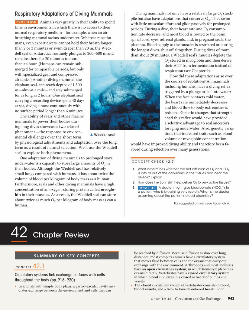
Diving mammals not only have a relatively large O2 stock-pile but also have adaptations that conserve O2. They swim with little muscular effort and glide passively for prolonged periods. During a dive, their heart rate and O2 consump-tion rate decrease, and most blood is routed to the brain, spinal cord, eyes, adrenal glands, and, in pregnant seals, the placenta. Blood supply to the muscles is restricted or, during the longest dives, shut off altogether. During dives of more than about 20 minutes, a Weddell seal’s muscles deplete the
O2 stored in myoglobin and then derive their ATP from fermentation instead of respiration (see Chapter 9).
How did these adaptations arise over the course of evolution? All mammals, including humans, have a diving reflex triggered by a plunge or fall into water: When the face contacts cold water, the heart rate immediately decreases and blood flow to body extremities is reduced. Genetic changes that strength-ened this reflex would have provided a selective advantage to seal ancestors foraging underwater. Also, genetic varia-tions that increased traits such as blood volume or myoglobin concentration
would have improved diving ability and therefore been fa-vored during selection over many generations.
Respiratory Adaptations of Diving Mammals E VO L U T I O N Animals vary greatly in their ability to spend
time in environments in which there is no access to their normal respiratory medium—for example, when an air-breathing mammal swims underwater. Whereas most hu-mans, even expert divers, cannot hold their breath longer than 2 or 3 minutes or swim deeper than 20 m, the Wed-dell seal of Antarctica routinely plunges to 200–500 m and remains there for 20 minutes to more than an hour. (Humans can remain sub-merged for comparable periods, but only with specialized gear and compressed air tanks.) Another diving mammal, the elephant seal, can reach depths of 1,500 m—almost a mile—and stay submerged for as long as 2 hours! One elephant seal carrying a recording device spent 40 days at sea, diving almost continuously with no surface period longer than 6 minutes.
The ability of seals and other marine mammals to power their bodies dur-ing long dives showcases two related phenomena—the response to environ-mental challenges over the short term by physiological adjustments and adaptation over the long term as a result of natural selection. We’ll use the Weddell seal to explore both phenomena.
One adaptation of diving mammals to prolonged stays underwater is a capacity to store large amounts of O2 in their bodies. Although the Weddell seal has relatively small lungs compared with humans, it has about twice the volume of blood per kilogram of body mass as a human. Furthermore, seals and other diving mammals have a high concentration of an oxygen-storing protein called myoglo-bin in their muscles. As a result, the Weddell seal can store about twice as much O2 per kilogram of body mass as can a human.
▲ Weddell seal
C O N C E P T C H E C K 4 2 . 7
1. What determines whether the net diffusion of O2 and CO2 is into or out of the capillaries in the tissues and near the alveoli? Explain.
2. How does the Bohr shift help deliver O2 to very active tissues? 3. W H AT I F ? A doctor might give bicarbonate (HCO3
-) to a patient who is breathing very rapidly. What is the doctor assuming about the patient’s blood chemistry?
For suggested answers, see Appendix A.
42 Chapter Review
be reached by diffusion. Because diffusion is slow over long distances, most complex animals have a circulatory system that moves fluid between cells and the organs that carry out exchange with the environment. Arthropods and most molluscs have an open circulatory system, in which hemolymph bathes organs directly. Vertebrates have a closed circulatory system, in which blood circulates in a closed network of pumps and vessels.
t� The closed circulatory system of vertebrates consists of blood, blood vessels, and a two- to four-chambered heart. Blood
SUMMARY OF KEY CONCEPTS
C O N C E P T 42.1Circulatory systems link exchange surfaces with cells throughout the body (pp. 916–920)t� In animals with simple body plans, a gastrovascular cavity me-
diates exchange between the environment and cells that can
C H A P T E R 4 2 Circulation and Gas Exchange 943

contain thick elastic walls that maintain blood pressure. Veins contain one-way valves that contribute to the return of blood to the heart. Blood pressure is altered by changes in cardiac output and by variable constriction of arterioles.
t� Fluid leaks out of capillaries and is returned to blood by the lymphatic system, which also defends against infection.
? If you placed your forearm on your head, how, if at all, would the blood pressure in that arm change? Explain.
C O N C E P T 42.4Blood components function in exchange, transport, and defense (pp. 928–933)t� Whole blood consists of cells and cell fragments (platelets)
suspended in a liquid matrix called plasma. Plasma proteins influence blood pH, osmotic pressure, and viscosity, and they function in lipid transport, immunity (antibodies), and blood clotting (fibrinogen). Red blood cells, or erythrocytes, transport O2. Five types of white blood cells, or leukocytes, function in defense against microorganisms and foreign substances in the blood. Platelets function in blood clotting, a cascade of reactions that converts plasma fibrinogen to fibrin.
t� A variety of diseases impair function of the circulatory system. In sickle-cell disease, an aberrant form of hemoglobin disrupts erythrocyte shape and function, leading to blockage of small blood vessels and a decrease in the oxygen-carrying capacity of the blood. In cardiovascular disease, inflammation of the arterial lining enhances deposition of lipids and cells, resulting in the potential for life-threatening damage to the heart or brain.
? In the absence of infection, what percentage of cells in human blood are leukocytes?
C O N C E P T 42.5Gas exchange occurs across specialized respiratory surfaces (pp. 933–938)t� At all sites of gas exchange, a gas undergoes net diffusion from
where its partial pressure is higher to where it is lower. Air is more conducive to gas exchange than water because air has a higher O2 content, lower density, and lower viscosity.
t� The structure and organization of respiratory surfaces differ among animal species. Gills are outfoldings of the body surface specialized for gas exchange in water. The effectiveness of gas exchange in some gills, including those of fishes, is increased by ventilation and countercurrent exchange between blood and water. Gas exchange in insects relies on a tracheal system, a branched network of tubes that bring O2 directly to cells. Spi-ders, land snails, and most terrestrial vertebrates have internal lungs. In mammals, air inhaled through the nostrils passes through the pharynx into the trachea, bronchi, bronchioles, and dead-end alveoli, where gas exchange occurs.
? Why does altitude have almost no effect on an animal’s ability to rid itself of CO2 through gas exchange?
C O N C E P T 42.6Breathing ventilates the lungs (pp. 938–940)t� Breathing mechanisms vary substantially among vertebrates.
An amphibian ventilates its lungs by positive pressure breath-ing, which forces air down the trachea. Birds use a system of air sacs as bellows to keep air flowing through the lungs in one direction only, preventing the mixing of incoming and outgoing air. Mammals ventilate their lungs by negative pressure breath-ing, which pulls air into the lungs when the rib muscles and
pumped by a heart ventricle passes to arteries and then to the capillaries, sites of chemical exchange between blood and inter-stitial fluid. Veins return blood from capillaries to an atrium, which passes blood to a ventricle. Fishes, rays, and sharks have a single pump in their circulation. Air-breathing vertebrates have two pumps combined in a single heart. Variations in ventricle number and separation reflect adaptations to different environ-ments and metabolic needs.
? How does the flow of a fluid in a closed circulatory system differ from the movement of molecules between cells and their environment with regard to distance traveled, direction traveled, and driving force?
C O N C E P T 42.2Coordinated cycles of heart contraction drive double circulation in mammals (pp. 920–923)t� The right ventricle pumps blood to the lungs, where it loads O2
and unloads CO2. Oxygen-rich blood from the lungs enters the heart at the left atrium and is pumped to the body tissues by the left ventricle. Blood returns to the heart through the right atrium.
Heart
Alveolarepithelialcells
CO2 O2
Alveolarcapillaries
Systemiccapillaries
Exhaled air Inhaled air
Alveolarspaces
Pulmonaryveins
Systemicarteries
Systemicveins
Body tissue
Pulmonaryarteries
O2CO2
© Pearson Education, Inc.
t� The cardiac cycle, a complete sequence of the heart’s pumping and filling, consists of a period of contraction, called systole, and a period of relaxation, called diastole. Heart function can be assessed by measuring the pulse (number of times the heart beats each minute) and cardiac output (volume of blood pumped by each ventricle per minute).
t� The heartbeat originates with impulses at the sinoatrial (SA) node (pacemaker) of the right atrium. They trigger atrial con-traction, are delayed at the atrioventricular (AV) node, and are then conducted along the bundle branches and Purkinje fibers, triggering ventricular contraction. The nervous system, hor-mones, and body temperature affect pacemaker activity.
? What changes in cardiac function might you expect after surgical replacement of a defective heart valve?
C O N C E P T 42.3Patterns of blood pressure and flow reflect the structure and arrangement of blood vessels (pp. 923–927)t� Blood vessels have structures well adapted to function. Cap-
illaries have narrow diameters and thin walls that facilitate exchange. The velocity of blood flow is lowest in the capillary beds as a result of their large total cross-sectional area. Arteries
944 U N I T S E V E N Animal Form and Function

C H A P T E R 4 2 Circulation and Gas Exchange 945
diaphragm contract. Incoming and outgoing air mix, decreasing the efficiency of ventilation.
t� Sensors detect the pH of cerebrospinal fluid (reflecting CO2 concentration in the blood), and a control center in the medulla oblongata adjusts breathing rate and depth to match metabolic demands. Additional input to the control center is provided by sensors in the aorta and carotid arteries that monitor blood lev-els of O2 as well as CO2 (via blood pH).
? How does air in the lungs differ from the fresh air that enters the body during inspiration?
C O N C E P T 42.7Adaptations for gas exchange include pigments that bind and transport gases (pp. 941–943)t� In the lungs, gradients of partial pressure favor the net diffusion
of O2 into the blood and CO2 out of the blood. The opposite situation exists in the rest of the body. Respiratory pigments such as hemocyanin and hemoglobin bind O2, greatly increasing the amount of O2 transported by the circulatory system.
t� Evolutionary adaptations enable some animals to satisfy ex-traordinary O2 demands. Deep-diving mammals stockpile O2 in blood and other tissues and deplete it slowly.
? How is the role of a respiratory pigment like that of an enzyme?
TEST YOUR UNDERSTANDING
LEVEL 1: KNOWLEDGE/COMPREHENSION
1. Which of the following respiratory systems is not closely associated with a blood supply? a. the lungs of a vertebrate b. the gills of a fish c. the tracheal system of an insect d. the skin of an earthworm
2. Blood returning to the mammalian heart in a pulmonary vein drains first into the a. left atrium. b. right atrium.
c. left ventricle. d. right ventricle.
c. cardiac output. d. heart rate.
c. rising CO2 d. falling CO2
c. the right atrium. d. the right ventricle.
7. Compared with the interstitial fluid that bathes active muscle cells, blood reaching these cells in arterioles has a a. higher PO2. b. higher PCO2. c. greater bicarbonate concentration. d. lower pH.
3. Pulse is a direct measure of a. blood pressure. b. stroke volume.
4. When you hold your breath, which of the following blood gas changes first leads to the urge to breathe? a. rising O2 b. falling O2
5. One feature that amphibians and humans have in common is a. the number of heart chambers. b. a complete separation of circuits for circulation. c. the number of circuits for circulation. d. a low blood pressure in the systemic circuit.
LEVEL 2: APPLICATION/ANALYSIS
6. If a molecule of CO2 released into the blood in your left toe is exhaled from your nose, it must pass through all of the following except a. the pulmonary vein. b. the trachea.
The diving bell spider (Argyroneta aquatica) stores air underwater in a net of silk. Explain why this adaptation could be more advan-tageous than having gills, taking into ac-count differences in gas exchange media and gas exchange or-gans among animals.
For selected answers, see Appendix A.
Students Go to MasteringBiology for assignments, the eText, and the Study Area with practice tests, animations, and activities.Instructors Go to MasteringBiology for automatically graded tutorials and questions that you can assign to your students, plus Instructor Resources.
LEVEL 3: SYNTHESIS/EVALUATION
8. D R AW I T Plot blood pressure against time for one cardiac cycle in humans, drawing separate lines for the pressure in the aorta, the left ventricle, and the right ventricle. Below the time axis, add a vertical arrow pointing to the time when you expect a peak in atrial blood pressure.
9. EVOLUTION CONNECTION One of the opponents of the movie monster Godzilla is Mothra, a giant mothlike creature with a wingspan of several dozen meters. The largest known insects were Paleozoic drag-onflies with half-meter wingspans. Focusing on respiration and gas exchange, explain why giant insects are improbable.
10. SCIENTIFIC INQUIRY I N T E R P R E T T H E DATA
The hemoglobin of a human fetus differs from adult he-moglobin. Compare the dis-sociation curves of the two hemoglobins in the graph at right. Describe how they differ, and propose a hy-pothesis to explain the ben-efit of this difference.
11. SCIENCE, TECHNOLOGY, AND SOCIETY Hundreds of studies have linked smoking with cardiovascular and lung disease. According to most health authorities, smok-ing is the leading cause of preventable, premature death in the United States. What are some arguments in favor of a total ban on cigarette advertising? What are arguments in opposition? Do you favor or oppose such a ban? Explain.
12. WRITE ABOUT A THEME: INTERACTIONS Some athletes prepare for competition at sea level by sleeping in a tent in which PO2 is kept low. When climbing high peaks, some mountaineers breathe from bottles of pure O2. In a short essay (100–150 words), relate these behaviors to the mecha-nism of O2 transport in the human body and to physiological interactions with our gaseous environment.
13. SYNTHESIZE YOUR KNOWLEDGE
0 20 40 60 80 1000
20
40
60
80
100
O2
satu
ratio
n of
hem
oglo
bin
(%) Fetus
Mother
PO2 (mm Hg)