Circulating Proteolytic Products of Carboxypeptidase N for Early Detection of Breast Cancer Y. Li,...
14
Circulating Proteolytic Products of Carboxypeptidase N for Early Detection of Breast Cancer Y. Li, Y. Li, T. Chen, A.S. Kuklina, P. Bernard, F.J. Esteva, H. Shen, M. Ferrari, and Y. Hu January 2014 www.clinchem.org/content/60/1/233.full © Copyright 2014 by the American Association for Clinical Chemistry
-
Upload
bruno-beasley -
Category
Documents
-
view
223 -
download
1
Transcript of Circulating Proteolytic Products of Carboxypeptidase N for Early Detection of Breast Cancer Y. Li,...
Circulating Proteolytic Products of Carboxypeptidase N for Early Detection of Breast Cancer
Y. Li, Y. Li, T. Chen, A.S. Kuklina, P. Bernard, F.J. Esteva, H. Shen, M. Ferrari, and Y. Hu
January 2014
www.clinchem.org/content/60/1/233.full
© Copyright 2014 by the American Association for Clinical Chemistry
© Copyright 2009 by the American Association for Clinical Chemistry
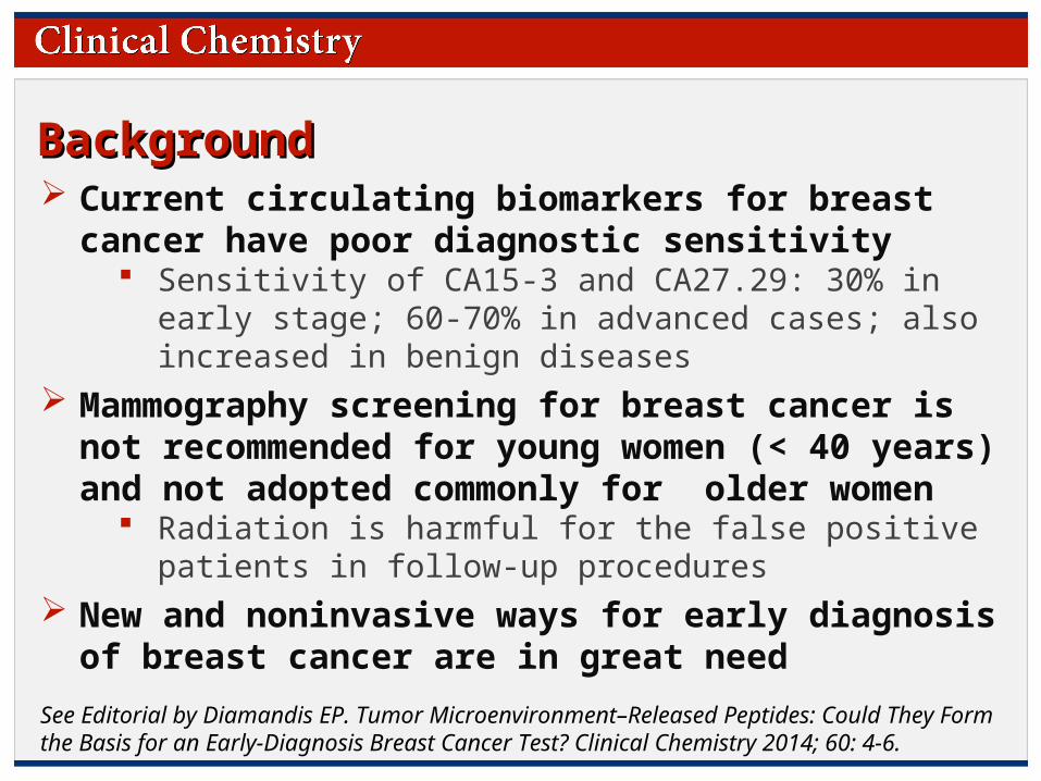
Background Background Current circulating biomarkers for breast cancer have
poor diagnostic sensitivity Sensitivity of CA15-3 and CA27.29: 30% in early stage; 60-
70% in advanced cases; also increased in benign diseases Mammography screening for breast cancer is not
recommended for young women (< 40 years) and not adopted commonly for older women
Radiation is harmful for the false positive patients in follow-up procedures
New and noninvasive ways for early diagnosis of breast cancer are in great need
See Editorial by Diamandis EP. Tumor Microenvironment–Released Peptides: Could They Form the Basis for an Early-Diagnosis Breast Cancer Test? Clinical Chemistry 2014; 60: 4-6.

© Copyright 2009 by the American Association for Clinical Chemistry
BackgroundBackground Proteolytic activity in the tumor microenviroment (not
necessarily deriving from tumor cells) may release peptides into circulation that are potential biomarkers for monitoring tumor progression
See Editorial by Diamandis EP. Tumor Microenvironment–Released Peptides: Could They Form the Basis for an Early-Diagnosis Breast Cancer Test? Clinical Chemistry 2014; 60: 4-6.

© Copyright 2009 by the American Association for Clinical Chemistry
Background Background
Carboxypeptidase N (CPN), also known as an arginine/lysine carboxypeptidase, is a member of a larger family of zinc metallopeptidases
CPN cleaves the carboxy (C)-terminal arginine or lysine of endogenous peptides
Substrates: bradykinin (BK9), anaphylatoxins (C3a, C4a, and C5a), and fibrinopeptides A and B
EDTA is an inhibitor of CPN The relationship between breast cancer, CPN, and circulating
concentrations of the proteolytic products of CPN has not been established
See Editorial by Diamandis EP. Tumor Microenvironment–Released Peptides: Could They Form the Basis for an Early-Diagnosis Breast Cancer Test? Clinical Chemistry 2014; 60: 4-6.

© Copyright 2009 by the American Association for Clinical Chemistry
Materials & Methods Materials & Methods
Injection 2 weeks 4 weeks 6 weeks
(Nude mice, n=8; breast cancer cell line: MDA-MD-231)Breast cancer mouse model :
8 weeks
Serum collection
Tissue collection
Breast cancer (BC) patients:
Control BC-I BC-II BC-III BC-IV Total
Blood plasma
10 11 11 14 12 58
Tissue slices
10 17 77 16 - 110

© Copyright 2009 by the American Association for Clinical Chemistry
Materials & MethodsMaterials & Methods
Data AnalysisMALDI-TOF MSNanopore fractionation
Circulating peptides were isolated by nanoporous silica chip and analyzed by mass spectrometry

© Copyright 2009 by the American Association for Clinical Chemistry© Copyright 2009 by the American Association for Clinical Chemistry
ResultsResults
Figure 1. Ex vivo peptide cleavage assay on the synthetic C3f peptide. A. C3f peptide cleavage assay in NIF (normal interstitial fluid), CM (conditioned cell-culture medium), and TIF (tumor interstitial fluid). B. Specific cleavage sites are indicated on the C3f peptide, a substrate of the enzymes factor I, chymotrypsin and CPN. C. Detecting CPN expression in different assay conditions. C3f, synthetic peptide His6-C3f_S1304-R1320-His6 only; NIF or TIF, normal or tumor interstitial fluid only; EDTA, an inhibitor of CPN; NIF (CPN -) and TIF (CPN -), CPN-depleted normal and tumor tissue interstitial fluid; CM (CPN -), CPN-depleted conditioned medium.
A
B C

© Copyright 2009 by the American Association for Clinical Chemistry© Copyright 2009 by the American Association for Clinical Chemistry
Result Result
Figure 2. CPN expression in mouse tissues and sera. A. CPN immunoblotting expression in normal breast interstitial fluid (NIF) and tumor interstitial fluid (TIF). Coomassie Brilliant Blue (CBB) stained gel was used for the evaluation of equal loading. Both immunoblot and CBB stained gel were subjected to quantitative image analysis by software Image J, and the result was presented by histogram. B. Immunohistochemistry analysis of CPN in normal breast tissue and tumor tissue collected at the 2nd and 8th week. C. Immunoblotting of CPN in mouse sera. Ponceau S-stained serum albumin (SA) serves as internal control for loading. (Groups: 0, 2nd, 4th, 6th, 8th week; n = 4 / group).
A
B C
© Copyright 2009 by the American Association for Clinical Chemistry© Copyright 2009 by the American Association for Clinical Chemistry
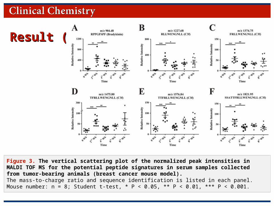
Result (3)Result (3)
Figure 3. The vertical scattering plot of the normalized peak intensities in MALDI TOF MS for the potential peptide signatures in serum samples collected from tumor-bearing animals (breast cancer mouse model). The mass-to-charge ratio and sequence identification is listed in each panel. Mouse number: n = 8; Student t-test, * P < 0.05, ** P < 0.01, *** P < 0.001.

© Copyright 2009 by the American Association for Clinical Chemistry© Copyright 2009 by the American Association for Clinical Chemistry
Result (4)Result (4)
Figure 4. CPN expression in human tissues and plasma. A. IHC stains for CPN in human control cohort (cancer adjunct tissue or adenosis) and tumor tissues. B. The histogram shows the percentage of the scoring results of IHC stains for CPN (n = 10, 7, 77 and 16 for control, BC-I, BC-II and BC-III slides, respectively). Scoring method of IHC staining pattern refers to the protocol reported by Zhang et al. (30). C. CPN expression by immunoblotting in human plasma from healthy controls and breast cancer (BC) patients at different stages of disease (I, II, III and IV). Ponceau S-stained serum albumin (SA) served as internal control. n = 4 / group.
A
B C

© Copyright 2009 by the American Association for Clinical Chemistry© Copyright 2009 by the American Association for Clinical Chemistry
Result (5)Result (5)
Figure 5. Profiles of the potential peptide signatures in plasma from healthy women and women with breast cancer of different pathological stages. Sample number: n = 10, healthy controls; n = 11, stage I breast cancer (BC-I); n = 12, stage II breast cancer (BC-II); n = 15, stage III breast cancer (BC-III); n = 10, stage IV breast cancer (BC-IV); Student t-test, * P < 0.05, *** P < 0.001.

© Copyright 2009 by the American Association for Clinical Chemistry
DiscussionDiscussion
CPN expression was increased in tumor as compared with normal breast tissue, but no changes in blood levels were observed
Six circulating CPN-catalyzed peptides reflect the higher CPN activity in tumors as early as the first pathologic stage of breast cancer
© Copyright 2009 by the American Association for Clinical Chemistry
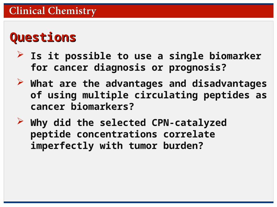
QuestionsQuestions Is it possible to use a single biomarker for cancer
diagnosis or prognosis?
What are the advantages and disadvantages of using multiple circulating peptides as cancer biomarkers?
Why did the selected CPN-catalyzed peptide concentrations correlate imperfectly with tumor burden?
© Copyright 2009 by the American Association for Clinical Chemistry
Thank you for participating in this month’sClinical Chemistry Journal Club.
Additional Journal Clubs are available atwww.clinchem.org
Download the free Clinical Chemistry app on iTunes for additional content!
Follow us