CIED Practice Advisory Update 2019 Pre-Final Draft
46
Embargoed for release until approved by ASA House of Delegates. No part of this document may be released, distributed or reprinted until approved. Any unauthorized copying, reproduction, appropriation or communication of the contents of this document without the express written consent of the American Society of Anesthesiologists is subject to civil and criminal prosecution to the fullest extent possible, including punitive damages Practice Advisory for the Perioperative Management of Patients with Cardiac Implantable Electronic Devices: Pacemakers and Implantable Cardioverter-Defibrillators 2020 An Updated Report by the American Society of Anesthesiologists Task Force on Perioperative Management of Patients with Cardiac Implantable Electronic Devices* PRACTICE advisories are systematically developed reports that are intended to assist decision- 1 making in areas of patient care. Advisories provide a synthesis of scientific literature and analysis of 2 expert opinion, clinical feasibility data, open forum commentary, and consensus surveys. Practice 3 advisories developed by the American Society of Anesthesiologists (ASA) are not intended as standards, 4 guidelines, or absolute requirements, and their use cannot guarantee any specific outcome. They may be 5 adopted, modified, or rejected according to clinical needs and constraints, and they are not intended to 6 replace local institutional policies. 7 Practice advisories summarize the state of the literature and report opinions obtained from expert 8 consultants and ASA members. They are not supported by scientific literature to the same degree as 9 standards or guidelines because of the lack of sufficient numbers of adequately controlled studies. 10 Practice advisories are subject to periodic revision as warranted by the evolution of medical knowledge, 11 technology, and practice. 12 This document updates the “Practice Advisory for the Perioperative Management of Patients with 13 Cardiac Implantable Electronic Devices: Pacemakers and Implantable Cardioverter-Defibrillators. An 14 Updated Report by the American Society of Anesthesiologists Task Force on Perioperative Management 15 of Patients with Cardiac Implantable Electronic Devices, adopted by the ASA in 2010 and published in 16 2011. 1 17 Methodology 18 Definition of Cardiac Implantable Electronic Devices 19 For this Advisory, a cardiac implantable electronic device (CIED) refers to any permanently implanted 20 * Updated by the Committee on Standards and Practice Parameters: Jeffrey L. Apfelbaum, M.D. (Committee Chair), Chicago, Illinois; Peter M. Schulman, M.D. (Task Force Co-Chair), Portland, Oregon, Aman Mahajan M.D. Ph.D. (Task Force Co-Chair), Pittsburg, Pennsylvania; Richard T. Connis, Ph.D. (Chief Methodologist), Woodinville, Washington; and Madhulika Agarkar, M.P.H. (Methodologist), Schaumburg, Illinois. Received from the American Society of Anesthesiologists, Schaumburg, Illinois. Submitted for publication October __, 2019. Accepted for publication October __, 2019. Supported by the American Society of Anesthesiologists and developed under the direction of the Committee on Standards and Practice Parameters, Jeffrey L. Apfelbaum, M.D. (Chair). Approved by the ASA House of Delegates on October __, 2019. A complete bibliography used to develop this updated Advisory, arranged alphabetically by author, is available as Supplemental Digital Content 1, http://links.lww.com/ALN/XXXXXXXX. Address correspondence to the American Society of Anesthesiologists: 1061 American Lane, Schaumburg, Illinois 60173. This Practice Advisory, as well as all published ASA Practice Parameters, may be obtained at no cost through the Journal Web site, www.anesthesiology.org.
Transcript of CIED Practice Advisory Update 2019 Pre-Final Draft
Embargoed for release until approved by ASA House of Delegates. No part of this document may be released, distributed or reprinted until approved. Any unauthorized copying, reproduction, appropriation or communication of the contents of this document without the express written consent of the American Society of Anesthesiologists is subject to civil and criminal prosecution to the fullest extent possible, including punitive damages
Practice Advisory for the Perioperative Management of Patients with
Cardiac Implantable Electronic Devices: Pacemakers and Implantable
Cardioverter-Defibrillators 2020
An Updated Report by the American Society of Anesthesiologists Task Force on Perioperative Management of Patients with Cardiac Implantable Electronic Devices*
PRACTICE advisories are systematically developed reports that are intended to assist decision-1
making in areas of patient care. Advisories provide a synthesis of scientific literature and analysis of 2
expert opinion, clinical feasibility data, open forum commentary, and consensus surveys. Practice 3
advisories developed by the American Society of Anesthesiologists (ASA) are not intended as standards, 4
guidelines, or absolute requirements, and their use cannot guarantee any specific outcome. They may be 5
adopted, modified, or rejected according to clinical needs and constraints, and they are not intended to 6
replace local institutional policies. 7
Practice advisories summarize the state of the literature and report opinions obtained from expert 8
consultants and ASA members. They are not supported by scientific literature to the same degree as 9
standards or guidelines because of the lack of sufficient numbers of adequately controlled studies. 10
Practice advisories are subject to periodic revision as warranted by the evolution of medical knowledge, 11
technology, and practice. 12
This document updates the “Practice Advisory for the Perioperative Management of Patients with 13
Cardiac Implantable Electronic Devices: Pacemakers and Implantable Cardioverter-Defibrillators. An 14
Updated Report by the American Society of Anesthesiologists Task Force on Perioperative Management 15
of Patients with Cardiac Implantable Electronic Devices, adopted by the ASA in 2010 and published in 16
2011.1 17
Methodology 18
Definition of Cardiac Implantable Electronic Devices 19
For this Advisory, a cardiac implantable electronic device (CIED) refers to any permanently implanted 20
* Updated by the Committee on Standards and Practice Parameters: Jeffrey L. Apfelbaum, M.D. (Committee
Chair), Chicago, Illinois; Peter M. Schulman, M.D. (Task Force Co-Chair), Portland, Oregon, Aman Mahajan M.D. Ph.D. (Task Force Co-Chair), Pittsburg, Pennsylvania; Richard T. Connis, Ph.D. (Chief Methodologist), Woodinville, Washington; and Madhulika Agarkar, M.P.H. (Methodologist), Schaumburg, Illinois.
Received from the American Society of Anesthesiologists, Schaumburg, Illinois. Submitted for publication October __, 2019. Accepted for publication October __, 2019. Supported by the American Society of Anesthesiologists and developed under the direction of the Committee on Standards and Practice Parameters, Jeffrey L. Apfelbaum, M.D. (Chair). Approved by the ASA House of Delegates on October __, 2019.
A complete bibliography used to develop this updated Advisory, arranged alphabetically by author, is available as Supplemental Digital Content 1, http://links.lww.com/ALN/XXXXXXXX. Address correspondence to the American Society of Anesthesiologists: 1061 American Lane, Schaumburg, Illinois 60173. This Practice Advisory, as well as all published ASA Practice Parameters, may be obtained at no cost through the Journal Web site, www.anesthesiology.org.

PRACTICE ADVISORY
2
cardiac pacemaker or any implantable cardioverter-defibrillator (ICD). The term CIED also refers to any 21
cardiac resynchronization therapy (CRT) device.†22
Purposes of the Advisory 23
The purposes of this Advisory update are to: (1) facilitate safe and effective perioperative 24
management of the patient with a CIED, and (2) reduce the incidence of adverse outcomes. 25
Perioperative management refers to the preoperative, intraoperative, postoperative or recovery period 26
in any setting where an anesthesia provider will be delivering anesthesia care. Adverse outcomes 27
associated with CIED function, but are not limited to, damage to the device, inability of the device to 28
deliver pacing or shocks, lead-tissue interface damage, changes in pacing behavior, electrical reset to 29
the backup pacing mode, and inappropriate ICD therapies.‡30
Adverse clinical outcomes include, but are not limited to, hypotension, tachyarrhythmia and 31
bradyarrhythmia, myocardial tissue damage, and myocardial ischemia and infarction. Other related 32
adverse outcomes may include extended hospital stay, delay and cancellation of surgery, readmission 33
to manage device malfunction, and additional hospital resource utilization and cost. 34
Focus of the Advisory 35
This updated Advisory focuses on the perioperative management of the patient who has a 36
preexisting, permanently implanted CIED for treatment of bradyarrhythmia, tachyarrhythmia or heart 37
failure. This Advisory applies to all CIED patients receiving general or regional anesthesia, sedation or 38
monitored anesthesia care. Both inpatient and outpatient procedures are addressed by this update. 39
This update does not address the perioperative management of the patient without a permanently 40
implanted CIED, including those: (1) with a temporary CIED, (2) with a noncardiac implantable device 41
(e.g., neurological or spinal cord stimulator), (3) with an implantable mechanical cardiac assist device 42
(e.g., ventricular assist device), or (4) undergoing CIED implantation or revision. This update does not 43
address procedures where there are no known perioperative CIED concerns (e.g., plain radiography, 44
fluoroscopy, mammograms, or ultrasound). In addition, this update does not address patient comfort or 45
management of pain during a procedure. 46
Application of the Advisory 47
This updated Advisory is intended for use by anesthesiologists and all other individuals who 48
deliver or who are responsible for anesthesia care. The update may also serve as a resource for other 49
physicians and health care professionals who manage patients with CIEDs. 50
51
† Generic pacemaker and defibrillator codes are provided in Tables 1 and 2. Note that every ICD includes both
pacing and shock therapies for the management of bradyarrhythmias and tachyarrhythmias. ‡ Inappropriate ICD therapy refers to the delivery of antitachycardia therapy (paced or shock) in the absence of a
clinically indicated tachyarrhythmia. Inappropriate ICD therapy can harm a patient by inducing ischemia, worsening the arrhythmia, or causing the patient to move during a delicate procedure.

PRACTICE ADVISORY
3
Task Force Members and Consultants 52
The original Advisory was developed by an ASA appointed task force of 12 members, consisting of 53
anesthesiologists and cardiologists in private and academic practices from various geographic areas of 54
the United States, and two methodologists from the ASA Committee on Standards and Practice 55
Parameters. In 2017, the ASA Committee on Standards and Practice Parameters requested that the 56
Advisory be updated. This update is a revision developed by an ASA-appointed task force of 5 members, 57
including 3 anesthesiologists and two methodologists. Conflict of interest documentation regarding 58
current or potential financial and other interests pertinent to the practice guideline were disclosed by all 59
task force members and managed. 60
61
Process and Evaluation of Evidence 62
This updated Advisory was developed by means of a five-step process. First, consensus was 63
reached on the criteria for evidence. Second, original published articles from peer-reviewed journals 64
relevant to the perioperative management of CIEDs were evaluated and added to literature reported in 65
the previous update. Third, consultants who had expertise or interest in central venous catheterization, 66
and who practiced or worked in various settings (e.g., private and academic practice) were asked to 67
participate in opinion surveys addressing the appropriateness, completeness, and feasibility of 68
implementation of the draft recommendations, and to review and comment on a draft of the Guidelines. 69
Fourth, additional opinions were solicited from random samples of active ASA members. Fifth, all 70
available information was used to build consensus to finalize the Advisory. A summary of 71
recommendations can be found in appendix 1. 72
Preparation of this updated Advisory followed a rigorous methodological process. Evidence was 73
obtained from two principal sources: scientific evidence and opinion-based evidence. Detailed 74
descriptions of the ASA process and methodology used in these Guidelines may be found in other related 75
publications.2-5 Appendix 5 contains information on the evidence model, the literature search process, 76
literature findings, and survey results. 77
Within the text of the Advisory, literature classifications are reported for each intervention using the 78
following classifications: Category A, level 1: Meta-analysis of randomized controlled trials (RCTs); 79
Category A level 2, multiple RCTs, and Category A, level 3: a single RCT. Category B, level 1: 80
nonrandomized studies with group comparisons, Category B, level 2: nonrandomized studies with 81
associative findings; Category B, level 3: nonrandomized studies with descriptive findings, and level 4: 82
case series or case reports. Outcomes are designated as either beneficial (B) or harmful (H) for the 83
patient; statistically nonsignificant findings are designated as equivocal (E). Survey findings from task 84
force–appointed expert consultants and a random sample of the ASA membership are fully reported in 85
the text of these Guidelines. Survey responses for each recommendation are reported using a 5-point 86
scale based on median values from strongly agree to strongly disagree. 87
88

PRACTICE ADVISORY
4
Advisory Evidence and Recommendations 89
Preoperative Evaluation 90
A focused preoperative evaluation of the patient with a CIED consists of the following topics: (1) 91
determining whether a patient has a CIED, (2) determining the CIED type, manufacturer and primary 92
indication for placement, (3) determining whether a patient is pacing dependent, and (4) determining the 93
CIED’s current settings and that it is functioning properly by interrogating the CIED or obtaining the most 94
recent interrogation report. 95
Literature Findings. Although the literature is insufficient to evaluate the clinical benefit of 96
performing a focused preoperative evaluation of CIED patients, case reports indicate that adverse 97
outcomes (e.g., inappropriate shock, CIED switch to “end of life mode,” acute ventricular lead dysfunction, 98
and corrupted device memory) may occur when a complete preoperative examination is not performed to 99
determine whether the patient has a CIED (Category B4-H evidence).6-9 The literature is insufficient to 100
evaluate whether preoperatively determining the CIED type, manufacturer and primary indication for 101
placement, or determining whether a patient is pacing dependent affects perioperative outcomes. A case 102
series reported inappropriate antitachycardia pacing or shocks, premature battery depletion, and CIED 103
damage when the CIED’s current settings were not adequately assessed preoperatively. (Category B4-H 104
evidence).10 The literature is insufficient to evaluate the benefit of any specific time interval to determine 105
recency for review of a previous CIED interrogation. 106
Survey Findings. The expert consultants and ASA members strongly agree with the 107
recommendation that a preoperative evaluation should include determining whether a patient has a CIED, 108
determining the CIED type (i.e. PM, ICD, CRT), determining the primary indication for CIED placement, 109
and determining whether the patient is pacing dependent. The consultants strongly agree and ASA 110
members agree that a preoperative evaluation should include determining the CIED manufacturer. 111
The consultants strongly agree and ASA members agree that a preoperative evaluation should 112
include determining the CIED’s current settings and confirming that the CIED is functioning properly (i.e. 113
by interrogating the CIED or obtaining the most recent interrogation report). The consultants selected 114
preferred time spans for determining proper ICD functioning prior to a procedure, as follows: immediately 115
= 6% of consultants, at least 3 months prior = 48% of consultants, at least 6 months prior = 36% of 116
consultants, and at least 12 months prior = 6% of consultants. For a pacemaker the following time spans 117
were selected by consultants: immediately = 3% of consultants, at least 3 months prior = 39% of 118
consultants, at least 6 months prior = 30% of consultants, and at least 12 months prior = 27% of 119
consultants. The ASA members selected the following time spans preferred to determine proper 120
functioning of an ICD prior to the procedure: immediately = 10% of members, at least 3 months prior = 121
39% of members, at least 6 months prior = 44% of members, and at least 12 months prior = 7% of 122
members. For a pacemaker the following time spans were selected by members - immediately = 9% of 123
PRACTICE ADVISORY
5
members, at least 3 months prior = 38%, at least 6 months prior = 36%, and at least 12 months prior = 124
18% of consultants.§ 125
Advisory Recommendations for Preoperative Evaluation. 126
• Determine whether a patient has a CIED. 127
o Conduct a focused history (e.g., interview the patient or other source, review medical record, 128
chest x-ray, and electrocardiogram if available). 129
o Perform a focused physical examination (e.g., check for scars, palpate for device). 130
• Determine the CIED type, manufacturer and primary indication for placement.131
o Obtain the manufacturer’s ID card from the patient or other source. 132
o Review the medical record. 133
o Obtain and review the most recent CIED interrogation report. 134
o Refer to supplemental resources (e.g., manufacturer’s databases, CIED clinic records). 135
o Order a chest x-ray if no other data are available.**136
• Determine whether the patient is pacing dependent. 137
o From the focused history and medical record, assess for one or more of the following 138
indicators: 139
▪ Bradycardia that caused syncope or other symptoms resulting in CIED implantation. 140
▪ Successful atrioventricular (A-V) nodal ablation resulting in CIED implantation. 141
▪ A CIED interrogation showing no evidence of spontaneous ventricular activity when the 142
CIED's pacing function is temporarily programmed to a non-tracking mode (i.e., VVI) at 143
the lowest programmable rate. 144
• Determine the CIED’s current settings and that it is functioning properly (i.e., by interrogating the 145
CIED or obtaining the most recent interrogation report.††‡‡ 146
Preoperative Preparation. 147
Preoperative preparation for patient safety and proper maintenance of the CIED during a planned 148
procedure includes the following topics: (1) sources of electromagnetic interference (EMI), (2) 149
preoperative reprogramming of the CIED's pacing function to an asynchronous pacing mode or disabling 150
any special algorithms, including rate adaptive pacing functions, (3) suspending the anti-tachyarrhythmia 151
functions for an ICD, and (4) availability of temporary pacing and defibrillation equipment. 152
§ To view a bar chart with the above findings, refer to Supplemental Digital Content 5, http://links.lww.com/ALN/XXXXXX. ** Most CIEDs have an x-ray code inscribed on the generator that can be used to identify the CIED manufacturer. †† A CIED specialist might need to be consulted to help determine key information about the CIED, whether the
patient is pacing dependent, the CIED's current settings and that it is functioning properly. ‡‡ In many patients, determining proper CIED function can be accomplished by accessing the patient's most recent CIED interrogation report. Note that the majority of consultants and ASA members agree that a CIED should be interrogated within 3-6 months before a procedure.
PRACTICE ADVISORY
6
Literature Findings. The literature was evaluated for the following potential sources of EMI from 153
monopolar electrosurgery, bipolar electrosurgery, radiofrequency (RF) ablation, lithotripsy, external 154
cardioversion or defibrillation, magnetic resonance imaging (MRI), radiation therapy, radiofrequency 155
scanners, cardiac monitors, and electroconvulsive therapy (ECT). 156
Observational studies report that EMI may occur during monopolar electrosurgery,11-15 RF ablation,16-157
21 MRI,22-35 and radiation therapy36-42 (Category B3-H evidence). Case reports also indicate the 158
occurrence of EMI during monopolar electrosurgery,43-50 bipolar electrosurgery,51 RF ablation,52-54 MRI,6-159
9,55,56 and radiation therapy57-59 (Category B4-H evidence). 160
Case reports indicate that inappropriately high pacing rates may occur due to EMI effects between 161
cardiac monitoring equipment and CIEDs with active minute ventilation sensors (Category B4-H 162
evidence).60-62 An observational study reports a significantly higher occurrence of EMI when 163
electrosurgery above the umbilicus is performed compared with electrosurgery below the umbilicus 164
(Category B1-H evidence).15 The literature is insufficient to evaluate the benefit of the availability of 165
temporary pacing and defibrillation equipment during a procedure. 166
Survey Findings. 167
The consultants and ASA members strongly agree that a preoperative evaluation should include 168
determining whether EMI from monopolar electrosurgery or other sources is likely to occur, and strongly 169
agree with the recommendation to alter the pacing function of a CIED to an asynchronous pacing mode in 170
the pacing dependent patient if monopolar electrosurgery ("bovie") use is planned superior to the 171
umbilicus. The consultants disagree and ASA members are equivocal with the recommendation to alter 172
the pacing function of a CIED to an asynchronous pacing mode in the pacing dependent patient if 173
monopolar electrosurgery ("bovie") use is planned inferior to the umbilicus. The consultants and ASA 174
members strongly agree with the recommendation to suspend an ICD’s anti- tachycardia function, when 175
present if monopolar electrosurgery ("bovie") use is planned superior to the umbilicus. The consultants 176
agree and ASA members are equivocal with the recommendation to suspend an ICD’s anti- tachycardia 177
function, when present If monopolar electrosurgery ("bovie") use is planned inferior to the umbilicus. The 178
consultants and ASA members strongly agree with the recommendation to assure that the patient is in a 179
monitored environment before suspending the anti-tachycardia function of an ICD. The consultants are 180
equivocal and ASA members agree with the recommendation to avoid the routine use of a magnet over 181
an ICD. The consultants and ASA members strongly agree that if needed, a specialist should be 182
consulted to alter the pacing function of a CIED or to suspend the antitachycardia function of an ICD. The 183
consultants and ASA members strongly agree that the proceduralist should be advised to use bipolar 184
electrosurgery or an ultrasonic scalpel when feasible. The consultants and ASA members strongly agree 185
with the recommendation that temporary pacing and defibrillation equipment should be immediately 186
available before, during, and after all procedures with EMI potential. Finally, the consultants and ASA 187
members agree with the recommendation that a CIED's active sensor for rate- responsive pacing should 188
be suspended to prevent undesirable tachycardia. 189

PRACTICE ADVISORY
7
Advisory Recommendations for Preoperative Preparation. 190
• Determine whether intraoperative electromagnetic interference (EMI) is likely to occur. 191
• If EMI is likely to occur (e.g.,monopolar electrosurgery ["bovie"] use, or radiofrequency ablation) is 192
planned superior to the umbilicus, alter the pacing function of a CIED to an asynchronous pacing 193
mode in the pacing dependent patient and suspend an ICD’s anti-tachycardia function, if 194
present.§§***††† 195
o Before suspending the anti-tachycardia function, assure that the patient is in a monitored 196
environment.197
• Avoid the routine use of a magnet over an ICD.‡‡‡ 198
• If needed, consult a specialist to alter the pacing function of a CIED or to suspend the 199
antitachycardia function of an ICD. 200
• Assure that temporary pacing and defibrillation equipment are immediately available before, 201
during, and after all procedures with EMI potential. 202
• Suspend the CIED's active sensor for rate-responsive pacing to prevent undesirable tachycardia, 203
if present. 204
Intraoperative Monitoring 205
Intraoperative monitoring topics include (1) continuous ECG monitoring, (2) continuous SpO2 206
monitoring, and (3) peripheral pulse monitoring (e.g., pulse palpitation, pulse oximeter plethysmogram, or 207
arterial line). 208
Literature Findings. Case reports indicate that continuous ECG monitoring may detect EMI related 209
pacemaker function abnormalities,49,56,63 and cardiac abnormalities64-65 during a procedure (Category B4-210
B evidence). The literature is insufficient to examine the clinical impact of continuous perioperative 211
monitoring of SpO2, or peripheral pulse for CIED patients. 212
Survey Findings. The consultants and ASA members strongly agree with the recommendations to 213
(1) continuously monitor and display a patient’s ECG as required by ASA standards from the beginning of 214
anesthesia until the patient is transferred out of the anesthetizing location, with additional ECG monitoring 215
in the postoperative period as indicated by the patient’s medical condition, (2) perform continuous 216
peripheral pulse monitoring for all CIED patients receiving anesthesia care, and (3) if unanticipated CIED 217
§§ If EMI is unlikely it might be unnecessary to alter the pacing function of a CIED or suspend the antitachycardia
function of an ICD *** Note that the majority of consultants disagree and ASA members are equivocal regarding the recommendation to
alter the pacing function of a CIED to an asynchronous pacing mode in the pacing dependent patient if monopolar electrosurgery ("bovie") use is planned inferior to the umbilicus,
††† To view a bar chart with these findings, refer to Supplemental Digital Content 2, http://links.lww.com/ALN/XXXXXX. ‡‡‡ A magnet will not alter the pacing mode of an ICD. A magnet correctly applied to an ICD often results in
suspension of antitachycardia therapy. For most ICDs, there is no reliable means to confirm the magnet response. Some ICDs may have no magnet response. In obese patients or those with a deep CIED implant (i.e., subcutaneous ICD), magnet application might fail to elicit the magnet response. Some older ICDs can be permanently disabled by magnet application.
PRACTICE ADVISORY
8
interactions occur, discontinue the procedure until the source of interference can be eliminated or 218
managed. 219
Advisory Recommendations for Intraoperative Monitoring. 220
• Continuously monitor and display a patient’s ECG and SpO2 as required by ASA standards66,67 221
from the beginning of anesthesia until the patient is transferred out of the anesthetizing 222
location.§§§223
• Perform continuous peripheral pulse monitoring for all CIED patients receiving anesthesia care. 224
• If unanticipated CIED interactions occur, discontinue the procedure until the source of 225
interference can be identified and eliminated or managed. 226
Managing Potential Sources of EMI 227
Procedures using electrosurgery, radio frequency ablation, radiofrequency identification devices, 228
lithotripsy, MRI, radiation therapy, nerve conduction studies, cardioversion, or ECT may damage CIEDs 229
or interfere with CIED function, potentially resulting in severe adverse outcomes. Sources of EMI are 230
often unique to specific procedures, and the management of each of these potential EMI sources is 231
reported separately below. 232
Electrosurgery 233
Management of potential sources of EMI associated with electrosurgery includes the following topics: 234
(1) positioning the electrosurgical unit’s (ESU) dispersive electrode so that the current pathway does not 235
pass through or near the CIED generator and leads, (2) avoiding proximity of the ESU’s electrical current 236
to the generator or leads, (3) using intermittent and irregular bursts of monopolar electrosurgery at the 237
lowest feasible energy levels, (4) using bipolar electrosurgery and (5) using ultrasonic (harmonic) scalpel. 238
Literature Findings. The literature is insufficient to evaluate whether positioning the current pathway 239
away from the CIED generator and leads reduces the occurrence of EMI. A case report indicates that EMI 240
occurred when the ESU’s electrical current was placed in proximity to the generator or leads (Category 241
B4-H evidence).68 An observational study reports that EMI may occur in spite of positioning the 242
dispersive electrode to divert the return path away from the generator and leads (Category B3-H),15 case 243
reports also indicate that EMI may still occur when proximity is avoided (Category B4-H evidence)46,66 244
No controlled studies were found that examine the benefit of using short intermittent bursts of 245
electrosurgery at the lowest feasible energy levels. One case report describes pacemaker failure when 246
short bursts of current were used with a bipolar electrosurgery system (Category B4-H evidence).51 247
Case reports indicate that cardiac arrhythmias and asystole occurred when monopolar electrosurgery 248
was initiated, and after changing to bipolar electrosurgery the procedures proceeded uneventfully 249
§§§ The term “continuous” means “prolonged without any interruption at any time” (see Standards for Basic Anesthetic
Monitoring, American Society of Anesthesiologists. Approved by the ASA House of Delegates October 21, 1986, and last amended October 28, 2015.

PRACTICE ADVISORY
9
(Category B4-B evidence).46,64,65 A case report indicated that arrhythmia and asystole occurred when 250
monopolar electrosurgery was initiated, and after changing to a harmonic scalpel the procedure was 251
completed successfully (Category B4-B evidence).44 252
Survey Findings. The consultants and ASA members strongly agree with the recommendations to 253
(1) minimize the risk of EMI by positioning the electrosurgical instrument and dispersive electrode (“bovie 254
pad”) so the current pathway does not pass through or near the CIED system, (2) avoid proximity of the 255
electrosurgery electrical field to the generator and leads, including the avoidance of waving the activated 256
electrode over the generator, and (3) use short, intermittent, and irregular bursts of electrosurgery at the 257
lowest feasible energy levels. The consultants agree and ASA members strongly agree with the 258
recommendations to use bipolar electrosurgery or an ultrasonic (harmonic) scalpel, if possible. 259
Radiofrequency (RF) Ablation 260
Management of potential sources of EMI associated with RF ablation primarily involves keeping the 261
RF current path (electrode tip to current return pad) as far away from the generator and lead system as 262
possible. 263
Literature Findings. The literature is insufficient to examine the benefit of avoiding direct contact 264
between the ablation catheter and the generator and leads, or of keeping the RF current path (electrode 265
tip to current return pad) as far away from the generator and lead system as possible. 266
Survey Findings. The consultants and ASA members strongly agree with the recommendations to 267
avoid direct contact between the ablation catheter and the generator and leads and to keep the RF’s 268
current path (electrode tip to current return pad) as far away from the generator and leads as possible. 269
Lithotripsy 270
Management of potential sources of EMI associated with lithotripsy consists of avoiding focus of the 271
lithotripsy beam near the generator 272
Literature Findings. The literature insufficient to evaluate the benefits of focusing the lithotripsy 273
beam away from the generator. 274
Survey Findings. The consultants and ASA members strongly agree with the recommendation to 275
avoid focusing the lithotripsy beam near the generator. 276
Magnetic Resonance Imaging 277
Management of potential sources of EMI associated with MRI include the topics of (1) moving the 278
patient outside of the immediate MRI area when the use of an external defibrillator/monitor, CIED 279
programming system or any other MRI-unsafe equipment is used, (2) interrogating the CIED before the 280
MRI, (3) suspending the antitachycardia function of an ICD before the MRI, (4) altering the pacing 281
function of the CIED to an asynchronous pacing mode in the pacing dependent patient before the MRI, 282
(5) assuring that an individual capable of programming the CIED remains in attendance for the duration of 283

PRACTICE ADVISORY
10
the MRI, and (6) re-interrogating the CIED and restoring its permanent settings after the MRI is 284
completed.**** 285
Literature Findings. Observational studies evaluating the effects of suspending the antitachycardia 286
function of an ICD report that EMI may still occur (Category B3-E evidence).25,30,32,33 Observational 287
studies of MRI conditional CIEDs report that EMI does not occur when a CIED is programmed to “MRI 288
mode” and the antitachycardia function is suspended (Category B3-E evidence).22-24 289
The literature is insufficient to examine the necessity of: (1) moving the patient outside of the MRI 290
area when an external defibrillator/monitor, CIED programming system or any other MRI-unsafe 291
equipment is used, (2) interrogating a CIED before an MRI is performed, (3) having an individual capable 292
of programming the CIED remain in attendance for the duration of an MRI, and (4) re-interrogating the 293
CIED and restoring its permanent settings after the MRI is completed. 294
Survey Findings. The consultants and ASA members strongly agree with the recommendations to 295
move the patient outside of the immediate MRI area when the use of an external defibrillator/monitor, 296
CIED programming system, or any other MRI-unsafe equipment is required and monitor the patient’s 297
ECG and/or SpO2 continuously throughout the MRI. The consultants agree and ASA members are 298
equivocal regarding the recommendation to have an individual capable of programming the CIED remain 299
in attendance for the duration of the MRI. 300
For MRI conditional CIEDs, the consultants strongly agree and ASA members agree with the 301
recommendations to interrogate the CIED, program the CIED to "MRI Mode," suspend the 302
antitachycardia function of an ICD, and alter the pacing function of the CIED to an asynchronous pacing 303
mode in the pacing dependent patient before the MRI. The consultants and ASA members strongly 304
agree with the recommendation that, following the MRI, to re-interrogate the CIED and restore its 305
permanent settings after the MRI. 306
For MRI non-conditional CIEDs, the consultants strongly agree and ASA members agree with the 307
recommendations to interrogate the CIED before the MRI, alter the pacing function of the CIED to an 308
asynchronous pacing mode in the pacing dependent patient, and suspend the antitachycardia function of 309
an ICD if present. The consultants and ASA members strongly agree with the recommendation that, 310
following the MRI, to re-interrogate the CIED and restore its permanent settings. 311
Radiation Therapy 312
Management of potential sources of EMI associated with radiation therapy include the topics of 313
positioning the CIED outside the radiation field, shielding the CIED from direct radiation, relocating the 314
generator to the patient’s contralateral side, and determining whether the manufacturer recommends 315
verification of CIED function both before and immediately after completion of the radiation therapy. 316
**** Note that some CIEDs are labeled by the FDA as MRI conditional. Any CIED system not labeled as such by the
FDA is considered MRI non-conditional.

PRACTICE ADVISORY
11
Literature Findings. No comparative studies were found that evaluated the effects of specific 317
management activities related to CIED patients undergoing radiation therapy. Case reports indicate that 318
CIED malfunction may still occur when the procedure is conducted inside the radiation field (Category B4-319
H).57,58 Observational studies report that EMI and device malfunction may still occur when a procedure is 320
conducted outside the radiation field (Category B3-E).37,39 One case report indicates that CIED 321
malfunction still occurred when a procedure was conducted outside the radiation field (Category B4-E).59 322
One case report indicated that shock impedance suggestive of shock coil failure occurred when the ICD 323
was shielded from radiation (Category B4-E).57 The literature is insufficient to evaluate the benefits of 324
relocating the generator to the patient’s contralateral side during radiation therapy or to evaluate the 325
benefit of verifying CIED function before and immediately after completion of radiation therapy. 326
Survey Findings. The consultants strongly agree and ASA members agree with the 327
recommendations to avoid exposing the CIED to radiation whenever possible by positioning the CIED 328
outside the radiation field, shielding the CIED from direct radiation, relocating the generator to the 329
patient’s contralateral side, and determining whether the manufacturer recommends verification of CIED 330
function before and at the completion of radiation. 331
Radiofrequency Identification Devices 332
Radiofrequency identification devices are scanners used to detect retained surgical items. 333
Management of potential sources of EMI associated with radiofrequency identification devices (RFIDs) 334
addresses the topic of avoiding the use of these devices in close proximity to the CIED. 335
Literature Findings. The literature is insufficient to evaluate either the impact of RFIDs as a source 336
of EMI or to evaluate whether EMI depends on the RF frequency or distance between the RF source and 337
CIED in the perioperative setting. 338
Survey Findings. For RFIDs, the consultants strongly agree and ASA members agree with the 339
recommendations to avoid using RFIDs in close proximity to the CIED whenever possible. 340
Electroconvulsive Therapy 341
Management of potential sources of EMI associated with electroconvulsive therapy includes the 342
topics of altering the pacing function of a CIED to an asynchronous pacing mode in the pacing dependent 343
patient, suspending an ICD’s antitachycardia functions, and monitoring and treating ventricular 344
arrhythmias that may occur secondary to the hemodynamic effects of ECT. 345
Literature Findings. The literature is insufficient to evaluate the effects of specific management 346
activities related to electroconvulsive therapy. 347
Survey Findings. The consultants and ASA members agree with the recommendations to alter the 348
pacing function of a CIED to an asynchronous pacing mode in the pacing dependent patient, and to 349
suspend an ICD’s anti-tachycardia functions, if present. The consultants and ASA members strongly 350
agree with the recommendation to monitor for and treat ventricular arrhythmias that may occur secondary 351
to the hemodynamic effects of ECT. 352

PRACTICE ADVISORY
12
Advisory Recommendations for Managing Potential Sources of EMI 353
Electrosurgery 354
• If monopolar electrosurgery is planned superior to the umbilicus, assure that the pacing 355
function of a CIED to an asynchronous pacing mode in the pacing dependent patient and 356
suspend an ICD’s anti-tachycardia function, if present. 357
o Before suspending the anti-tachycardia function, assure that the patient is in a 358
monitored environment.359
• Minimize the risk of EMI from monopolar electrosurgery. 360
o Position the electrosurgical instrument and dispersive electrode (“bovie pad”) so the current 361
pathway does not pass through or near the CIED system.††††362
o Avoid proximity of the electrosurgery electrical field to the generator and leads, including the 363
avoidance of waving the activated electrode over the generator.‡‡‡‡364
o Use short, intermittent, and irregular bursts of electrosurgery at the lowest feasible energy 365
levels. 366
• Use bipolar electrosurgery or an ultrasonic (harmonic) scalpel, if possible. 367
Radiofrequency (RF) Ablation 368
• If radiofrequency ablation is planned superior to the umbilicus, assure that the pacing function 369
of a CIED is altered to an asynchronous pacing mode in the pacing dependent patient and 370
suspend an ICD’s anti-tachycardia function, if present. 371
o Before suspending the anti-tachycardia function, assure that the patient is in a 372
monitored environment.373
• Avoid direct contact between the ablation catheter and the generator and leads. 374
• Keep the RF’s current path (electrode tip to current return pad) as far away from the generator 375
and leads as possible. 376
Lithotripsy 377
• Do not focus the lithotripsy beam near the generator. 378
Magnetic Resonance Imaging (MRI). 379
• Move the patient outside of the immediate MRI area when the use of an external 380
defibrillator/monitor, CIED programmer or any other MRI-unsafe equipment is required. 381
• Before the MRI, perform the following: 382
o Interrogate the CIED. 383
o Suspend the antitachycardia function of an ICD, if present. 384
†††† For some cases, the electrosurgical dispersive electrode will need to be placed on a site different from the thigh. For example, in head and neck cases, the dispersive electrode can be placed on the posterior superior aspect of the shoulder contralateral to the generator position. ‡‡‡‡ An inhibitory effect could occur even when the active electrode of the electrosurgery instrument is not touching the patient.

PRACTICE ADVISORY
13
▪ For MRI conditional ICDs , program to "MRI Mode" to suspend the antitachycardia 385
function.§§§§386
o In the pacing dependent patient, alter the pacing function of the CIED to an asynchronous 387
pacing mode. 388
• Assure that an individual capable of programming the CIED remains in attendance for the 389
duration of the MRI. 390
• After the MRI is completed, re-interrogate the CIED and restore its permanent settings. 391
Radiation Therapy 392
• Avoid exposing the CIED to radiation whenever possible. 393
o Position the CIED outside the radiation field.*****394
o Shield the CIED from direct radiation. 395
o Relocate the generator to the patient’s contralateral side. 396
• Determine whether the manufacturer recommends verification of CIED function before and 397
immediately after completion of the radiation. 398
Radiofrequency Identification Devices (RFIDs) 399
• Avoid using RFIDs in close proximity to the CIED whenever possible. 400
• Monitor for signs of interference with the CIED and be prepared to stop using the RFID if 401
interference occurs. 402
Electroconvulsive Therapy (ECT) 403
• Alter the pacing function of a CIED to an asynchronous pacing mode in the pacing dependent 404
patient. 405
• Suspend an ICD’s antitachycardia functions, if present. 406
• Monitor for and treat ventricular arrhythmias that may occur secondary to the hemodynamic 407
effects of ECT. 408
Emergency External Defibrillation or Cardioversion 409
During the perioperative period, the CIED patient might require emergency external defibrillation or 410
cardioversion. In this case, the primary concern is to minimize the current flowing through the pulse 411
generator and leads. 412
Literature Findings. The literature is insufficient to evaluate the effects of specific management 413
activities related to emergency defibrillation or cardioversion. 414
§§§§ Some CIEDs are labeled by the Food and Drug Administration (FDA) as MRI conditional. These systems have been approved for MRI under specific conditions of use. CIEDs that do not meet these criteria are MRI non-conditional. In many centers, MRI remains contraindicated in the presence of an MRI non-conditional CIED, however some centers have implemented specific protocols allowing patients with a non-conditional CIED to undergo MRI ***** Radiation shielding may not be feasible for some patients due to the size and weight of the shield. This may be compensated for by relocating the generator.
PRACTICE ADVISORY
14
Survey Findings. The consultants and ASA members agree with the recommendation that before 415
emergently defibrillating or cardioverting a the patient with an ICD and magnet-disabled therapies, all 416
sources of EMI should be terminated and the magnet should be removed to re-enable the ICD’s 417
antitachycardia therapies, then the patient should be observed for the delivery of appropriate 418
antitachycardia therapy from the ICD. The consultants agree and ASA members strongly agree with the 419
recommendation to determine whether the antitachycardia therapy should be re-enabled when an ICD 420
and antitachycardia therapy has been disabled by programming, The consultants and ASA members 421
strongly agree that if the above activities fail to restore ICD antitachycardia function, emergency external 422
defibrillation or cardioversion should be performed when needed using ACLS guidelines for delivered 423
energy level and pad placement. The consultants and ASA members strongly agree with the 424
recommendation to minimize the current flowing through the generator and leads by positioning the 425
defibrillation or cardioversion pads so they are not directly over the CIED. The consultants strongly agree 426
and ASA members agree with the recommendation to use anterior-posterior rather than anterior-lateral 427
pad positioning whenever possible. The consultants and ASA members strongly agree with the 428
recommendations to use a clinically appropriate energy output regardless of the presence of the CIED, 429
and to interrogate the CIED immediately after external cardioversion or defibrillation is performed. 430
Advisory Recommendations for Emergency Defibrillation or Cardioversion 431
• Before attempting to emergently externally defibrillate or cardiovert a patient with an ICD and 432
magnet-disabled therapies, terminate all sources of EMI and remove the magnet to re-enable the 433
ICD’s antitachycardia therapies. 434
o Observe the patient for appropriate antitachycardia therapy from the ICD. 435
o Determine the need for re-enabling an ICD's antitachycardia therapy if it was disabled by 436
programming 437
• If the above activities fail to restore ICD function or if ICD function cannot be restored 438
expeditiously, proceed with emergency external defibrillation or cardioversion when needed. 439
o Follow ACLS guidelines for delivered energy level and pad placement. 440
o Position the defibrillation or cardioversion pads so they are not directly over the CIED to 441
minimize the current flowing through the generator and leads. 442
o Use a clinically appropriate energy output regardless of the presence of the CIED. 443
o Interrogate the CIED immediately after external cardioversion or defibrillation is performed. 444
Postoperative Management 445
Postoperative management of CIED patients primarily consists of interrogating and restoring CIED 446
function. 447
Literature Findings. An observational study reports that a postoperative interrogation may reveal 448
CIED malfunctions that occur during a procedure (Category B3-B evidence).41 Case reports also indicate 449
that postoperative interrogation can reveal intraoperative changes in the CIED settings; subsequently the 450
PRACTICE ADVISORY
15
devices were programmed back to their original settings, except in one case where the device was 451
damaged to the point it had to be replaced (Category B4-B evidence).10,52 The literature is insufficient to 452
evaluate the benefits of; (1) continuing to monitor and display a patient’s ECG, (2) monitoring cardiac rate 453
and rhythm throughout the immediate postoperative period, (3) assuring that back-up pacing and 454
cardioversion-defibrillation equipment are immediately available, and (4) restoring the CIED to its 455
permanent setting before the patient is discharged from a monitored environment when the CIED has 456
been reprogrammed pre- or intraoperatively. 457
Survey Findings. The consultants and ASA members strongly agree with the following 458
recommendations: (1) continuously monitor cardiac rate and rhythm throughout the immediate 459
postoperative period, (2) for a CIED that was reprogrammed pre-or-intraoperatively, assure that back-up 460
pacing and cardioversion-defibrillation equipment are immediately available until its permanent settings 461
are restored (3) for a CIED that was reprogrammed pre-or-intraoperatively, restore the CIED to its 462
permanent settings before the patient is discharged from a monitored environment (4) if interrogation 463
determines that the CIED settings are inappropriate, then reprogram the CIED to newly appropriate 464
settings (5) perform a postoperative CIED interrogation if emergency surgery occurred without 465
appropriate preoperative CIED evaluation (5) perform a postoperative CIED interrogation if there is 466
suspicion that antitachycardia therapy might have been disabled rather than temporarily suspended with 467
magnet placement (6) perform a postoperative CIED interrogation if significant EMI occurred in close 468
proximity to the CIED, and (7) perform a postoperative CIED interrogation if the delivery of antitachycardia 469
therapy was observed, or there is concern for CIED malfunction. The consultants strongly agree and 470
ASA members agree that if the CIED is not interrogated during the immediate postoperative period, 471
interrogate within 30 days following the procedure. 472
Advisory Recommendations for Postoperative Management: 473
• Continue to monitor and display a patient’s cardiac rate and rhythm throughout the immediate 474
postoperative period as required by ASA standards and as indicated by the patient’s medical 475
condition. 476
• For a CIED that was reprogrammed pre-or-intraoperatively: 477
o Assure that back-up pacing and cardioversion-defibrillation equipment are immediately 478
available until the CIED’s permanent settings are restored.XVIII479
480
o Assure the patient's cardiac rate and rhythm are continuously monitored and displayed, and 481
the patient remains in a monitored environment until the CIED's permanent settings are 482
restored.‡‡‡‡‡483
XVIII Postoperative checks of CIEDs may not be needed in low risk situations (e.g., appropriate preoperative
CIED check, no EMI-generating devices used during case, no perioperative reprogramming took place, and no problems identified during case).
‡‡‡‡‡ In some instances new settings may be needed.

PRACTICE ADVISORY
16
• Perform a postoperative CIED interrogation whenever:
o Emergency surgery occurred without appropriate preoperative CIED evaluation. 484
o There is suspicion that antitachycardia therapy might have been disabled rather than 485
temporarily suspended with magnet placement. 486
o Significant EMI occurred in close proximity to the CIED. 487
o The delivery of antitachycardia therapy was observed 488
o There is concern for CIED malfunction. 489
• If interrogation determines that the CIED settings are inappropriate, reprogram to newly 490
appropriate settings. §§§§§ 491
§§§§§ If the CIED is not interrogated during the immediate postoperative period, an interrogation after the patient is
discharged may be warranted. Note that the expert consultants strongly agree and ASA members agree that interrogation should occur within 30 days after a procedure.

PRACTICE ADVISORY
17
References
1. Practice advisory for the perioperative management of patients with cardiac implantable electronic
devices: pacemakers and implantable cardioverter-defibrillators: an updated report by the American
Society of Anesthesiologists task force on perioperative management of patients with cardiac
implantable electronic devices. Anesthesiology 2011; 114:247-61
2. Apfelbaum JL, Connis RT: The American Society of Anesthesiologists practice parameter
methodology. Anesthesiology: 2019; 130:367-84
3. Connis RC ND, Caplan RA, Apfelbaum JL: Evaluation and Classification of Evidence for the ASA
Clinical Practice Guidelines, Miller's Anesthesia, 8th edition. Edited by Miller RD. Philadelphia, PA,
Elsevier 2014, pp 3257-70
4. Apfelbaum JL, Connis RT, Nickinovich DG: 2012 Emery A. Rovenstine Memorial Lecture: The
genesis, development, and future of the American Society of Anesthesiologists evidence-based
practice parameters. Anesthesiology: 2013; 118:767-8
5. Connis RT, Nickinovich DG, Caplan RA, Arens JF: The development of evidence-based clinical
practice guidelines. Integrating medical science and practice. Int J Technol Assess Health Care 2000;
16:1003-12
6. Anfinsen OG, Berntsen RF, Aass H, Kongsgaard E, Amlie JP: Implantable cardioverter defibrillator
dysfunction during and after magnetic resonance imaging. Pacing Clin Electrophysiol 2002; 25:1400-
2
7. Atar I, Bal U, Ertan C, Ozin B, Muderrisoglu H: Inappropriate shock and battery switching to "End of
Life" in a patient with biventricular ICD during magnetic resonance imaging. Turk Kardiyol Dern Ars
2016; 44:79-81
8. Baser K, Guray U, Durukan M, Demirkan B: High ventricular lead impedance of a DDD pacemaker
after cranial magnetic resonance imaging. Pacing Clin Electrophysiol 2012; 35:e251-3
9. Fiek M, Remp T, Reithmann C, Steinbeck G: Complete loss of ICD programmability after magnetic
resonance imaging. Pacing Clin Electrophysiol 2004; 27:1002-4
10. Schulman PM, Rozner MA: Case report: use caution when applying magnets to pacemakers or
defibrillators for surgery. Anesth Analg 2013; 117:422-7
11. Baeg MK, Kim S-W, Ko S-H, Lee YB, Hwang S, Lee B-W, Choi HJ, Park JM, Lee I-S, Oh Y-S, Choi
M-G: Endoscopic electrosurgery in patients with cardiac implantable electronic devices. Clin Endosc
2016; 49:176-81
12. Gifford J, Larimer K, Thomas C, May P, Stanhope S, Gami A: Randomized controlled trial of
perioperative ICD management: magnet application versus reprogramming. Pacing Clin
Electrophysiol 2014; 37:1219-24
13. Gifford J, Larimer K, Thomas C, May P: ICD-ON registry for perioperative management of CIEDs:
Most require no change. Pacing Clin Electrophysiol 2017; 40:128-34
14. Mahlow WJ, Craft RM, Misulia NL, Cox JW, Jr., Hirsh JB, Snider CC, Nabers JO, Dickson ZA,
Muenchen RA, Wortham DC: A perioperative management algorithm for cardiac rhythm management
devices: The PACED-OP protocol. Pacing Clin Electrophysiol 2013; 36:238-48
15. Schulman PM, Treggiari MM, Yanez ND, Henrikson CA, Jessel PM, Dewland TA, Merkel MJ, Sera V,
Harukuni I, Anderson RB, Kahl E, Bingham A, Alkayed N, Stecker EC: Electromagnetic interference
with protocolized electrosurgery dispersive electrode positioning in patients with implantable
cardioverter defibrillators. Anesthesiology 2018; doi10.1097/ALN.0000000000002571
16. Chang AC, McAreavey D, Tripodi D, Fananapazir L: Radiofrequency catheter atrioventricular node
ablation in patients with permanent cardiac pacing systems. Pacing Clin Electrophysiol 1994; 17:65-9
17. Ellenbogen KA, Wood MA, Stambler BS: Acute effects of radiofrequency ablation of atrial arrhythmias
on implanted permanent pacing systems. Pacing Clin Electrophysiol 1996; 19:1287-95
18. Pfeiffer D, Tebbenjohanns J, Schumacher B, Jung W, Luderitz B: Pacemaker function during
radiofrequency ablation. Pacing Clin Electrophysiol 1995; 18:1037-44

PRACTICE ADVISORY
18
19. Sadoul N, Blankoff I, de Chillou C, Beurrier D, Messier M, Bizeau O, Magnin I, Dodinot B, Aliot E:
Effects of radiofrequency catheter ablation on patients with permanent pacemakers. J Interv Card
Electrophysiol 1997; 1:227-33
20. Skonieczki BD, Wells C, Wasser EJ, Dupuy DE: Radiofrequency and microwave tumor ablation in
patients with implanted cardiac devices: Is it safe? Eur J Radiol 2011; 79:343-6
21. Trohman RG, Simmons TW, Moore SL, Firstenberg MS, Williams D, Maloney JD: Catheter ablation
of the atrioventricular junction using radiofrequency energy and a bilateral cardiac approach. Am J
Cardiol 1992; 70:1438-43
22. Awad K, Griffin J, Crawford TC, Lane Cox S, Ferrick K, Mazur A, Pena RE, Lloyd SG, Michalski J,
Johnson W, Bailey WM: Clinical safety of the Iforia implantable cardioverter-defibrillator system in
patients subjected to thoracic spine and cardiac 1.5-T magnetic resonance imaging scanning
conditions. Heart Rhythm 2015; 12:2155-61
23. Bailey WM, Rosenthal L, Fananapazir L, Gleva M, Mazur A, Rinaldi CA, Kypta A, Merkely B,
Woodard PK: Clinical safety of the ProMRI pacemaker system in patients subjected to head and
lower lumbar 1.5-T magnetic resonance imaging scanning conditions. Heart Rhythm 2015; 12:1183-
91
24. Bailey WM, Mazur A, McCotter C, Woodard PK, Rosenthal L, Johnson W, Mela T: Clinical safety of
the ProMRI pacemaker system in patients subjected to thoracic spine and cardiac 1.5-T magnetic
resonance imaging scanning conditions. Heart Rhythm 2016; 13:464-71
25. Buendía F, Sánchez-Gómez JM, Sancho-Tello MJ, Olagüe J, Osca J, Cano Ó, Arnau MA, Igual B:
Nuclear magnetic resonance imaging in patients with cardiac pacing devices. Rev Esp Cardiol (Engl
Ed) 2010; 63:735-9
26. Camacho JC, Moreno CC, Shah AD, Mittal PK, Mengistu A, Lloyd MS, El-Chami MF, Lerakis S,
Saindane AM: Safety and quality of 1.5-T MRI in patients with conventional and MRI-conditional
cardiac implantable electronic devices after implementation of a standardized protocol. Am J
Roentgenol 2016; 207:599-604
27. Gimbel JR, Kanal E, Schwartz KM, Wilkoff BL: Outcome of magnetic resonance imaging (MRI) in
selected patients with implantable cardioverter defibrillators (ICDs). Pacing Clin Electrophysiol 2005;
28:270-3
28. Halshtok O, Goitein O, Abu Sham'a R, Granit H, Glikson M, Konen E: Pacemakers and magnetic
resonance imaging: No longer an absolute contraindication when scanned correctly. Isr Med Assoc J
2010; 12:391-5
29. Higgins JV, Sheldon SH, Watson RE, Dalzell C, Acker N, Cha Y-M, Asirvatham SJ, Kapa S, Felmlee
JP, Friedman PA: “Power-on resets” in cardiac implantable electronic devices during magnetic
resonance imaging. Heart Rhythm 2015; 12:540-4
30. Horwood L, Attili A, Luba F, Ibrahim EH, Parmar H, Stojanovska J, Gadoth-Goodman S, Fette C, Oral
H, Bogun F: Magnetic resonance imaging in patients with cardiac implanted electronic devices: Focus
on contraindications to magnetic resonance imaging protocols. Europace 2017; 19:812-7
31. Martin ET, Coman JA, Shellock FG, Pulling CC, Fair R, Jenkins K: Magnetic resonance imaging and
cardiac pacemaker safety at 1.5-Tesla. J Am Coll Cardiol 2004; 43:1315-24
32. Mollerus M, Albin G, Lipinski M, Lucca J: Magnetic resonance imaging of pacemakers and
implantable cardioverter-defibrillators without specific absorption rate restrictions. Europace 2010;
12:947-51
33. Naehle CP, Strach K, Thomas D, Meyer C, Linhart M, Bitaraf S, Litt H, Schwab JO, Schild H,
Sommer T: Magnetic resonance imaging at 1.5-T in patients with implantable cardioverter-
defibrillators. J Am Coll Cardiol 2009; 54:549-55
34. Vahlhaus C, Sommer T, Lewalter T, Schimpf R, Schumacher B, Jung W, Luderitz B: Interference with
cardiac pacemakers by magnetic resonance imaging: Are there irreversible changes at 0.5 Tesla?
Pacing Clin Electrophysiol 2001; 24:489-95
35. Nazarian S, Hansford R, Rahsepar AA, Weltin V, McVeigh D, Gucuk Ipek E, Kwan A, Berger RD,
Calkins H, Lardo AC, Kraut MA, Kamel IR, Zimmerman SL, Halperin HR: Safety of magnetic
resonance imaging in patients with cardiac devices. N Engl J Med 2017; 377:2555-64

PRACTICE ADVISORY
19
36. Brambatti M, Mathew R, Strang B, Dean J, Goyal A, Hayward JE, Long L, DeMeis P, Smoke M,
Connolly SJ, Morillo CA, Amit G, Capucci A, Healey JS: Management of patients with implantable
cardioverter-defibrillators and pacemakers who require radiation therapy. Heart Rhythm 2015;
12:2148-54
37. Elders J, Kunze-Busch M, Smeenk RJ, Smeets JL: High incidence of implantable cardioverter
defibrillator malfunctions during radiation therapy: Neutrons as a probable cause of soft errors.
Europace 2013; 15:60-5
38. Gelblum DY, Amols H: Implanted cardiac defibrillator care in radiation oncology patient population. Int
J Radiat Oncol Biol Phys 2009; 73:1525-31
39. Gomez DR, Poenisch F, Pinnix CC, Sheu T, Chang JY, Memon N, Mohan R, Rozner MA, Dougherty
AH: Malfunctions of implantable cardiac devices in patients receiving proton beam therapy: Incidence
and predictors. Int J Radiat Oncol Biol Phys 2013; 87:570-5
40. Grant JD, Jensen GL, Tang C, Pollard JM, Kry SF, Krishnan S, Dougherty AH, Gomez DR, Rozner
MA: Radiotherapy-induced malfunction in contemporary cardiovascular implantable electronic
devices: clinical incidence and predictors. JAMA Oncol 2015; 1:624-32
41. Zaremba T, Jakobsen AR, Sogaard M, Thogersen AM, Johansen MB, Madsen LB, Riahi S: Risk of
device malfunction in cancer patients with implantable cardiac device undergoing radiotherapy: A
population-based cohort study. Pacing Clin Electrophysiol 2015; 38:343-56
42. Makkar A, Prisciandaro J, Agarwal S, Lusk M, Horwood L, Moran J, Fox C, Hayman JA, Ghanbari H,
Roberts B, Belardi D, Latchamsetty R, Crawford T, Good E, Jongnarangsin K, Bogun F, Chugh A,
Oral H, Morady F, Pelosi F: Effect of radiation therapy on permanent pacemaker and implantable
cardioverter-defibrillator function. Heart Rhythm 2012; 9:1964-8
43. Bailey AG, Lacey SR: Intraoperative pacemaker failure in an infant. Can J Anaesth 1991; 38:912-913
44. Bales JG, Colon J, Ramadhyani U, LeDoux E, Bennett JT: Electrocautery-induced asystole in a
scoliosis patient with a pacemaker. J Pediatr Orthop B 2007; 16:19-22
45. Casavant D, Haffajee C, Stevens S, Pacetti P: Aborted implantable cardioverter defibrillator shock
during facial electrosurgery. Pacing Clin Electrophysiol 1998; 21:1325-6
46. Kleinman B, Hamilton J, Hariman R, Olshansky B, Justus D, Desai R: Apparent failure of a precordial
magnet and pacemaker programmer to convert a DDD pacemaker to VOO mode during the use of
the electrosurgical unit. Anesthesiology 1997; 86:247-50
47. Mangar D, Atlas GM, Kane PB: Electrocautery-induced pacemaker malfunction during surgery. Can J
Anaesth 1991; 38:616-8
48. Rozner MA: Review of electrical interference in implanted cardiac devices. Pacing Clin Electrophysiol
2003; 26:923-5
49. Rubin JM, Miller ED, Jr.: Intraoperative pacemaker malfunction during total hip arthroplasty. Anesth
Analg 1995; 80:410-2
50. Wong DT, Middleton W: Electrocautery-induced tachycardia in a rate-responsive pacemaker.
Anesthesiology 2001; 94:710-1
51. Peters RW, Gold MR: Reversible prolonged pacemaker failure due to electrocautery. J Interv Card
Electrophysiol 1998; 2:343-4
52. Donohoo JH, Anderson MT, Mayo-Smith WW: Pacemaker reprogramming after radiofrequency
ablation of a lung neoplasm. Am J Roentgenol 2007; 189:890-2
53. Hammwöhner M, Stachowitz J, Willich T, Goette A: Induction of ventricular tachycardia during
radiofrequency ablation via pulmonary vein ablation catheter in a patient with an implanted
pacemaker. Europace 2012; 14:298-9
54. Tong NY, Ru HJ, Ling HY, Cheung YC, Meng LW, Chung PC: Extracardiac radiofrequency ablation
interferes with pacemaker function but does not damage the device. Anesthesiology 2004; 100:1041
55. Gimbel JR, Johnson D, Levine PA, Wilkoff BL: Safe performance of magnetic resonance imaging on
five patients with permanent cardiac pacemakers. Pacing Clin Electrophysiol 1996; 19:913-9
56. Gimbel JR: Unexpected asystole during 3T magnetic resonance imaging of a pacemaker-dependent
patient with a 'modern' pacemaker. Europace 2009; 11:1241-2
PRACTICE ADVISORY
20
57. John J, Kaye GC: Shock coil failure secondary to external irradiation in a patient with implantable
cardioverter defibrillator. Pacing Clin Electrophysiol 2004; 27:690-1
58. Nemec J: Runaway implantable defibrillator--a rare complication of radiation therapy. Pacing Clin
Electrophysiol 2007; 30:716-8
59. Raitt MH, Stelzer KJ, Laramore GE, Bardy GH, Dolack GL, Poole JE, Kudenchuk PJ: Runaway
pacemaker during high-energy neutron radiation therapy. Chest 1994; 106:955-7
60. Chew EW, Troughear RH, Kuchar DL, Thorburn CW: Inappropriate rate change in minute ventilation
rate responsive pacemakers due to interference by cardiac monitors. Pacing Clin Electrophysiol
1997; 20:276-82
61. Lau W, Corcoran SJ, Mond HG: Pacemaker tachycardia in a minute ventilation rate-adaptive
pacemaker induced by electrocardiographic monitoring. Pacing Clin Electrophysiol 2006; 29:438-40
62. Southorn PA, Kamath GS, Vasdev GM, Hayes DL: Monitoring equipment induced tachycardia in
patients with minute ventilation rate-responsive pacemakers. Br J Anaesth 2000; 84:508-9
63. Kellow NH: Pacemaker failure during transurethral resection of the prostate. Anaesthesia 1993;
48:136-8
64. Rodriguez-Blanco YF, Souki F, Tamayo E, Candiotti K: Magnets and implantable cardioverter
defibrillators: What's the problem? Ann Card Anaesth 2013; 16:54-7
65. Smith CL, Frawley G, Hamer A: Diathermy and the telectronics "META" pacemaker. Anaesth
Intensive Care 1993; 21:452-4
66. American Society of Anesthesiologists: Standards for Basic Anesthetic Monitoring, Last Amended
October 28, 2015 (original approval: October 21, 1986),
67. American Society of Anesthesiologists: Standards for Postanesthesia Care, Last Amended October
15, 2014 (original approval October 27, 2004),
68. Mychaskiw G, 2nd, Eichhorn JH: Interaction of an implanted pacemaker with a transesophageal atrial
pacemaker: Report of a case. J Clin Anesth 1999; 11:669-71
69. Heller LI: Surgical electrocautery and the runaway pacemaker syndrome. Pacing Clin Electrophysiol
1990; 13:1084-5
PRACTICE ADVISORY
21
Appendix 1: Summary of Advisory Recommendations******
Preoperative Evaluation
• Determine whether a patient has a CIED. o Conduct a focused history (e.g., interview the patient or other source, review medical record,
chest x-ray, and electrocardiogram if available). o Perform a focused physical examination (e.g., check for scars, palpate for device).
• Determine the CIED type, manufacturer and primary indication for placement.o Obtain the manufacturer’s ID card from the patient or other source. o Review the medical record. o Obtain and review the most recent CIED interrogation report. o Refer to supplemental resources (e.g., manufacturer’s databases, CIED clinic records). o Order a chest x-ray if no other data are available.††††††
• Determine whether the patient is pacing dependent. o From the focused history and medical record, assess for one or more of the following
indicators: ▪ Bradycardia that caused syncope or other symptoms resulting in CIED implantation. ▪ Successful atrioventricular (A-V) nodal ablation resulting in CIED implantation. ▪ A CIED interrogation showing no evidence of spontaneous ventricular activity when the
CIED's pacing function is temporarily programmed to a non-tracking mode (i.e., VVI) at the lowest programmable rate.
• Determine the CIED’s current settings and that it is functioning properly (i.e., by interrogating the CIED or obtaining the most recent interrogation report.‡‡‡‡‡‡§§§§§§
Preoperative Preparation
• Determine whether intraoperative electromagnetic interference (EMI) is likely to occur. • If EMI is likely to occur (e.g.,monopolar electrosurgery ["bovie"] use, or radiofrequency ablation) is
planned superior to the umbilicus, alter the pacing function of a CIED to an asynchronous pacing mode in the pacing dependent patient and suspend an ICD’s anti-tachycardia function, if present.*******†††††††‡‡‡‡‡‡‡
o Before suspending the anti-tachycardia function, assure that the patient is in a monitored environment.
• Avoid the routine use of a magnet over an ICD.§§§§§§§
• If needed, consult a specialist to alter the pacing function of a CIED or to suspend the antitachycardia function of an ICD.
****** Refer to table 3 for an example of a stepwise approach to the perioperative management of the patient with a CIED. †††††† Most CIEDs have an x-ray code inscribed on the generator that can be used to identify the CIED manufacturer. ‡‡‡‡‡‡ A CIED specialist might need to be consulted to help determine key information about the CIED, whether the
patient is pacing dependent, the CIED's current settings and that it is functioning properly. §§§§§§ In many patients, determining proper CIED function can be accomplished by accessing the patient's most recent CIED interrogation report. Note that the majority of consultants and ASA members agree that a CIED should be interrogated within 3-6 months before a procedure. ******* If EMI is unlikely it might be unnecessary to alter the pacing function of a CIED or suspend the antitachycardia
function of an ICD ††††††† Note that the majority of consultants disagree and ASA members are equivocal regarding the recommendation
to alter the pacing function of a CIED to an asynchronous pacing mode in the pacing dependent patient if monopolar electrosurgery ("bovie") use is planned inferior to the umbilicus,
‡‡‡‡‡‡‡ To view a bar chart with these findings, refer to Supplemental Digital Content 2, http://links.lww.com/ALN/XXXXXX. §§§§§§§ A magnet will not alter the pacing mode of an ICD. A magnet correctly applied to an ICD often results in
suspension of antitachycardia therapy. For most ICDs, there is no reliable means to confirm the magnet response. Some ICDs may have no magnet response. In obese patients or those with a deep CIED implant (i.e., subcutaneous ICD), magnet application might fail to elicit the magnet response. Some older ICDs can be permanently disabled by magnet application.

PRACTICE ADVISORY
22
• Assure that temporary pacing and defibrillation equipment are immediately available before, during, and after all procedures with EMI potential.
• Suspend the CIED's active sensor for rate-responsive pacing to prevent undesirable tachycardia, if present.
Intraoperative Monitoring
• Continuously monitor and display a patient’s ECG and SpO2 as required by ASA standards66,67 from the beginning of anesthesia until the patient is transferred out of the anesthetizing location.********
• Perform continuous peripheral pulse monitoring for all CIED patients receiving anesthesia care.
• If unanticipated CIED interactions occur, discontinue the procedure until the source of interference can be identified and eliminated or managed.
Managing Potential Sources of EMI
Electrosurgery
• Minimize the risk of EMI from monopolar electrosurgery. o Position the electrosurgical instrument and dispersive electrode (“bovie pad”) so the current
pathway does not pass through or near the CIED system.††††††††
o Avoid proximity of the electrosurgery electrical field to the generator and leads, including the avoidance of waving the activated electrode over the generator.‡‡‡‡‡‡‡‡
o Use short, intermittent, and irregular bursts of electrosurgery at the lowest feasible energy levels.
• Use bipolar electrosurgery or an ultrasonic (harmonic) scalpel, if possible.
Radiofrequency (RF) Ablation
• Avoid direct contact between the ablation catheter and the generator and leads.
• Keep the RF’s current path (electrode tip to current return pad) as far away from the generator and leads as possible.
Lithotripsy
• Do not focus the lithotripsy beam near the generator.
Magnetic Resonance Imaging (MRI)
• Move the patient outside of the immediate MRI area when the use of an external defibrillator/monitor, CIED programmer or any other MRI-unsafe equipment is required.
• Before the MRI, perform the following: o Interrogate the CIED. o Suspend the antitachycardia function of an ICD, if present.
▪ For MRI conditional ICDs , program to "MRI Mode" to suspend the antitachycardia function.§§§§§§§§
o In the pacing dependent patient, alter the pacing function of the CIED to an asynchronous pacing mode.
• Assure that an individual capable of programming the CIED remains in attendance for the
******** The term “continuous” means “prolonged without any interruption at any time” (see Standards for Basic
Anesthetic Monitoring, American Society of Anesthesiologists. Approved by the ASA House of Delegates October 21, 1986, and last amended October 28, 2015.
†††††††† For some cases, the electrosurgical dispersive electrode will need to be placed on a site different from the thigh. For example, in head and neck cases, the dispersive electrode can be placed on the posterior superior aspect of the shoulder contralateral to the generator position. ‡‡‡‡‡‡‡‡ An inhibitory effect could occur even when the active electrode of the electrosurgery instrument is not touching the patient. §§§§§§§§ Any CIED system not labeled by the FDA as MRI conditional is considered MRI non-conditional.

duration of the MRI.
• After the MRI is completed, re-interrogate the CIED and restore its permanent settings.
Radiation Therapy
• Avoid exposing the CIED to radiation whenever possible. o Position the CIED outside the radiation field.*********
o Shield the CIED from direct radiation. o Relocate the generator to the patient’s contralateral side.
• Determine whether the manufacturer recommends verification of CIED function before and immediately after completion of the radiation.
Radiofrequency Identification Devices (RFIDs)
• Avoid using RFIDs in close proximity to the CIED whenever possible.
• Monitor for signs of interference with the CIED and be prepared to stop using the RFID if interference occurs.
Electroconvulsive Therapy (ECT)
• Alter the pacing function of a CIED to an asynchronous pacing mode in the pacing dependent patient.
• Suspend an ICD’s antitachycardia functions, if present.
• Monitor for and treat ventricular arrhythmias that may occur secondary to the hemodynamic effects of ECT.
Emergency Defibrillation or Cardioversion
• Before attempting to emergently externally defibrillate or cardiovert a patient with an ICD and magnet-disabled therapies, terminate all sources of EMI and remove the magnet to re-enable the ICD’s antitachycardia therapies. o Observe the patient for appropriate antitachycardia therapy from the ICD. o Determine the need for re-enabling an ICD's antitachycardia therapy if it was disabled by
programming
• If the above activities fail to restore ICD function or if ICD function cannot be restored expeditiously, proceed with emergency external defibrillation or cardioversion when needed. o Follow ACLS guidelines for delivered energy level and pad placement. o Position the defibrillation or cardioversion pads so they are not directly over the CIED to
minimize the current flowing through the generator and leads. o Use a clinically appropriate energy output regardless of the presence of the CIED. o Interrogate the CIED immediately after external cardioversion or defibrillation is performed.
Postoperative Management:
• Continue to monitor and display a patient’s cardiac rate and rhythm throughout the immediate postoperative period as required by ASA standards and as indicated by the patient’s medical condition.
• For a CIED that was reprogrammed pre-or-intraoperatively: o Assure that back-up pacing and cardioversion-defibrillation equipment are immediately
available until the CIED’s permanent settings are restored.XXXIV
********* Radiation shielding may not be feasible for some patients due to the size and weight of the shield. This may be compensated for by relocating the generator. XXXIV Postoperative checks of CIEDs may not be needed in low risk situations (e.g., appropriate preoperative
CIED check, no EMI-generating devices used during case, no perioperative reprogramming took place, and no problems identified during case).

o Assure the patient's cardiac rate and rhythm are continuously monitored and displayed, and the patient remains in a monitored environment until the CIED's permanent settings are restored.‡‡‡‡‡‡‡‡‡
• Perform a postoperative CIED interrogation whenever: o Emergency surgery occurred without appropriate preoperative CIED evaluation. o There is suspicion that antitachycardia therapy might have been disabled rather than
temporarily suspended with magnet placement. o Significant EMI occurred in close proximity to the CIED. o The delivery of antitachycardia therapy was observed o There is concern for CIED malfunction.
• If interrogation determines that the CIED settings are inappropriate, reprogram to newly appropriate settings.§§§§§§§§§
‡‡‡‡‡‡‡‡‡ In some instances new settings may be needed. §§§§§§§§§ If the CIED is not interrogated during the immediate postoperative period, an interrogation after the patient is
discharged may be warranted. Note that the expert consultants strongly agree and ASA members agree that interrogation should occur within 30 days after a procedure.

Appendix 2: Methods and Analyses
For this updated practice advisory, a systematic search and review of peer reviewed published
literature was conducted, with scientific findings summarized and reported below and in the document.
Assessment of conceptual issues, and the practicality and feasibility of the advisory recommendations
was also evaluated, with opinion data collected from surveys and other sources. Both the systematic
literature review and the opinion data are based on evidence linkages, or statements regarding potential
relationships between perioperative interventions and EMI outcomes associated with CIEDs. The
evidence linkage interventions are listed below. The evidence model below guided the search, providing
inclusion and exclusion information regarding patients, procedures, practice settings, providers, clinical
interventions, and outcomes. After review of all evidentiary information, the task force placed each
recommendation into one of three categories: (1) provide the intervention or treatment, (2) the
intervention or treatment may be provided to the patient based on circumstances of the case and the
practitioner’s clinical judgment, or (3) do not provide the intervention or treatment. The ASA Committee
on Standards and Practice Parameters reviews all practice parameters at the ASA annual meeting and
determines update and revision timelines. The policy of the ASA Committee on Standards and Practice
Parameters is to update practice guidelines every 5 yr.
Evidence Model.
Patients
• Inclusion criteria: o Patients with preexisting, permanently implanted CIED (cardiac implantable electronic
device) for treatment of bradyarrhythmia, tachyarrhythmia, or heart failure ▪ Cardiac pacemakers ▪ Cardioverter-defibrillators (ICD) ▪ Cardiac resynchronization devices
• Exclusion criteria: o Patients undergoing CIED implantation or revision o Patients without a permanently implanted pacemaker or ICD o Patients with a temporary CIED o Patients with a non-cardiac implantable device
▪ Neurologic or spinal cord stimulators o Patients with an implantable mechanical cardiac-assist device
▪ Ventricular-assist devices
Procedures
• Inclusion criteria: o Inpatient procedures o Outpatient procedures
• Exclusion criteria: o Procedures where there are no known perioperative CIED concerns
▪ Plain radiography ▪ Fluoroscopy ▪ Mammograms ▪ Ultrasound
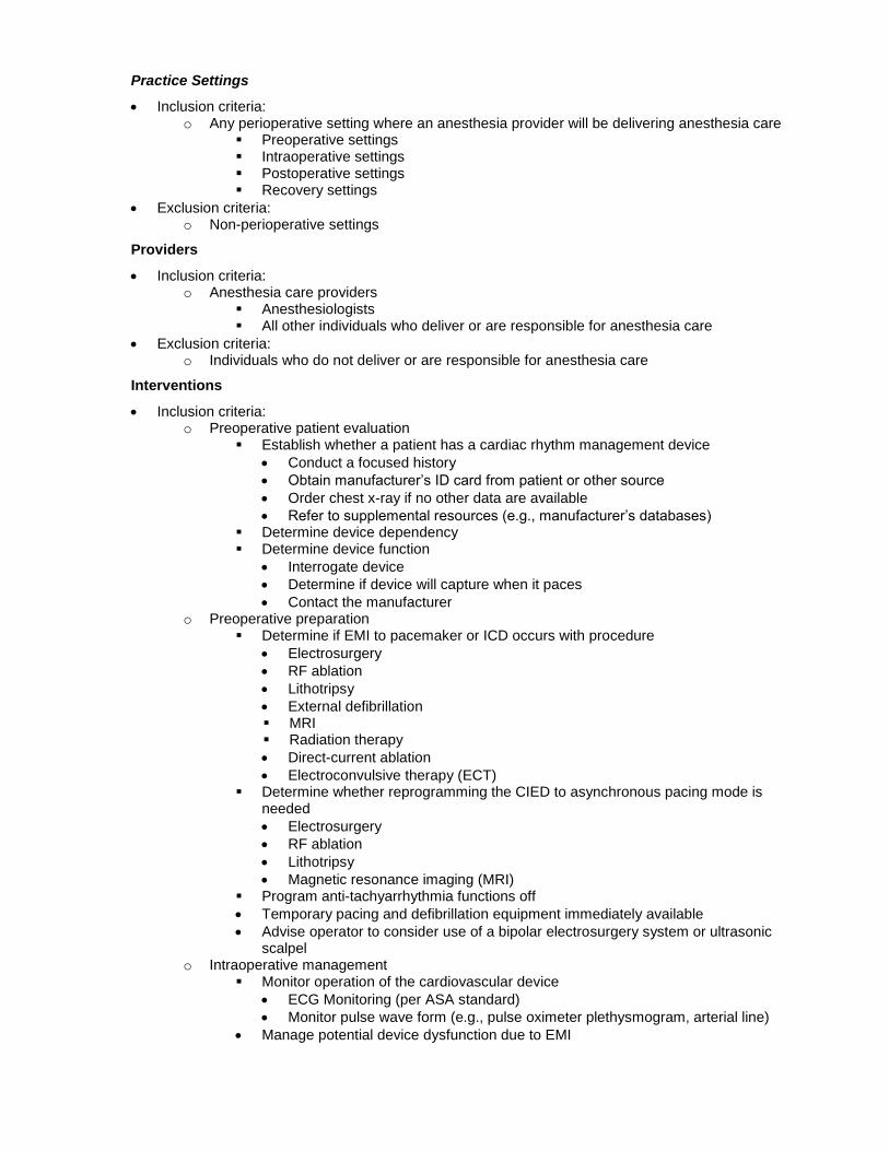
Practice Settings
• Inclusion criteria: o Any perioperative setting where an anesthesia provider will be delivering anesthesia care
▪ Preoperative settings ▪ Intraoperative settings ▪ Postoperative settings ▪ Recovery settings
• Exclusion criteria: o Non-perioperative settings
Providers
• Inclusion criteria: o Anesthesia care providers
▪ Anesthesiologists ▪ All other individuals who deliver or are responsible for anesthesia care
• Exclusion criteria: o Individuals who do not deliver or are responsible for anesthesia care
Interventions
• Inclusion criteria: o Preoperative patient evaluation
▪ Establish whether a patient has a cardiac rhythm management device
• Conduct a focused history
• Obtain manufacturer’s ID card from patient or other source
• Order chest x-ray if no other data are available
• Refer to supplemental resources (e.g., manufacturer’s databases) ▪ Determine device dependency ▪ Determine device function
• Interrogate device
• Determine if device will capture when it paces
• Contact the manufacturer o Preoperative preparation
▪ Determine if EMI to pacemaker or ICD occurs with procedure
• Electrosurgery
• RF ablation
• Lithotripsy
• External defibrillation ▪ MRI ▪ Radiation therapy
• Direct-current ablation
• Electroconvulsive therapy (ECT) ▪ Determine whether reprogramming the CIED to asynchronous pacing mode is
needed
• Electrosurgery
• RF ablation
• Lithotripsy
• Magnetic resonance imaging (MRI) ▪ Program anti-tachyarrhythmia functions off
• Temporary pacing and defibrillation equipment immediately available
• Advise operator to consider use of a bipolar electrosurgery system or ultrasonic scalpel
o Intraoperative management ▪ Monitor operation of the cardiovascular device
• ECG Monitoring (per ASA standard)
• Monitor pulse wave form (e.g., pulse oximeter plethysmogram, arterial line)
• Manage potential device dysfunction due to EMI

• Electrosurgery o Position the receiving plate so that the current pathway does not
pass through or near the pacemaker system o Avoid direct contact with the pacemaker or leads o Use short, intermittent and irregular bursts at the lowest feasible
energy levels o Use a bipolar electrosurgery system (a bipolar electrosurgery system
can be safely used without affecting a pacemaker or ICD) o Use an ultrasonic (harmonic) scalpel (an ultrasonic scalpel can be
safely used without affecting a pacemaker or ICD)
• Radiofrequency (RF) ablation o Keep the current path (electrode tip to ground plate) as far away
from the pulse generator and lead system as possible o Avoid proximity of the ablation catheter to the leads (intercardiac
ablative procedures)
• Lithotripsy o Avoid focusing the lithotripsy beam near the pulse generator
• Magnetic resonance imaging
• Move the patient outside of the immediate MRI area when the use of an external defibrillator/monitor, CIED programming system, or any other MRI-unsafe equipment is required.
• Monitor the patient’s ECG and/or SpO2 continuously throughout the MRI.
• An individual capable of programming the CIED should remain in attendance for the duration of the MRI.
MRI Conditional CIEDs
• Prior to the MRI, interrogate the CIED and program to "MRI Mode" to suspend the antitachycardia function for an ICD.
• Alter the pacing function of the CIED to an asynchronous pacing mode in the pacing dependent patient.
• After the MRI is completed, re-interrogate the CIED and restore its permanent settings.
MRI Non-Conditional CIEDs
• Interrogate the CIED prior to and after the MRI is completed.
• Alter the pacing function of the CIED to an asynchronous pacing mode in the pacing dependent patient
• Suspend the antitachycardia function of an ICD, if present.
• After the MRI is completed, re-interrogate the CIED and restore its permanent settings.
• Radiation therapy
• Avoid exposing the CIED to radiation whenever possible. o Position the CIED outside the radiation field. o Shield the CIED from direct radiation. o Relocate the generator to the patient’s contralateral side.
• Determine whether the manufacturer recommends verification of CIED function before and at the completion of radiation.
• Radiofrequency identification devices
• Avoid using this equipment in close proximity to the CIED whenever possible.
• Monitor for signs of interference with the CIED and be prepared to stop using the RFID if interference occurs.
• Electroconvulsive therapy
• Alter the pacing function of a CIED to an asynchronous pacing mode in the pacing dependent patient,
• Suspend an ICD’s anti-tachyarrhythmia functions, if present.

• Monitor for and treat ventricular arrhythmias that may occur secondary to the hemodynamic effects of ECT.
o Emergency defibrillation or cardioversion ▪ Patients with an ICD and magnet-disabled therapies:
• Remove the magnet to re-enable antitachycardia therapies
• Terminate all sources of EMI while magnet is removed
• Observe the patient for appropriate CIED therapy ▪ Patients with an ICD and antiarrhythmic therapies that have been disabled by
programming
• Re-enable therapies through programming ▪ Minimize the current flowing through the pulse generator and lead system
• Position defibrillation/cardioversion pads or paddles as far as possible from the pulse generator
• Use apex – (anterior-) posterior position
• Position current flow between the pads/paddles perpendicular to the major lead axis of the CIED
• Use a clinically appropriate energy output regardless of the device o Postoperative management
▪ Confirm or restore device function (i.e., in PACU or ICU).
• Interrogate device
• Reprogram device to appropriate settings
• Restore all anti-arrhythmic therapies
• Patients with ICD antitachycardia functions disabled o Continuously monitor cardiac function o Keep defibrillation equipment immediately available until
antitachycardia function has been restored anesthesia Outcomes
• Expected benefits: o Successful procedure o Reduced frequency/severity of adverse outcomes:
▪ Adverse outcomes associated with a CIED device
• Damage to device
• Inability of device to deliver pacing or shocks
• Lead-tissue interface damage
• Changes in pacing behavior
• Electrical reset to the backup pacing mode
• Inappropriate ICD therapies ▪ Adverse clinical outcomes
• Hypotension
• Tachyarrhythmia
• Bradyarrhythmia
• Myocardial tissue damage Evidence collection
• Literature inclusion criteria: o Randomized controlled trials o Prospective nonrandomized comparative studies (e.g., quasi-experimental, cohort) o Retrospective comparative studies (e.g., case-control) o Observational studies (e.g., correlational or descriptive statistics) o Case reports, case series
• Literature exclusion criteria (except to obtain new citations): o Editorials o Literature reviews o Meta-analyses conducted by others o Abstracts greater than 5 years old o Unpublished studies

o Studies in non-peer reviewed journals o Newspaper articles
• Survey evidence: o Expert consultant survey o ASA membership survey o Other participating organization surveys o Reliability survey o Feasibility survey
State of the Literature
For the systematic review, potentially relevant clinical studies were identified via electronic and
manual searches. Healthcare database searches included PubMed, EMBASE, Web of Science, Google
Books, and the Cochrane Central Register of Controlled Trials. The searches covered a 9.5-yr period
from January 1, 2010, through July 1, 2019. Accepted studies from the previous advisory were also re-
reviewed, covering the period of January 1, 1990, through July 31, 2010. Only studies containing original
findings from peer-reviewed journals were acceptable. Editorials, letters, and other articles without data
were excluded. A literature search strategy and PRISMA********** flow diagram are available as
Supplemental Digital Content 2, http://links.lww.com/ALN/XXXXXXXX.
In total, 1143 new citations were identified, with 810 articles assessed for eligibility. After review, 746
were excluded, with 24 new studies meeting the above stated criteria. These studies were combined with
40 pre-2010 articles used in the previous advisory and 8 provided by task force members, resulting in a
total of 72 articles accepted as evidence for these guidelines. In this document, 63 peer-reviewed articles,
2 ASA Standards and 1 ASA practice advisory are referenced, with a complete bibliography of articles
used to develop these guidelines, organized by section, available as Supplemental Digital Content 3,
http://links.lww.com/ALN/XXXXXXXX.
Each pertinent outcome reported in a study was classified by evidence category and level and
designated as beneficial, harmful, or equivocal. Findings were then summarized for each evidence
linkage and reported in the text of the updated Advisory, with evidence tables available as Supplemental
Digital Content 4, http://links.lww.com/ALN/XXXXXX.
Evidence categories refer specifically to the strength and quality of the research design of the studies.
Category A evidence represents results obtained from randomized controlled trials (RCTs), and category
B evidence represents observational results obtained from nonrandomized study designs or RCTs
without pertinent comparison groups. When available, category A evidence is given precedence over
category B evidence for any particular outcome. These evidence categories are further divided into
evidence levels. Evidence levels refer specifically to the strength and quality of the summarized study
findings (i.e., statistical findings, type of data, and the number of studies reporting/replicating the findings).
In this document, only the highest level of evidence is included in the summary report for each
intervention—outcome pair, including a directional designation of benefit, harm, or equivocality.
********** Preferred reporting items of systematic reviews and meta-analyses

Category A: RCTs report comparative findings between clinical interventions for specified outcomes.
Statistically significant (P < 0.01) outcomes are designated as either beneficial (B) or harmful (H) for the
patient; statistically nonsignificant findings are designated as equivocal (E).
Level 1: The literature contains a sufficient number of RCTs to conduct meta-analysis, ††††††††††
and meta-analytic findings from these aggregated studies are reported as evidence.
Level 2: The literature contains multiple RCTs, but the number of RCTs is not sufficient to
conduct a viable meta-analysis for the purpose of these Guidelines. Findings from these
RCTs are reported separately as evidence.
Level 3: The literature contains a single RCT, and findings from this study are reported as
evidence.
Category B: Observational studies or RCTs without pertinent comparison groups may permit
inference of beneficial or harmful relationships among clinical interventions and clinical outcomes.
Inferred findings are given a directional designation of beneficial (B), harmful (H), or equivocal (E). For
studies that report statistical findings, the threshold for significance is P < 0.01.
Level 1: The literature contains nonrandomized comparisons (e.g., quasiexperimental, cohort
[prospective or retrospective], or case-control research designs) with comparative
statistics between clinical interventions for a specified clinical outcome.
Level 2: The literature contains noncomparative observational studies with associative statistics
(e.g., correlation, sensitivity, and specificity).
Level 3: The literature contains noncomparative observational studies with descriptive statistics
(e.g., frequencies, percentages).
Level 4: The literature contains case reports.
Insufficient Literature. The lack of sufficient scientific evidence in the literature may occur when the
evidence is either unavailable (i.e., no pertinent studies found) or inadequate. Inadequate literature
cannot be used to assess relationships among clinical interventions and outcomes because a clear
interpretation of findings is not obtained due to methodological concerns (e.g., confounding of study
design or implementation) or the study does not meet the criteria for content as defined in the “Focus” of
the guidelines.
Although interobserver agreement among task force members and two methodologists was not
assessed for this update, the original Guidelines reported agreement levels using a κ statistic for two-rater
agreement pairs as follows: (1) type of study design, κ = 0.72 to 0.90; (2) type of analysis, k = 0.80 to
0.90; (3) evidence linkage assignment, κ = 0.84 to 1.00; and (4) literature inclusion for database, κ = 0.70
to 1.00. Three-rater agreement values were as follows: (1) study design, Sav = 0.81, Var (Sav) = 0.010;
(2) type of analysis, Sav = 0.86, Var (Sav) = 0.009; (3) linkage assignment, Sav = 0.82 Var (Sav) = 0.005;
and (4) literature database inclusion Sav=0.78 Var (Sav) = 0.031. These values represent moderate to-
high levels of agreement.
†††††††††† All meta-analyses are conducted by the ASA methodology group. Meta-analyses from other sources are reviewed but not included as evidence in this document. A minimum of five independent RCTs (i.e., sufficient for fitting a random-effects model253) is required for meta-analysis.
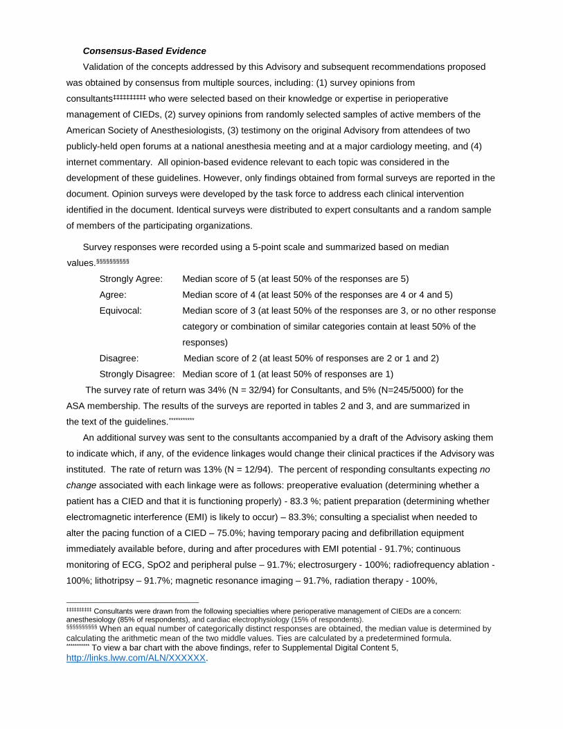
Consensus-Based Evidence
Validation of the concepts addressed by this Advisory and subsequent recommendations proposed
was obtained by consensus from multiple sources, including: (1) survey opinions from
consultants‡‡‡‡‡‡‡‡‡‡ who were selected based on their knowledge or expertise in perioperative
management of CIEDs, (2) survey opinions from randomly selected samples of active members of the
American Society of Anesthesiologists, (3) testimony on the original Advisory from attendees of two
publicly-held open forums at a national anesthesia meeting and at a major cardiology meeting, and (4)
internet commentary. All opinion-based evidence relevant to each topic was considered in the
development of these guidelines. However, only findings obtained from formal surveys are reported in the
document. Opinion surveys were developed by the task force to address each clinical intervention
identified in the document. Identical surveys were distributed to expert consultants and a random sample
of members of the participating organizations.
Survey responses were recorded using a 5-point scale and summarized based on median
values.§§§§§§§§§§
Strongly Agree: Median score of 5 (at least 50% of the responses are 5)
Agree: Median score of 4 (at least 50% of the responses are 4 or 4 and 5)
Equivocal: Median score of 3 (at least 50% of the responses are 3, or no other response
category or combination of similar categories contain at least 50% of the
responses)
Disagree: Median score of 2 (at least 50% of responses are 2 or 1 and 2)
Strongly Disagree: Median score of 1 (at least 50% of responses are 1)
The survey rate of return was 34% (N = 32/94) for Consultants, and 5% (N=245/5000) for the
ASA membership. The results of the surveys are reported in tables 2 and 3, and are summarized in
the text of the guidelines.***********
An additional survey was sent to the consultants accompanied by a draft of the Advisory asking them
to indicate which, if any, of the evidence linkages would change their clinical practices if the Advisory was
instituted. The rate of return was 13% (N = 12/94). The percent of responding consultants expecting no
change associated with each linkage were as follows: preoperative evaluation (determining whether a
patient has a CIED and that it is functioning properly) - 83.3 %; patient preparation (determining whether
electromagnetic interference (EMI) is likely to occur) – 83.3%; consulting a specialist when needed to
alter the pacing function of a CIED – 75.0%; having temporary pacing and defibrillation equipment
immediately available before, during and after procedures with EMI potential - 91.7%; continuous
monitoring of ECG, SpO2 and peripheral pulse – 91.7%; electrosurgery - 100%; radiofrequency ablation -
100%; lithotripsy – 91.7%; magnetic resonance imaging – 91.7%, radiation therapy - 100%,
‡‡‡‡‡‡‡‡‡‡ Consultants were drawn from the following specialties where perioperative management of CIEDs are a concern: anesthesiology (85% of respondents), and cardiac electrophysiology (15% of respondents). §§§§§§§§§§ When an equal number of categorically distinct responses are obtained, the median value is determined by calculating the arithmetic mean of the two middle values. Ties are calculated by a predetermined formula. *********** To view a bar chart with the above findings, refer to Supplemental Digital Content 5,
http://links.lww.com/ALN/XXXXXX.

Radiofrequency identification devices - 100%, electroconvulsive therapy - 100%, emergency defibrillation
or cardioversion - 91.7%; postoperative management (continuing to monitor and display ECG, cardiac
rate and rhythm) - 100%, postoperative management (for a CIED that was reprogrammed pre-or-
intraoperatively, restore the CIED to its permanent settings before the patient is discharged from a
monitored environment) – 83.3%, and postoperative CIED interrogation– 91.7%. Sixty-seven percent of
the respondents indicated that the Advisory would have no effect on the amount of time spent on a typical
case with the implementation of this Advisory. Twenty-five percent of respondents indicated that there
would be an increase and 8.3% indicated that there would be a decrease.
Research Support
Support was provided solely by the American Society of Anesthesiologists.
Competing Interests
The authors declare no competing interests.
Correspondence
Address correspondence to the American Society of Anesthesiologists: 1061 American Lane,
Schaumburg, Illinois 60173. These updated Practice Advisories, and all ASA Practice Parameters, may
be obtained at no cost through the Journal Web site, www.anesthesiology.org.
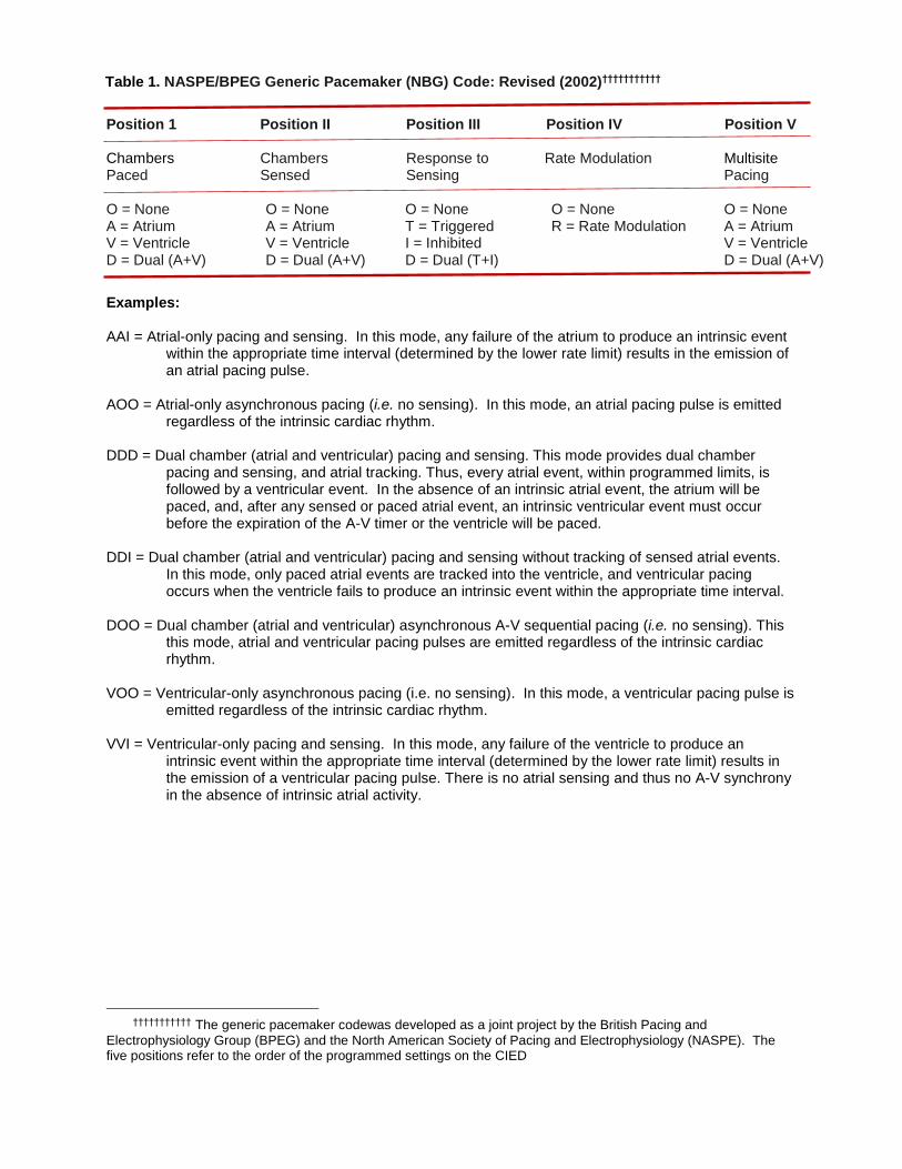
Table 1. NASPE/BPEG Generic Pacemaker (NBG) Code: Revised (2002)†††††††††††
Position 1 Position II Position III Position IV Position V Chambers Chambers Response to Rate Modulation Multisite Paced Sensed Sensing Pacing O = None O = None O = None O = None O = None A = Atrium A = Atrium T = Triggered R = Rate Modulation A = Atrium V = Ventricle V = Ventricle I = Inhibited V = Ventricle D = Dual (A+V) D = Dual (A+V) D = Dual (T+I) D = Dual (A+V) Examples: AAI = Atrial-only pacing and sensing. In this mode, any failure of the atrium to produce an intrinsic event
within the appropriate time interval (determined by the lower rate limit) results in the emission of an atrial pacing pulse.
AOO = Atrial-only asynchronous pacing (i.e. no sensing). In this mode, an atrial pacing pulse is emitted
regardless of the intrinsic cardiac rhythm. DDD = Dual chamber (atrial and ventricular) pacing and sensing. This mode provides dual chamber
pacing and sensing, and atrial tracking. Thus, every atrial event, within programmed limits, is followed by a ventricular event. In the absence of an intrinsic atrial event, the atrium will be paced, and, after any sensed or paced atrial event, an intrinsic ventricular event must occur before the expiration of the A-V timer or the ventricle will be paced.
DDI = Dual chamber (atrial and ventricular) pacing and sensing without tracking of sensed atrial events.
In this mode, only paced atrial events are tracked into the ventricle, and ventricular pacing occurs when the ventricle fails to produce an intrinsic event within the appropriate time interval.
DOO = Dual chamber (atrial and ventricular) asynchronous A-V sequential pacing (i.e. no sensing). This
this mode, atrial and ventricular pacing pulses are emitted regardless of the intrinsic cardiac rhythm.
VOO = Ventricular-only asynchronous pacing (i.e. no sensing). In this mode, a ventricular pacing pulse is
emitted regardless of the intrinsic cardiac rhythm. VVI = Ventricular-only pacing and sensing. In this mode, any failure of the ventricle to produce an
intrinsic event within the appropriate time interval (determined by the lower rate limit) results in the emission of a ventricular pacing pulse. There is no atrial sensing and thus no A-V synchrony in the absence of intrinsic atrial activity.
††††††††††† The generic pacemaker codewas developed as a joint project by the British Pacing and
Electrophysiology Group (BPEG) and the North American Society of Pacing and Electrophysiology (NASPE). The five positions refer to the order of the programmed settings on the CIED

Table 2. NASPE / BPEG Generic Defibrillator (NBD) Code‡‡‡‡‡‡‡‡‡‡‡
Position 1 Position II Position III Position IV Shock Antitachycardia Tachycardia Antibradycardia Chambers Pacing Chambers Detection Pacing Chambers O = None O = None E = Electrocardiogram O = None A = Atrium A = Atrium H = Hemodynamic A = Atrium V = Ventricle V = Ventricle V = Ventricle D = Dual (A+V) D = Dual (A+V) D = Dual (AV)
For robust identification, Position IV is expanded into its complete NBG code. For example, a
biventricular cardioverter-defibrillator with ventricular shock and antitachycardia pacing functionality would
be identified as VVE-DDDRV, assuming it was programmed to pace and sense in the DDD mode with
rate response. Currently, no hemodynamic sensors have been approved for tachycardia detection
(Position III).
‡‡‡‡‡‡‡‡‡‡‡ The generic defibrillator code was developed as a joint project by the British Pacing and
Electrophysiology Group (BPEG) and the North American Society of Pacing and Electrophysiology (NASPE). The five positions refer to the order of the programmed settings on the CIED
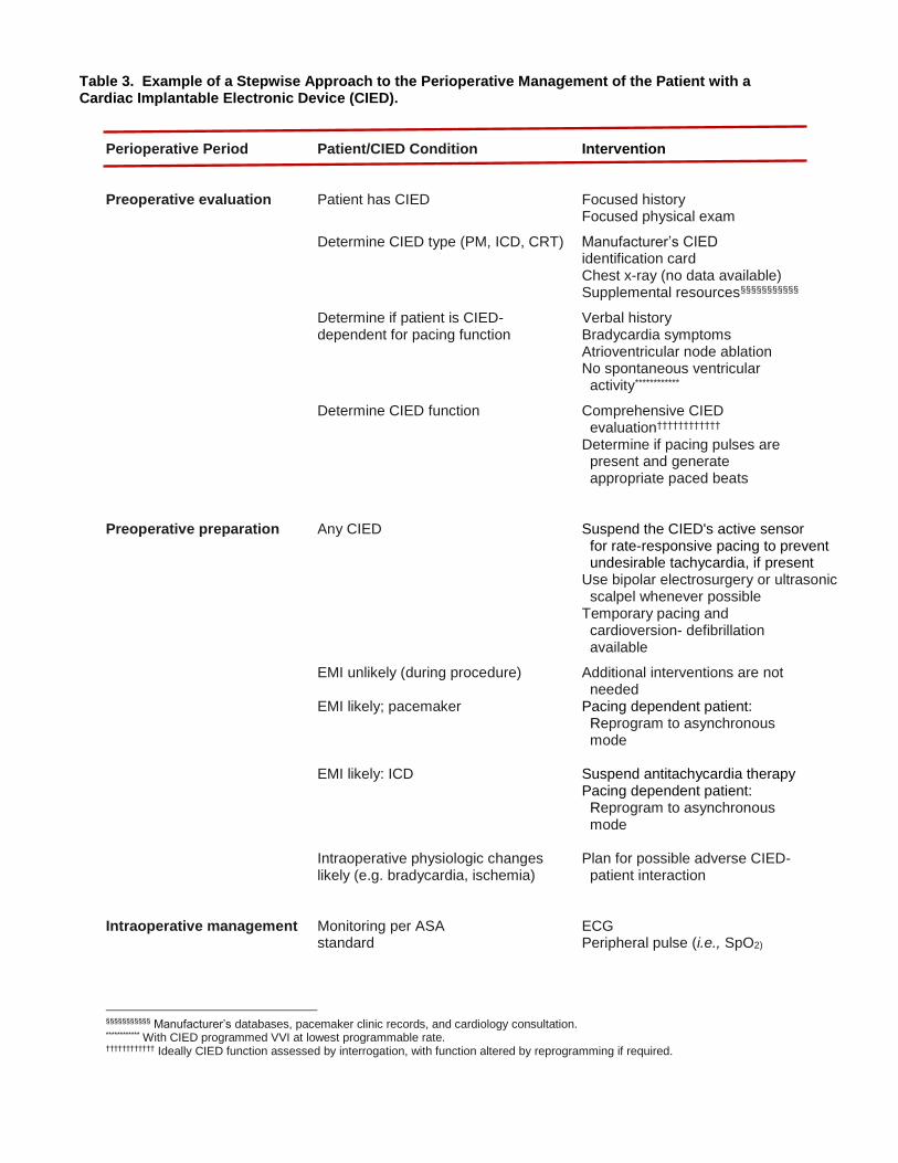
Table 3. Example of a Stepwise Approach to the Perioperative Management of the Patient with a Cardiac Implantable Electronic Device (CIED).
Perioperative Period Patient/CIED Condition Intervention Preoperative evaluation Patient has CIED Focused history Focused physical exam
Determine CIED type (PM, ICD, CRT) Manufacturer’s CIED identification card Chest x-ray (no data available) Supplemental resources§§§§§§§§§§§
Determine if patient is CIED- Verbal history dependent for pacing function Bradycardia symptoms Atrioventricular node ablation No spontaneous ventricular activity************
Determine CIED function Comprehensive CIED evaluation††††††††††††
Determine if pacing pulses are present and generate appropriate paced beats Preoperative preparation Any CIED Suspend the CIED's active sensor
for rate-responsive pacing to prevent undesirable tachycardia, if present Use bipolar electrosurgery or ultrasonic scalpel whenever possible
Temporary pacing and cardioversion- defibrillation available
EMI unlikely (during procedure) Additional interventions are not needed EMI likely; pacemaker Pacing dependent patient: Reprogram to asynchronous mode EMI likely: ICD Suspend antitachycardia therapy Pacing dependent patient: Reprogram to asynchronous mode Intraoperative physiologic changes Plan for possible adverse CIED- likely (e.g. bradycardia, ischemia) patient interaction Intraoperative management Monitoring per ASA ECG standard Peripheral pulse (i.e., SpO2)
§§§§§§§§§§§ Manufacturer’s databases, pacemaker clinic records, and cardiology consultation. ************ With CIED programmed VVI at lowest programmable rate. †††††††††††† Ideally CIED function assessed by interrogation, with function altered by reprogramming if required.

Electrosurgery Direct current return path away from pulse generator and leads Avoid proximity of ESU to pulse generator / leads Short bursts at lowest possible energy Use bipolar electrosurgery or ultrasonic scalpel whenever possible RF catheter ablation Avoid contact RF catheter with pulse generator / leads RF current path far away from pulse generator / leads Discuss these concerns with operator
Lithotripsy Do not focus lithotripsy beam near generator
MRI Move the patient outside of the immediate MRI area when the use of an external defibrillator/monitor, CIED programmer or any other MRI- unsafe equipment is required. Before the MRI, perform the following: Interrogate the CIED. Suspend the antitachycardia function of an ICD, if present. For MRI Conditional ICDs , program to "MRI Mode" to suspend the antitachycardia function In the pacing dependent patient, alter the pacing function of the CIED to an asynchronous pacing mode. Assure that an individual capable of programming the CIED remains in attendance for the duration of the MRI. After the MRI is completed, re- interrogate the CIED and restore its permanent settings. Radiation therapy Pulse generator / leads must be outside of RT field Possible surgical relocation of pulse generator Verify PG function during/after radiation therapy course ECT Alter the pacing function to an asynchronous pacing mode in the pacing dependent patient Suspend an ICD’s antitachycardia functions, if present Monitor for and treat ventricular arrhythmias that may occur secondary to the hemodynamic effects of ECT
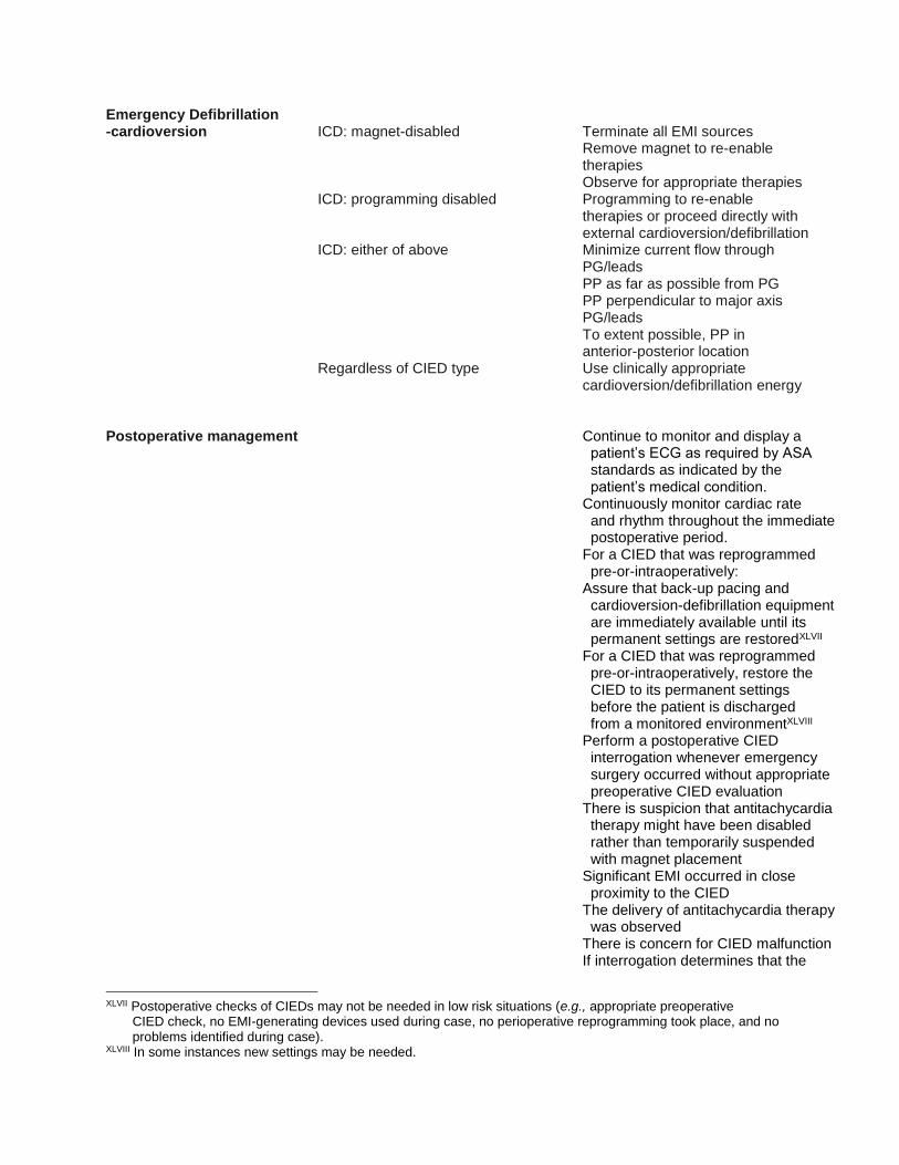
Emergency Defibrillation -cardioversion ICD: magnet-disabled Terminate all EMI sources Remove magnet to re-enable therapies Observe for appropriate therapies ICD: programming disabled Programming to re-enable therapies or proceed directly with external cardioversion/defibrillation ICD: either of above Minimize current flow through PG/leads PP as far as possible from PG PP perpendicular to major axis PG/leads To extent possible, PP in anterior-posterior location Regardless of CIED type Use clinically appropriate cardioversion/defibrillation energy Postoperative management Continue to monitor and display a patient’s ECG as required by ASA standards as indicated by the patient’s medical condition. Continuously monitor cardiac rate and rhythm throughout the immediate postoperative period. For a CIED that was reprogrammed pre-or-intraoperatively: Assure that back-up pacing and cardioversion-defibrillation equipment are immediately available until its permanent settings are restoredXLVII For a CIED that was reprogrammed pre-or-intraoperatively, restore the CIED to its permanent settings before the patient is discharged from a monitored environmentXLVIII Perform a postoperative CIED interrogation whenever emergency surgery occurred without appropriate preoperative CIED evaluation There is suspicion that antitachycardia therapy might have been disabled rather than temporarily suspended with magnet placement Significant EMI occurred in close proximity to the CIED The delivery of antitachycardia therapy was observed There is concern for CIED malfunction If interrogation determines that the
XLVII Postoperative checks of CIEDs may not be needed in low risk situations (e.g., appropriate preoperative
CIED check, no EMI-generating devices used during case, no perioperative reprogramming took place, and no problems identified during case).
XLVIII In some instances new settings may be needed.

CIED settings are inappropriate, reprogram to newly appropriate settings______________________________________________________________________________________ CIED: cardiac implantable electronic device; DE: dispersive electrode; CRT: cardiac resynchronization therapy; ESU: electrosurgical unit; ECT: electroconvulsive therapy; EMI: electromagnetic interference; ICD: implantable cardioverter-defibrillator; MRI: magnetic resonance imaging; PG: pulse generator; PP: defibrillation or cardioversion pads; R&R: rhythm and rate; RT: radiation therapy.

Table 4. Expert Consultant Survey Results*************
Recommendations N Strongly Agree (%)
Agree (%)
Equivocal (%)
Disagree (%)
Strongly Disagree (%)
Preoperative Evaluation
1. Determine whether a patient has a CIED. 32 100* 0 0 0 0 2. Determine the CIED type (i.e. PM, ICD, CRT). 32 97* 3 0 0 0 3. Determine the CIED manufacturer. 32 66* 28 6 0 0 4. Determine the primary indication for CIED placement. 32 69* 28 3 0 0 5. Determine whether the patient is pacing dependent. 32 91* 9 0 0 0
Preoperative Preparation
6. Determine the CIED’s current settings. 32 63* 31 6 0 0 7. Confirm that the CIED is functioning properly (i.e. by interrogating the CIED or obtaining the most recent interrogation report.)
32 72* 22 6 0 0
8. Determine whether intraoperative electromagnetic interference (EMI) from monopolar electrosurgery or other sources is likely to occur.
32 81* 19 0 0 0
9. If monopolar electrosurgery ("bovie") use is planned superior to the umbilicus, alter the pacing function of a CIED to an asynchronous pacing mode in the pacing dependent patient.
32 63* 22 13 3 0
10. If monopolar electrosurgery ("bovie") use is planned inferior to the umbilicus, alter the pacing function of a CIED to an asynchronous pacing in the patient is pacing dependent.
32 34 6 25 34* 28
11. If monopolar electrosurgery ("bovie") use is planned superior to the umbilicus, suspend an ICD’s anti-tachycardia function, if present. 32 78* 19 3 0 0 12. If monopolar electrosurgery ("bovie") use is planned inferior to the umbilicus, suspend an ICD’s anti-tachycardia function, if present. 31 23 29* 13 23 13 13. Before suspending the anti-tachycardia function of an ICD, assure that the patient is in a monitored environment. 32 75* 25 0 0 0 14. Avoid the routine use of a magnet over an ICD. 32 16 31 9* 25 19 15. If needed, consult a specialist to alter the pacing function of a CIED or suspend the antitachycardia function of an ICD. 32 81* 9 6 3 0 16. Advise the proceduralist to use bipolar electrosurgery or an ultrasonic scalpel when feasible. 32 25 41* 31 3 0 17. Assure that temporary pacing and defibrillation equipment are immediately available before, during, and after all procedures with EMI potential. 32 72* 25 3 0 0 18. Suspend the CIED's active sensor for rate-responsive pacing to prevent undesirable tachycardia. 32 28 44* 22 6 0
Intraoperative Monitoring
19. Continuously monitor and display a patient’s ECG as required by ASA standards, from the beginning of anesthesia until the patient is transferred out of the anesthetizing location, with additional ECG monitoring in the postoperative period as indicated by the patient’s medical condition.
32 91* 9 0 0 0
20. Perform continuous peripheral pulse monitoring for all CIED patients receiving anesthesia care. 32 69* 13 9 3 6 21. If unanticipated CIED interactions occur, discontinue the procedure until the source of interference can be eliminated or managed.
32 56* 34 6 3 0
************* N = number of members who responded to each item. An asterisk beside a percentage score indicates the median.
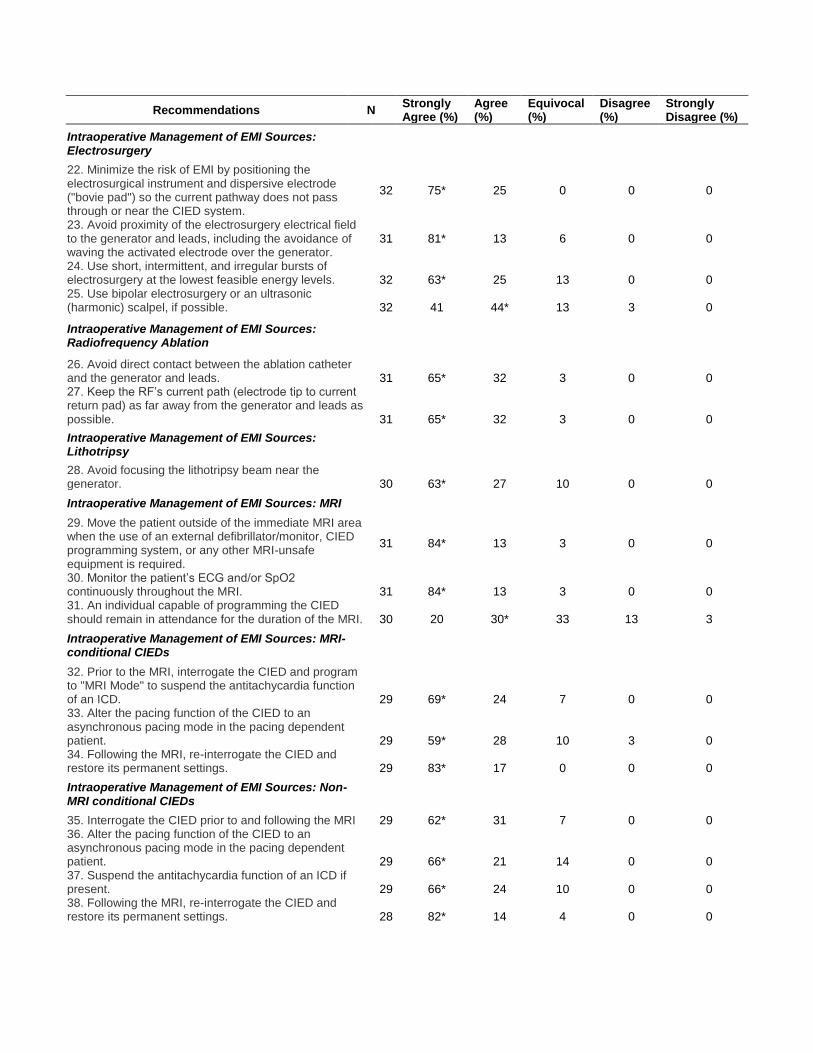
Recommendations N Strongly Agree (%)
Agree (%)
Equivocal (%)
Disagree (%)
Strongly Disagree (%)
Intraoperative Management of EMI Sources: Electrosurgery
22. Minimize the risk of EMI by positioning the electrosurgical instrument and dispersive electrode ("bovie pad") so the current pathway does not pass through or near the CIED system.
32 75* 25 0 0 0
23. Avoid proximity of the electrosurgery electrical field to the generator and leads, including the avoidance of waving the activated electrode over the generator.
31 81* 13 6 0 0
24. Use short, intermittent, and irregular bursts of electrosurgery at the lowest feasible energy levels. 32 63* 25 13 0 0 25. Use bipolar electrosurgery or an ultrasonic (harmonic) scalpel, if possible. 32 41 44* 13 3 0
Intraoperative Management of EMI Sources: Radiofrequency Ablation
26. Avoid direct contact between the ablation catheter and the generator and leads. 31 65* 32 3 0 0 27. Keep the RF’s current path (electrode tip to current return pad) as far away from the generator and leads as possible. 31 65* 32 3 0 0
Intraoperative Management of EMI Sources: Lithotripsy
28. Avoid focusing the lithotripsy beam near the generator. 30 63* 27 10 0 0
Intraoperative Management of EMI Sources: MRI
29. Move the patient outside of the immediate MRI area when the use of an external defibrillator/monitor, CIED programming system, or any other MRI-unsafe equipment is required.
31 84* 13 3 0 0
30. Monitor the patient’s ECG and/or SpO2 continuously throughout the MRI. 31 84* 13 3 0 0 31. An individual capable of programming the CIED should remain in attendance for the duration of the MRI. 30 20 30* 33 13 3
Intraoperative Management of EMI Sources: MRI-conditional CIEDs
32. Prior to the MRI, interrogate the CIED and program to "MRI Mode" to suspend the antitachycardia function of an ICD. 29 69* 24 7 0 0 33. Alter the pacing function of the CIED to an asynchronous pacing mode in the pacing dependent patient. 29 59* 28 10 3 0 34. Following the MRI, re-interrogate the CIED and restore its permanent settings. 29 83* 17 0 0 0
Intraoperative Management of EMI Sources: Non-MRI conditional CIEDs
35. Interrogate the CIED prior to and following the MRI 29 62* 31 7 0 0 36. Alter the pacing function of the CIED to an asynchronous pacing mode in the pacing dependent patient. 29 66* 21 14 0 0 37. Suspend the antitachycardia function of an ICD if present. 29 66* 24 10 0 0 38. Following the MRI, re-interrogate the CIED and restore its permanent settings. 28 82* 14 4 0 0

Recommendations N Strongly Agree (%)
Agree (%)
Equivocal (%)
Disagree (%)
Strongly Disagree (%)
Intraoperative Management of EMI Sources: Radiation Therapy
39. Avoid exposing the CIED to radiation whenever possible by positioning the CIED outside the radiation field, shielding the CIED from direct radiation, and relocating the generator to the patient’s contralateral side.
29 55* 17 24 3 0
40. Determine whether the manufacturer recommends verification of CIED function before and at the completion of radiation. 29 59* 31 10 0 0
Intraoperative Management of EMI Sources: Radiofrequency Identification Devices (RFID)
41. Avoid using this equipment in close proximity to the CIED whenever possible. 29 52* 31 14 0 3 42. Monitor for signs of interference with the CIED and be prepared to stop using the RFID if interference occurs. 28 71* 21 4 0 4
Intraoperative Management of EMI Sources: Electroconvulsive Therapy (ECT)
43. Alter the pacing function of a CIED to an asynchronous pacing mode in the pacing dependent patient. 27 33 33* 22 7 4 44. Suspend an ICD’s anti-tachyarrhythmia functions, if present. 27 48 41* 7 4 0 45. Monitor for and treat ventricular arrhythmias that may occur secondary to the hemodynamic effects of ECT. 28 89* 7 4 0 0
Intraoperative Management: Emergency External Defibrillation or Cardioversion
46. Before performing emergency defibrillation or cardioversion of the patient with an ICD and magnet-disabled therapies, terminate all sources of EMI and remove the magnet to re-enable the ICD’s antitachycardia therapies, then observe the patient for the delivery of appropriate antitachycardia therapy from the ICD.
28 54* 32 4 7 4
47. For the patient with an ICD and antitachycardia therapy that have been disabled by programming, determine whether the antitachycardia therapy should be re-enabled.
28 43 32* 7 18 0
48. If the above activities fail to restore ICD function perform, emergency external defibrillation or cardioversion when needed. 28 89* 7 4 0 0 49. Follow ACLS and emergency protocols to provide rapid cardioversion or defibrillation when needed. 28 86* 14 0 0 0 50. Follow ACLS guidelines for delivered energy level and pad placement. 28 75* 21 0 4 0 51. Attempt to minimize the current flowing through the pulse generator and leads by positioning the defibrillation or cardioversion pads so they are not directly over the CIED. 28 75* 21 4 0 0 52. Use anterior-posterior rather than anterior-lateral pad positioning whenever possible. 27 52* 22 22 4 0 53. Use a clinically appropriate energy output regardless of the presence of the CIED. 28 79* 21 0 0 0 54. Interrogate the CIED immediately after external cardioversion or defibrillation is performed. 27 67* 22 11 0 0
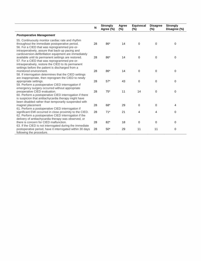
N Strongly Agree (%)
Agree (%)
Equivocal (%)
Disagree (%)
Strongly Disagree (%)
Postoperative Management
55. Continuously monitor cardiac rate and rhythm throughout the immediate postoperative period. 28 86* 14 0 0 0 56. For a CIED that was reprogrammed pre-or-intraoperatively, assure that back-up pacing and cardioversion-defibrillation equipment are immediately available until its permanent settings are restored. 28 86* 14 0 0 0 57. For a CIED that was reprogrammed pre-or-intraoperatively, restore the CIED to its permanent settings before the patient is discharged from a monitored environment. 28 86* 14 0 0 0 58. If interrogation determines that the CIED settings are inappropriate, then reprogram the CIED to newly appropriate settings. 28 57* 43 0 0 0 59. Perform a postoperative CIED interrogation if emergency surgery occurred without appropriate preoperative CIED evaluation. 28 75* 11 14 0 0 60. Perform a postoperative CIED interrogation if there is suspicion that antitachycardia therapy might have been disabled rather than temporarily suspended with magnet placement 28 68* 29 0 0 4 61. Perform a postoperative CIED interrogation if significant EMI occurred in close proximity to the CIED. 28 71* 21 4 4 0 62. Perform a postoperative CIED interrogation if the delivery of antitachycardia therapy was observed, or there is concern for CIED malfunction. 28 82* 18 0 0 0 63. If the CIED is not interrogated during the immediate postoperative period, have it interrogated within 30 days following the procedure.
28 50* 29 11 11 0
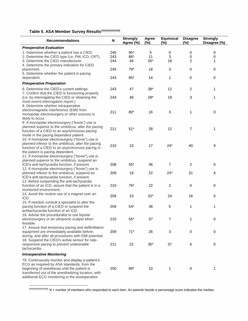
Table 5. ASA Member Survey Results†††††††††††††
Recommendations N Strongly Agree (%)
Agree (%)
Equivocal (%)
Disagree (%)
Strongly Disagree (%)
Preoperative Evaluation
1. Determine whether a patient has a CIED. 245 95* 5 0 0 0 2. Determine the CIED type (i.e. PM, ICD, CRT). 243 86* 11 3 0 0 3. Determine the CIED manufacturer. 244 44 35* 18 2 1 4. Determine the primary indication for CIED placement. 245 79* 18 3 0 0 5. Determine whether the patient is pacing dependent. 243 85* 14 1 0 0
Preoperative Preparation
6. Determine the CIED’s current settings. 243 47 38* 12 2 1 7. Confirm that the CIED is functioning properly (i.e. by interrogating the CIED or obtaining the most recent interrogation report.)
243 49 28* 19 3 1
8. Determine whether intraoperative electromagnetic interference (EMI) from monopolar electrosurgery or other sources is likely to occur.
211 80* 16 3 1 0
9. If monopolar electrosurgery ("bovie") use is planned superior to the umbilicus, alter the pacing function of a CIED to an asynchronous pacing mode in the pacing dependent patient.
211 51* 28 12 7 1
10. If monopolar electrosurgery ("bovie") use is planned inferior to the umbilicus, alter the pacing function of a CIED to an asynchronous pacing in the patient is pacing dependent.
210 10 17 24* 40 9
11. If monopolar electrosurgery ("bovie") use is planned superior to the umbilicus, suspend an ICD’s anti-tachycardia function, if present. 208 55* 36 7 2 0 12. If monopolar electrosurgery ("bovie") use is planned inferior to the umbilicus, suspend an ICD’s anti-tachycardia function, if present.
209 19 22 24* 31 4
13. Before suspending the anti-tachycardia function of an ICD, assure that the patient is in a monitored environment.
210 76* 22 2 0 0
14. Avoid the routine use of a magnet over an ICD.
209 23 32* 24 16 5
15. If needed, consult a specialist to alter the pacing function of a CIED or suspend the antitachycardia function of an ICD.
209 54* 38 5 1 1
16. Advise the proceduralist to use bipolar electrosurgery or an ultrasonic scalpel when feasible.
210 55* 37 7 1 0
17. Assure that temporary pacing and defibrillation equipment are immediately available before, during, and after all procedures with EMI potential.
209 71* 26 3 0 0
18. Suspend the CIED's active sensor for rate-responsive pacing to prevent undesirable tachycardia.
211 22 35* 37 6 0
Intraoperative Monitoring
19. Continuously monitor and display a patient’s ECG as required by ASA standards, from the beginning of anesthesia until the patient is transferred out of the anesthetizing location, with additional ECG monitoring in the postoperative
200 89* 10 1 0 1
††††††††††††† N = number of members who responded to each item. An asterisk beside a percentage score indicates the median.
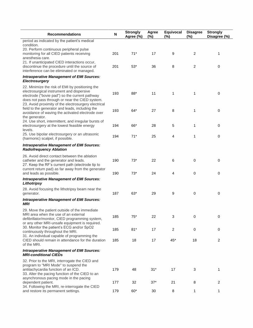
Recommendations N Strongly Agree (%)
Agree (%)
Equivocal (%)
Disagree (%)
Strongly Disagree (%)
period as indicated by the patient’s medical condition. 20. Perform continuous peripheral pulse monitoring for all CIED patients receiving anesthesia care.
201 71* 17 9 2 1
21. If unanticipated CIED interactions occur, discontinue the procedure until the source of interference can be eliminated or managed.
201 53* 36 8 2 0
Intraoperative Management of EMI Sources: Electrosurgery
22. Minimize the risk of EMI by positioning the electrosurgical instrument and dispersive electrode ("bovie pad") so the current pathway does not pass through or near the CIED system.
193 88* 11 1 1 0
23. Avoid proximity of the electrosurgery electrical field to the generator and leads, including the avoidance of waving the activated electrode over the generator.
193 64* 27 8 1 0
24. Use short, intermittent, and irregular bursts of electrosurgery at the lowest feasible energy levels.
194 66* 28 5 1 0
25. Use bipolar electrosurgery or an ultrasonic (harmonic) scalpel, if possible.
194 71* 25 4 1 0
Intraoperative Management of EMI Sources: Radiofrequency Ablation
26. Avoid direct contact between the ablation catheter and the generator and leads. 190 73* 22 6 0 0 27. Keep the RF’s current path (electrode tip to current return pad) as far away from the generator and leads as possible. 190 73* 24 4 0 0
Intraoperative Management of EMI Sources: Lithotripsy
28. Avoid focusing the lithotripsy beam near the generator. 187 63* 29 9 0 0
Intraoperative Management of EMI Sources: MRI
29. Move the patient outside of the immediate MRI area when the use of an external defibrillator/monitor, CIED programming system, or any other MRI-unsafe equipment is required.
185 75* 22 3 0 0
30. Monitor the patient’s ECG and/or SpO2 continuously throughout the MRI.
185 81* 17 2 0 0
31. An individual capable of programming the CIED should remain in attendance for the duration of the MRI.
185 18 17 45* 18 2
Intraoperative Management of EMI Sources: MRI-conditional CIEDs
32. Prior to the MRI, interrogate the CIED and program to "MRI Mode" to suspend the antitachycardia function of an ICD. 179 48 31* 17 3 1 33. Alter the pacing function of the CIED to an asynchronous pacing mode in the pacing dependent patient. 177 32 37* 21 8 2 34. Following the MRI, re-interrogate the CIED and restore its permanent settings. 179 60* 30 8 1 1

Recommendations N Strongly Agree (%)
Agree (%)
Equivocal (%)
Disagree (%)
Strongly Disagree (%)
Intraoperative Management of EMI Sources: Non-MRI conditional CIEDs
35. Interrogate the CIED prior to and following the MRI 172 44 31* 22 2 1 36. Alter the pacing function of the CIED to an asynchronous pacing mode in the pacing dependent patient. 172 38 35* 20 5 2 37. Suspend the antitachycardia function of an ICD if present. 172 41 36* 17 4 2 38. Following the MRI, re-interrogate the CIED and restore its permanent settings. 173 59* 28 12 1 1
Intraoperative Management of EMI Sources: Radiation Therapy
39. Avoid exposing the CIED to radiation whenever possible by positioning the CIED outside the radiation field, shielding the CIED from direct radiation, and relocating the generator to the patient’s contralateral side.
165 31 35* 31 2 1
40. Determine whether the manufacturer recommends verification of CIED function before and at the completion of radiation.
165 42 41* 15 1 1
Intraoperative Management of EMI Sources: Radiofrequency Identification Devices (RFID)
41. Avoid using this equipment in close proximity to the CIED whenever possible.
163 40 39* 17 2 1
42. Monitor for signs of interference with the CIED and be prepared to stop using the RFID if interference occurs.
163 52* 39 8 1 0
Intraoperative Management of EMI Sources: Electroconvulsive Therapy (ECT)
43. Alter the pacing function of a CIED to an asynchronous pacing mode in the pacing dependent patient. 159 33 36* 20 9 1 44. Suspend an ICD’s anti-tachyarrhythmia functions, if present. 157 41 27* 22 10 0 45. Monitor for and treat ventricular arrhythmias that may occur secondary to the hemodynamic effects of ECT. 159 70* 23 7 0 0
Intraoperative Management: Emergency External Defibrillation or Cardioversion
46. Before performing emergency defibrillation or cardioversion of the patient with an ICD and magnet-disabled therapies, terminate all sources of EMI and remove the magnet to re-enable the ICD’s antitachycardia therapies, then observe the patient for the delivery of appropriate antitachycardia therapy from the ICD.
147 57* 30 9 3 1
47. For the patient with an ICD and antitachycardia therapy that have been disabled by programming, determine whether the antitachycardia therapy should be re-enabled.
146 51* 37 8 3 1
48. If the above activities fail to restore ICD function perform, emergency external defibrillation or cardioversion when needed.
146 85* 12 3 0 0

Recommendations N Strongly Agree (%)
Agree (%)
Equivocal (%)
Disagree (%)
Strongly Disagree (%)
49. Follow ACLS and emergency protocols to provide rapid cardioversion or defibrillation when needed.
146 88* 10 2 0 0
50. Follow ACLS guidelines for delivered energy level and pad placement.
146 77* 13 9 0 1
51. Attempt to minimize the current flowing through the pulse generator and leads by positioning the defibrillation or cardioversion pads so they are not directly over the CIED.
147 71* 24 4 0 1
52. Use anterior-posterior rather than anterior-lateral pad positioning whenever possible.
145 48 30* 20 3 0
53. Use a clinically appropriate energy output regardless of the presence of the CIED.
147 60* 33 5 1 0
54. Interrogate the CIED immediately after external cardioversion or defibrillation is performed. 27 67* 22 11 0 0
Postoperative Management
55. Continuously monitor cardiac rate and rhythm throughout the immediate postoperative period. 145 83* 14 2 0 0 56. For a CIED that was reprogrammed pre-or-intraoperatively, assure that back-up pacing and cardioversion-defibrillation equipment are immediately available until its permanent settings are restored. 145 82* 16 2 0 0 57. For a CIED that was reprogrammed pre-or-intraoperatively, restore the CIED to its permanent settings before the patient is discharged from a monitored environment. 145 83* 15 2 0 0 58. If interrogation determines that the CIED settings are inappropriate, then reprogram the CIED to newly appropriate settings. 145 72* 23 5 1 0 59. Perform a postoperative CIED interrogation if emergency surgery occurred without appropriate preoperative CIED evaluation. 145 56* 28 16 1 0 60. Perform a postoperative CIED interrogation if there is suspicion that antitachycardia therapy might have been disabled rather than temporarily suspended with magnet placement 145 77* 21 1 0 0 61. Perform a postoperative CIED interrogation if significant EMI occurred in close proximity to the CIED. 145 51* 34 14 1 0 62. Perform a postoperative CIED interrogation if the delivery of antitachycardia therapy was observed, or there is concern for CIED malfunction. 145 82* 17 1 0 0 63. If the CIED is not interrogated during the immediate postoperative period, have it interrogated within 30 days following the procedure. 145 42 33* 17 6 1