
Chytrid infections of Daphnia pulicaria: development ...
15
Chytrid infections of Daphnia pulicaria: development, ecology, pathology and phylogeny of Polycaryum laeve PIETER T. J. JOHNSON,* JOYCE E. LONGCORE, † DANIEL E. STANTON,* RYAN B. CARNEGIE, ‡ JEFFREY D. SHIELDS ‡ AND ERIC R. PREU* *Center for Limnology, University of Wisconsin, Madison, WI, U.S.A. † Department of Biological Sciences, University of Maine, Orono, ME, U.S.A. ‡ Virginia Institute of Marine Science, College of William and Mary, Gloucester Point, VA, U.S.A. SUMMARY 1. We combined ecological surveys, life table studies, microscopy and molecular sequencing to determine the development, ecology, pathology and phylogeny of Polycaryum laeve, an endoparasite of cladocerans. We report the first records of P. laeve from North America, where we have used a polymerase chain reaction primer and microscopic examination to confirm infections in 14 lakes. Infections are highly pathogenic and caused increased mortality, reduced growth, and reproductive castration in Daphnia pulicaria during life table studies. 2. Biweekly data from Allequash Lake (Wisconsin, U.S.A.) throughout 2003 indicated that fecundity and infection prevalence were inversely correlated. Infection prevalence was highest in late winter and early spring (up to 80%) and lowest during late summer. Epidemics were generally followed by sharp declines in host population density (up to 99%). 3. Within the haemocoel of its host, P. laeve forms thick-walled sporangia, which occur systemically in later stages of infection. Fungal thalli associate closely with muscle fibres and connective tissue, leading to degeneration as the infection becomes advanced. Following death of the host, flagellated zoospores are released through an exit papilla on the sporangium. Based on the infection-induced castration of the host and increases in infection prevalence with Daphnia size, we postulate that transmission is horizontal, but may be indirect through an additional host or free-living stage. 4. Molecular and morphological data clearly indicate that P. laeve belongs in the fungal phylum Chytriodiomycota, order Blastocladiales. Based on ribosomal RNA gene sequences and morphological features, we transfer the genus Polycaryum from the Haplosporidia to the Chytridiomycota, and designate a lectotype and epitype for P. laeve. Considering the high prevalence of P. laeve infection within Daphnia populations, the frequency with which we detected infections among lakes, and the keystone importance of large-bodied Daphnia in aquatic food webs, we suggest that P. laeve may exert a regulatory influence on Daphnia populations in lake ecosystems. Keywords: Blastocladiales, Chytridiomycota, fungus, parasite, zooplankton mortality Introduction Freshwater cladocerans host a variety of ecto- and endoparasites, including viruses, bacteria, algae, microsporidians, haplosporidians, ciliates, fungi, oomycetes and helminths (reviewed in Green, 1974). Although many pathogens have a low prevalence in a Correspondence: Pieter Johnson, Center for Limnology, University of Wisconsin, 680 North Park Street, Madison, WI 53706–1492, U.S.A. E-mail: [email protected] Freshwater Biology (2006) 51, 634–648 doi:10.1111/j.1365-2427.2006.01517.x 634 ȑ 2006 The Authors, Journal compilation ȑ 2006 Blackwell Publishing Ltd
Transcript of Chytrid infections of Daphnia pulicaria: development ...
Chytrid infections of Daphnia pulicaria: development,ecology, pathology and phylogeny of Polycaryum laeve
PIETER T. J . JOHNSON,* JOYCE E. LONGCORE, † DANIEL E. STANTON,* RYAN B. CARNEGIE,‡
JEFFREY D. SHIELDS ‡ AND ERIC R. PREU*
*Center for Limnology, University of Wisconsin, Madison, WI, U.S.A.†Department of Biological Sciences, University of Maine, Orono, ME, U.S.A.‡Virginia Institute of Marine Science, College of William and Mary, Gloucester Point, VA, U.S.A.
SUMMARY
1. We combined ecological surveys, life table studies, microscopy and molecular
sequencing to determine the development, ecology, pathology and phylogeny of
Polycaryum laeve, an endoparasite of cladocerans. We report the first records of P. laeve
from North America, where we have used a polymerase chain reaction primer and
microscopic examination to confirm infections in 14 lakes. Infections are highly pathogenic
and caused increased mortality, reduced growth, and reproductive castration in Daphnia
pulicaria during life table studies.
2. Biweekly data from Allequash Lake (Wisconsin, U.S.A.) throughout 2003 indicated that
fecundity and infection prevalence were inversely correlated. Infection prevalence was
highest in late winter and early spring (up to 80%) and lowest during late summer.
Epidemics were generally followed by sharp declines in host population density (up to 99%).
3. Within the haemocoel of its host, P. laeve forms thick-walled sporangia, which occur
systemically in later stages of infection. Fungal thalli associate closely with muscle fibres
and connective tissue, leading to degeneration as the infection becomes advanced.
Following death of the host, flagellated zoospores are released through an exit papilla on
the sporangium. Based on the infection-induced castration of the host and increases in
infection prevalence with Daphnia size, we postulate that transmission is horizontal, but
may be indirect through an additional host or free-living stage.
4. Molecular and morphological data clearly indicate that P. laeve belongs in the fungal
phylum Chytriodiomycota, order Blastocladiales. Based on ribosomal RNA gene
sequences and morphological features, we transfer the genus Polycaryum from the
Haplosporidia to the Chytridiomycota, and designate a lectotype and epitype for P. laeve.
Considering the high prevalence of P. laeve infection within Daphnia populations, the
frequency with which we detected infections among lakes, and the keystone importance of
large-bodied Daphnia in aquatic food webs, we suggest that P. laeve may exert a regulatory
influence on Daphnia populations in lake ecosystems.
Keywords: Blastocladiales, Chytridiomycota, fungus, parasite, zooplankton mortality
Introduction
Freshwater cladocerans host a variety of ecto- and
endoparasites, including viruses, bacteria, algae,
microsporidians, haplosporidians, ciliates, fungi,
oomycetes and helminths (reviewed in Green, 1974).
Although many pathogens have a low prevalence in a
Correspondence: Pieter Johnson, Center for Limnology,
University of Wisconsin, 680 North Park Street, Madison,
WI 53706–1492, U.S.A. E-mail: [email protected]
Freshwater Biology (2006) 51, 634–648 doi:10.1111/j.1365-2427.2006.01517.x
634 � 2006 The Authors, Journal compilation � 2006 Blackwell Publishing Ltd

given host population, some can achieve a high
prevalence, often with substantial, population-level
consequences for the hosts (Burns, 1989). Pathogenic
effects include reduced growth rates, inhibited repro-
duction (including castration), and increased morta-
lity, leading some researchers to suggest that parasites
can regulate zooplankton populations (Brambilla,
1983; Ebert, Lipsitch & Mangin, 2000; Bittner,
Rothhaupt & Ebert, 2002). Infection also may have
indirect consequences. Infected zooplankton are often
visually conspicuous (Willey, Cantrell & Threlkeld,
1990; Willey, Willey & Threlkeld, 1993) and may
suffer reduced mobility or altered diel-vertical migra-
tion behaviour (Torgersen, Karlsbakk & Kaartvedt,
2002; Fels, Lee & Ebert, 2004), each of which may
further increase the mortality caused by predation.
Research on diseases of cladocerans has focused
primarily on European populations of Daphnia from
shallow ponds and rock pools with simplified food
webs (Ebert, 1995; Stirnadel & Ebert, 1997; Bengtsson
& Ebert, 1998; Ebert et al., 2000; Ebert, Hottinger &
Pajunen, 2001; Decaestecker, De Meester & Ebert,
2002). Few researchers have explored the importance
of parasitic infection in Daphnia populations in lakes,
particularly in North America. The few existing
reports document an epidemic in a single or small
number of lakes over a limited time. For example, Yan
& Larsson (1988) reported on the effects of micro-
sporidian infections in Holopedium gibberum Zaddach,
1855 from a Canadian Shield lake during 1985.
Similarly, Duffy et al. (2005) investigated the role of
predation in controlling bacterial infections of Daphnia
dentifera Taylor and Hebert, 1996 in Michigan lakes in
2003. Based on their observations, researchers in both
studies suggested that pathogens had strong direct
and indirect effects on host population dynamics.
Given the keystone importance of Daphnia as grazers
of phytoplankton and as a food resource for plank-
tivorous fishes, pathogens that negatively affect the
dynamics of Daphnia populations have the potential to
significantly influence food webs, water clarity, and
fish production in lakes.
During routine limnological sampling by the North
Temperate Lakes Long-Term Ecological Research
programme, we discovered high levels of infection
(>50% prevalence) by an unusual parasite of Daphnia
pulicaria Forbes, 1893 from Allequash Lake, Wiscon-
sin. Infected animals were visibly conspicuous (Fig. 1-
a,b), often moribund, and rarely gravid, suggesting
that the infection was pathogenic. Examination of
archived zooplankton samples from Allequash
revealed no evidence of infection in previous years
(1982–2001). Comparison of the parasite with draw-
ings and descriptions in Green (1974) suggested
similarities with Polycaryum laeve Stempell, 1903, a
presumed haplosporidian described by Stempell
(1903) from preserved samples of Daphnia longispina
Muller, 1785 collected in Germany. Green (1974)
reported a second observation of this parasite in
Daphnia pulex Leydig, 1860 from Greenland in 1961.
However, neither author provided information on the
ecology, pathology or life cycle of the parasite, nor are
we aware of any other published accounts on P. laeve.
Here we present the first records of P. laeve infection
in Daphnia populations from North America and
describe the development, basic ecology, partial life
cycle, pathology and phylogeny of this parasite. Our
objectives were to (i) determine the effects of infection
on individual hosts using life table studies and
histopathology, (ii) evaluate the importance of epi-
demics for Daphnia populations using biweekly infec-
tion data from Allequash Lake, (iii) study the
development and life cycle of P. laeve through micros-
copy and development of a polymerase chain reaction
(PCR) primer specific to Polycaryum sp., and (iv) use
morphological and molecular data to resolve the
phylogenetic relationships of Polycaryum.
Methods
Collection and processing of field samples
Allequash Lake is a 172 ha lake located in Vilas
County, Wisconsin (46.041�N, 89.628�W). It is one of
eleven lakes in the North Temperate Lakes Long-
Term Ecological Research (LTER) programme run by
the University of Wisconsin. Detailed information on
the physicochemical and biological features of this
lake is available at http://lter.limnology.wisc.edu.
During the ice-free period of 2003 (April to Novem-
ber), we collected vertical zooplankton tows biweekly
from the deepest point (7 m) of Allequash Lake with
an 80 lm Wisconsin plankton net. During winter
months (December to March), samples were collected
monthly or as ice conditions permitted. Samples were
preserved immediately in 80% ethanol and subse-
quently inspected for infection with a stereomicro-
scope. For each sample, we examined a minimum of
Chytrid Parasitism of Daphnia 635
� 2006 The Authors, Journal compilation � 2006 Blackwell Publishing Ltd, Freshwater Biology, 51, 634–648
200 mature (>1.2 mm) female D. pulicaria. Infected
animals were isolated, measured with an ocular
micrometer, and ranked for intensity of infection.
Intensity was based on a semi-quantitative estimate of
the density of chytrid sporangia in an infected indi-
vidual: 0 ¼ no infection, 1 ¼ light infection (1–500
sporangia), 2 ¼ moderate infection (501–1000), and
3 ¼ severe infection (>1000). We calibrated these
intensity estimates by dissecting representative Daph-
nia and counting sporangia. The mean size of
uninfected D. pulicaria was determined from meas-
urements of 50 or more randomly selected adults. We
also quantified the percentage of adult females with
eggs, the number of eggs per gravid female, and the
abundance of other cladocerans (e.g. D. mendotae Birge
1918 and H. gibberum). To obtain additional material
for validating the specificity of PCR primers, we
collected zooplankton from 14 lakes in which P. laeve
had been identified visually and six lakes from which
no infection was observed.
Development and microscopy
We examined a range of infected D. pulicaria to
evaluate the development of P. laeve. Living or
recently dead infected Daphnia were isolated and
dissected under a stereomicroscope. We examined
morphological features at different developmental
stages of the parasite with light and electron micro-
scopy. To examine zoospores and their release by
mature sporangia, we disrupted the carapace of
heavily infected, recently dead or dying Daphnia and
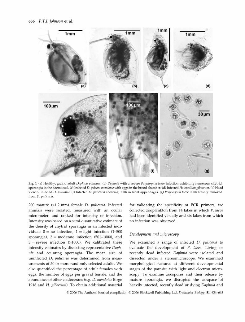
Fig. 1 (a) Healthy, gravid adult Daphnia pulicaria. (b) Daphnia with a severe Polycaryum laeve infection exhibiting numerous chytrid
sporangia in the haemocoel. (c) Infected D. galeata mendotae with eggs in the brood chamber. (d) Infected Holopedium gibberum. (e) Head
view of infected D. pulicaria. (f) Infected D. pulicaria showing thalli in front appendages. (g) Polycaryum laeve thalli freshly removed
from D. pulicaria.
636 P.T.J. Johnson et al.
� 2006 The Authors, Journal compilation � 2006 Blackwell Publishing Ltd, Freshwater Biology, 51, 634–648
placed them at 5 �C. After 24–48 h, sporangia with
well-developed exit pores and abundant zoospores
were usually present.
For histology, we fixed whole D. pulicaria in 10%
neutral-buffered formalin or Bouins solution. Samples
were decalcified for 2 h using the formic acid-sodium
citrate method, then processed through routine par-
affin procedures using Harris haematoxylin and eosin
Y (e.g. Luna, 1968; Humason, 1979). Tissues were
examined with an Olympus BX51 compound micro-
scope equipped with a Nikon DXM1200 and ACT-1
image capture program (Nikon, Melville, NY, U.S.A.).
For scanning electron microscopy, we fixed Daphnia
tissues or isolated sporangia in 2.5% glutaraldehyde
buffered in artificial pond water (1 mMM KH2PO4,
0.1 mMM MgCl2, 0.02 mMM CaCl2) overnight at 4 �C and
post-fixed tissues for 1 h in 1% OsO4. Material was
then dehydrated through an ethanol series and critical
point dried. Specimens were coated with approxi-
mately 20 nm 60 : 40 gold : palladium (See Vac Auto-
conductavac sputter coater, Pittsburgh, PA, U.S.A.)
and observed with a Hitachi S-570 scanning electron
microscope. For transmission electron microscopy,
samples were primary fixed in Karnovsky’s fixative,
post-fixed in OsO4, and dehydrated through a graded
ethanol series. Samples were infiltrated and embedded
in Spurr’s low viscosity resin. Ultra-thin sections were
collected and viewed on a JEOL 100CX (Peabody, MA,
U.S.A.) at 80 kV. Images were captured using a SIS
MegaView III (Lakewood, CO, U.S.A.).
We unsuccessfully attempted to isolate P. laeve into
pure culture on various chytrid media including
PmTG, (Barr & Desaulniers, 1987) and mTGh (tryp-
tone, 8 g L)1; gelatin hydrolysate, 2 g L)1) and a
medium for growing insect tissue (Grace’s insect
medium and crayfish saline, with and without added
FBS).
Pathology
To evaluate the effects of P. laeve infection on host
survival, growth, and reproduction, we conducted a
life table experiment with D. pulicaria from Tenderfoot
Lake, Vilas County, Wisconsin. We randomly selected
30 infected and 30 uninfected Daphnia and recorded
the size, infection intensity, and fecundity of each
individual. Initial sizes ± 1 SE of infected and unin-
fected Daphnia were 1.88 ± 0.05 and 1.87 ± 0.05,
respectively. Daphnia were isolated individually into
50 mL containers and acclimated to artificial lake
water (COMBO; Kilham et al., 1998) over 24 h. Indi-
viduals that died prior to initiation of the experiment
were replaced. Daphnia were maintained on a 16 : 8 h
light : dark cycle at 18 ± 2 �C. Every other day,
Daphnia were transferred into new containers filled
with clean water and 1 mL of a Scenedesumus solution
(approximately 6.5 · 104 cell count). Containers were
autoclaved before re-use. We recorded the numbers of
offspring and moults produced between water chan-
ges. Dead individuals were measured and examined
to evaluate infection status and intensity before
preservation. We concluded the experiment when all
of the infected individuals had died.
Sequencing
DNA extraction. Moderately to heavily infected
D. pulicaria from Birch Lake (Vilas County, Wisconsin)
were fixed in 95% ethanol for molecular analyses;
uninfected individuals were fixed separately. DNA
was extracted from two pools of ten infected and two
pools of five uninfected D. pulicaria using a QIAGEN
(Valencia, CA, U.S.A.) DNeasy Tissue Kit according to
the manufacturer’s instructions and with a final
elution in 50 lL Buffer AE. Small sub-unit (SSU)
rDNA was amplified using a protocol designed for
amplification of protistan and fungal sequences from
metazoan hosts (see Carnegie et al., 2003; Bower et al.,
2004 for reaction conditions). Amplification products
from the presumptive Polycaryum sp. were excised
from the gel, purified using a S.N.A.P. Gel Purification
Kit (Invitrogen, Carlsbad, CA, U.S.A.), and cloned
using a TOPO TA Cloning Kit (Invitrogen). Products
were sequenced on a LICOR 4200 following a
sequencing reaction with vector-specific (M13 for-
ward and reverse) primers and compared against the
GenBank database using the BLAST search algorithm.
Polymerase chain reaction probes. We designed PCR
primers that were presumptively specific to Poly-
caryum sp. to (i) verify correspondence between the
visual identification of infection and the molecular
signal and (ii) screen for additional stages or hosts of
the parasite. We selected primers Polycaryum-F1
(5¢-TAGTCGAACCTCAAGAGCG-3¢) and Poly-
caryum-R2 (5¢-CGGCATCGTTTATGGTTGTG-3¢) from
among several candidates for continued application.
DNA for presumptive Polycarum sp.-specific PCR was
Chytrid Parasitism of Daphnia 637
� 2006 The Authors, Journal compilation � 2006 Blackwell Publishing Ltd, Freshwater Biology, 51, 634–648
obtained from numerous sources: lake sediment, lake
seston (filtered onto a 0.2 lm glass fibre filter),
calanoid and cyclopoid copepods, Chaoborus larvae
and D. pulicaria from lakes with visually-identified
infections [Allequash, Katinka, Nelson, Oxbow,
Horsehead, Crab, Annabelle, Tenderfoot, Big Muskel-
lunge, South Turtle, Spider (all in Vilas County),
Owen (Bayfield County), Nebagamon (Douglas
County), and Devils (Sauk County)] and lakes without
discernible infection [Crystal, Stormy (Vilas County),
Lulu, Beulah, Green (Walworth County), and Ripley
(Jefferson County)]. One microliter (1 lL) of template
DNA was used in 25-lL PCR reactions that also
included 1X PCR Buffer, MgCl2 at 1.5 mMM, each dNTP
at 0.2 mMM, primers Polycaryum-F1 and R2 at 0.5 lMM,
and Taq polymerase at 0.08 units l L)1 (all purchased
from Invitrogen). Control reactions included either
Polycaryum-positive D. pulicaria DNA from Birch Lake
(positive) or ddH20 (negative). Cycling in an MJ
Research PTC-200 thermal cycler began with dena-
turation at 95 �C for 7 min, continued with 40 cycles
of 95 �C for 1 min, 58.9 �C for 1 min, and 72 �C for
1 min and ended with a final extension at 72 �C for
7 min. Products were electrophoresed in a 1.5%
agarose (in 1X TBE) gel, post-stained with ethidium
bromide, and visualised under UV light.
Phylogeny. A nucleotide BLAST search of the ribo-
somal sequence suggested a close homology to
Catenaria anguillulae (Chytridiomycota, Blastocladia-
les). This preliminary information enabled a more
thorough and directed phylogenetic analysis using
available sequences of members of Blastocladiales as
well as a wide range of other fungal taxa. All other
sequences were obtained from GenBank: Allomyces
arbuscula (AY55254), Allomyces macrogynus (U23936),
Ascobolus lineolatus (L37533), Blastocladiella emersonii
(AY635842), C. anguillulae (AF164339), Catenophlyctis
sp. JEL298 (AY635822), Coelomomyces stegomyiae
(AF322406), Cronartium ribicola (M94338), Gigaspora
albida (Z14009), Microallomyces sp. CR74 (AY635840),
Neocallimastix sp. LM-2 (M59761), Neolecta vitellina
(Z27393), Orbilia delicatula (U72603), Peridermium hark-
nessii (M94339), Peziza badia (L37539), Physoderma
maydis (AY601708), Pneumocystis carinii (L27658), Sac-
charomyces cerevisiae (J01353), Schizosaccharomyces
pombe (X54866), Spizellomyces acuminatus (M59759),
Sporobolomyces roseus (X60181), Taphrina wiesneri
(D12531), Tilletia caries (U00972), Ustilago hordei
(U00973), Zoophagus insidians (AB016009), Zoophthora
radicans (D29949). Sequences other than those from P.
laeve were aligned using MacClade v4.05 (Maddison
& Maddison, 2002) and analysed with PAUP* v4.0b10
(Swofford, 2002).
To mitigate the biases inherent in each algorithm, we
used two analyses to explore the phylogenetic position
of Polycaryum. A maximum parsimony (MP) heuristic
search was conducted with 1000 random addition
replicates with tree branch reconnection (TBR) branch
swapping, equal character weighting, MULTREES
activated and steepest descent option not activated. A
Bayesian heuristic search was also conducted with
MrBayes v3.1 (Huelsenbeck & Ronquist, 2001). Four
replicates of four chains were run for 1 000 000 gener-
ations (nst ¼ 6, rates ¼ gamma, savebrlens ¼ yes, bur-
nin ¼ 100 000, heat ¼ 0.2). Trees were sampled every
100 generations. To better resolve relationships within
the order, we constructed an additional tree within the
clade Blastocladiales using a MP search (settings as
above) and a reduced data set (nine member taxa and
one outgroup).
Results
Ecological observations
We observed individuals of D. pulicaria from Alle-
quash Lake infected with P. laeve on all but two
sample dates in 2003 (Fig. 2a). Prevalence of infection
in mature D. pulicaria, however, varied considerably
among seasons, from a maximum prevalence in
March (>80%) to a minimum in late summer (<1%;
Fig. 2b). The highest infection levels occurred between
January and March when the lake’s surface was
frozen. The population density of D. pulicaria subse-
quently declined by two orders of magnitude before
recovering in early June (Fig. 2a).
Infection prevalence increased linearly as a function
of Daphnia size (Fig. 3a) and hosts with discernable
P. laeve infection were significantly larger than unin-
fected individuals (Fig. 3b; P < 0.0001, ANOVAANOVA, F ¼107.251; N ¼ 2306). Immature D. pulicaria were rarely
infected. On average, larger infected Daphnia also
exhibited more severe infections than smaller infected
individuals (P < 0.0001; r ¼ 0.242,N ¼ 1454). Infection
was negatively associated with host fecundity
(P < 0.0001; v2 ¼ 565.416; N ¼ 2306). Only five
(0.35%) infected D. pulicaria contained eggs, indicating
638 P.T.J. Johnson et al.
� 2006 The Authors, Journal compilation � 2006 Blackwell Publishing Ltd, Freshwater Biology, 51, 634–648

that infection castrated the host. Correspondingly, the
percentage of fecund hosts was low during epidemics
(Fig. 2b).
We observed P. laeve infection in other species of
zooplankton only rarely. Throughout 2003, only 15
D. galeata mendotae were found infected (Fig. 1c; N ¼2683). Four of these (26.7%) supported eggs, suggest-
ing the infection was less pathogenic in this species
than in D. pulicaria (P < 0.001, v2 ¼ 132.41; N ¼ 1455).
One infected H. gibberum was also observed
(N >> 1000; Fig. 1d).
Development and life cycle
The haemocoel of infected Daphnia individuals was
filled with between 50 and 2500 spherical sporangia,
depending on the intensity of infection. Infections
were initially observed on and around the heart and
subsequently in the head. In severe infections, spor-
angia occurred throughout the host body cavity and
into the feeding and swimming appendages, causing
a markedly altered appearance (Fig. 1b,c). The distri-
bution and distinctive appearance of these sporangia
made infection rapidly identifiable in both living and
preserved samples. Dissection and subsequent enu-
meration of sporangia within 15 randomly selected,
obviously infected Daphnia yielded an average (±1 SE)
of 920.9 ± 91.3 sporangia per host.
Dissection of infected Daphnia revealed many of the
life history stages described by previous authors
(Stempell, 1903; Green, 1974). We followed Stempell’s
(1903) division of development into seven growth
stages and a sporulation stage. In stages I–III, from
first appearance to final size (approximately 30 lm),
thalli are characterised by the absence of a visible cell
wall, as revealed by light microscopy (Fig. 4a–c), TEM
(Fig. 5a,b) and histological preparations (Fig. 6c). The
0
0.1
0.2
0.3
0.4
0.5
0.6
0.7
0.8
0.9
1
1.2 1.5 1.7 1.9 2.2 2.4 2.7
Size class (mm)
Size (mm)
Infe
ctio
n pr
eval
ence
1.50 2.00 2.50 3.00
0
5
10
15
Fre
quen
cy (
%)
1.50 2.00 2.50 3.00
Uninfected Infected
(a)
(b)
Fig. 3 (a) Infection prevalence as a function of carapace size
and (b) size-frequency distribution of infected and uninfected
D. pulicaria from Allequash Lake in 2003. Error bars in (a) rep-
resent the upper value of the 95% confidence interval for pro-
portions.
0
0.1
0.2
0.3
0.4
0.5
0.6
0.7
0.8
0.9
1Lo
g de
nsity
(L–1
)
Total
Infected
0
10
20
30
40
50
60
70
80
90
100
1/24
/200
3
2/24
/200
3
3/24
/200
3
4/24
/200
3
5/24
/200
3
6/24
/200
3
7/24
/200
3
8/24
/200
3
9/24
/200
3
10/2
4/20
03
Pre
vale
nce
(%)
Gravid
Infected
(a)
(b)
Ice cover
Fig. 2 Temporal patterns in (a) the density (logarithmically-
transformed) of D. pulicaria infected with P. laeve and the total
D. pulicaria population density and (b) the percentages of gravid
(egg-bearing) and infected Daphnia. All data are from Allequash
Lake, Vilas Co., Wisconsin. Black bar indicates the period of time
in which the lake was frozen.
Chytrid Parasitism of Daphnia 639
� 2006 The Authors, Journal compilation � 2006 Blackwell Publishing Ltd, Freshwater Biology, 51, 634–648

Fig. 4 Light microscopy of stages of development of Polycaryum laeve. (a) Smallest determined size class of thalli within living host. (b)
Intermediate-sized thalli and (c) large thalli without visible walls. All developing thalli were recognisable by the accumulation of lipid
globules in the core of the thallus. (d) Early walled thalli, with lipid globules smaller and more dispersed. (e) Increased number of
smaller globules throughout spherical thallus. (f) Ovate and lobate zoosporangia. Membrane-covered sporangium has broken through
wall of thallus leaving torn wall (arrow heads). (g) Open sporangium with exiting zoospore (arrowhead). Arrow shows edge of wall.
(h) Montage of five zoospores, each with a posterior flagellum (i) Clumping of zoospores. (j) Montage of three biflagellate zoospores.
Arrowheads indicate flagella.
640 P.T.J. Johnson et al.
� 2006 The Authors, Journal compilation � 2006 Blackwell Publishing Ltd, Freshwater Biology, 51, 634–648
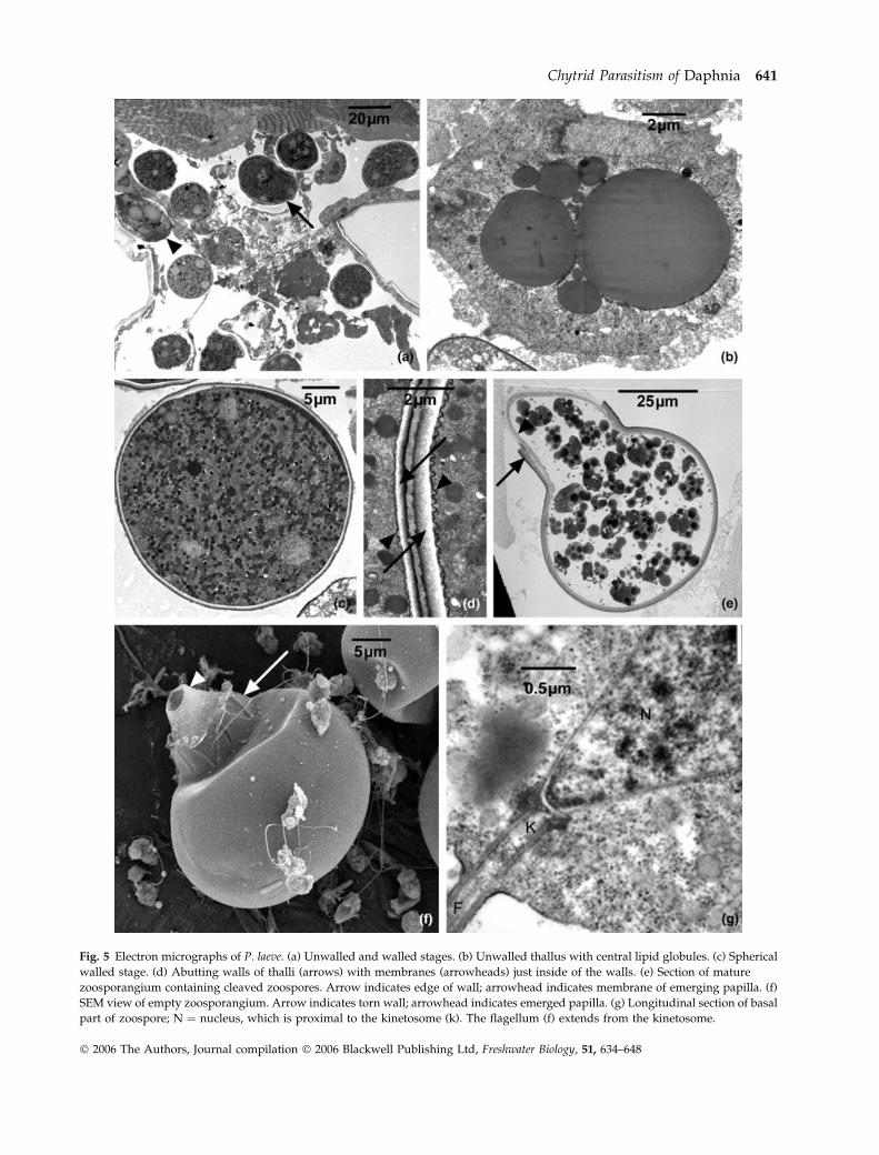
Fig. 5 Electron micrographs of P. laeve. (a) Unwalled and walled stages. (b) Unwalled thallus with central lipid globules. (c) Spherical
walled stage. (d) Abutting walls of thalli (arrows) with membranes (arrowheads) just inside of the walls. (e) Section of mature
zoosporangium containing cleaved zoospores. Arrow indicates edge of wall; arrowhead indicates membrane of emerging papilla. (f)
SEM view of empty zoosporangium. Arrow indicates torn wall; arrowhead indicates emerged papilla. (g) Longitudinal section of basal
part of zoospore; N ¼ nucleus, which is proximal to the kinetosome (k). The flagellum (f) extends from the kinetosome.
Chytrid Parasitism of Daphnia 641
� 2006 The Authors, Journal compilation � 2006 Blackwell Publishing Ltd, Freshwater Biology, 51, 634–648
earliest stages were difficult to observe and were seen
primarily in lightly infected individuals. A central
clustering of lipid globules characterised both early
unwalled stages (Fig. 4a) and later walled stages
(Stage IV, Figs 1e & 1g) of fungal thalli within living
Daphnia. Before cell wall formation, thalli tended to be
flattened (Fig. 4b,c; Fig. 5a,b); once walls were visible,
however, thalli appeared round (Fig. 4d,e; Fig. 5c).
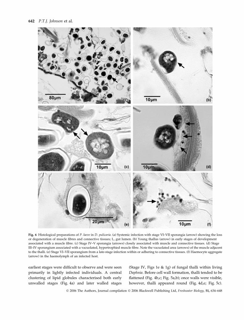
Fig. 6 Histological preparations of P. laeve in D. pulicaria. (a) Systemic infection with stage VI–VII sporangia (arrow) showing the loss
or degeneration of muscle fibres and connective tissues; L, gut lumen. (b) Young thallus (arrow) in early stages of development
associated with a muscle fibre. (c) Stage IV–V sporangia (arrows) closely associated with muscle and connective tissues. (d) Stage
III–IV sporangium associated with a vacuolated, hypertrophied muscle fibre. Note the vacuolated area (arrows) of the muscle adjacent
to the thalli. (e) Stage VI–VII sporangium from a late-stage infection within or adhering to connective tissues. (f) Haemocyte aggregate
(arrow) in the haemolymph of an infected host.
642 P.T.J. Johnson et al.
� 2006 The Authors, Journal compilation � 2006 Blackwell Publishing Ltd, Freshwater Biology, 51, 634–648
Some walled thalli were irregularly lobate (Fig. 4f).
Thalli walls varied in thicknesses (Fig. 5d) and were
underlain by a membrane (Fig. 5d–f), which emerged
through the wall during sporulation.
Sporulation. Zoosporogenesis occurred following dis-
ruption of the carapace of dead or dyingDaphnia. Stages
V and VI (Fig. 4e,f) were precursors of sporulation and
represented stages of internal reorganisation, during
which the lipid bodies were redistributed and zoo-
spores were formed. During this reorganisation, an exit
papilla formed, either in conjunction with protrusion of
the thallus wall (Fig. 5e), or through rupture of the cell
wall with the sporangial membrane emerging through
the jagged opening (Fig. 4f). Motile zoospores (Fig. 4h)
began to exit sporangia usually within 48 h after the
disruption of the host carapace when incubated at 5 �C.
Motile spores were 5 · 7 lm (n ¼ 12) with a approxi-
mately 34 lm flagellum (Fig. 4h). The kinetosome
(basal body) of the zoospore flagellum is proximal to
the cone-shaped nucleus (Fig. 5g), similar to those
found in members of the Blastocladiales (Barr, 1990).
After several hours of motility, zoospores lost their
flagellum and encysted on whatever substrate was
available. Zoospores sometimes joined together
(Fig. 4i), and the presence of biflagellate spores (Fig. 4j)
suggests that fusion may have occurred (i.e. zoospores
are isogametes). A larger, multiflagellated stage was
observed only infrequently.
Attempts to infect D. pulicaria with free-swimming
zoospores were unsuccessful, despite treatments
involving different clones and ages of Daphnia, vari-
able temperatures, variable light regimes and differ-
ent water mixtures. Individual thalli kept in sterile
pond water augmented with penicillin and strepto-
mycin (0.5 mg ml)1 each) developed somewhat but
our efforts to culture P. laeve sporangia or zoospores
on artificial media were unsuccessful.
Pathology
Histopathology. Early stages of infection associated
closely with – but did not penetrate into – muscle
fibres and connective tissue cells (Fig. 6a–d).
Although rhizoids were not observed, early develop-
ment stages, which had poorly defined protoplasmic
regions, closely adhered to host tissues (Fig. 6b,d). As
the cell walls of the sporangial thalli developed, many
sporangia no longer adhered to muscle fibres, but
some remained associated with muscle or connective
tissues (Fig. 6c). Connective tissue cells that surroun-
ded muscle fibre bundles with associated sporangia
sometimes appeared hypertrophied or vacuolated
(Fig. 6d). Degeneration and loss of muscle fibres and
connective tissues were apparent in moderate and
heavy infections. Heavy infections were systemic;
sporangia occurred throughout the haemocoel and in
some cases were surrounded by remnant muscle
fibres or connective tissues (Fig. 6e). In moderate and
heavy infections, haemocytic infiltrates were occa-
sionally observed either surrounding a sporangium or
as aggregates in the haemolymph (Fig. 6f). Further,
the connective tissues contained no reserve inclusion
cells, suggesting that infected animals lacked meta-
bolic reserves and starved. Histologically, heavily
infected hosts lacked significant gonadal develop-
ment, but the sample size was relatively small (n ¼ 12
infected animals).
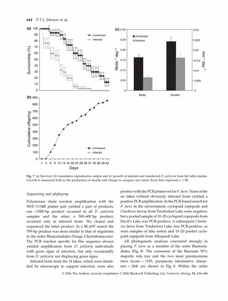
Life table. The life table study ended after 42 days, by
which time all of the infected (n ¼ 30) and 26 of 30
uninfected Daphnia had died. Within the infected
group, infections became more severe over time and
no individuals recovered from infection. One puta-
tively uninfected Daphnia developed a visible infec-
tion during the study and was excluded from
analyses. Infected and uninfected Daphnia differed
significantly with respect to growth, survival, and
reproduction. Infected individuals died sooner
(12 ± 2.21 days) than uninfected individuals
(26 ± 2.04 days; Fig. 7a; Kaplan–Meier Survival Ana-
lysis; P < 0.0001; Log Rank ¼ 28.46). By day 17, only
20% of infected Daphnia remained alive, whereas
>70% of the uninfected individuals were alive
(Fig. 7a). Differences in reproduction were similarly
extreme. Uninfected Daphnia produced, on average,
0.901 ± 0.074 offspring day)1 and a cumulative total
of 852 offspring. Infected Daphnia completely failed
to reproduce (Fig. 7b; P < 0.0001; F ¼ 147.823). One
infected individual produced a single clutch of eggs
but no viable offspring were released. Although
there were no differences between the initial lengths
of each group (ANOVAANOVA, P ¼ 0.953; F ¼ 0.003; N ¼60),
uninfected Daphnia moulted more frequently and
grew significantly more than infected individuals
during the course of the study (Fig. 7c; MANOVAMANOVA;
Wilks’ k ¼ 49.333; P < 0.0001; growth: F ¼ 5.572;
P ¼ 0.022; moults: F ¼ 27.453; P < 0.0001).
Chytrid Parasitism of Daphnia 643
� 2006 The Authors, Journal compilation � 2006 Blackwell Publishing Ltd, Freshwater Biology, 51, 634–648
Sequencing and phylogeny
Polymerase chain reaction amplification with the
581F/1134R primer pair yielded a pair of products;
one >1000 bp product occurred in all D. pulicaria
samples and the other, a 500–600 bp product,
occurred only in infected hosts. We cloned and
sequenced the latter product. In a BLAST search the
559-bp product was most similar to that of organisms
in the order Blastocladiales (Fungi; Chytridiomycota).
The PCR reaction specific for this sequence always
yielded amplification from D. pulicaria individuals
with gross signs of infection, but only occasionally
from D. pulicaria not displaying gross signs.
Infected hosts from the 14 lakes, which were identi-
fied by microscopy to support infection, were also
positive with the PCR primer set forP. laeve. None of the
six lakes without obviously infected hosts yielded a
positive PCR amplification. In the PCR-based search for
P. laeve in the environment, cyclopoid copepods and
Chaoborus larvae from Tenderfoot Lake were negative,
but a pooled sample of 10–20 cyclopoid copepods from
Devil’s Lake was PCR-positive. A subsequent Chaobo-
rus larva from Tenderfoot Lake was PCR-positive, as
were samples of lake seston and 10–20 pooled cyclo-
poid copepods from Allequash Lake.
All phylogenetic analyses concurred strongly in
placing P. laeve as a member of the order Blastocla-
diales (Fig. 8). The consensus of the Bayesian 50%
majority rule tree and the two most parsimonious
trees (score ¼ 1193, parsimony informative charac-
ters ¼ 264) are shown in Fig. 8. Within the order
0
100
200
300
400
500
600
700
800
900
1 3 6 9 12 14 16 20 23 26 28 31 33 36 39 42
Days
Cum
ulat
ive
offs
prin
g
Uninfected
Infected
0
10
20
30
40
50
60
70
80
90
100S
urvi
vors
hip
(%)
Uninfected
Infected
0
0.05
0.10
0.15
0.20
0.25
0.30
Molts Growth
Mol
ts *
day
–1
Uninfected
Infected
mm
* day–1
0.03
0.025
0.02
0.015
0.005
0.01
0
(c)
(b)
(a)
Fig. 7 (a) Survival, (b) cumulative reproductive output and (c) growth of infected and uninfected D. pulicaria from life table studies.
Growth is measured both as the production of moults and change in carapace size (mm). Error bars represent ± 1 SE.
644 P.T.J. Johnson et al.
� 2006 The Authors, Journal compilation � 2006 Blackwell Publishing Ltd, Freshwater Biology, 51, 634–648
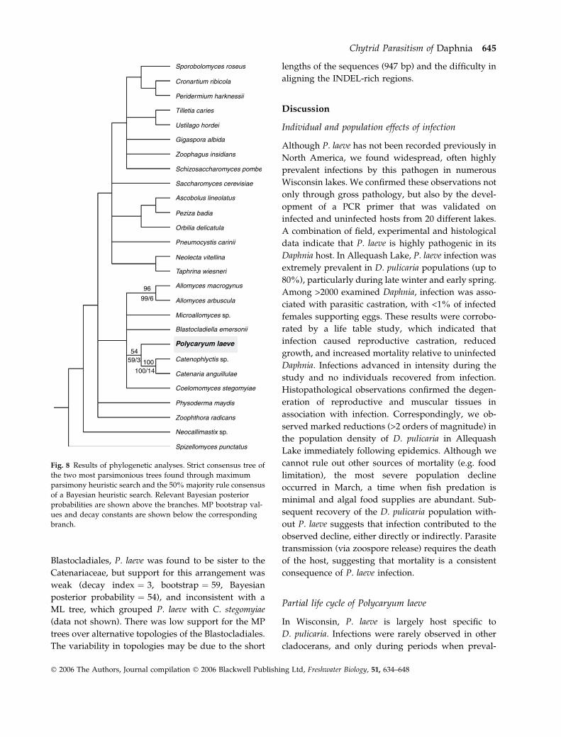
Blastocladiales, P. laeve was found to be sister to the
Catenariaceae, but support for this arrangement was
weak (decay index ¼ 3, bootstrap ¼ 59, Bayesian
posterior probability ¼ 54), and inconsistent with a
ML tree, which grouped P. laeve with C. stegomyiae
(data not shown). There was low support for the MP
trees over alternative topologies of the Blastocladiales.
The variability in topologies may be due to the short
lengths of the sequences (947 bp) and the difficulty in
aligning the INDEL-rich regions.
Discussion
Individual and population effects of infection
Although P. laeve has not been recorded previously in
North America, we found widespread, often highly
prevalent infections by this pathogen in numerous
Wisconsin lakes. We confirmed these observations not
only through gross pathology, but also by the devel-
opment of a PCR primer that was validated on
infected and uninfected hosts from 20 different lakes.
A combination of field, experimental and histological
data indicate that P. laeve is highly pathogenic in its
Daphnia host. In Allequash Lake, P. laeve infection was
extremely prevalent in D. pulicaria populations (up to
80%), particularly during late winter and early spring.
Among >2000 examined Daphnia, infection was asso-
ciated with parasitic castration, with <1% of infected
females supporting eggs. These results were corrobo-
rated by a life table study, which indicated that
infection caused reproductive castration, reduced
growth, and increased mortality relative to uninfected
Daphnia. Infections advanced in intensity during the
study and no individuals recovered from infection.
Histopathological observations confirmed the degen-
eration of reproductive and muscular tissues in
association with infection. Correspondingly, we ob-
served marked reductions (>2 orders of magnitude) in
the population density of D. pulicaria in Allequash
Lake immediately following epidemics. Although we
cannot rule out other sources of mortality (e.g. food
limitation), the most severe population decline
occurred in March, a time when fish predation is
minimal and algal food supplies are abundant. Sub-
sequent recovery of the D. pulicaria population with-
out P. laeve suggests that infection contributed to the
observed decline, either directly or indirectly. Parasite
transmission (via zoospore release) requires the death
of the host, suggesting that mortality is a consistent
consequence of P. laeve infection.
Partial life cycle of Polycaryum laeve
In Wisconsin, P. laeve is largely host specific to
D. pulicaria. Infections were rarely observed in other
cladocerans, and only during periods when preval-
Sporobolomyces roseus
Cronartium ribicola
Peridermium harknessii
Tilletia caries
Ustilago hordei
Gigaspora albida
Zoophagus insidians
Schizosaccharomyces pombe
Saccharomyces cerevisiae
Ascobolus lineolatus
Peziza badia
Orbilia delicatula
Pneumocystis carinii
Neolecta vitellina
Taphrina wiesneri
Allomyces macrogynus96
99/6
5459/3 100
100/14
Allomyces arbuscula
Microallomyces sp.
Catenophlyctis sp.
Catenaria anguillulae
Coelomomyces stegomyiae
Physoderma maydis
Zoophthora radicans
Neocallimastix sp.
Spizellomyces punctatus
Blastocladiella emersonii
Polycaryum laeve
Fig. 8 Results of phylogenetic analyses. Strict consensus tree of
the two most parsimonious trees found through maximum
parsimony heuristic search and the 50% majority rule consensus
of a Bayesian heuristic search. Relevant Bayesian posterior
probabilities are shown above the branches. MP bootstrap val-
ues and decay constants are shown below the corresponding
branch.
Chytrid Parasitism of Daphnia 645
� 2006 The Authors, Journal compilation � 2006 Blackwell Publishing Ltd, Freshwater Biology, 51, 634–648
ence of the parasite was high in populations of
D. pulicaria (i.e. ‘spillover’ infections). Evidence for
horizontal transmission may be inferred from the
rarity of infected individuals with eggs and the
overall increase in prevalence with host size class.
Larger individuals may be more likely to become
infected either because of their age, increased filtra-
tion rate relative to smaller (younger) Daphnia, or
because of a necessary incubation period before
infection becomes visible. Attempts to raise offspring
from the few infected Daphnia with eggs yielded only
uninfected neonates, indicating that vertical transmis-
sion is unlikely.
The complete life cycle of P. laeve and the conditions
necessary for transmission remain conjectural. Initial
attempts to transmit or culture P. laeve failed, suggest-
ing that the parasite has an indirect life cycle involving
additional host species or free-living stages. In support
of this possibility, species in the closely related
blastocladialean genus Coelomomyces use mosquito
larvae as definitive hosts and copepods as intermedi-
ate hosts (Whisler, Zebold & Shemanchuk, 1975;
Whisler et al., 1999). Within the mosquito host the
coenocytic, weakly-branched thallus of Coelomomyces
gives rise to large numbers of thick-walled sporangia
(Whisler et al., 1975). Meiosis occurs in the resting
spores, releasing zoospores that infect copepods and
develop into unwalled gametangia. Gametes, which
are uniflagellate and microscopically indistinguish-
able from zoospores, fuse to form a biflagellate zygote
that infects a new, larval mosquito host.
The portion of the life cycle of P. laeve occurring in
D. pulicaria could be analogous to either the mosquito
(2N) or copepod (1N) stage of Coelomomyces. If the
thalli in Daphnia are diploid zoosporangia, then
zoospores might be infectious to another host species
or encyst in lake sediments, and the bi- and multi-
flagellated stages that we observed would reflect
irregular cleavage during zoosporogenesis. Alternat-
ively, if what we have called zoosporangia are
actually gametangia, the haploid isogametes (cf. zoo-
spores) could fuse to form biflagellated zygotes and
infect an alternate host; this interpretation would
explain the pairing and clumping behaviour that we
observed. Our preliminary results indicating a posit-
ive PCR amplification for cyclopoid copepods and a
single Chaoborus larva may be interpreted as support
for a multi-host life cycle for P. laeve, but this requires
further verification.
Taxonomic and phylogenetic position of Polycaryum
The stages of P. laeve observed in D. pulicaria were
remarkably similar to those described by Stempell
(1903), although his observations were primarily from
fixed material in D. longispina. Whereas Polycaryum
was previously considered a member of the Haplos-
poridia (Stempell, 1903; Green, 1974; Radek & Herth,
1999), based on our TEM observations of the
zoospores and our molecular sequencing results, we
transfer P. laeve to the Blastocladiales (Chytridiomy-
cota) and designate Stempell’s figures [1903, Archiv
fur Protistenkunde, 2: Tafel (Plate) IX, Figs 1–27] as
the appropriate lectotype. Further, we designate
collection Allequash Lake 26 June 2003 as the epitype
of P. laeve. The epitype material is deposited at
the New York Botanical Garden as 00705250. We
cannot assign P. laeve to a specific family within
Blastocladiales without additional molecular data,
particularly from other pathogenic members of the
Blastocladiales.
The first species appropriately described for this
genus was P. branchipodianum Stempell, 1902, a para-
site of fairy shrimp (Branchipus grubei; Branchiopoda,
Anostraca) that forms an ornamented resting spore.
This ornamented resting spore, which somewhat
resembles the 2N resting spores of Coelomomyces
spp. and the lipid-globule configuration that domin-
ates sporangia of P. laeve, indicates that this fungus
also is a member of the Blastocladiales. Therefore,
we designate Figs 1–8 [Stempell, 1902 Zoologische
Jahrbucher. Abteilung fur Systematik, Geographie
und Biologie der Tiere 15: Tafel (Plate) 31, Figs 1–8]
as the lectotype for P. branchipodianum and place it in
the Blastocladiales. Because P. branchipodianum was
published previous to P. laeve, we consider it as the
type species of the genus. Two other species, Poly-
caryum ecdyonuris and P. legeri, were described as
parasites of mayflies (Weiser, 1977, 1984). Little
information is available on these species and their
taxonomic affinities remain uncertain (J. Weiser,
personal communication).
Implications
Fungal pathogens can cause marked changes in the
population dynamics of many aquatic invertebrates,
including copepods, midges and crayfish (Green,
1974; Burns, 1979; Redfield & Vincent, 1979; Martin,
646 P.T.J. Johnson et al.
� 2006 The Authors, Journal compilation � 2006 Blackwell Publishing Ltd, Freshwater Biology, 51, 634–648

1981, 1984; Reynolds, 1988). To the best of our
knowledge, however, P. laeve is the first recorded
chytrid disease of Daphnia. Given the epidemic
outbreaks of P. laeve and its pathogenicity in Daphnia,
we believe that the parasite has a significant regu-
latory effect on host population dynamics. In Alle-
quash Lake, for example, the most severe epidemic
in March was followed by a catastrophic (99%)
decline in Daphnia population density. This occurred
well before the expected seasonal decline in Daphnia,
which is typically around late June or early July in
Allequash (see Fig. 2). Although P. laeve appears to
be a relatively recent arrival in Allequash Lake, we
have subsequently recorded infections from >25
lakes in Wisconsin, indicating that this parasite is
enzootic in Wisconsin lakes. Given the keystone
importance of Daphnia in lake food webs, epidemics
of P. laeve could have ecosystem-scale consequences
under certain conditions. Large-bodied Daphnia such
as D. pulicaria are effective grazers of phytoplankton
and can have important effects on nutrient cycling,
primary production and water clarity (e.g. Carpenter
et al., 1987). Correspondingly, D. pulicaria is an
important food resource for many young-of-the-year
and planktivorous fishes; epidemics of P. laeve could
therefore lead to shifts in food web structure or fish
production. Additional research should focus on: (i)
the long-term effects of P. laeve infection on Daphnia
populations, (ii) how environmental factors influence
P. laeve epidemics, (iii) completion of the parasite life
cycle and (iv) field and modelling efforts aimed at
evaluating the consequences of infection for lake
food webs, including both fish production and water
clarity.
Acknowledgments
We are indebted to the many individuals who
provided assistance with sampling and logistical
support, including Steve Carpenter, Tim Kratz, James
Rusak, Scott van Egeren and the UW Trout Lake
Station staff. We would also like to thank Barbara
Benson and the UW Zoology Museum, which
archives zooplankton samples for the North Temper-
ate Lakes LTER project. James Ellwein and Julia
Logeman provided invaluable assistance with sample
processing, Philip Oshel dedicated his time and
energy for scanning electron microscopy and Randy
Massey offered his expertise in transmission electron
microscopy. Chris Lucarotti offered valuable assist-
ance with interpretation of micrographs. Kersten
Wheeler provided histological support at VIMS. This
project was funded, in part, by grants from the
National Science Foundation (DEB-0411760, DEB-
0217533, DEB-0083545, DEB-9978094), an NSF Gradu-
ate Research Fellowship to PTJJ, the Anna Grant Birge
Foundation and the Research Experience for Under-
graduates (REU) programme.
References
Barr D. J. S. (1990) Phylum Chytridiomycota. In: Hand-
book of Protoctista, (Eds Margulis L., Corliss J. O.,
Melkonian M. & Chapman D. J.), pp. 454–466. Jones
and Bartlett Publishers, Boston.
Barr D. J. S. & Desaulniers N. L. (1987) Allochytridium
luteum n. sp.: morphology, physiology and zoospore
ultrastructure. Mycologia, 79, 193–199.
Bengtsson J. & Ebert D. (1998) Distribution and impacts
of microparasites on Daphnia in a rockpool metapopu-
lation. Oecologia, 115, 213–221.
Bittner K., Rothhaupt K.O. & Ebert D. (2002) Ecological
interactions of the microparasite Caullerya mesnili and
its host Daphnia galeata. Limnology and Oceanography,
47, 300–305.
Bower S. M., Carnegie R. B., Goh B., Jones S. R. M., Lowe
G. J. & Mak M. W. S. (2004) Preferential PCR
amplification of parasitic protistan small subunit
rDNA from metazoan tissues. Journal of Eukaryotic
Microbiology, 51, 325–332.
Brambilla D. J. (1983) Microsporidiosis in a Daphnia pulex
population. Hydrobiologia, 99, 175–188.
Burns C. W. (1979) Fungal parasitism in a copepod
population: the effects of Aphanomyces on the popula-
tion dynamics of Boeckella dilatata Sars. Journal of
Plankton Research, 7, 201–205.
Burns C. W. (1989) Parasitic regulation in a population of
Boeckella hamata Brehm (Copepoda: Calanoida). Fresh-
water Biology, 21, 421–426.
Carnegie R. B., Meyer G. R., Blackbourn J., Cochennec-
Laureau N., Berthe F. C. J. & Bower S. M. (2003)
Molecular detection of the oyster parasite Mikrocytos
mackini, and a preliminary phylogenetic analysis.
Diseases of Aquatic Organisms, 54, 219–227.
Carpenter S. R., Kitchell J. F., Hodgson J. R., Cochran P.
A., Elser J. J., Elser M. M., Lodge D. M., Kretchmer D.,
He X. & von Ende C. (1987) Regulation of lake primary
productivity by food-web structure. Ecology, 68, 1863–
1876.
Decaestecker E., De Meester L. & Ebert D. (2002) In deep
trouble: habitat selection constrained by multiple
Chytrid Parasitism of Daphnia 647
� 2006 The Authors, Journal compilation � 2006 Blackwell Publishing Ltd, Freshwater Biology, 51, 634–648
enemies in zooplankton. Proceedings of the National
Academy of Sciences (USA), 99, 5481–5485.
Duffy M. A., Hall S. R., Tessier A. J. & Huebner M. (2005)
Selective predators and their parasitized prey: are
epidemics in zooplankton under top-down control?
Limnology and Oceanography, 50, 412–420.
Ebert D. (1995) The ecological interaction between a
microsporidian parasite and its host Daphnia magna.
Journal of Animal Ecology, 64, 361–369.
Ebert D., Lipsitch M. & Mangin K. L. (2000) The effect of
parasites on host population density and extinction:
experimental epidemiology with Daphnia and six
microparasites. American Naturalist, 156, 459–477.
Ebert D., Hottinger J.W. & Pajunen V. I. (2001) Temporal
and spatial dynamics of parasites in a Daphnia
metapopulation: which factors explain parasite rich-
ness? Ecology, 82, 3417–3434.
Fels D., Lee V. A. & Ebert D. (2004) The impact of
microparasites on the vertical distribution of Daphnia
magna. Archiv fur Hydrobiologie, 161, 65–80.
Green J. (1974) Parasites and epibionts of Cladocera.
Transactions of the Royal Society of London, 32, 417–515.
Huelsenbeck J. P. & Ronquist F. (2001) MrBayes:
Bayesian inference of phylogeny. Bioinformatics, 17,
754–755.
Humason G. L. (1979) Animal Tissue Techniques, 4th edn.
WH Freeman & Co, San Francisco.
Kilham S. S., Kreeger D. A., Lynn S. G., Goulden C. E. &
Hen-era L. (1998) COMBO: a defined freshwater
culture medium for algae and zooplankton. Hydro-
biologia, 377, 147–159.
Luna L. G. (1968) Manual of Histologic Staining Methods of
the Armed Forces Institutes of Pathology, 3rd edn.
McGraw-Hill Book Company, New York.
Maddison D. R. & Maddison W. P. (2002) MacClade.
Version 4.05. Sinauer, Sunderland, Massachusetts.
Martin W. W. (1981) The natural regulation of midge
populations by aquatic fungi. Virginia Journal of the
Elisha Mitchell Society, 97, 162–170.
Martin W. W. (1984) The dynamics of aquatic fungi
parasitic in a stream population of the midge, Chirono-
mus attenuatus. Journal of Invertebrate Pathology, 44, 36–45.
Radek R. & Herth W. (1999) Ultrastructural investigation
of the spore-forming protist Nephridiophaga blattellae in
the Malpighian tubules of the German cockroach
Blattella germanica. Parasitology Research, 85, 216–231.
Redfield G.W. & Vincent W. F. (1979) Stages of infection
and ecological effects of a fungal epidemic on the eggs
of a limnetic copepod. Freshwater Biology, 9, 503–510.
Reynolds J. D. (1988) Crayfish extinctions and crayfish
plague in central Ireland. Biological Conservation, 45,
279–285.
Stempell W. (1902) Uber Jahrbucher Polycaryum branchipo-
dianum n. g., n. sp. Zoologische. Abteilung fur System-
atik, Geographie und Biologie der Tiere, 15, 591–596.
and Tafel, 31.
Stempell W. (1903) Beitrage zur Kenntnis der Gattung
Polycaryum. Archives for Protistenkunde, 2, 349–363.
Stirnadel H. A. & Ebert D. (1997) Prevalence, host
specificity and impact on host fecundity of micropar-
asites and epibionts in three sympatric Daphnia
species. Journal of Animal Ecology, 66, 212–222.
Swofford D. L. (2002) PAUP*: Phylogenetic analysis using
parsimony (*and other methods). Version 4. Sinauer,
Sunderland, Massachusetts.
Torgersen T., Karlsbakk E. & Kaartvedt S. (2002)
Deviating vertical distribution and increased conspicu-
ousness of parasitized Calanus. Limnology and Oceano-
graphy, 47, 1187–1191.
Weiser J. (1977) An atlas of insect diseases. (Ed. W. Junk),
B.V. Publishers, The Hague, Netherlands, 240 pp.
Weiser J. (1984) Ecology of mayfly infections. In: Proceed-
ings of the 4th International Conference on Ephemeroptera.
(Eds V. Landa, T. Soldan & M. Tonner), pp. 291–296. Inst.
Entomol., Czech. Acad. Sci., Praha, Czechoslovakia.
Whisler H. C., Zebold S. L. & Shemanchuk J. A. (1975)
Life-history of Coelomomyces psorophorae. Proceedings of
the National Academy of Sciences (USA), 72, 693–696.
Whisler H. C., Gabriel B. P., Chanpaisaeng J, Zebold S. L. &
Padua L. E. (1999) Observations on the life cycle of
Coelomomyces indicus (Blastocladiales: Coelomomyceta-
ceae) in anopheline mosquitoes from the Philippines
and Thailand. Journal ofMedical Entomology, 36, 695–701.
Willey R. L., Cantrell P. A. & Threlkeld S. T. (1990)
Epibiotic euglenoid flagellates increase the suscept-
ibility of some zooplankton to fish predation. Limnol-
ogy and Oceanography, 35, 952–959.
Willey R. L., Willey R. B. & Threlkeld S. T. (1993)
Planktivore effects on zooplankton epibiont commu-
nities: epibiont pigmentation effects. Limnology and
Oceanography, 38, 1818–1822.
Yan N. D. & Larsson J. I. R. (1988) Prevalence and
inferred effects of microsporidia of Holopedium gibber-
um (Crustacea: Cladocera) in a Canadian Shield lake.
Journal of Plankton Research, 10, 875–886.
(Manuscript accepted 20 December 2005)
648 P.T.J. Johnson et al.
� 2006 The Authors, Journal compilation � 2006 Blackwell Publishing Ltd, Freshwater Biology, 51, 634–648


![Centennial clonal stability of asexual Daphnia in ... · 7/22/2020 · 88 Daphnia, in particular the large-bodied Daphnia pulex-complex [7]. Arctic Daphnia 89 populations are generally](https://static.fdocuments.in/doc/165x107/5fb33315ffe483517d15d37c/centennial-clonal-stability-of-asexual-daphnia-in-7222020-88-daphnia-in.jpg)