Chronic Obstructive Pulmonary Disease file•In the UK COPD was found to reduce life expectancy by...
76
Chronic Obstructive Pulmonary Disease Erica Child Ashley Blaylock Kirsten Wilkinson Hannah Price
-
Upload
nguyenlien -
Category
Documents
-
view
213 -
download
0
Transcript of Chronic Obstructive Pulmonary Disease file•In the UK COPD was found to reduce life expectancy by...
Chronic Obstructive Pulmonary
Disease
Erica Child
Ashley Blaylock
Kirsten Wilkinson
Hannah Price
Chronic Obstructive Pulmonary Disease (COPD)
Basic Disease:
Progressive disease of airflow obstruction
Types:
Emphysema and Chronic Bronchitis
Prevalence
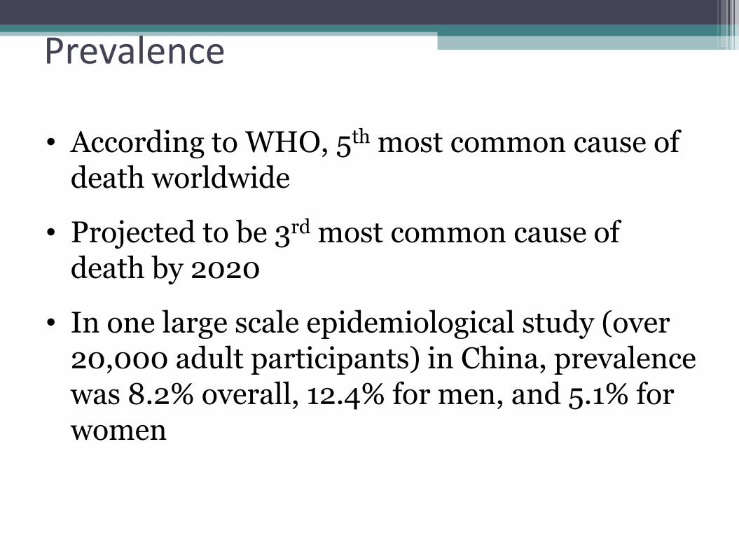
• According to WHO, 5th most common cause of death worldwide
• Projected to be 3rd most common cause of death by 2020
• In one large scale epidemiological study (over 20,000 adult participants) in China, prevalence was 8.2% overall, 12.4% for men, and 5.1% for women
Prevalence
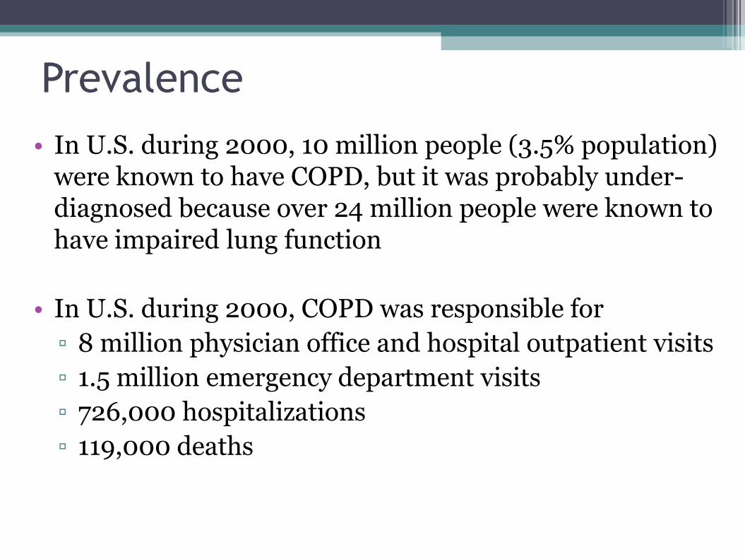
• In U.S. during 2000, 10 million people (3.5% population) were known to have COPD, but it was probably under-diagnosed because over 24 million people were known to have impaired lung function
• In U.S. during 2000, COPD was responsible for
▫ 8 million physician office and hospital outpatient visits
▫ 1.5 million emergency department visits
▫ 726,000 hospitalizations
▫ 119,000 deaths
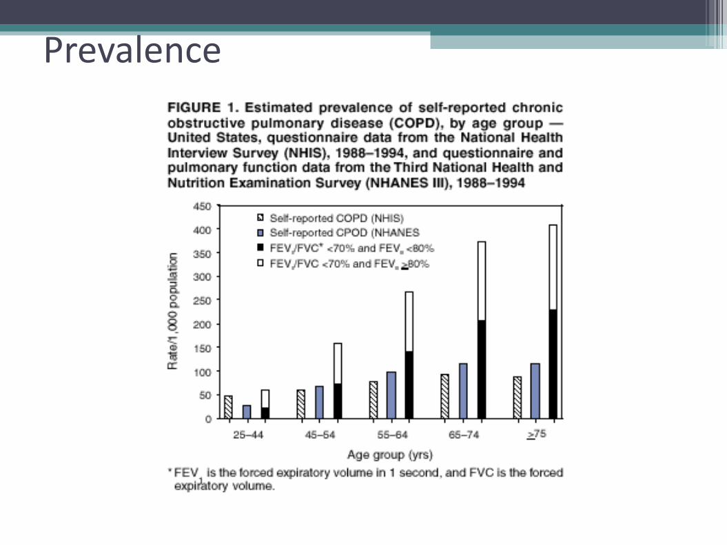
Prevalence
Incidence
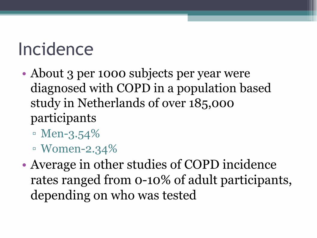
• About 3 per 1000 subjects per year were diagnosed with COPD in a population based study in Netherlands of over 185,000 participants
▫ Men-3.54%
▫ Women-2.34%
• Average in other studies of COPD incidence rates ranged from 0-10% of adult participants, depending on who was tested
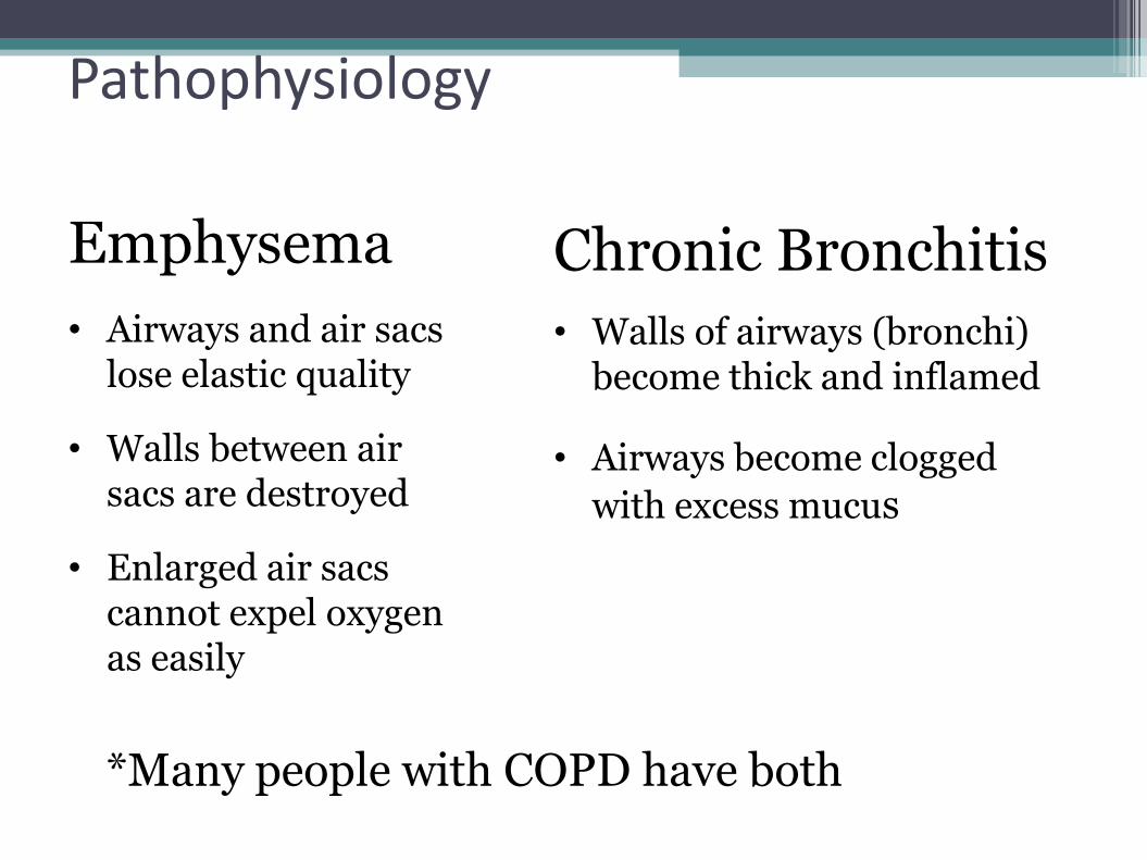
Pathophysiology
Emphysema
• Airways and air sacs lose elastic quality
• Walls between air sacs are destroyed
• Enlarged air sacs cannot expel oxygen as easily
Chronic Bronchitis
• Walls of airways (bronchi) become thick and inflamed
• Airways become clogged
with excess mucus
*Many people with COPD have both
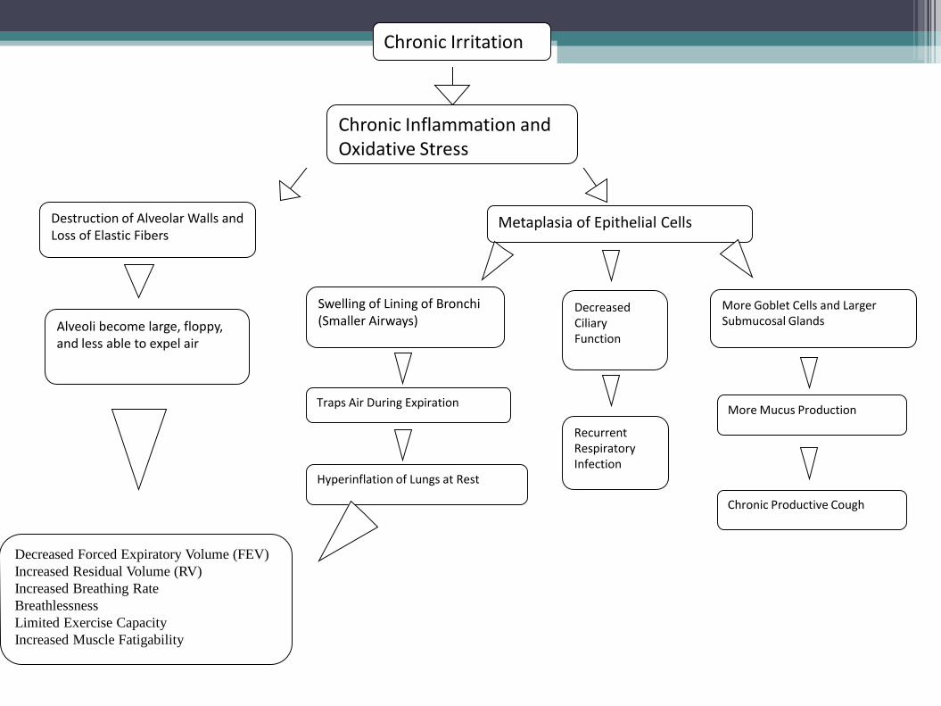
Metaplasia of Epithelial Cells
Swelling of Lining of Bronchi (Smaller Airways)
Decreased Ciliary Function
More Goblet Cells and Larger Submucosal Glands
Recurrent Respiratory Infection
More Mucus Production
Chronic Productive Cough
Traps Air During Expiration
Hyperinflation of Lungs at Rest
Chronic Irritation
Chronic Inflammation and Oxidative Stress
Destruction of Alveolar Walls and Loss of Elastic Fibers
Decreased Forced Expiratory Volume (FEV)
Increased Residual Volume (RV)
Increased Breathing Rate
Breathlessness
Limited Exercise Capacity
Increased Muscle Fatigability
Alveoli become large, floppy, and less able to expel air
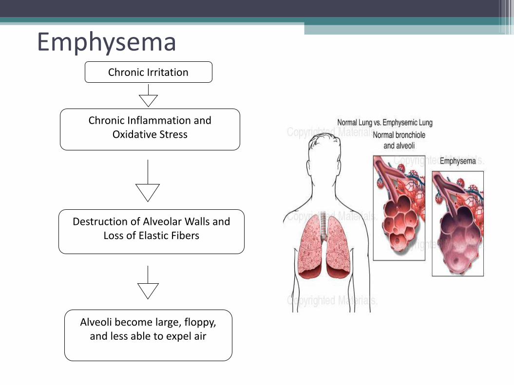
Chronic Irritation
Chronic Inflammation and Oxidative Stress
Destruction of Alveolar Walls and Loss of Elastic Fibers
Alveoli become large, floppy, and less able to expel air
Emphysema

Chronic Bronchitis
Metaplasia of Epithelial Cells
Swelling of Lining of Bronchi (Smaller Airways)
Decreased Ciliary Function
More Goblet Cells and Larger Submucosal Glands
Recurrent Respiratory Infection
More Mucus Production
Chronic Productive Cough
Traps Air During Expiration
Hyperinflation of Lungs at Rest
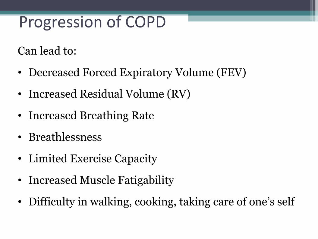
Progression of COPD
Can lead to:
• Decreased Forced Expiratory Volume (FEV)
• Increased Residual Volume (RV)
• Increased Breathing Rate
• Breathlessness
• Limited Exercise Capacity
• Increased Muscle Fatigability
• Difficulty in walking, cooking, taking care of one’s self
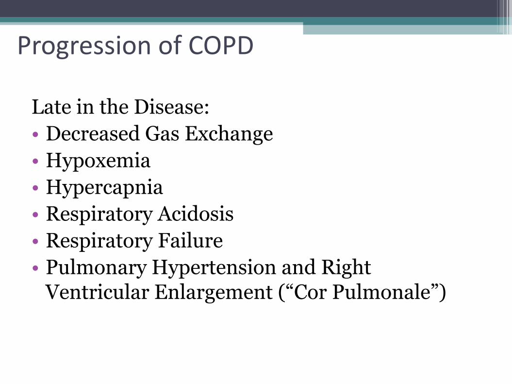
Late in the Disease:
• Decreased Gas Exchange
• Hypoxemia
• Hypercapnia
• Respiratory Acidosis
• Respiratory Failure
• Pulmonary Hypertension and Right Ventricular Enlargement (“Cor Pulmonale”)
Progression of COPD

Cor Pulmonale
Contributing Factors:
• Low oxygen exchange
(blood is shunted to alveoli with the most oxygen; if all of them have little oxygen, then all pulmonary arteries vasoconstrict, increasing blood pressure and stress on the right ventricle)
• Endothelial dysfunction
• Hypertrophy and hyperplasia of pulmonary arteries
• Systemic Inflammation

Prognosis
• Poor: No cure or reversal of disease
• Worse if malnourished
• Goals of treatment are mainly to manage the exacerbations and enhance overall well-being
• Decreased life expectancy
• In the UK COPD was found to reduce life expectancy by 1.8 years
• In the Netherlands study 26% of patients with very severe COPD died after 1 year of follow-up

Etiology

COPD Causes
COPD is characterized by slowly progressive obstruction of the airways, often the result of long-term exposure to irritants.
In rare occasions, genetics may play a major role in causing COPD.
Causes of COPD
Tobacco Use
Exposure to Other Irritants
Genetic Factors

Tobacco Use
Cigarette smoking is the single most important risk factor, attributing to 80-90% of cases.
Pipe and cigar smoking also increase risk.
10-20% of smokers develop clinically significant COPD.
Even if you quite smoking, your still at risk to develop COPD.

% Prevalence of Airflow Obstruction in
U.S. Adults
Never Smoked Former Smokers Current Smokers
0
2
4
6
8
10
12
14
How Tobacco Effects the Body
http://video.about.com/copd/COPD.htm

How Tobacco Effects the Body Cigarettes contain many hazardous substances that damage the lung when inhaled, including tar nicotine, carbon monoxide, and cyanide.
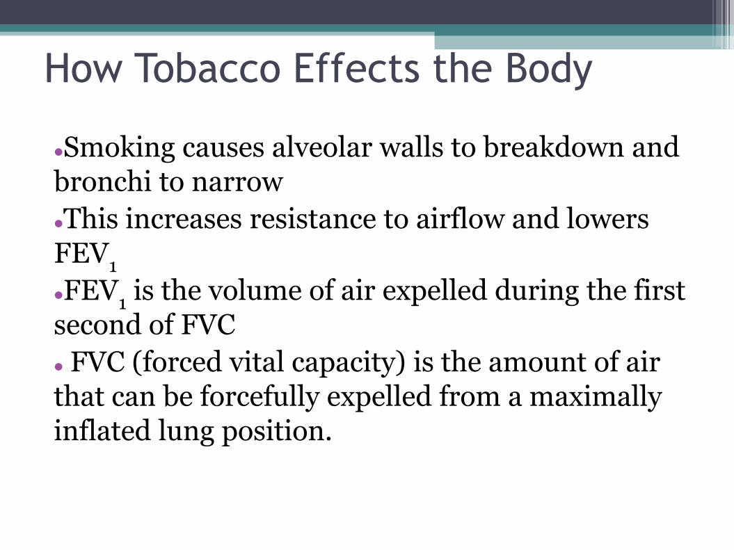
How Tobacco Effects the Body
Smoking causes alveolar walls to breakdown and bronchi to narrow
This increases resistance to airflow and lowers FEV
1
FEV1 is the volume of air expelled during the first
second of FVC
FVC (forced vital capacity) is the amount of air that can be forcefully expelled from a maximally inflated lung position.
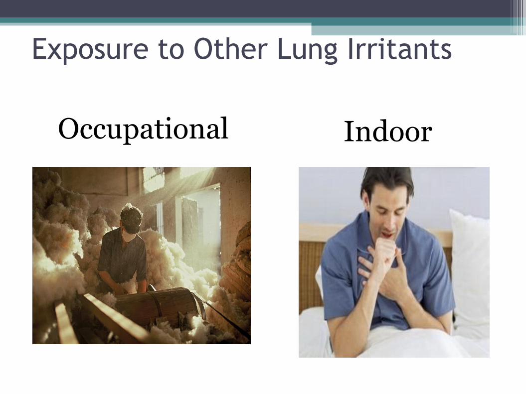
Exposure to Other Lung Irritants
Indoor
Occupational
Occupation Exposure/Outdoor
Pollutants Found in work place and outdoors environment
Vapors
Fumes
Dust
Examples include air pollution in urban areas, cadmium, coal, other mineral dusts, and welding fumes.

Indoor Air Pollutants
More common a cause of COPD in underdeveloped, low income areas
Due to exposure to biomass fuels such as coal, straw, animal dung, crop residues, and wood, which are used to heat and cook in poorly ventilated homes
Includes second hand smoke
Globally, perhaps the most important risk factor
Genetics
As only 10–20% of smokers develop COPD, a genetic susceptibility to COPD seems reasonable.
The best understood risk is α1 antitrypsin
deficiency.
Probably accounts for only 1-2% of COPD patients
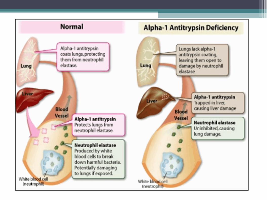
α1 Antitrypsin (AAT) Deficiency
Inherited Disease
Protein made in the liver. It travels in the bloodstream and protects the lungs from other harmful proteins.
In AAT deficiency, the number of functioning AAT proteins is reduced and adequate amounts can't enter the bloodstream to protect the lungs.
Many people live normal lives with an AAT deficiency.
Other Possible Genetic
Genes coding polymorphisms of... Growth Factor β1,
Tumor Necrosis Factor α
Microsomal Epoxide Hydrolase 1.
None of these yet proven to contribute to COPD

Risk Factors
Smoking/Pollutant Exposure
Family History
Age (lung function peaks in 20s, decline 30-40)
Gender
Respiratory Infection/Asthma
Socioeconomic Status

Comorbidities
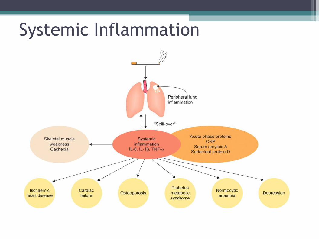
Systemic Inflammation

Common Comorbidities
Cardiovascular Problems Due to Inflammation
Cor Pulmonale
Pulmonary Hypertension
Lung Cancer
Increased inflammation and Oxidative Stress
Osteoporosis
Advanced age, poor mobility, smoking, poor nutrition, low BMI
Diabetes
Unknown but due to inflammation

Preventative Measures
Do Not Smoke!
Limit Exposure to Second Hand Smoke and Other Pollutants
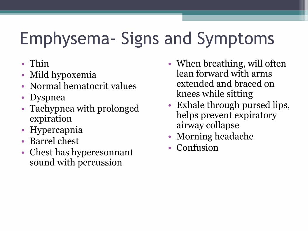
Emphysema- Signs and Symptoms
• Thin • Mild hypoxemia • Normal hematocrit values • Dyspnea • Tachypnea with prolonged
expiration • Hypercapnia • Barrel chest • Chest has hyperesonnant
sound with percussion
• When breathing, will often lean forward with arms extended and braced on knees while sitting
• Exhale through pursed lips, helps prevent expiratory airway collapse
• Morning headache • Confusion
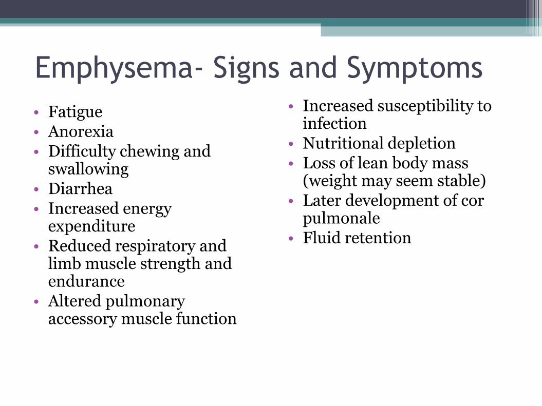
• Increased susceptibility to infection
• Nutritional depletion • Loss of lean body mass
(weight may seem stable) • Later development of cor
pulmonale • Fluid retention
• Fatigue • Anorexia • Difficulty chewing and
swallowing • Diarrhea • Increased energy
expenditure • Reduced respiratory and
limb muscle strength and endurance
• Altered pulmonary accessory muscle function
Emphysema- Signs and Symptoms
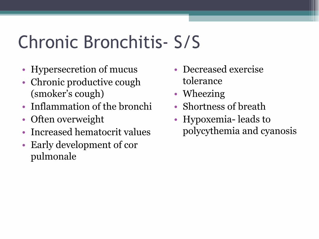
Chronic Bronchitis- S/S
• Hypersecretion of mucus
• Chronic productive cough (smoker’s cough)
• Inflammation of the bronchi
• Often overweight
• Increased hematocrit values
• Early development of cor pulmonale
• Decreased exercise tolerance
• Wheezing
• Shortness of breath
• Hypoxemia- leads to polycythemia and cyanosis

As disease progresses:
• Pulmonary hypertension
• Large amounts of sputum produced
• Frequent pulmonary infections
• FVC and FEV1 decrease
• FRC and RV increase since airway obstruction and air trapping are more pronounced
Chronic Bronchitis- S/S
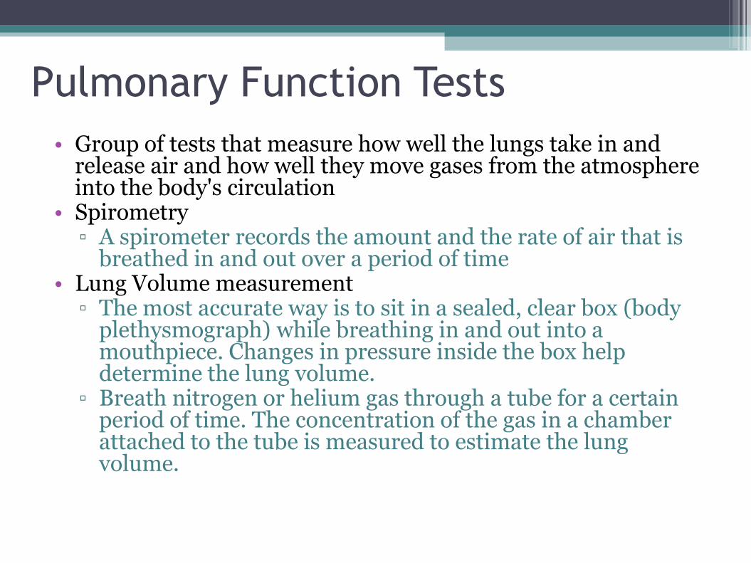
Pulmonary Function Tests
• Group of tests that measure how well the lungs take in and release air and how well they move gases from the atmosphere into the body's circulation
• Spirometry ▫ A spirometer records the amount and the rate of air that is
breathed in and out over a period of time • Lung Volume measurement
▫ The most accurate way is to sit in a sealed, clear box (body plethysmograph) while breathing in and out into a mouthpiece. Changes in pressure inside the box help determine the lung volume.
▫ Breath nitrogen or helium gas through a tube for a certain period of time. The concentration of the gas in a chamber attached to the tube is measured to estimate the lung volume.
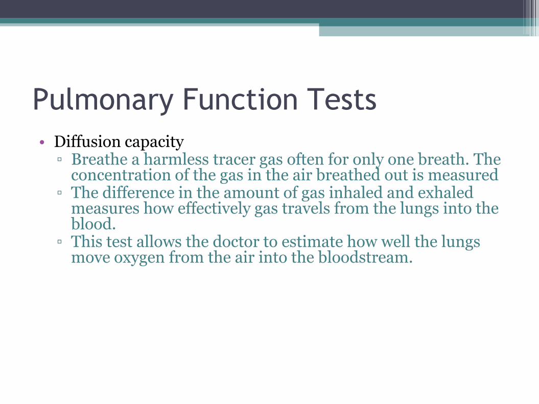
Pulmonary Function Tests
• Diffusion capacity ▫ Breathe a harmless tracer gas often for only one breath. The
concentration of the gas in the air breathed out is measured ▫ The difference in the amount of gas inhaled and exhaled
measures how effectively gas travels from the lungs into the blood.
▫ This test allows the doctor to estimate how well the lungs move oxygen from the air into the bloodstream.

Pulmonary Function Tests
• Forced expiratory volume (FEV1) ▫ The volume of air that can be forced out in one second after taking
a deep breath • Forced vital capacity (FVC)
▫ The amount of air that can be forcible exhaled after taking the deepest breath
• Functional residual capacity (FRC) ▫ Volume of air left in the lungs after a normal, passive exhalation
• Residual volume (RV) ▫ The volume of air remaining in the lungs after a maximum
exhalation • Total lung capacity (TLC)
▫ The volume of gas that is contained in the lungs at the end of maximal inspiration
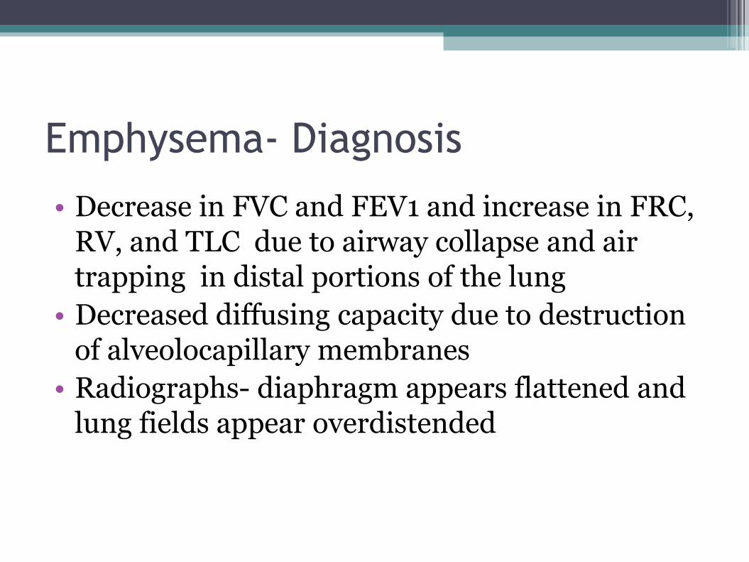
Emphysema- Diagnosis
• Decrease in FVC and FEV1 and increase in FRC, RV, and TLC due to airway collapse and air trapping in distal portions of the lung
• Decreased diffusing capacity due to destruction of alveolocapillary membranes
• Radiographs- diaphragm appears flattened and lung fields appear overdistended
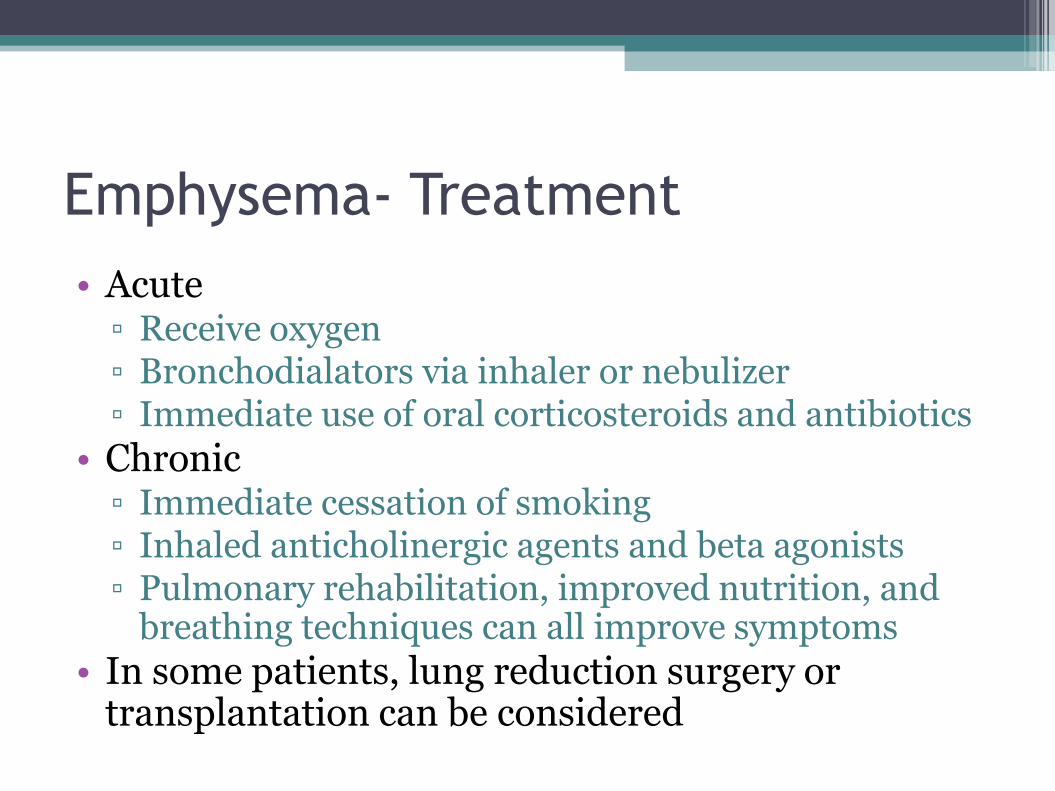
Emphysema- Treatment
• Acute ▫ Receive oxygen ▫ Bronchodialators via inhaler or nebulizer ▫ Immediate use of oral corticosteroids and antibiotics
• Chronic ▫ Immediate cessation of smoking ▫ Inhaled anticholinergic agents and beta agonists ▫ Pulmonary rehabilitation, improved nutrition, and
breathing techniques can all improve symptoms
• In some patients, lung reduction surgery or transplantation can be considered
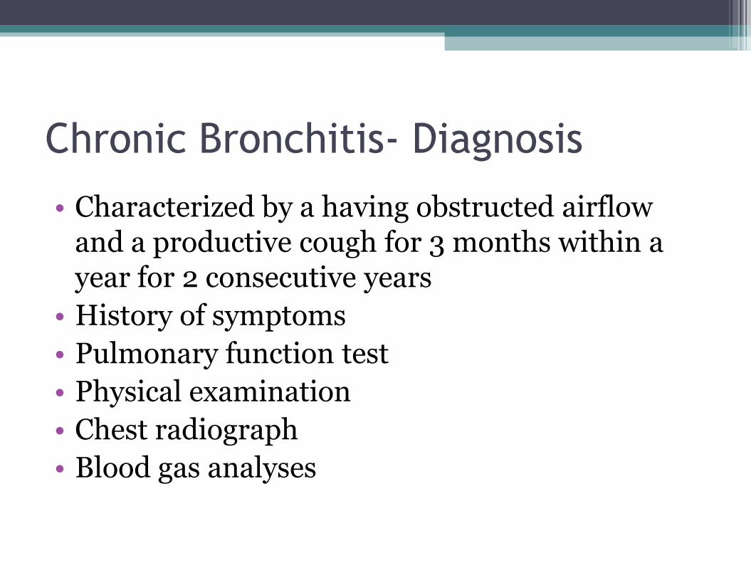
Chronic Bronchitis- Diagnosis
• Characterized by a having obstructed airflow and a productive cough for 3 months within a year for 2 consecutive years
• History of symptoms
• Pulmonary function test
• Physical examination
• Chest radiograph
• Blood gas analyses

Chronic Bronchitis- Treatment
• Pathologic changes are not reversible
• Bronchodilators and expectorants
▫ Control cough and reduce dyspnea
• Nutritional counseling
• Respiratory hygiene
• Oxygen therapy with severe hypoxemia
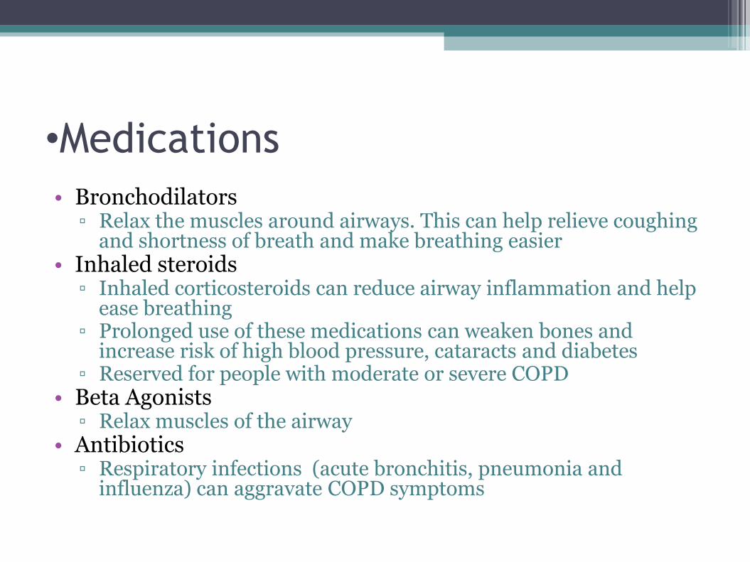
•Medications
• Bronchodilators ▫ Relax the muscles around airways. This can help relieve coughing
and shortness of breath and make breathing easier • Inhaled steroids
▫ Inhaled corticosteroids can reduce airway inflammation and help ease breathing
▫ Prolonged use of these medications can weaken bones and increase risk of high blood pressure, cataracts and diabetes
▫ Reserved for people with moderate or severe COPD • Beta Agonists
▫ Relax muscles of the airway • Antibiotics
▫ Respiratory infections (acute bronchitis, pneumonia and influenza) can aggravate COPD symptoms
Lung Transplants
• COPD is the most common cause in adults
• Aren’t very common due to small amount of donor organs
• A last resort treatment
• The are several tests that are done to make sure a patient is healthy enough to receive one.
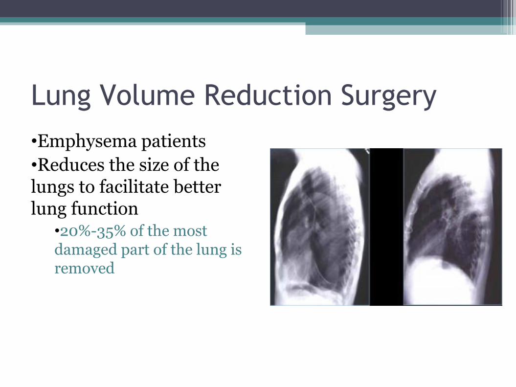
Lung Volume Reduction Surgery
•Emphysema patients
•Reduces the size of the lungs to facilitate better lung function
•20%-35% of the most damaged part of the lung is removed

Breathing Techniques
• Bending forward at the waist
• Allows diaphragm to move more easily
• Pursed Lip Breathing
• Breath in through nose and out through pursed lips
• Helps control dyspnea
Breathing Techniques
•Diaphragmatic Breathing •Lie on back on a flat surface with a pillow under knees and head
•Place left hand on upper chest and right hand on abdomen
•Inhale slowly through nose so stomach moves out against right hand
•Left hand shouldn’t move at all
•Tighten stomach muscles and exhale with pursed lip technique
•Left hand should not move
•Helps lungs expand and strengthens diaphragm

Nutrition Assessment
Nutritional Assessment

Nutritional Assessment
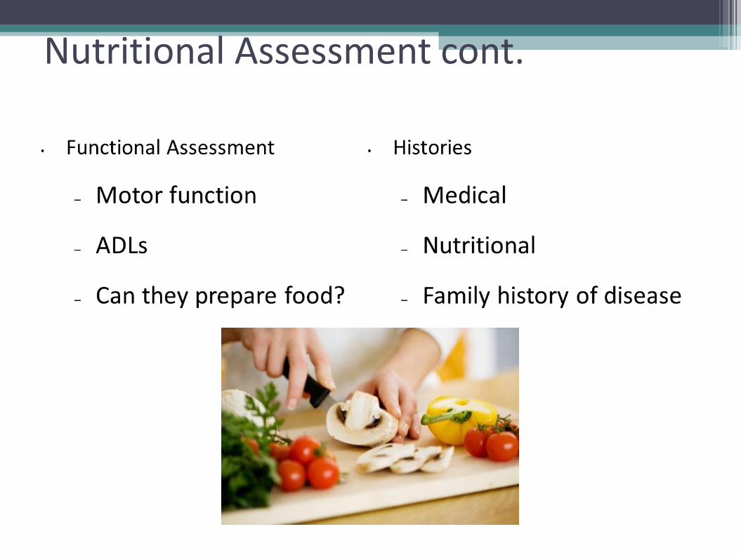
Nutritional Assessment cont.

Energy Expenditure Elevated
• Affected by:
– Airflow obstruction
– Gas diffusing capacity
– CO2 Retention
– Respiratory inflammation
– Biochemical status
• Cytokines
• Hormones
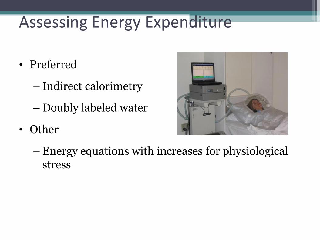
Assessing Energy Expenditure
• Preferred
– Indirect calorimetry
– Doubly labeled water
• Other
– Energy equations with increases for physiological stress
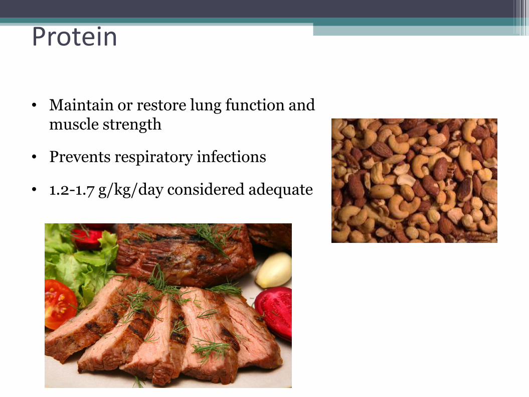
Protein
• Maintain or restore lung function and muscle strength
• Prevents respiratory infections
• 1.2-1.7 g/kg/day considered adequate
Vitamins and Minerals • Vitamin C
– For people still smoking
• Magnesium
– Muscle contraction and relaxation
• Calcium
– Maintain bone density
• Vitamin D and K
– If on glucocorticoids or have decreased bone mineral density
• Sodium restriction
– For cor pulmonale and fluid retention
Feeding Difficulties
Problems
• Adequate calorie intake
• Distended/bloated stomach
• Short of breath
• Small appetite
• Aspiration

Solutions to Feeding Difficulties

Enteral Supplementation
• Feeding tube
– Can increase nutrition intake
– Health can improve
– Once supplementation is removed, nutrition status will revert

Case Study
Patient
• Stella Bernhardt (SB)
• Age: 62
• Female
• Diagnosed with Stage 1 COPD 5 yrs ago
• Complaining of being short of breathe and not able to do anything for herself
• Quit smoking 1 yr ago

Anthropometric
• Ht: 5’3”
• Wt: 119 lb
• Usual weight: 145-150lbs
• Wt 1 yr ago: 139 lbs
– 14% wt loss
• BMI: 21.1 kg/m2
• % IBW: 103%
• MAC: 19.05 cm
• TSF: 15mm
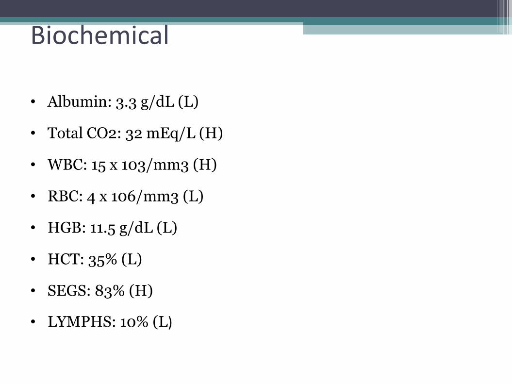
Biochemical
• Albumin: 3.3 g/dL (L)
• Total CO2: 32 mEq/L (H)
• WBC: 15 x 103/mm3 (H)
• RBC: 4 x 10⁶/mm3 (L)
• HGB: 11.5 g/dL (L)
• HCT: 35% (L)
• SEGS: 83% (H)
• LYMPHS: 10% (L)
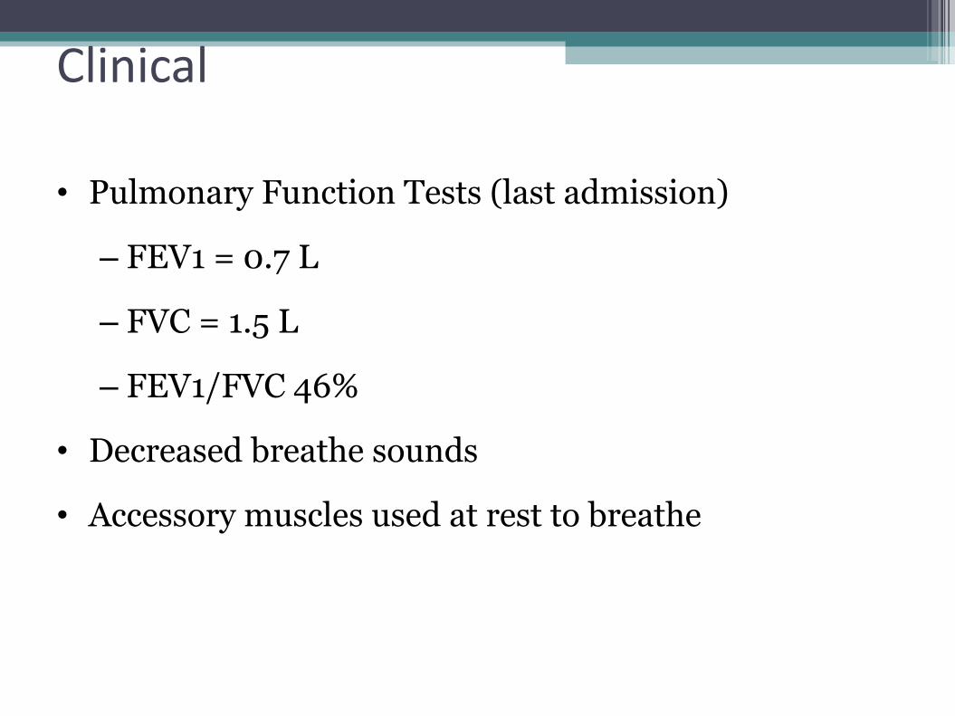
Clinical
• Pulmonary Function Tests (last admission)
– FEV1 = 0.7 L
– FVC = 1.5 L
– FEV1/FVC 46%
• Decreased breathe sounds
• Accessory muscles used at rest to breathe
Dietary
• Poor appetite stated
• Meal prep is hard because it makes her tired
– Daughters sometimes help
• Coughing decreases intake
• Avoids milk
– Claims increases mucus production
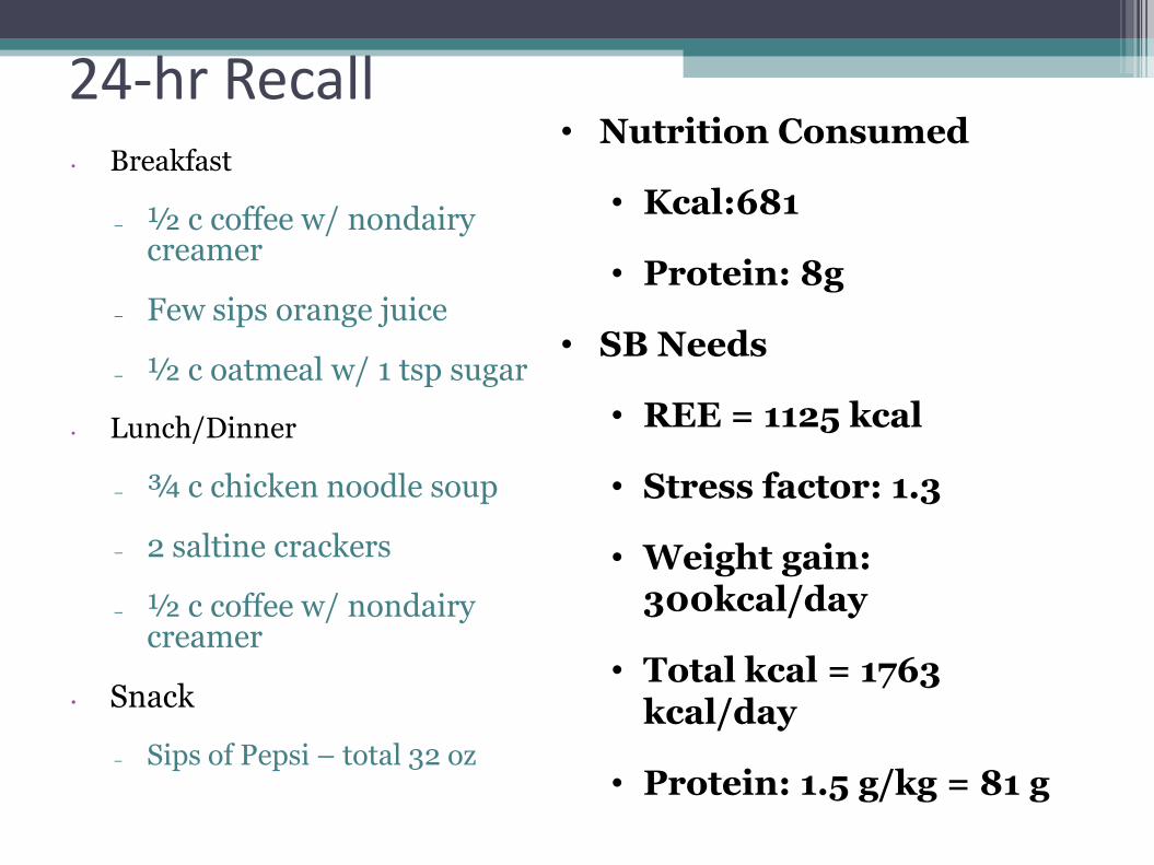
24-hr Recall • Breakfast
– ½ c coffee w/ nondairy creamer
– Few sips orange juice
– ½ c oatmeal w/ 1 tsp sugar
• Lunch/Dinner
– ¾ c chicken noodle soup
– 2 saltine crackers
– ½ c coffee w/ nondairy creamer
• Snack
– Sips of Pepsi – total 32 oz
• Nutrition Consumed
• Kcal:681
• Protein: 8g
• SB Needs
• REE = 1125 kcal
• Stress factor: 1.3
• Weight gain: 300kcal/day
• Total kcal = 1763 kcal/day
• Protein: 1.5 g/kg = 81 g
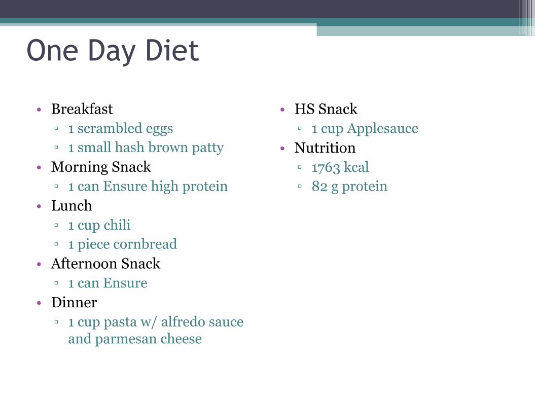
One Day Diet
• Breakfast
▫ 1 scrambled eggs
▫ 1 small hash brown patty
• Morning Snack
▫ 1 can Ensure high protein
• Lunch
▫ 1 cup chili
▫ 1 piece cornbread
• Afternoon Snack
▫ 1 can Ensure
• Dinner
▫ 1 cup pasta w/ alfredo sauce and parmesan cheese
• HS Snack
▫ 1 cup Applesauce
• Nutrition
▫ 1763 kcal
▫ 82 g protein
Functional History
• She can prepare food but tires her out
– Too tired to eat
• No other disabilities
• Diagnosed 5 yrs ago w/ COPD
• Quit smoking 1 yr ago
• Had a cold in the past two weeks
Doctor’s Diagnosis and Treatment
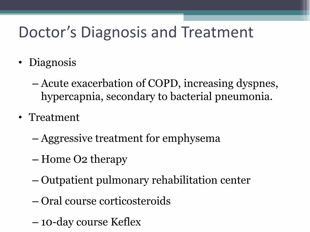
• Diagnosis
– Acute exacerbation of COPD, increasing dyspnes, hypercapnia, secondary to bacterial pneumonia.
• Treatment
– Aggressive treatment for emphysema
– Home O2 therapy
– Outpatient pulmonary rehabilitation center
– Oral course corticosteroids
– 10-day course Keflex
Nutrition Diagnosis
• SB has an inadequate energy intake related to a decreased ability to prepare food and anorexia as evidenced by her 14% unintentional weight loss in the last year.
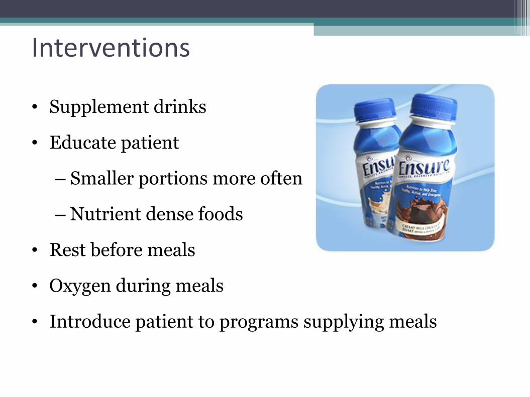
Interventions
• Supplement drinks
• Educate patient
– Smaller portions more often
– Nutrient dense foods
• Rest before meals
• Oxygen during meals
• Introduce patient to programs supplying meals
Monitoring and Evaluation
• Watch weight every three months
• Schedule a follow up appointment to see how she has implemented nutrition advice
• Encourage physical activity in order to allow easier breathing

References • Grant B. Medical nutrition therapy for pulmonary disease. In:
Mahan LK, Escott-Stump S, ed. Krause’e Food, Nutrition, & Diet Therapy. 12th ed. Philadelphia: Elsevier; 2008:900-916.
• PubMed Health. Chronic obstructive pulmonary disease. Available at http://www.ncbi.nlmnih.gov/pubmedhealth/PMH0001153/. Accessed on February 23, 2012.
• Currie P. ABC’s of COPD. 205;332:1142-1144.\ • Mannino DM, Buist SA. Global burden of COPD: Risk factors,
prevalence, and future trends. Lancet. 2007;370(9589):765-73. • Anto JM, Vermeire P, Vestboz J, Sunyer J. Epidemiology of chronic
obstructive pulmonary disease. Eur Respir J. 2001; 17(5): 982–994. • National Heart Lung and Blood Institute. What is alpha-1
antitrypsin deficiency?. Available at http://www.nhlbi.nih.gov/health/health-topics/topics/aat/. Accessed on February 23, 2012.
• Gary G. COPD Reference Guide. Boca Raton, FL: BarCharts Publishing Inc; 2008.
• Barnes PJ, Celli BR. Systemic manifestations and comorbidities of COPD. Eur Respir J. 2009; 33(5): 1165–1185.
References • O'Neill P. Nutrition for a patient with COPD can be complicated. Nursing.
2004;34(12):32hn6-32hn8. • Barnett M. Improving nursing management of nutrition in COPD patients. J Community
Nurs. 2009;23(3):32. • Global surveillance, prevention and control of chronic respiratory diseases: A
comprehensive approach; Geneva, Switzerland; World Health Organization (WHO); 2007.
• Chapman KR, Mannino DM, Soriano JB, Vermeire JB, Buist AS, Thun MJ. Epidemiology and costs of chronic obstructive pulmonary disease. Eur Respir J. 2006;27:188-207.
• Mannino DM, Gagnon RC, Petty TL, Lydick E. Obstructive lung disease and low lung function in adults in the United States: Data from the national health and nutrition examination survey, 1988–1994. Arch Intern Med. 2000;160:1683-1689.
• Zhong N, Wang C, Yao W, Chen P, Kang J, Huang S, Chen B, Wang C, Ni D, Zhou Y, Liu S, Wang X, Wang D, Lu J. Prevalence of chronic obstructive pulmonary disease in China: A large, population-based survey. Am J Respir Crit Care Med. 2007; 176:753–760.
• Mannino DM, Homa DM, Akinbami LJ, Ford ES, Redd SC. Chronic Obstructive Pulmonary Disease Surveillance—United States, 1971—2000. Morb Mortal Wkly Rep. 2002;51(6):1-16.
• Afonso AS, Verhamme KM, Sturkenboom MC, Brusselle GG. COPD in the general population: Prevalence, incidence, and survival. Respir Med. 2011;105:1872-1884.
• National Heart Lung and Blood Institute. What is COPD?. Available at http://www.nhlbi.nih.gov/health/health-topics/topics/copd/. Accessed on February 21, 2012.
• Judd AM. Lecture notes. Pathophysiology, Brigham Young University, February 21, 2012.