Chronic obstructive pulmonary disease by aminu arzet
47
Chronic Obstructive Pulmonary Disease (COPD) ______________________________ Aminu Arzet Department of Internal Medicine Nelson Mandela School of Medicine University of K-Natal Durban 27 th March,2015
-
Upload
aminuarzet -
Category
Health & Medicine
-
view
263 -
download
1
Transcript of Chronic obstructive pulmonary disease by aminu arzet

Chronic Obstructive Pulmonary Disease
(COPD)______________________________
Aminu ArzetDepartment of Internal Medicine
Nelson Mandela School of MedicineUniversity of K-Natal
Durban
27th March,2015

ICU 2B/Respiratory unit presentation
Inkosi Albert Luthuli Central Hospital (IALCH)
DURBAN

Introduction
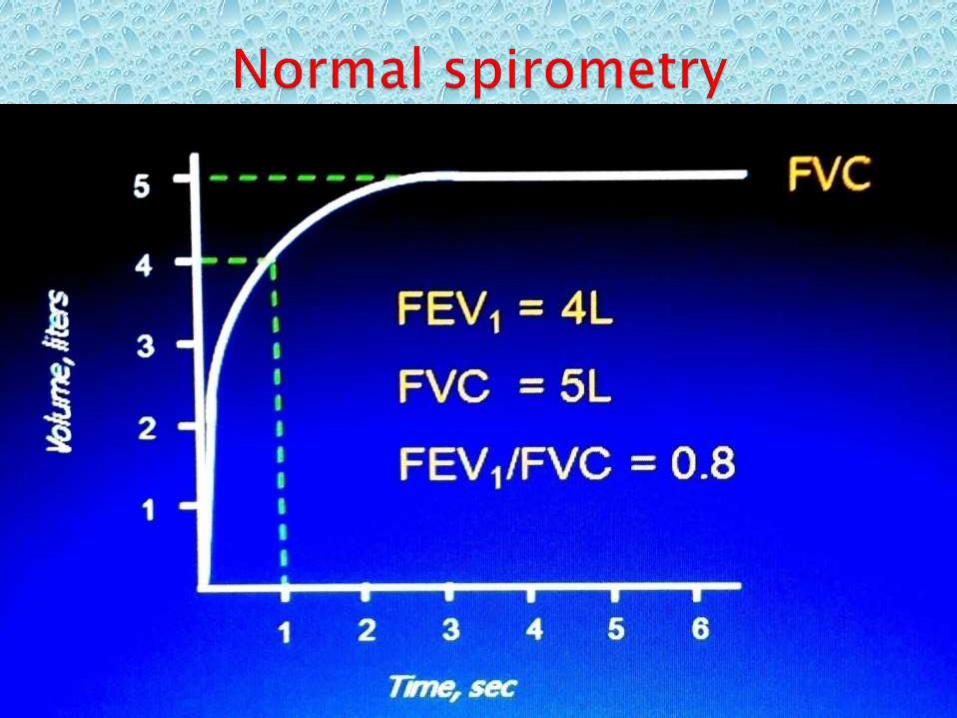
COPD Is a lung disease characterized by persistent, progressive airway obstruction, that leads to poor airflow in and out of lungs, with resultantreduction in FEV1and FEV1/FVC ratio.
The lung function impairment is fixed, but some reversibility can be achieved by using bronchodilator/other therapies

Introduction Continuation
Its associated with shortness of breath, cough, and sputum production, which is due to chronic inflammatory response in the airways and lungs to noxious substances.
It commonly involve narrowing of small airways(small airway disease) and breakdown of lung tissue,with resultant entrapment of air (emphysema).

Epidemiology
COPD is a leading course of morbidity and mortality world over
It affects about 5% of world population (330 million people)
In 2012, it ranked as the 3rd leading cause of death, killing over 3 million people globally.
Epidemiology Continuation
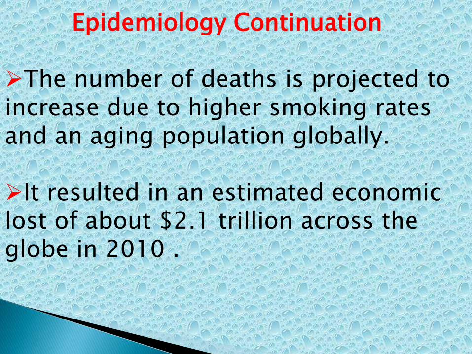
The number of deaths is projected to increase due to higher smoking rates and an aging population globally.
It resulted in an estimated economic lost of about $2.1 trillion across the globe in 2010 .

Risk FactorsCigarette smoking is the major risk factor for COPD
Occupational exposure to dust and chemicals
Environmental pollution from Car exhaust, tobacco smoke, wild fire/bush burning, poorly ventilated indoor cooking fires using biomass fuel(BMF).

Risk factor continuation
Genetic makeup of individual- Alpha1 antitrypsin deficiency
Recurrent bronchopulmonary infections
Socioeconomic status-commoner among less privileged.

Pathophysiology
Air ways obstruction occur as a result of chronic inflammatory response to inhaled noxious substances and recurrent infection.
There is excessive release of inflammatory mediators like Nuetrophils, Macrophages, Lymphocytes, Histamines, Leukotrienes, Cytokines,Chemokines, free radicals,etc
Pathophysiology Cont.
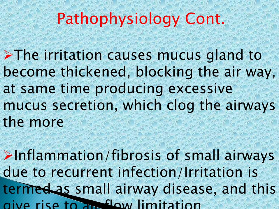
The irritation causes mucus gland to become thickened, blocking the air way, at same time producing excessive mucus secretion, which clog the airways the more
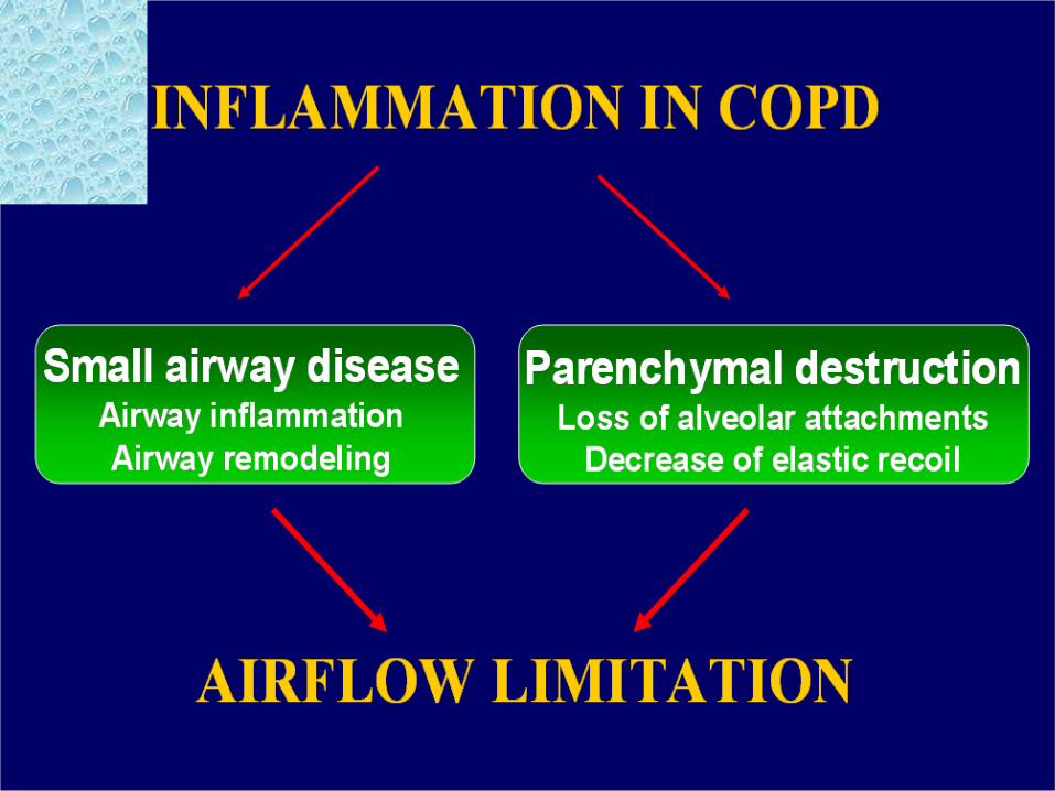
Inflammation/fibrosis of small airways due to recurrent infection/Irritation is termed as small airway disease, and this give rise to air flow limitation
Pathophysiology Cont.
Alveolar wall destruction due to breakage of alveolar attachment, and lost of elasticity with resultant decrease in elastic recoil, are another cause of air entrapment, and emphysema ensued.
In addition to aforementioned, Pulmonary capillary bed damage and attendant pulmonary edema, amplify the airflow limitation.

Pathophysiology Cont.Lung damage also occur due to break down of lung tissue by inflammatory cells and released proteases, which are insufficiently inhibited, due to lack of Alpha1antitrypsin(anti protease).
Alpha1antrypsin deficiency is genetically mediated, but cigarette smoking is believed to potentiate that, by stimulating release of free radicals and inflammatory cells.

Clinical features Symptoms: Include shortness of
breath, cough, sputum production, dyspnoea, and wheeze.
Signs:
Pink puffers-Thin body build, with expiratory pursed -lip breathing
Blue bloaters–cyanosis with mild activity

Clinical features Patients
Patient who have chronic cough and sputum production with a history of exposure to risk factors, should be tested for airflow limitation, even if they do not have dyspnea .

Physical examination Pt has large, barrel shaped chest,
Prominent accessory respiratory muscles in the neck.
Low, flat diaphragm, causing costal margin retractions on inspiration.
Hyperimplated lungs with diminished breath sounds, distant HS, prolonged expiration with generalized wheezes predominantly on expiration.

Physical examinations cont
Depressed liver, which is not enlarged.
In ‘blue bloater’ type of COPD, patient may also have:
Cyanosis at rest or mild exertion.Pedal oedemaCrackles at lung bases.Loud second heart sound in pulmonary area (difficult to hear in COPD).

Physical examinations cont
In ‘pink puffer’ type of COPD patient may also have:
expiratory pursed-lip breathing, thin body build and tendency to lean forward over a support to assist breathing
Investigations
Plain chest X-ray shows
1. Low flattened diaphragms.
2. An obtuse costophrenic angle.
4. A reduction in size and numbers of pulmonary vessels, particularly in the periphery of the lung.
5. Vessel distortion producing increased branching, angles or bowing of vessels
CT CHEST It shows areas of low attenuation
without obvious margins or walls.
Abnormal vascular configuration.
CT Scan is the most sensitive and specific imaging technique for assessing Emphysema

Diagnosis Clinically based on dyspnoea,
Chronic cough and exposure to risk factors.
Spirometry is the gold standard.
Post bronchodilator FEV1/FVC < 70% or FEV1 < 80% of predicted value, confirms the presence of airflow limitation that is not fully reversible.
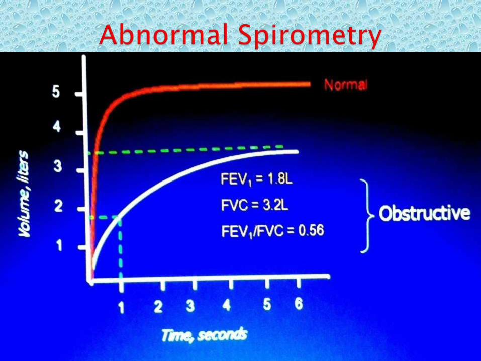
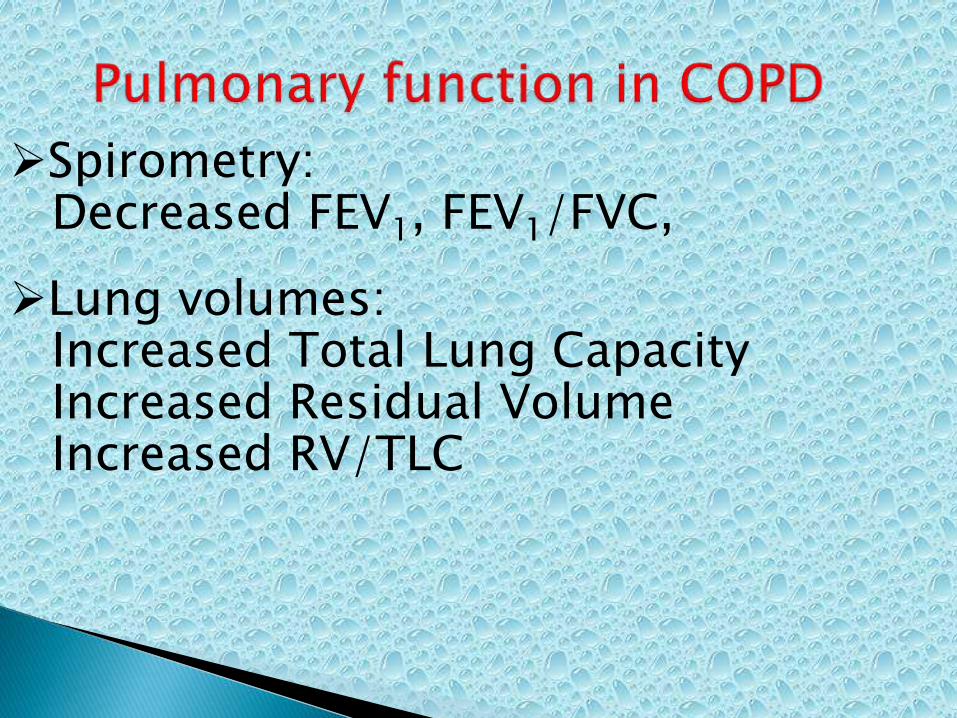
Spirometry:Decreased FEV1, FEV1/FVC,
Lung volumes:Increased Total Lung CapacityIncreased Residual VolumeIncreased RV/TLC

Additional investigations
1.Bronchodilator reversibility testing
2. Glucocorticosteroid reversibility testing
3. Arterial blood gas measurement
4. Alpha1 antitrypsin deficiency screening

Additional investigations cont.
Alpha 1 antitrypsin screening is done in the fallowing settings:
- COPD develops under 45
- COPD develops in non-smoker
- Strong family history of COPD
Normal: >150 mg/dL , In disease: <45 mg/dL
Differential diagnosis
Asthma-due to bronchoconstriction
Congestive Heart Failure
Bronchiectasis
Tuberculosis
Obliterative Bronchiolitis
Management
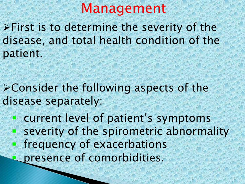
First is to determine the severity of the disease, and total health condition of the patient.
Consider the following aspects of the disease separately:
current level of patient’s symptoms severity of the spirometric abnormality frequency of exacerbations
presence of comorbidities.
Symptoms assessment
COPD symptoms are chronic and progressive dyspnea, cough, and sputum production that can vary from day to day.
In symptoms assessment these features are objectively graded using the fallowing scoring systems:

Stmptoms assesement cont.
1. COPD Assessment Test (CAT): An 8 items measure of health status impairment in COPD
2. Clinical COPD Questionnaire (CCQ): This is self administered questionnaire, developed to measure clinical control in patients with COPD.

Symptoms assesement cont
3. Modified British Medical Research Council(mMRC) Questionnaire: It also assesses health status and predicts future mortality risk. It has a score of 0-4

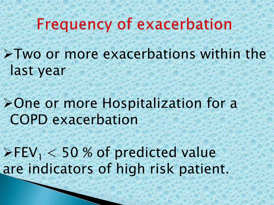
Two or more exacerbations within the last year
One or more Hospitalization for a COPD exacerbation
FEV1 < 50 % of predicted value are indicators of high risk patient.


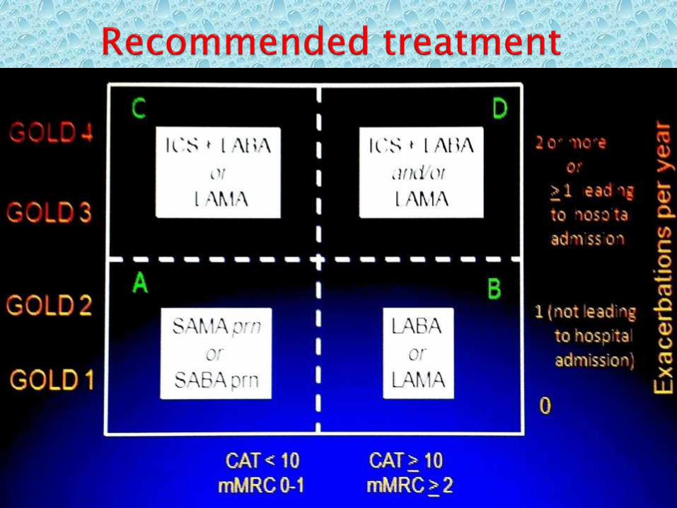
Stage Characteristics Recommended Treatment
All * Avoidance of risk factor (s)
* Influenza vaccination
0: At risk * Chronic Symptoms
(cough, Sputum)
* Exposure to risk factors
* Normal spirometry
Mild COPD * FEV1/FVC < 70% *Short acting B/dilator
* FEV1 80% predicted when needed or SAMA
* With or without symptoms

Therapy at Each Stage of COPD
Stage Characteristics Recommended Treatment
Moderate FEV1 50 -79% *Regular treatment * Inhaled Gluccocorti COPDD with one or more costeorodis +
bronchodilators LABA or LAMA
* Rehabilitation Symptoms and lung
function response

Therapy at Each Stage of COPD
Stage Characteristics Recommended Treatment
Severe COPD FEV1 30-49% * Regular treatment with LABA+
and or LAMA
Very Severe FEV1<30% * Inhaled glucorticosteroids if
COPD significant symptoms or repeated
exacerbations.
* Treatment of complications.
* Consider surgical treatments -
lung volume reduction /transplant
* Long-term oxygen therapy if
in respiratory failure
* Rehabilitation- exercise,
*Health Education-cessation of smoking


COPD Comorbidities
COPD patients are at increased risk for:
CVS disease like Corpulmonale
Osteoporosis
Respiratory infections
Anxiety and Depression
Diabetes
Lung cancer
Bronchiectasis
Prognosis
Prognosis is generally poor, unless if detected early and management start
in early stage.

THANK YOU

Reference
•GOLD 2014/2006 document
•Kumar and ckerk
•Harrison
•Davidsons
•British medical journal
•Wikipedia