
Chronic Lymphocytic Leukemia FISH Panel Impact on Diagnosiscllcanada.ca/2010/pages/CLL_FISH...
10
Am J Clin Pathol 2007;128:323-332 323 323 DOI: 10.1309/21TN2RUWKR827UW2 323 © American Society for Clinical Pathology Hematopathology / CLL, FISH, AND 14Q32 TRANSLOCATIONS Chronic Lymphocytic Leukemia FISH Panel Impact on Diagnosis Beverly P. Nelson, MD, 1 Rohit Gupta, MD, 1 Gordon W. Dewald, PhD, 2 Sarah F. Paternoster, 2 Steven T. Rosen, MD, 3 and LoAnn C. Peterson, MD 1 Key Words: Chronic lymphocytic leukemia; Fluorescence in situ hybridization; FISH; 14q32 Translocation DOI: 10.1309/21TN2RUWKR827UW2 Abstract Interphase fluorescence in situ hybridization (FISH) is an alternative to conventional chromosome analysis of chronic lymphocytic leukemia (CLL) cells. We analyzed 172 samples from 136 possible CLL cases using a FISH panel. Reflex testing with probes to CCND1, BCL2, BCL3, BCL11A, c-MYC, MALT1, and a break-apart immunoglobulin heavy chain (IGH) probe was done if more than 2 signals for 14q32 occurred. For 111 cases, there were sufficient data for analysis. Of 111 cases, 81 (72.9%) had 1 or more genetic abnormalities. The most frequent abnormality was 13q–, followed by trisomy 12, 11q–, and 17p–. In 13 cases, there were IGH abnormalities. Two cases with CCND1/IGH fusion were reclassified as mantle cell lymphoma. Four CLL cases had IGH fusion with BCL2, BCL3 (2 cases), and BCL11A; no fusion partner was detected in 7 cases. Morphologic features were atypical for CLL in 2 cases with IGH fusion (BCL11A and BCL3). The FISH CLL panel is useful to identify prognostic aberrations and to clarify diagnosis in cases with unusual morphologic features. B-cell chronic lymphocytic leukemia (CLL) is the most common leukemia in adults. The morphologic features and immunophenotype of CLL cells are well characterized. A unique, recurring genetic alteration has not been identified in CLL, but chromosomal aberrations occurring in CLL have been linked to prognosis. 1-3 For example, deletions of chromo- somes 17p and 11q are associated with an adverse clinical out- come, with overall survival of 3 years and 6 to 7 years, respec- tively. Patients with isolated 13q deletions have a favorable clinical course and best overall survival, 11 years. The clinical course of patients with CLL with trisomy 12q is intermediate between the groups with inferior and favorable outcomes. Early genetic studies of CLL using conventional chromo- some banding analysis detected chromosomal aberrations in 40% to 60% of cases. 4,5 However, only dividing cells are eval- uated by chromosome banding techniques. Because it has been difficult to stimulate CLL cells to divide, novel stimula- tion techniques have been reported to improve the detection of chromosomal aberrations, particularly translocations, in CLL lymphocytes. 1 Nevertheless, fluorescence in situ hybridization (FISH), which allows analysis of dividing and nondividing cells, is increasingly being offered as an alternative to conven- tional chromosome banding. With FISH, up to 80% of CLL cases demonstrate genetic alterations. 2 In this report, we review the Northwestern Memorial Hospital (NMH; Chicago, IL) experience with a FISH panel for CLL after it was implemented as part of the routine procedure to assess prognosis. Morphologic and flow cytometric evalua- tion of peripheral blood and/or bone marrow samples is routine- ly performed at our institution in all new cases in which CLL is considered a possible diagnosis. When FISH is performed, the results are correlated with the initial diagnostic impression.
-
Upload
nguyenxuyen -
Category
Documents
-
view
220 -
download
0
Transcript of Chronic Lymphocytic Leukemia FISH Panel Impact on Diagnosiscllcanada.ca/2010/pages/CLL_FISH...

Am J Clin Pathol 2007;128:323-332 323323 DOI: 10.1309/21TN2RUWKR827UW2 323
© American Society for Clinical Pathology
Hematopathology / CLL, FISH, AND 14Q32 TRANSLOCATIONS
Chronic Lymphocytic Leukemia FISH Panel
Impact on Diagnosis
Beverly P. Nelson, MD,1 Rohit Gupta, MD,1 Gordon W. Dewald, PhD,2 Sarah F. Paternoster,2
Steven T. Rosen, MD,3 and LoAnn C. Peterson, MD1
Key Words: Chronic lymphocytic leukemia; Fluorescence in situ hybridization; FISH; 14q32 Translocation
DOI: 10.1309/21TN2RUWKR827UW2
A b s t r a c tInterphase fluorescence in situ hybridization
(FISH) is an alternative to conventional chromosomeanalysis of chronic lymphocytic leukemia (CLL) cells.We analyzed 172 samples from 136 possible CLL casesusing a FISH panel. Reflex testing with probes toCCND1, BCL2, BCL3, BCL11A, c-MYC, MALT1, anda break-apart immunoglobulin heavy chain (IGH)probe was done if more than 2 signals for 14q32occurred. For 111 cases, there were sufficient data foranalysis. Of 111 cases, 81 (72.9%) had 1 or moregenetic abnormalities. The most frequent abnormalitywas 13q–, followed by trisomy 12, 11q–, and 17p–. In13 cases, there were IGH abnormalities. Two cases withCCND1/IGH fusion were reclassified as mantle celllymphoma. Four CLL cases had IGH fusion with BCL2,BCL3 (2 cases), and BCL11A; no fusion partner wasdetected in 7 cases. Morphologic features were atypicalfor CLL in 2 cases with IGH fusion (BCL11A andBCL3). The FISH CLL panel is useful to identifyprognostic aberrations and to clarify diagnosis in caseswith unusual morphologic features.
B-cell chronic lymphocytic leukemia (CLL) is the mostcommon leukemia in adults. The morphologic features andimmunophenotype of CLL cells are well characterized. Aunique, recurring genetic alteration has not been identified inCLL, but chromosomal aberrations occurring in CLL havebeen linked to prognosis.1-3 For example, deletions of chromo-somes 17p and 11q are associated with an adverse clinical out-come, with overall survival of 3 years and 6 to 7 years, respec-tively. Patients with isolated 13q deletions have a favorableclinical course and best overall survival, 11 years. The clinicalcourse of patients with CLL with trisomy 12q is intermediatebetween the groups with inferior and favorable outcomes.
Early genetic studies of CLL using conventional chromo-some banding analysis detected chromosomal aberrations in40% to 60% of cases.4,5 However, only dividing cells are eval-uated by chromosome banding techniques. Because it hasbeen difficult to stimulate CLL cells to divide, novel stimula-tion techniques have been reported to improve the detection ofchromosomal aberrations, particularly translocations, in CLLlymphocytes.1 Nevertheless, fluorescence in situ hybridization(FISH), which allows analysis of dividing and nondividingcells, is increasingly being offered as an alternative to conven-tional chromosome banding. With FISH, up to 80% of CLLcases demonstrate genetic alterations.2
In this report, we review the Northwestern MemorialHospital (NMH; Chicago, IL) experience with a FISH panel forCLL after it was implemented as part of the routine procedureto assess prognosis. Morphologic and flow cytometric evalua-tion of peripheral blood and/or bone marrow samples is routine-ly performed at our institution in all new cases in which CLL isconsidered a possible diagnosis. When FISH is performed, theresults are correlated with the initial diagnostic impression.

324 Am J Clin Pathol 2007;128:323-332324 DOI: 10.1309/21TN2RUWKR827UW2
© American Society for Clinical Pathology
Nelson et al / CLL, FISH, AND 14Q32 TRANSLOCATIONS
In this study, the FISH results not only provided prognosticinformation, but also added data that in some cases supportedCLL when the morphologic features were not classic for CLLand in others changed the initial diagnostic impression.
Materials and Methods
Between June 2002 and November 2005, samples ofperipheral blood, bone marrow, or lymph nodes from 136patients with a working or established diagnosis of CLL wereanalyzed with a FISH panel at Mayo Medical Laboratories,Rochester, MN, using the following probes: c-MYB (6q23),D6Z1 (6cen), ATM (11q22.3), CCND1 (11q13), D11Z1(11cen), D12Z3 (12cen), MDM2 (12q15), D13S319 (13q14),LAMP1 (13q34), p53 (17p13.1), D17Z1 (17cen), andimmunoglobulin heavy chain (IGH; 14q32). When more than2 signals for chromosome 14q32 were identified, reflex test-ing using additional FISH probes was performed to detectfusion of IGH with CCND1 (11q13), BCL2 (18q21), BCL3(19q13), BCL11A (2p13), c-MYC (8q24), or MALT1(18q21). A break-apart probe for IGH was also used to deter-mine if the additional IGH signal was due to trisomy 14 orIGH translocations involving loci other than those tested.Normal values, sensitivity, and specificity for each probe andsets of probes were established at Mayo Medical Laboratoriesand have been previously published.6,7 Commercial probes forcyclin D1, bcl-2, bcl-6, c-myc, and MALT-1 were purchasedfrom Vysis, Des Plaines, IL. “Homebrew” probes for bcl-3and bcl-11a were made at Mayo Medical Laboratories. Caseswere stratified into 3 risk groups as follows: good, normal or13q– only; intermediate, +12 only; or poor, 6q–,11q–, or17p–. CLL cases with more than 1 genetic abnormality butwith 17p– were placed in the 17p– group, and those with 11q–but not 17p– were placed in the 11q– group.
Immunophenotyping using flow cytometry was performedon blood samples, bone marrow aspirates, or lymph node biop-sy specimens in all cases using previously published proce-dures.8 Briefly, 4-color immunophenotyping was performedevaluating surface antigens using antibodies in the followingcombinations: CD45/CD19/CD56/CD3; CD2/CD3/CD7/CD5;CD8/CD3/CD4/CD25; CD10/CD19/!/"; CD19/CD20/CD11c/CD103; CD38/CD19/CD5/CD79b; CD25/CD19/CD23/FMC7;CD34/CD45/CD10/CD19; and CD33/CD45/CD13/CD14. Inaddition, after the cells were incubated with antibodies directedagainst surface antigens, cells were permeabilized using theIntra-Prep kit (Beckman Coulter, Miami, FL) to evaluate forintracellular antigens terminal deoxynucleotidyl transferase(TdT), myeloperoxidase, and CD79a in the following combi-nations: CD45/CD19/CD3/TdT and CD45/CD3/CD79a/myeloperoxidase. The antibodies were directly labeled with flu-orescein isothiocyanate, phycoerythrin, phycoerythrin–cyanin
5, or phycoerythrin–Texas red. CD79b was purchased fromDAKO, Fort Collins, CO, and CD52 from Caltag (Invitrogen),Carlsbad, CA; the remainder of the antibodies were purchasedfrom Beckman Coulter, Miami, FL.
The following immunophenotype was interpreted astypical for CLL: CD5+, dim to moderate CD20+, andCD23+ monotypic B cells. Brightly positive staining forCD20, FMC7, and/or CD79b or negative staining forCD23 was regarded as an atypical immunophenotype forCLL. CD38 was regarded as positive if a distinct popula-tion of the lymphocytes displayed greater staining intensi-ty than the granulocytes in the sample and as dim if thestaining intensity overlapped with the granulocytes andwas consistent even following blocking with 100% normalmouse serum. The #-associated protein (ZAP-70) stainingpattern was evaluated in selected cases using previouslypublished procedures.9
CBC count and leukocyte differentials were performedon peripheral blood samples. Peripheral blood samplesand/or bone marrow aspirate smears were stained withWright-Giemsa for morphologic evaluation. B-5–fixed,decalcified bone marrow trephine biopsy specimens werestained with H&E before histologic evaluation. The morpho-logic features of lymphocytes in blood and/or bone marrowwere reviewed in all cases without knowledge ofimmunophenotype or FISH results.
Lymphocyte morphologic features were consideredtypical for CLL if the lymphocytes were small with roundnuclei, condensed chromatin, and scant cytoplasm and pro-lymphocytes were fewer than 10%; morphologic featureswere considered atypical if lymphocytes displayed irregu-larly shaped nuclei or dispersed chromatin or prolympho-cytes were greater than 10%. The morphologic featureswere correlated with the immunophenotype to arrive at aninitial diagnostic impression. When the FISH resultsbecame available, they were correlated with the initial diag-nostic impression.
Results
Summary of CasesA total of 136 cases of presumed CLL were referred for
the FISH panel. Three cases were excluded from analysisbecause neither immunophenotypic nor morphologic datawere available to correlate with the FISH results, and 22 werenot CLL based on review of the morphologic features andimmunophenotype. The remaining 111 cases included 71 menand 40 women ranging in age from 25 to 84 years (median, 61years). Specimens used for FISH analysis were peripheralblood (61), bone marrow (49), and lymph node (1).

Am J Clin Pathol 2007;128:323-332 325325 DOI: 10.1309/21TN2RUWKR827UW2 325
© American Society for Clinical Pathology
FISH Results
Cases With CCND1/IGH Fusion Classified as Mantle CellLymphoma
Two cases initially regarded as CLL based on clinicalmanifestations demonstrated CCND1/IGH fusion, indicatingbcl-1 gene rearrangement. One patient, a 41-year-old man,had marked lymphocytosis in the peripheral blood (WBCcount, 155,400/µL [155.4 ! 109/L]; hemoglobin, 10.6 g/dL[106 g/L]; mean corpuscular volume [MCV], 90 µm3 [90 fL];platelet count, 343 ! 103/µL [343 ! 109/L]; lymphocytes,96.5% [0.97]; and neutrophils, 3.5% [0.04]). The lymphocytesvaried from small cells with condensed chromatin and roundnuclei to medium-sized cells with slightly irregular nuclei!Image 1!. Flow cytometric immunophenotyping showedmonotypic B cells that were CD19+, CD20+, CD5+, brightCD23+, bright FMC7+, and bright CD79b+. In addition toCCND1/IGH fusion, FISH also showed deletion of chromo-somes 11q and 13q.
The second patient, a 76-year-old man, had absolute lym-phocytosis (WBC count, 11,500/µL [11.5 ! 109/L]; hemoglo-bin, 10.4 g/dL [104 g/L]; MCV, 76 µm3 [76 fL]; platelet count,155 ! 103/µL [155 ! 109/L]; lymphocytes, 50.2% [0.50]; neu-trophils 35.4% [0.35]; monocytes, 10.3% [0.10]; eosinophils,3.1% [0.03]; and basophils, 1.0% [0.01]). Lymphocyte morpho-logic features were unusual for CLL; the cells were primarilysmall with condensed chromatin and scant cytoplasm butincluded occasional larger cells with visible nucleoli. Flowcytometric immunophenotyping of the blood showed monotyp-ic B cells that were CD5+, CD10–, CD20+, dim CD23+, brightFMC7+, and bright CD79b+. In addition to CCND1/IHGfusion, 13q and 17p deletions were also identified.
Although both cases were clinically regarded as CLL,their morphologic features and immunophenotype were atypi-cal for CLL. Even though both cases were CD20+, CD5+, andCD23+, the bright FMC7 and CD79b positivity are unusual forCLL. FISH demonstrated CCND1/IGH fusion, and the caseswere classified as mantle cell lymphoma (MCL).
CLL CasesOf 109 CLL cases, 79 (72.5%) had 1 or more genetic
abnormalities; the remaining 30 cases (27.5%) had normalFISH results. The majority of the patients with genetic alter-ations, 67% (53/79), had a single abnormality. Two alterationswere present in 25% (20/79), and 8% (6/79) had 3 or moregenetic alterations.
Chromosome 13q deletion (13q–) was the most commonaberration !Table 1!. Deletion of chromosome 13q was theonly abnormality detected in more than half (33/53 [62%]) ofthe cases with 13q–. Trisomy 12 was the second most frequentabnormality and was the sole abnormality in 13 cases.Deletion of chromosome 11q was the third most common
finding. Abnormalities of chromosomes 17p and 14q32occurred with similar frequencies. The least common abnor-mality was deletion of chromosome 6q.
An abnormal signal pattern for the IGH gene was presentin 11 CLL cases. Translocations were identified in 4 of thesecases !Table 2!. One or more extra IGH signals without anidentifiable fusion partner were present in 6 cases, and the 3'end of the IGH variable region (IGHv) signal was absent in 1case !Table 3!.
FISH Results Correlated With Phenotypic, Morphologic,and Other Pathologic Findings
Immunophenotypic and Morphologic FindingsA total of 109 cases displayed typical phenotypes, and 2
were atypical with bright CD20+ results. The 2 CLL cases
Hematopathology / ORIGINAL ARTICLE
!Image 1! Mantle cell lymphoma in peripheral blood. Thelymphocytes show condensed chromatin, with round nuclei,but cells with slightly irregular nuclei and visible nucleoli thatare unusual for chronic lymphocytic leukemia are also present(Wright-Giemsa, !1,000).
!Table 1!Chromosomal Aberration Frequency by Fluorescence In SituHybridization for 79 Clonal Chronic Lymphocytic LeukemiaCases*
Chromosomal Abnormality No. (%) of Cases
13q– 53 (67)Trisomy 12 25 (32)11q– 17 (22)14q32 11 (14)17p– 12 (15)6q– 4 (5)
* More than 1 aberration was present in 26 cases.

326 Am J Clin Pathol 2007;128:323-332326 DOI: 10.1309/21TN2RUWKR827UW2
© American Society for Clinical Pathology
Nelson et al / CLL, FISH, AND 14Q32 TRANSLOCATIONS
with bright CD20 staining had isolated trisomy 12. Nine CLLcases displayed atypical morphologic features for CLL; theyincluded 2 with translocations involving chromosome 14q32(bcl-3 and bcl-11a; discussed in the next section), 3 with iso-lated trisomy 12, 2 with isolated 13q deletion, and 2 with nor-mal FISH results.
CLL Cases With Chromosome 14q32 TranslocationsEleven CLL cases demonstrated abnormalities involving
the IGH gene located at chromosome 14q32. Of these, 4 hadhad translocations (Table 2). These cases are discussed inmore detail because the FISH results were informative for thediagnoses.
The first case, a 46-year-old man, had absolute lympho-cytosis in the blood (WBC count, 32,100/µL [32.1 ! 109/L];hemoglobin, 14.3 g/dL [143 g/L]; hematocrit, 43.8% [0.44];MCV, 84 µm3 [84 fL]; platelet count, 286 ! 103/µL [286 !109/L]; lymphocytes, 86% [0.86]; neutrophils, 10% [0.10];bands, 2% [0.02]; monocytes, 1% [0.01]; and eosinophils, 1%[0.01]) associated with lymphadenopathy and splenomegaly.A bone marrow biopsy showed a lymphoid infiltrate with adiffuse growth pattern involving approximately 80% of thesection. The lymphocytes were primarily small with con-densed chromatin but included many cells with angulated orcleaved nuclei !Image 2A!. The morphologic features of thelymphocytes were not typical for CLL and raised the possibil-ity of another type of non-Hodgkin lymphoma.
Flow cytometric immunophenotyping findings, however,were characteristic of CLL: " surface immunoglobulin (sIg)light chain–restricted B cells that were CD19+/CD20+/CD5+/CD10–/CD23+/CD79b–/FMC7–. Small lymphocytic lym-phoma/CLL was confirmed with a lymph node biopsy thatshowed effacement of the normal architecture by a proliferationof small lymphocytes with a diffuse growth pattern that includ-ed proliferation centers !Image 2B!. FISH analysis revealedfusion of IGH and bcl-11a, indicating t(2;14)(p13;q32), whichrarely occurs in CLL.10,11 Deletion of the long arm of chromo-some 11 was also identified.
His illness was characterized by development of massivegeneralized lymphadenopathy that encased major blood ves-sels, including the inferior vena cava, aorta, common iliacarteries, portal vein, splenic vein, and superior mesentericvein. Multiple bilateral pulmonary nodules, splenomegalythat required radiation therapy for symptom management,severe thrombocytopenia (platelet count, 4 ! 103/µL [4 !109/L]), and anemia (hemoglobin, 7.4 g/dL [74 g/L]) alsodeveloped. He was treated with several different chemother-apeutic regimens throughout the disease course. The thera-pies included fludarabine; fludarabine/cyclophosphamide;cyclophosphamide, doxorubicin, vincristine, and prednisone;and pentostatin/rituximab. He died of progressive disease 51months after initial diagnosis.
The second case was a 52-year-old man who had absolutelymphocytosis in the blood (WBC count, 85,000/µL [85.0 !
!Table 3!Chronic Lymphocytic Leukemia Cases With IgH Abnormalities and No Translocations Identified
Chromosomal AbnormalityCase No./Sex/ TissueAge (y) Source IgH Abnormality 6q– 11q– +12 13q– 17p– CD38 ZAP-70 Rai Stage
1/M/72 BM 3 signals – – – – – – NA 02/M/62 PB 4 signals – – – – – – NA IV3/M/53 BM 3 signals – – – + – – – IV4/F/55 PB 3 signals – – + – – – – IV5/F/59 BM 3 signals – – – + + – NA IV6/F/74 LN 3 signals – + – + – – NA I7/M/29 BM 3' signal deletion – – + – – + – III
BM, bone marrow; IgH, immunoglobulin heavy chain gene; LN, lymph node; NA, not available; PB, peripheral blood; ZAP, #-associated protein; +, positive result; –, negative result.
!Table 2!Chronic Lymphocytic Leukemia Cases With Translocations Involving Chromosome 14q32
Chromosomal Abnormality
Case No./Sex/Age (y) Tissue Source 6q– 11q– +12 13q– 17p– CD38 ZAP-70 Gene
1/M/46 BM – + – – – + NA bcl-11a2/M/52 PB – – + – – + NA bcl-33/F/47 PB – – + + + + NA bcl-34/M/52 PB – – – + – – – bcl-2
BM, bone marrow; NA, not available; PB, peripheral blood; ZAP, #-associated protein; +, positive result; –, negative result.

Am J Clin Pathol 2007;128:323-332 327327 DOI: 10.1309/21TN2RUWKR827UW2 327
© American Society for Clinical Pathology
109/L]; hemoglobin, 14.3 g/dL [143 g/L]; MCV, 90 µm3 [90fL]; platelet count, 170 ! 103/µL [170 ! 109/L]; lymphocytes,87% [0.87]; neutrophils, 11% [0.11]; and monocytes, 2%[0.02]) and lacked lymphadenopathy. The lymphocytes weremorphologically atypical for CLL and included small cells withcondensed chromatin and round nuclei !Image 3A! and manycells with irregular nuclear contours and larger lymphocytes
with more abundant cytoplasm and visible nucleoli. The bonemarrow trephine biopsy section was hypercellular with anextensive lymphoid infiltrate that displayed interstitial !Image3B! and diffuse growth patterns. Flow cytometricimmunophenotyping showed " sIg light chain–restricted Bcells that were CD19+, CD20+, CD5+, CD10–, dim CD23+,and dim FMC7+.
Hematopathology / ORIGINAL ARTICLE
A B
!Image 2! A, Chronic lymphocytic leukemia (CLL) with bcl-11a translocation involving the bone marrow aspirate. Thelymphocytes are primarily small with condensed chromatin. However, many display angulated nuclei and do not resemble typicalCLL cells (Wright-Giemsa, !600). B, CLL with bcl-11a translocation involving the lymph node. The infiltrate is composed of smalllymphocytes with a diffuse growth pattern and includes proliferation centers that are typical of CLL (H&E, !100).
A B
!Image 3! A, Chronic lymphocytic leukemia (CLL) with bcl-3 translocation involving the blood. The lymphocytes aremorphologically heterogeneous and include small cells with condensed chromatin and round nuclei, many cells with irregularlyshaped nuclei, and also larger cells with visible nucleoli (Wright-Giemsa, !600). B, CLL with bcl-3 translocation involving thebone marrow core biopsy section. The infiltrate includes increased numbers of lymphocytes with visible nucleoli and moredispersed chromatin (H&E, !200).
328 Am J Clin Pathol 2007;128:323-332328 DOI: 10.1309/21TN2RUWKR827UW2
© American Society for Clinical Pathology
Nelson et al / CLL, FISH, AND 14Q32 TRANSLOCATIONS
The phenotype was compatible with CLL, but the unusu-al morphologic features and dim CD23 staining raised thepossibility of MCL. Although conventional chromosomebanding of the bone marrow demonstrated a normal malekaryotype, FISH showed trisomy 12 and fusion of the bcl-3gene located at 19q13 to the IGH gene, resulting int(14;19)(q32;q13), a translocation reported in rare CLL cases.Absence of CCND1/IGH fusion in this case excluded MCL.CLL with atypical morphologic features was diagnosed. Afteralmost 2 years with a stable clinical course, he was treatedwith fludarabine, cyclophosphamide, and rituximab for pro-gressive disease that responded well to therapy. He was alivewith disease at 38 months.
The third patient with CLL with IGH fusion was a 47-year-old woman who also had a bcl-3 translocation. She wasfirst given a diagnosis of Rai stage 0 CLL in May 1997 whena CBC count showed absolute lymphocytosis, anemia, and anormal platelet count (WBC count, 50,200/µL [50.2 ! 109/L];hemoglobin, 10.8 g/dL [108 g/L]; platelet count, 236 ! 103/µL[236 ! 109/L]; lymphocytes, 85% [0.85]; neutrophils, 12%[0.12]; and monocytes, 3% [0.03]). Flow cytometry per-formed on the blood in 2001 showed that the lymphocyteswere dim " sIg light chain restricted, dim CD20+, CD5+,CD10–, CD23+, CD38+, CD52+, FMC7–, and dim CD79b+.A bone marrow biopsy specimen was hypercellular (80%)with a dense lymphoid infiltrate composed of small, mature-appearing cells.
Fludarabine was given for 6 months beginning in August1997. She achieved and remained in remission until March2000, when lymphocytosis recurred. Additional combination
chemotherapy was given, resulting in a partial response.Autologous stem cell transplantation was performed in June2002 because of progressive disease that was no longerresponsive to chemotherapy; she achieved a complete mor-phologic and molecular remission with negative IGH generearrangement shown by polymerase chain reaction of bloodand bone marrow specimens.
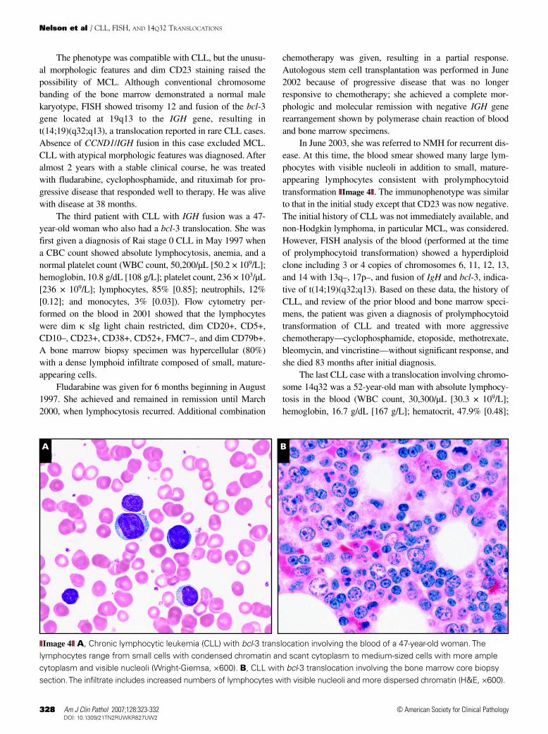
In June 2003, she was referred to NMH for recurrent dis-ease. At this time, the blood smear showed many large lym-phocytes with visible nucleoli in addition to small, mature-appearing lymphocytes consistent with prolymphocytoidtransformation !Image 4!. The immunophenotype was similarto that in the initial study except that CD23 was now negative.The initial history of CLL was not immediately available, andnon-Hodgkin lymphoma, in particular MCL, was considered.However, FISH analysis of the blood (performed at the timeof prolymphocytoid transformation) showed a hyperdiploidclone including 3 or 4 copies of chromosomes 6, 11, 12, 13,and 14 with 13q–, 17p–, and fusion of IgH and bcl-3, indica-tive of t(14;19)(q32;q13). Based on these data, the history ofCLL, and review of the prior blood and bone marrow speci-mens, the patient was given a diagnosis of prolymphocytoidtransformation of CLL and treated with more aggressivechemotherapy—cyclophosphamide, etoposide, methotrexate,bleomycin, and vincristine—without significant response, andshe died 83 months after initial diagnosis.
The last CLL case with a translocation involving chromo-some 14q32 was a 52-year-old man with absolute lymphocy-tosis in the blood (WBC count, 30,300/µL [30.3 ! 109/L];hemoglobin, 16.7 g/dL [167 g/L]; hematocrit, 47.9% [0.48];
A B
!Image 4! A, Chronic lymphocytic leukemia (CLL) with bcl-3 translocation involving the blood of a 47-year-old woman. Thelymphocytes range from small cells with condensed chromatin and scant cytoplasm to medium-sized cells with more amplecytoplasm and visible nucleoli (Wright-Giemsa, !600). B, CLL with bcl-3 translocation involving the bone marrow core biopsysection. The infiltrate includes increased numbers of lymphocytes with visible nucleoli and more dispersed chromatin (H&E, !600).

Am J Clin Pathol 2007;128:323-332 329329 DOI: 10.1309/21TN2RUWKR827UW2 329
© American Society for Clinical Pathology
MCV, 92 µm3 [92 fL]; platelet count, 300 ! 103/µL [300 !109/L]; lymphocytes, 84% [0.84]; neutrophils, 15% [0.15];and monocytes, 1% [0.01]) that was first identified during anannual physical examination. Left axillary lymph nodes werealso enlarged, but neither a bone marrow nor a lymph nodebiopsy was performed. The lymphocytes in the blood weresmall with condensed chromatin and scant cytoplasm !Image5!. Flow cytometric analysis of the blood showed CD19+,CD20+, CD5+, CD10–, CD23+, CD79b–, FMC7–, and " sIglight chain–restricted B cells. CLL was diagnosed.
FISH analysis demonstrated fusion of the bcl-2 and IGHgenes, resulting in t(14;18)(q32;q21), as well as deletion ofthe long arm of chromosome 13. Although an unusual presen-tation of follicular lymphoma with leukemic phase could notbe completely excluded, the immunophenotype and morpho-logic features of the lymphocytes were considered most con-sistent with CLL. His disease was stable without treatment,and he was alive 44 months after initial diagnosis.
CLL Cases With Chromosome 14q32 Aberrations With NoTranslocation Partner Identified
Seven CLL cases without translocations had an abnor-mality involving the IGH gene. Six CLL cases had at least 1extra signal for chromosome 14q32 without fusion of IGHwith CCND1, bcl-2, bcl-3, bcl-11A, c-myc, or MALT1. Theyincluded 3 men and 3 women ranging in age from 53 to 74years (Table 3). All had typical lymphocyte morphologic fea-tures and immunophenotype for CLL. Four had Rai stage IVCLL, 1 had stage I, and 1 had stage 0. Deletion of chromo-some 13q was detected in 3 cases, trisomy 12 in 2 cases, and17p deletion in 1 case; no additional aberration was present in2 cases.
The final CLL case with a chromosome 14q32 aberrationand no identified translocation had loss of the 3' IGHv signaland trisomy 12. The results of chromosome banding analysiswere similar; in 12 cells, they showed an interstitial deletionin chromosome 14 with breakpoints at 14q24 and 14q32, aswell as trisomy 12. This patient, a 29-year-old man, had typi-cal lymphocyte morphologic features and immunophenotypefor CLL, Rai stage III disease at initial examination, and anaggressive clinical course that required therapy at the time ofdiagnosis because of bulky lymphadenopathy.
Correlation of FISH Prognostic Groups With CD38/ZAP-70Status
The majority of the 109 CLL cases (63 [57.8%]) were inthe favorable prognostic group with isolated 13q– or normalFISH results !Table 4!. Thirteen cases (11.9%) were in theintermediate prognostic group with isolated trisomy 12; 25(22.9%) were in the least favorable prognostic group, with17p– or 11q–; this group included the 4 patients with 6q–because all 4 also had 17p–.
Of the 90 CLL cases analyzed for CD38, 81 were placedin prognostic groups. Nineteen (23%) of the 81 were CD38+.A similar percentage of CD38+ cases was present in caseswith 17p (2/6 [33%]) and 11q deletion (4/11 [36%]) and caseswith normal FISH results (9/27 [33%]) (Table 4). CLL caseswith trisomy 12 and isolated 13q– had the lowest percentageof CD38+ cases; 15% (2/13) and 8% (2/24), respectively.ZAP-70 was tested in 36 cases; 10 were positive. In this lim-ited number of cases tested for ZAP-70, the ZAP-70 stainingpattern did not correlate with different genetic prognosticgroups and was negative in all 3 cases with IGH anomaliesthat were tested (Table 3).
The majority of cases in the poor prognostic FISH groupsof isolated 17p– (10/12 [83%]) and isolated 11q– (8/13[62%]) had advanced disease with Rai stage III or IV. Morepatients in the intermediate and good prognostic groups haddisease with a low Rai stage, 0 to II: trisomy 12 (7/11[64%]),
Hematopathology / ORIGINAL ARTICLE
!Image 5! Chronic lymphocytic leukemia with bcl-2translocation involving the blood. The lymphocytes are smallwith condensed chromatin, scant cytoplasm, and mostlyround nuclei (Wright-Giemsa, !600).
!Table 4!Chronic Lymphocytic Leukemia FISH Prognostic GroupsCorrelated With CD38 and ZAP-70
ZAP-70+/FISH Group No. of Cases* CD38+/No. Tested No. Tested
17p– 12 2/6 1/211q– 13 4/11 1/4Trisomy 12 only 13 2/13 2/3Normal 30 9/27 3/1213q– only 33 2/24 3/15
FISH, fluorescence in situ hybridization; ZAP, #-associated protein.* Does not include the 4 CLL cases with chromosome 14q32 translocations and 5
cases with extra immunoglobulin heavy chain gene signals.

330 Am J Clin Pathol 2007;128:323-332330 DOI: 10.1309/21TN2RUWKR827UW2
© American Society for Clinical Pathology
Nelson et al / CLL, FISH, AND 14Q32 TRANSLOCATIONS
normal FISH results (16/25 [64%]), and isolated 13q– (20/32[62%]) !Table 5!. Interestingly, the majority (5/7) of CLLcases with an IGH anomaly but without an identifiabletranslocation partner had Rai stage III or IV disease.
Discussion
We evaluated our experience with a FISH CLL panelafter it had been implemented by clinicians at NMH to assessprognosis. We found that FISH results were also useful to clar-ify the diagnosis in cases in which the phenotype and/or mor-phologic features were atypical. This was especially true forcases with chromosome 14q32 aberrations.
Two cases were classified as MCL after FISH analysisshowed CCND1/IGH fusion, indicating t(11;14)(q13;q32).Both patients had blood lymphocytosis and someimmunophenotypic features that resembled CLL. Both caseswere CD20+, CD5+, and CD23+ (one bright and one dim).However, both cases had atypical morphologic features forCLL and were also FMC7+ and CD79b+, features that areunusual for CLL. The FISH results confirmed the diagnosis ofMCL.
Although aberrations of chromosome 14q32 are reportedto be rare in CLL,12 we found translocations of chromosome14q32 in about 15% of CLL cases. In 4 of these cases, translo-cations involving chromosome 14q32 that have only rarelybeen encountered in CLL were found; some of these cases hadmorphologic features that suggested another non-Hodgkinlymphoma. One of these cases was a CLL with a bcl-11atranslocation.
The bcl-11a gene codes for a transcription factor that isexpressed in many hematopoietic tissues, including bone mar-row cells and germinal center B cells, and is required for nor-mal lymphoid development.13 The bcl-11a translocation isextremely rare in CLL. Prior reported cases include the initialdescription of 2 children with CLL.10 Both children died 1 to4 years after receiving bone marrow transplants for progres-sive CLL. Two adults with CLL and bcl-11a translocations
have also been described.11 Both had an aggressive clinicalcourse and died with progressive disease. Our patient with abcl-11a translocation also had a relatively aggressive coursecharacterized by massive generalized lymphadenopathy,splenomegaly, and extensive bone marrow involvement asso-ciated with severe thrombocytopenia and anemia. His diseaseshowed no response to chemotherapy, and he died 51 monthsafter diagnosis.
Deletion of chromosome 11q was also present in thepatient with a bcl-11a translocation. Chromosome 11q dele-tion is associated with extensive lymphadenopathy and a moreadvanced stage of CLL.14 The extent to which chromosome11q deletion contributed to the aggressive clinical behavior isunclear. The prior reported CLL cases with bcl-11a transloca-tions also had complex karyotypes that included chromosome11q deletion in at least 1 adult.11
Recognition of a bcl-11a translocation occurring in CLLcases is important for at least 2 reasons. First, as in our case,the morphologic features of the lymphocytes may suggestnon-Hodgkin lymphoma, making it difficult to establish adefinitive diagnosis of CLL based on analysis of only periph-eral blood samples and bone marrow trephine biopsy sections.This is particularly true when proliferation centers are notpresent in the trephine biopsy section. Although theimmunophenotype of the lymphocytes may be typical forCLL, a lymph node or other extramedullary tissue biopsy maybe necessary to confirm the diagnosis. Second, because CLLcases with the bcl-11a translocation seem to be clinicallyaggressive, their recognition may allow for initiation of earlyand possibly innovative therapy.
Two CLL cases with a chromosome 14q32 translocationhad t(14;19)(q32;q13), resulting in fusion of the bcl-3 andIGH genes; trisomy 12 was also present in both cases, and13q– and 17p– were also present in 1 case. Prior studies havesuggested that atypical morphologic features, trisomy 12, andan aberrant immunophenotype are features of CLL cases witha bcl-3 translocation.3,15 The findings in our cases are consis-tent with the prior observations. Both cases had relativelyaggressive courses requiring therapy within 2 years, similar to
!Table 5!FISH Prognostic Group Correlated With Rai Clinical Stage
Chromosomal Status
Rai Stage at FISH Analysis 17p– (n = 12) 11q– (n = 13) Trisomy 12q Only (n = 13) Normal (n = 30) 13q– Only (n = 33)
0 2 1 2 8 10I 0 3 3 6 9II 0 1 2 2 1III 3 4 3 4 7IV 7 4 1 5 5Not available 0 0 2 5 2
FISH, fluorescence in situ hybridization.

Am J Clin Pathol 2007;128:323-332 331331 DOI: 10.1309/21TN2RUWKR827UW2 331
© American Society for Clinical Pathology
what has been observed in some CLL cases with bcl-3 translo-cations.3,16 The bcl-3 translocation has also been described inother non-Hodgkin lymphomas.1
In the fourth CLL case with a translocation, FISH analy-sis demonstrated fusion of bcl-2 and IGH, indicatingt(14;18)(q32;q21); deletion of the long arm of chromosome13 was also present. The MALT1 gene was not involvedbecause it showed a normal signal pattern. The lymphocyteshad the typical morphologic features and immunophenotypeof CLL, and lymphocytosis was incidentally found during aroutine physical examination. Therefore, this case wasthought to be more consistent with CLL than follicular lym-phoma, even though bcl-2 gene rearrangement occurs muchmore frequently in follicular lymphoma.
Although it rarely occurs, CLL with bcl-2 generearrangement has been documented in several cases.1,17,18
Similar to our patient, all 7 CLL cases with t(14;18)(q32;q21)reported in 1 series had low-stage disease and the typicalimmunophenotype and morphologic features for CLL.17 Ourcase had an indolent course.
The bcl-2 translocations occurring in CLL may be differ-ent from those that typically occur in follicular lymphoma inthat the translocation partner may be an immunoglobulin lightchain rather than the heavy chain gene, and the fusion sitewithin the bcl-2 gene may occur at the 5' instead of the 3'regions.17,19 In addition, breakpoints within the bcl-2 genemay also occur between the major breakpoint region andminor cluster region instead of within these sites.17,20 The bcl-2 translocation in our patient was conventional in that itinvolved the IGH gene. However, more detailed molecularanalysis was not performed to determine the precise break-point within the bcl-2 gene.
Three or more IGH signals were present in 6 CLL casesusing the CCND1/IGH probe set in the initial phase of FISHanalysis. Multiple dual fusion probe sets were used to lookfor specific translocation partners (bcl-2, bcl-3, bcl-11a, c-myc, or MALT1) in these cases. A break-apart IGH probefor chromosome 14q32 was also used to distinguishbetween multiple copies of chromosome 14 and separationof the IGH locus due to an IGH translocation. Although thebreak-apart IGH probe demonstrated a separation of theIGH locus in 5 cases, a partner chromosome was not identi-fied. This finding indicates that IGH may be involved intranslocations with more loci than just the ones used in thisinvestigation. One case seemed to represent trisomy 14because all abnormal nuclei had 3 intact IGH signals withthe break-apart probes.
One of the 7 CLL cases with an aberration of chromo-some 14q32 had deletion of the 3' IGH signal involving theIGHv; no translocation was identified. This patient hadstage III disease at initial diagnosis and required therapy.Others have described deletion of the IGHv in CLL cases,
with progressive or stable CLL, but IGH deletion more com-monly occurred in the group with progressive disease.6
Our experience supports the use of a FISH panel withinclusion of probes directed at 14q32 in the evaluation ofCLL. Reflex testing using probes directed at previously iden-tified IGH fusion partners is particularly useful to excludeMCL that shows an immunophenotype and/or morphologicfeatures similar to those of CLL and to identify CLL caseswith atypical morphologic features that may otherwise beregarded as other types of non-Hodgkin lymphomas.
From the 1Department of Pathology and 3Robert H. LaurieComprehensive Cancer Center, Feinberg Medical School,Northwestern University, Chicago, IL; and 2Mayo MedicalLaboratories, Rochester, MN.
Address reprint requests to Dr Nelson: Dept of Pathology,Feinberg Medical School, Northwestern University, FeinbergPavilion, Room 7-209, 251 E Huron St, Chicago, IL 60611-2908.
References1. Mayr C, Speicher MR, Kofler DM, et al. Chromosomal
translocations are associated with poor prognosis in chroniclymphocytic leukemia. Blood. 2006;107:742-751.
2. Dohner H, Stilgenbauer S, Benner A, et al. Genomicaberrations and survival in chronic lymphocytic leukemia. N Engl J Med. 2000;343:1910-1916.
3. Dierlamm J, Michaux L, Criel A, et al. Genetic abnormalitiesin chronic lymphocytic leukemia and their clinical andprognostic implications. Cancer Genet Cytogenet. 1997;94:27-35.
4. Peterson LC, Lindquist LL, Church S, et al. Frequent clonalabnormalities of chromosome band 13q14 in B-cell chroniclymphocytic leukemia: multiple clones, subclones, andnonclonal alterations in 82 midwestern patients. GenesChromosomes Cancer. 1992;4:273-280.
5. Juliusson G, Oscier D, Gahrton G. Cytogenetic findings andsurvival in B-cell chronic lymphocytic leukemia: secondIWCCLL compilation of data on 662 patients. LeukLymphoma. 1991;5(suppl):21-25.
6. Fink SR, Paternoster SF, Smoley SA, et al. Fluorescent-labeledDNA probes applied to novel biological aspects of B-cellchronic lymphocytic leukemia. Leuk Res. 2005;29:253-262.
7. Dewald GW, Brockman SR, Paternoster SF, et al.Chromosome anomalies detected by interphase fluorescencein situ hybridization: correlation with significant biologicalfeatures of B-cell chronic lymphocytic leukaemia. Br JHaematol. 2003;121:287-295.
8. Nelson BP, Variakojis D, Peterson LC. Leukemic phase of B-cell lymphomas mimicking chronic lymphocytic leukemia andvariants at presentation. Mod Pathol. 2002;15:1111-1120.
9. Chen YH, Peterson LC, Dittmann D, et al. Comparativeanalysis of flow cytometric techniques in assessment of ZAP-70 expression in relation to IgVH mutational status in chroniclymphocytic leukemia. Am J Clin Pathol. 2007;127:182-191.
10. Yoffe G, Howard-Peebles PN, Smith RG, et al. Childhoodchronic lymphocytic leukemia with (2;14) translocation. J Pediatr. 1990;116:114-117.
Hematopathology / ORIGINAL ARTICLE

332 Am J Clin Pathol 2007;128:323-332332 DOI: 10.1309/21TN2RUWKR827UW2
© American Society for Clinical Pathology
Nelson et al / CLL, FISH, AND 14Q32 TRANSLOCATIONS
11. Satterwhite E, Sonoki T, Willis TG, et al. The BCL11 genefamily: involvement of BCL11A in lymphoid malignancies.Blood. 2001;98:3413-3420.
12. Dohner H, Stilgenbauer S, Dohner K, et al. Chromosomeaberrations in B-cell chronic lymphocytic leukemia:reassessment based on molecular cytogenetic analysis. J MolMed. 1999;77:266-281.
13. Liu P, Keller JR, Ortiz M, et al. Bcl11a is essential for normallymphoid development. Nat Immunol. 2003;4:525-532.
14. Fegan C, Robinson H, Thompson P, et al. Karyotypicevolution in CLL: identification of a new sub-group of patientswith deletions of 11q and advanced or progressive disease.Leukemia. 1995;9:2003-2008.
15. Schlette E, Rassidakis GZ, Canoz O, et al. Expression of bcl-3in chronic lymphocytic leukemia correlates with trisomy 12and abnormalities of chromosome 19. Am J Clin Pathol.2005;123:465-471.
16. Ueshima Y, Bird ML, Vardiman JW, et al. A 14;19translocation in B-cell chronic lymphocytic leukemia: a newrecurring chromosome aberration. Int J Cancer. 1985;36:287-290.
17. Dyer MJ, Zani VJ, Lu WZ, et al. BCL2 translocations inleukemias of mature B cells. Blood. 1994;83:3682-3688.
18. Nowakowski GS, Dewald GW, Hoyer JD, et al. Interphasefluorescence in situ hybridization with an IGH probe isimportant in the evaluation of patients with a clinicaldiagnosis of chronic lymphocytic leukaemia. Br J Haematol.2005;130:36-42.
19. Yabumoto K, Akasaka T, Muramatsu M, et al. Rearrangementof the 5' cluster region of the bcl2 gene in lymphoid neoplasm:a summary of nine cases. Leukemia. 1996;10:970-977.
20. Dyer MJ, Oscier DG. The configuration of theimmunoglobulin genes in B cell chronic lymphocyticleukemia. Leukemia. 2002;16:973-984.


![ReviewArticle Lenalidomide and Chronic Lymphocytic Leukemia · ReviewArticle Lenalidomide and Chronic Lymphocytic Leukemia AnaPilarGonzález-Rodríguez,1 AngelR.Payer,1 ... Ferrajoli[7]](https://static.fdocuments.in/doc/165x107/5acf388a7f8b9ad24f8c2cdd/reviewarticle-lenalidomide-and-chronic-lymphocytic-leukemia-lenalidomide-and-chronic.jpg)


