Chronic Lung Disease After Hematopoietic Stem Cell Transplantation
16
Chronic Lung Disease After Hematopoietic Stem Cell Transplantation Bekele Afessa, MD T , Steve G. Peters, MD Division of Pulmonary and Critical Care Medicine, Mayo Clinic College of Medicine, 200 First Street SW, Rochester, MN 55905, USA Tens of thousands of patients undergo hemato- poietic stem cell transplantation (HSCT) each year, mainly for hematologic disorders [1,2]. In addition to the underlying diseases, the chemotherapy and radiation therapy that HSCT recipients receive can result in damage to multiple organ systems. Pulmo- nary complications develop in 30% to 60% of HSCT recipients [3–5]. With the widespread use of prophy- laxis for certain infections, the spectrum of pulmo- nary complications after HSCT has shifted from more infectious to noninfectious complications [6]. This article reviews some of the noninfectious, chronic pulmonary complications (Box 1 and Table 1). Pulmonary function abnormalities Studies of pulmonary function tests in HSCT recipients have shown that restrictive and obstructive ventilatory defects and gas transfer abnormalities are frequent long-term sequelae, particularly after allo- geneic transplantation [6,7]. A systematic review of 20 reports published between 1996 and 2001 found decreased carbon monoxide diffusion (DLCO) in 83%, restriction in 35%, and obstruction in 23% of allogeneic HSCT recipients [8]. A retrospective co- hort study of more than 500 HSCT patients from the University of Toronto covering the period between 1980 and 1997 documented somewhat lower frequen- cies: impaired diffusion in 35%, restriction in 12%, and obstruction in only 6% of long-term survivors [9]. Although the frequency of restricted and im- paired diffusion seemed to be constant over time, this study suggested a declining frequency of airflow obstruction, from 15% in patients transplanted before 1987 to only 5% for those receiving transplantation after 1987. The extremely low incidence of airflow obstruction documented in this study probably re- flects the stringent diagnostic criteria used by the authors. Using a more sensitive definition in a con- temporary series from the Fred Hutchinson Cancer Center, Chien and colleagues [10] identified airflow obstruction in 26% of long-term survivors of alloge- neic HSCT. Notably, the development of airflow ob- struction serves as a marker for increased risk of mortality after transplantation [9 – 11]. Risk factors for the development of pulmonary function test abnormalities after HSCT include smok- ing history, pretransplantation pulmonary infection, and viral infection in the early posttransplantation period, older age, underlying disease, a pretransplan- tation chemotherapy and conditioning regimen, graft- versus-host-disease (GVHD), and human leukocyte antigen – mismatch [9,10,12 – 25]. Respiratory muscle weakness caused by GVHD-associated myositis has been described after allogeneic HSCT and, rarely, may be a contributing factor to restrictive pulmonary function abnormalities [26 – 28]. Several studies have suggested that abnormal pulmonary function may be a risk factor for the sub- sequent development of pulmonary complications in HSCT recipients [12,29 – 32]. Conflicting data exist, however. For example, a prospective study of 43 allogeneic HSCT recipients did not find a sig- 0272-5231/05/$ – see front matter D 2005 Elsevier Inc. All rights reserved. doi:10.1016/j.ccm.2005.06.012 chestmed.theclinics.com T Corresponding author. E-mail address: [email protected] (B. Afessa). Clin Chest Med 26 (2005) 571 – 586
Transcript of Chronic Lung Disease After Hematopoietic Stem Cell Transplantation

Clin Chest Med
Chronic Lung Disease After Hematopoietic Stem
Cell Transplantation
Bekele Afessa, MDT, Steve G. Peters, MD
Division of Pulmonary and Critical Care Medicine, Mayo Clinic College of Medicine, 200 First Street SW,
Rochester, MN 55905, USA
Tens of thousands of patients undergo hemato-
poietic stem cell transplantation (HSCT) each year,
mainly for hematologic disorders [1,2]. In addition to
the underlying diseases, the chemotherapy and
radiation therapy that HSCT recipients receive can
result in damage to multiple organ systems. Pulmo-
nary complications develop in 30% to 60% of HSCT
recipients [3–5]. With the widespread use of prophy-
laxis for certain infections, the spectrum of pulmo-
nary complications after HSCT has shifted from more
infectious to noninfectious complications [6]. This
article reviews some of the noninfectious, chronic
pulmonary complications (Box 1 and Table 1).
Pulmonary function abnormalities
Studies of pulmonary function tests in HSCT
recipients have shown that restrictive and obstructive
ventilatory defects and gas transfer abnormalities are
frequent long-term sequelae, particularly after allo-
geneic transplantation [6,7]. A systematic review of
20 reports published between 1996 and 2001 found
decreased carbon monoxide diffusion (DLCO) in
83%, restriction in 35%, and obstruction in 23% of
allogeneic HSCT recipients [8]. A retrospective co-
hort study of more than 500 HSCT patients from the
University of Toronto covering the period between
1980 and 1997 documented somewhat lower frequen-
cies: impaired diffusion in 35%, restriction in 12%,
0272-5231/05/$ – see front matter D 2005 Elsevier Inc. All rights
doi:10.1016/j.ccm.2005.06.012
T Corresponding author.
E-mail address: [email protected] (B. Afessa).
and obstruction in only 6% of long-term survivors
[9]. Although the frequency of restricted and im-
paired diffusion seemed to be constant over time, this
study suggested a declining frequency of airflow
obstruction, from 15% in patients transplanted before
1987 to only 5% for those receiving transplantation
after 1987. The extremely low incidence of airflow
obstruction documented in this study probably re-
flects the stringent diagnostic criteria used by the
authors. Using a more sensitive definition in a con-
temporary series from the Fred Hutchinson Cancer
Center, Chien and colleagues [10] identified airflow
obstruction in 26% of long-term survivors of alloge-
neic HSCT. Notably, the development of airflow ob-
struction serves as a marker for increased risk of
mortality after transplantation [9–11].
Risk factors for the development of pulmonary
function test abnormalities after HSCT include smok-
ing history, pretransplantation pulmonary infection,
and viral infection in the early posttransplantation
period, older age, underlying disease, a pretransplan-
tation chemotherapy and conditioning regimen, graft-
versus-host-disease (GVHD), and human leukocyte
antigen–mismatch [9,10,12–25]. Respiratory muscle
weakness caused by GVHD-associated myositis has
been described after allogeneic HSCT and, rarely,
may be a contributing factor to restrictive pulmonary
function abnormalities [26–28].
Several studies have suggested that abnormal
pulmonary function may be a risk factor for the sub-
sequent development of pulmonary complications in
HSCT recipients [12,29–32]. Conflicting data exist,
however. For example, a prospective study of
43 allogeneic HSCT recipients did not find a sig-
26 (2005) 571 – 586
reserved.
chestmed.theclinics.com

Box 1. Chronic noninfectious pulmonarycomplications in hematopoietic stem celltransplant recipients
Isolated abnormality inpulmonary function
AsthmaBronchiolitis obliteransBronchiolitis obliterans
organizing pneumoniaIdiopathic pneumonia syndromeDelayed pulmonary toxicity syndromePulmonary cytolytic thrombiPulmonary veno-occlusive diseaseProgressive pulmonary fibrosisPulmonary hypertensionHepatopulmonary syndromePulmonary alveolar proteinosisEosinophilic pneumonia
afessa & peters572
nificant association between baseline pulmonary
function values and subsequent pulmonary compli-
cations [21]. Moreover, there is considerable vari-
ability among published studies in the particular
pulmonary parameters considered predictive, the
posttransplantation complications that they predict,
and the strength of the association. In light of these
Table 1
Characteristics of selected pulmonary complications in hematopoie
Characteristics
Complications
BO BOOP
Incidence 3.8% in allogeneic
HSCT
Low
Rare in autologous
HSCT
Clinical features Dyspnea Dyspnea
Cough Cough
Wheezing Fever
PFT Airway obstruction # TLC
# DLCO
Radiographic
features
Air trapping Consolidation
Hyperinflation Ground glass opacity
Normal Nodular opacity
Definite diagnosis Surgical biopsy Surgical biopsy
Transbronchial biopsy
Treatment Immunosuppressant Steroids
Case fatality
[reference]
High (59%) Low (19%)
[6,16,40,43,44,46,47,
50,51,54,58,64,91]
[40,52,94–102,104]
Abbreviations: BO, bronchiolitis obliterans; BOOP, bronchiolitis
bon monoxide; DPTS, delayed pulmonary toxicity syndrome; P
PFT, pulmonary function test; PVOD, pulmonary veno-occlusive
limitations, the use of baseline pulmonary function
tests to identify recipients at risk for pulmonary
complications and to guide preventive and therapeu-
tic interventions is of limited utility; further inves-
tigations are warranted.
Asthma
There are case reports of asthma developing af-
ter allogeneic HSCT [33–36]. Limited data suggest
that the serum IgE level, of donor origin, is ele-
vated after transplantation [37]. Allergen-specific
IgE-mediated hypersensitivity can be transferred
by HSCT from donor to recipient by B cells with
allergen-specific memory leading to atopic dermatitis,
allergic rhinitis, and asthma [33,34,37,38]. The man-
agement of asthma in HSCT recipients is similar to
that in other populations.
Bronchiolitis obliterans
Chronic GVHD occurs in about 45% of allo-
geneic HSCT recipients who survive more than
3 months after transplantation [39]. Infectious and
noninfectious pulmonary complications are common
in HSCT recipients who have GVHD. The pulmonary
tic stem cell transplant recipients
DPTS PCT PVOD
Up to 72% of
autologous HSCT
for breast cancer
Rare
Allogeneic
Rare
Dyspnea Fever Dyspnea
Cough Cough Cough
Fever Lethargy
# TLC Not available # DLCO
# DLCO
Ground glass opacity Pulmonary
nodules
Kerley B
Linear-nodular opacity Prominent PA
Consolidation Pulmonary edema
Clinical/PFT Surgical
biopsy
Surgical biopsy
Steroids None None
0% 0% High
[114,115,119,121] [127,128] [142]
obliterans organizing pneumonia; DLCO, diffusion of car-
A, pulmonary artery; PCT, pulmonary cytolytic thrombi;
disease; TLC, total lung capacity.
chronic lung disease after hsct 573
histology of HSCT recipients who have GVHD may
show diffuse alveolar damage, lymphocytic bronchitis/
bronchiolitis with interstitial pneumonitis, bron-
chiolitis obliterans organizing pneumonia (BOOP),
and bronchiolitis obliterans (BO) [40]. BO is a
nonspecific inflammatory injury primarily affecting
the small airways, often sparing the interstitium [41].
The term ‘‘bronchiolitis obliterans’’ has been used to
describe a heterogeneous group of conditions [42].
In the clinical literature, it has been used to describe
various conditions that result in airflow limitation.
Philit and colleagues [43] have recommended the
term ‘‘post-transplant obstructive lung disease’’ in-
stead of ‘‘bronchiolitis obliterans’’ because it takes
into account both the functional definition and the
clinical context of the syndrome. Although BO can
be idiopathic, it is often associated with connective
tissue disease, inhaled toxins, infections, drugs, and
chronic GVHD [41]. In the HSCT recipient, BO in
most cases is considered a severe manifestation of
chronic GVHD, although infection and drug toxicity
may be contributing factors [44].
Incidence and risk factors
Airway disease as a complication of HSCT was
first recognized in the early 1980s [45]. BO almost
exclusively affects allogeneic HSCT recipients who
have GVHD; there are only rare case reports of BO
in autologous recipients [46]. In a recent publication
from the University of Minnesota, 47 of the 1789
allogeneic HSCT recipients (2.6%) developed BO
compared with none of the 1070 autologous HSCT
recipients [47]. The reported incidence of BO varies
among studies, related in part to patient population
and to variable definitions and diagnostic criteria.
Although some studies have included pathologic
findings, most of the reported cases of BO were
defined by the presence of airflow limitation in the
appropriate clinical setting. The frequency of BO
was 3.9% among 4180 allogeneic HSCT recipients
reported in 13 studies [16,21,23,43,44,47–54]. A
recent study employing a highly sensitive definition
of airflow obstruction documented an incidence of
26% [10]. The incidence varies between 6% and 35%
in long-term survivors who have GVHD [15,44,
47,49,50].
GVHD, older donor and recipient age, methotrex-
ate use, antecedent respiratory infection, and serum
immunoglobulin deficiency are risk factors for BO
[10,15,16,44,47,51]. Patients who receive allogeneic
HSCT after nonmyeloablative chemotherapy may
have a lower incidence of pulmonary complications,
including BO [55]. Although pulmonary infections
are identified in many patients who have BO, it is
not clear whether they are directly related to airway
disease or result from immune suppression.
Pathogenesis
The pathogenesis of BO in HSCT recipients is not
well understood. The strong association between BO
and chronic GVHD suggests that host bronchiolar
epithelial cells serve as targets for donor cytotoxic
T lymphocytes [56]. Recurrent aspiration of oral
material caused by esophagitis associated with
GVHD, abnormal local immunoglobulin secretory
function in the lungs, and unrecognized infections
may also be involved [44,56]. The variations in
histopathology, bronchoalveolar lavage cellular com-
position, and clinical course; the frequency of
associated pulmonary infection; the increased risk in
association with methotrexate, and the rare occur-
rence after autologous HSCT suggest a multifactorial
pathogenesis [46,47,56,57].
Clinical findings and diagnostic evaluation
BO can occur 2 months to 9 years after trans-
plantation [16,47,50,51,58]. Most patients present
insidiously with dry cough and dyspnea, 40%
develop wheezing, and 20% report antecedent ‘‘cold’’
symptoms; fever is notably absent [50,51]. Twenty
percent of the patients who had BO had no
respiratory symptoms at the time of the abnormal
pulmonary function testing [51]. Because the pre-
senting respiratory symptoms are nonspecific, a
complete history, including prior medications and
infections, and thorough physical examination focus-
ing on signs of chronic GVHD should be obtained.
Appropriate microbiology studies and laboratory
evaluations including complete blood count with
differential, blood urea nitrogen, creatinine, total
bilirubin, hepatic transaminases, gamma globulin
levels and subclasses, and urinalysis are recom-
mended to exclude infection and complications of
GVHD [56]. Because there is a high prevalence of
sinusitis in allogeneic HSCT recipients who have
GVHD, radiographic assessment of the paranasal
sinuses is also recommended [56,59–62]. If gastro-
intestinal GVHD or aspiration is a consideration,
esophageal studies should be performed [56,63].
Pulmonary function testing is the mainstay of
diagnosis of BO [64]. Although normal airflow has
been reported in HSCT recipients who have histo-
logically proven BO [46], irreversible airflow ob-
struction is the hallmark of clinically significant
disease. In lung transplant recipients, BO is classified

Fig. 1. CT of the chest in a patient with BO showing diffuse
areas of parenchymal hypoattenuation, proximal bronchiec-
tasis, and subsegemental bronchial dilatation. (From Afessa
B, Litzow MR, Tefferi A. Bronchiolitis obliterans and other
late onset non-infectious pulmonary complications in
hematopoietic stem cell transplantation. Bone Marrow
Transplant 2001;28(5):427; with permission.)
afessa & peters574
based on spirometry into stage 1 (forced expiratory
volume in 1 second [FEV1] 66% to 80% of peak
posttransplantation baseline), stage 2 (FEV1 51% to
65% of baseline) and stage 3 (FEV1< 51% of base-
line) [65]. This classification scheme has not been
applied to the HSCT population, and its utility in
defining severity or prognosis in this population
remains to be established.
Fig. 2. Lung pathology in bronchiolitis obliterans showing bronch
excess fibrous connective tissue. Alveoli and their ducts are spare
tissue stain). (From Afessa B, Litzow MR, Tefferi A. Bronchiolit
complications in hematopoietic stem cell transplantation. Bone Ma
The chest radiograph is usually normal or may
show hyperinflation [43,51,52,66]. High-resolution
CT of the chest may show decreased lung attenua-
tion, segmental or subsegmental bronchial dilatation,
diminution of peripheral vascularity, centrilobular
nodules, and expiratory air trapping (Fig. 1) [23,43,
52,67]. The most common finding on high-resolution
CT of the chest is decreased lung attenuation.
The decreased lung attenuation and bronchial dilata-
tions are more frequent and extensive in the lower
lobes [67].
Fiberoptic bronchoscopy with bronchoalveolar
lavage is used in HSCT recipients suspected of hav-
ing BO, mainly to exclude infection as the cause of
airflow obstruction [57]. Because BO involves the
respiratory and membranous bronchioles and is
patchy in distribution, transbronchial lung biopsy
rarely demonstrates the characteristic histologic
changes and consequently is of limited utility. Fur-
thermore, transbronchial biopsy is contraindicated in
the presence of severe airways obstruction or throm-
bocytopenia. Bronchoalveolar lavage shows neutro-
philic or lymphocytic inflammation, but this finding
is nonspecific [53,57].
Video-assisted thoracoscopic lung biopsy is re-
quired to make a definitive histologic diagnosis.
Lung biopsies show small airways involvement with
fibrinous obliteration of the lumen with or without
associated interstitial pneumonia, fibrosis, or diffuse
alveolar damage (Fig. 2) [43,50,68–70]. Necrotizing
bronchitis and bronchiolitis have been reported [68].
iolar inflammation and luminal obliteration associated with
d (Hematoxylin and eosin and Verhoeff-Van Gieson elastic
is obliterans and other late onset non-infectious pulmonary
rrow Transplant 2001;28(5):427; with permission.)

chronic lung disease after hsct 575
The inflammatory cellular infiltrates are usually
peribronchiolar and consist of neutrophils and lym-
phocytes in varying proportions [54]. Surgical biop-
sies are rarely indicated because the diagnosis usually
can be made on clinical grounds.
The diagnostic criteria for BO in HSCT recipients
have not been clearly defined. Obstructive airways
disease and BO may exist as distinct clinical entities
in HSCT recipients. Bronchiolitis may occur without
airway obstruction [56], and, conversely, airflow ob-
struction can occur for a number of reasons other than
BO (eg, asthma, pre-existent chronic obstructive pul-
monary disease, viral bronchiolitis). The diagnosis of
BO is established by the presence of irreversible
airflow obstruction and the exclusion of other causes
of this functional abnormality. BO should be sus-
pected in allogeneic HSCT recipients, particularly
those who have chronic GVHD or who present with
chronic cough, wheezing, dyspnea, or hypoxemia
with a normal chest radiograph and no evidence of
respiratory infection [56,71]. Similarly, the diagnosis
should be considered in asymptomatic patients who
have new onset of airflow obstruction on screening
spirometry. Other chronic diseases, such as pulmo-
nary veno-occlusive disease (PVOD) and early dif-
fuse interstitial disease, can present with similar signs
and symptoms. PVOD, however, is rare and unlikely
to show airflow obstruction. Interstitial lung disease
typically is associated with a restrictive defect on
pulmonary function testing and parenchymal abnor-
malities on high-resolution CT of the chest.
Treatment
The treatment of BO in the HSCT recipient is
empiric and typically consists of corticosteroids and
augmented immunosuppression, targeting chronic
GVHD. Only a minority of patients shows clinical
improvement in response to treatment [6,23,43,46,
47,51,52]. Typically, prednisone in a range of 1 to
1.5 mg/kg/d is given for 4 to 6 weeks [56]. If the
respiratory status remains stable, corticosteroid ther-
apy is tapered and discontinued over 6 to 12 months.
If no improvement is noted within 1 month, or if
deterioration continues, cyclosporin or azathioprine
can be added [56]. Antithymocyte globulin and high-
dose intravenous methylprednisolone have also been
used with mixed results [47].
Macrolide antibiotics are known to have anti-
inflammatory properties and have been shown to
improve symptoms, lung function, and mortality in
patients who have panbronchiolitis [72–78]. Addi-
tionally, there are recent reports of improved lung
function in lung transplant recipients who have
BO syndrome and who received macrolide therapy
[79,80]. Currently, there is only one published study
examining the effects of macrolides for post-HSCT
BO. Khalid and colleagues [81] reported on eight
HSCT patients who had BO and who were given
azithromycin at an initial dose of 500 mg daily for
3 days followed by 250 mg three times weekly for
12 weeks. All patients demonstrated improvement in
spirometric parameters, with a mean improvement in
FEV1 of 20.6% (range, 7.3%-42.9%). This prelimi-
nary evidence, coupled with the lack of significant
adverse effects, makes macrolide therapy an attractive
therapeutic option, but validation with a prospective,
randomized trial is necessary before this approach
can be strongly endorsed.
Although case reports have suggested beneficial
response to thalidomide in HSCT recipients who have
BO, a recent study showed no benefit [47,82,83].
Although hypogammaglobulinemia has been de-
scribed as a risk factor for chronic GVHD, prophy-
laxis with intravenous immunoglobulin has not
been shown to prevent BO [84]. Cyclosporin may
play a protective role against BO, but its role in es-
tablished BO is unproven [85]. A randomized
clinical trial showed inhaled corticosteroids to be
ineffective in lung transplant recipients who have
BO syndrome, and there is no reason to suspect
that this intervention would be any more effica-
cious when applied to post-HSCT BO [86].
In addition to immunosuppression and anti-
inflammatory therapy, adjunctive treatments are im-
portant in the management of HSCT recipients who
have BO. Prophylaxis for Pneumocystis jiroveci
pneumonia and Streptococcus pneumoniae should be
maintained. Bacterial infections, especially sinusi-
tis, are common in patients who have BO and
should be treated aggressively. Bronchodilator ther-
apy is recommended, although only a minority of
patients respond [51,60,68].
In selected HSCT recipients who have respiratory
failure secondary to BO and who are deemed to be
cured of the underlying malignancy for which HSCT
was performed, lung transplantation is an option
[40,87–90].
Clinical course
Serial pulmonary function tests in allogeneic
HSCT recipients who have BO have shown that the
rate of decline in FEV1 is widely variable [51]. De-
terioration in FEV1 correlates with increased mortal-
ity rate [51]. The FEV1 improves in only 8% to 20%
[50,52,56,64]. The reported case fatality rates vary
widely, ranging from 14% to 100%, with an aver-

afessa & peters576
age case fatality rate of 59% [6,16,40,43,44,46,47,
50,51,54,58,64,91]. In one study published 16 years
ago, the 3-year mortality of 35 allogeneic HSCT re-
cipients who had GVHD and obstructive airway dis-
ease was 65% compared with 44% for those who
GVHD but did not have obstructive airway disease
[51]. In a more recent study, the 5-year survival
rate of 47 HSCT recipients who had BO was 10%,
compared with 40% for those who did not have BO,
highlighting the lack of progress in the management
of BO during the last 2 decades [47].
Bronchiolitis obliterans organizing pneumonia
BOOP is an uncommon lung disease character-
ized by the presence of granulation tissue within the
terminal and respiratory bronchioles as well as the
alveolar ducts and alveoli. It was first described as a
distinct clinical entity in 1985 [92]. BOOP can be
either idiopathic or associated with other conditions
such as infections, drugs, radiation, or connective tis-
sue diseases [93]. The first cases of BOOP in HSCT
recipients were reported in the early 1990s [94,95].
BOOP is not as common as BO in HSCT recipients.
HSCT recipients accounted for 4 of the 296 pa-
tients who had BOOP (1.4%) reported in 63 pub-
lications [96]. At Vancouver General Hospital, 5 of
the 25 (20%) patients identified as having BOOP
over an 8-year period were allogeneic HSCT recipi-
ents [96]. At the Mayo Clinic, the incidence of
BOOP was 1.7% among 179 allogeneic HSCT recipi-
ents who survived for 3 months or longer [52]. The
published medical literature on BOOP in HSCT re-
cipients is limited to case reports with a maximum
number of five patients [40,52,94–102].
Idiopathic BOOP is an inflammatory lung disease
of unknown cause [93]. It occurs almost exclusively
in allogeneic HSCT recipients who have GVHD,
suggesting that it may represent a form of allo-
immune injury to the lung by the transplanted stem
cell [96]. The rare occurrence of BOOP in autologous
HSCT recipients suggests that other mechanisms
may be involved as well [97]. A recent case report
of BOOP associated with human herpes virus-6 in
a HSCT recipient suggests unrecognized infections
may play role [103].
Clinical presentation and diagnostic evaluation
Onset is usually 1 month to 2 years after trans-
plantation [52,94–96,98–102,104]. The presenting
symptoms of BOOP include dry cough, dyspnea, and
fever; rarely, the disease may be asymptomatic [52].
Physical examination may reveal inspiratory crackles.
BOOP should be included in the differential diag-
nosis of airspace disease in HSCT recipients who
do not respond to antibiotics. Although the airspace
disease usually is bilateral, it can be unilateral [97].
Pulmonary function tests usually show a restric-
tive defect, decreased DLCO, and normal flow rates
[94,96,99,105]. Arterial blood gas analysis may
reveal mild hypoxemia [96]. Chest radiographs and
CT show patchy air space consolidation, ground-glass
attenuation, and nodular opacities [94–96,99,106,
107]. The radiographic abnormalities usually have
a peripheral distribution [108]. One recent study
demonstrated increased exhaled nitric oxide concen-
tration in HSCT recipients who have BOOP that
decreases with response to treatment [99].
Although pulmonary function test and CT scan
findings in conjunction with the clinical features
may suggest the diagnosis, confirmation requires
either surgical or transbronchial lung biopsy. The di-
agnosis of BOOP in the HSCT recipient occasionally
can be made with transbronchial lung biopsy in the
appropriate clinical setting, but about 85% of the
patients require surgical lung biopsy [96]. Histo-
logic confirmation of the diagnosis is warranted
before the patient is subjected to long-term cortico-
steroid therapy. Surgical lung biopsy, usually by video-
assisted thoracoscopy, is considered the criterion for
the diagnosis of BOOP. The histologic hallmark of
BOOP is the presence of patchy intraluminal fibrosis
consisting of polypoid plugs of immature fibroblast
tissue resembling granulation tissue obliterating the
distal airways, alveolar ducts, and peribronchial al-
veolar space (Fig. 3) [42].
Treatment and prognosis
Corticosteroid therapy is the treatment of choice
for symptomatic patients who have BOOP [52,
93–96,99]. About 80% of HSCT recipients who
have BOOP respond favorably to treatment [52,94–
96,98–101,104]. The duration and dosage of cortico-
steroid therapy in the HSCT recipient who has BOOP
have not been clearly defined. Based on the expe-
rience from non-HSCT recipients, the initial dose of
prednisone is in the range of 0.75 to 1.5 mg/kg/d, to a
maximum daily dose of 100 mg, administered for 1 to
3 months, then 40 mg/d for 3 months, and then 10 to
20 mg/d or every other day for a total of 1 year
[93,109]. Radiographic abnormalities usually clear
within 1 to 3 months of initiating corticosteroid
therapy [95,96,99,100]. Erythromycin, 10 mg/kg/d
for 14 months, has been used in conjunction with

Fig. 3. Lung pathology in bronchiolitis obliterans organizing
pneumonia showing the presence of intraluminal granulation
tissue in bronchioli, alveolar ducts, and alveoli. There is also
interstitial infiltration with mononuclear cells and foamy
macrophages (Hematoxylin and eosin stain). (From Afessa
B, Litzow MR, Tefferi A. Bronchiolitis obliterans and other
late onset non-infectious pulmonary complications in hema-
topoietic stem cell transplantation. Bone Marrow Transplant
2001;28(5):429; with permission.)
chronic lung disease after hsct 577
corticosteroid to treat BOOP in one allogeneic HSCT
recipient [98].
The case fatality rate of treated BOOP is 7.9% in
the general population but seems to be higher in
HSCT recipients [96]. Among 27 HSCT recipients
who were documented in the literature as having
BOOP, the overall case fatality rate was 19% [40,52,
94–102,104].
Delayed pulmonary toxicity syndrome
Chemotherapeutic agents and radiation therapy
have been associated with pulmonary toxicities that
manifest weeks to years later [110,111]. In the 1990s,
many patients who had breast cancer were treated
with a high-dose chemotherapy regimen consisting
of cyclophosphamide, cisplatin, and bischloroethyli-
nitrosurea (BCNU) followed by autologous HSCT
[112]. After transplantation, a significant number of
these patients developed pulmonary complications,
one of the more common of which is the delayed
pulmonary toxicity syndrome (DPTS) [113–120].
DPTS develops in up to 72% of autologous HSCT
recipients who have received high-dose chemother-
apy for breast cancer [114,115,121]. The relatively
high frequency, low mortality, and good response to
corticosteroid treatment distinguish DPTS from idio-
pathic pneumonia syndrome [114,118,119,122].
Because recent studies have not shown a survival
benefit, the use of autologous HSCT after high-dose
chemotherapy for breast cancer has declined [1,123].
The pathogenesis of DPTS is not known. The
depletion of reduced glutathione and impaired anti-
oxidant defenses caused by cyclophosphamide and
BCNU have been implicated [118]. One study showed
no significant correlation between patient age, stage
of breast cancer, chemotherapy regimen, chest wall
radiotherapy, tobacco use, prior lung disease, or base-
line pulmonary function and the development of
DPTS [121].
Clinical findings and diagnostic evaluation
Patients who have DPTS present with cough, dys-
pnea, and fever [115,119,121]. The onset of symp-
toms ranges from 2 weeks to 4 months after
transplantation [119,121]. In the context of prior
breast cancer treated with high-dose chemotherapy
and autologous HSCT, DPTS is diagnosed by
demonstration of a decline in DLCO capacity and
exclusion of infectious causes [119,121]. The DLCO
declines in more than 70% of patients who have
breast cancer and who are treated with high-dose
chemotherapy and autologous HSCT [116]. In
patients who have DPTS, the median absolute DLCO
decrement is 26% (range, 10%–73%), and a na-
dir is reached 15 to 18 weeks after transplantation
[119,121]. The most common findings on CT of the
chest are ground-glass opacities [119]. Other abnor-
malities include linear or nodular opacities and
consolidation. Many patients may have a normal
chest CT at the onset of DPTS [119]. Because of the
typical clinical presentation and response to therapy,
invasive procedures such as bronchoscopy are not
usually required [114,115,119,121].
Treatment and prognosis
Corticosteroid therapy for DPTS usually re-
sults in resolution of symptoms and improvement in
DLCO without long-term pulmonary sequelae [115,
119,121]. Treatment usually consists of prednisone
initiated at a dose of 60 mg/d for 14 days and
subsequently tapered over 5 to 7 weeks [114]. While
patients are being treated with steroids, they should
also receive oral trimethoprim-sulfamethoxazole or
aerosolized pentamidine prophylaxis for Pneumo-
cystis jiroveci pneumonia [114,121]. One case of
DPTS refractory to steroid was treated successfully
with interferon-gamma [124].
A study using prophylactic inhaled fluticasone
propionate, 880 mg every 12 hours for 12 weeks
starting from the date of high-dose chemotherapy,
demonstrated a reduction in the frequency of DPTS to
35%, compared with 73% in historical controls [125].

afessa & peters578
No deaths attributable to DPTS have been re-
ported [114,115,119,121]. The 3-year survival of
stage II and III breast cancer patients who developed
DPTS is 84%, which is not significantly different from
that of patients who did not develop DPTS [121].
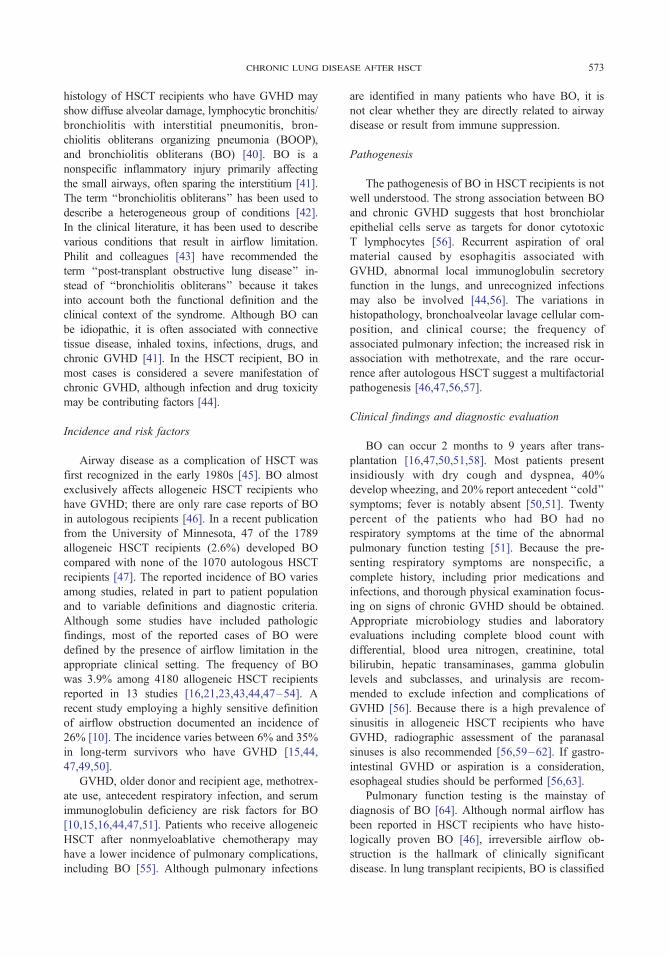
Fig. 4. CT of the chest in a patient with pulmonary cytolytic
thrombi showing multiple bilateral pulmonary nodules with
predominantly subpleural distribution. Inset shows magni-
fied (�3) view of right-sided nodules. (From Morales IJ,
Anderson PM, Tazelaar HD, et al. Pulmonary cytolytic
thrombi: unusual complication of hematopoietic stem cell
transplantation. J Pediatr Hematol Oncol 2003;25(1):90;
with permission.)
Pulmonary cytolytic thrombi
Pulmonary cytolytic thrombi (PCT) is a noninfec-
tious pulmonary complication of unknown origin.
Among HSCT recipients, PCT occurs exclusively
after allogeneic procedures, typically in the setting of
GVHD. PCT has also been described at autopsy in a
nontransplant patient who died after hip replacement
surgery [126]. All except 1 of the 16 HSCT recipi-
ents who had PCT reported in the medical literature
are from a single institution [127,128]. Fifteen of the
16 patients were under age 18 years at the time of
diagnosis [127,128]. Despite the seemingly rare and
previously unrecognized nature of PCT, it was found
in 15 of 33 (45%) HSCT recipients who underwent
surgical lung biopsy for diagnosis of pulmonary
nodules at the University of Minnesota [127].
Pathogenesis
The pathogenesis of PCT is not known. Although
the hemorrhagic infarcts in PCT are similar to those
seen in angioinvasive fungal infections, none of the
lung biopsies in the reported PCT cases had evi-
dence of infection [127,128]. The development of
PCT exclusively in allogeneic HSCT recipients, and
chiefly in those with GVHD, suggests that it may
be a manifestation of GVHD targeting the endothe-
lium of the lungs [127]. Demonstration of clinical and
radiological improvement after increased immuno-
suppression also favors that GVHD as the underlying
pathogenic mechanism [127,128].
Clinical findings and diagnostic evaluation
Most HSCT recipients who have PCT have active
GVHD at the time of presentation [128–130]. The
onset of PCT is between 8 and 343 days (median,
72 days) after transplantation [128–130]. All pa-
tients are febrile, and some have cough at presenta-
tion [128–130]. Dyspnea has not been reported
at presentation.
Chest radiographs, usually performed as part of
the evaluation of persistent fever, may be normal in
25% of the patients who have PCT [130]. Abnormal
chest radiographic findings include nodules, intersti-
tial prominence, and atelectasis [130]. Chest CT
shows multiple peripheral pulmonary nodules, rang-
ing from a few mm to 4 cm in size (Fig. 4). Pleural
effusion was reported in 1 of 13 patients who had
undergone chest CT examination [130].
Bronchoscopy with bronchoalveolar lavage is
used to exclude infection. Because of the peripheral
and intravascular location of the nodules in PCT,
transbronchial lung biopsy is unlikely to yield a
diagnosis. Histologic demonstration of PCT requires
surgical lung biopsy or necropsy [128,130]. The
histologic features of PCT include occlusive vascular
lesions and hemorrhagic infarcts caused by thrombi
that consist of intensely basophilic, amorphous ma-
terial that may extend into the adjacent tissue
through the vascular wall (Fig. 5) [127]. The amor-
phous material suggests cellular breakdown products
[129]. Immunohistochemical studies show a discon-
tinuous endothelial cell layer.
Treatment and prognosis
Because of the unclear causes of PCT and
concerns that lung nodules may have an infectious
cause, patients have been treated with broad-spectrum
antibiotics, antifungals, and systemic corticosteroids
[130]. In one recent case report, treatment with in-
travenous methylprednisolone, 2 mg/kg/d, and cy-
closporin, 3 mg/kg three times daily, for 1 week
followed by oral prednisone, 30 mg/d, and 125 mg cy-
closporin twice daily resulted in improvement [128].
Although it is unclear which of these various inter-
ventions is actually beneficial, most of the patients
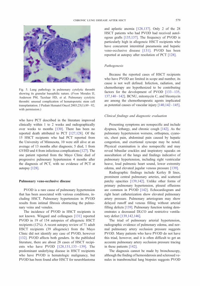
Fig. 5. Lung pathology in pulmonary cytolytic thrombi
showing its granular basophilic nature. (From Morales IJ,
Anderson PM, Tazelaar HD, et al. Pulmonary cytolytic
thrombi: unusual complication of hematopoietic stem cell
transplantation. J Pediatr Hematol Oncol 2003;25(1):89–92;
with permission.)
chronic lung disease after hsct 579
who have PCT described in the literature improved
clinically within 1 to 2 weeks and radiographically
over weeks to months [130]. There has been no
reported death attributed to PCT [127,128]. Of the
15 HSCT recipients who had PCT reported from
the University of Minnesota, 10 were still alive at an
average of 13 months after diagnosis; 5 died, 1 from
GVHD and 4 from infectious complications [127]. The
one patient reported from the Mayo Clinic died of
progressive pulmonary hypertension 4 months after
the diagnosis of PCT, with no evidence of PCT at
autopsy [128].
Pulmonary veno-occlusive disease
PVOD is a rare cause of pulmonary hypertension
that has been associated with various conditions, in-
cluding HSCT. Pulmonary hypertension in PVOD
results from intimal fibrosis obstructing the pulmo-
nary veins and venules.
The incidence of PVOD in HSCT recipients is
not known. Wingard and colleagues [131] reported
PVOD in 19 of 154 autopsies of allogeneic HSCT
recipients (12%). A recent autopsy review of 71 adult
HSCT recipients (39 allogeneic) from the Mayo
Clinic did not identify any case of PVOD, however
[132]. PVOD affects both genders. In the published
literature, there are about 28 cases of HSCT recipi-
ents who have PVOD [128,131,133–139]. The
predominant underlying disease in HSCT recipients
who have PVOD is hematologic malignancy, but
PVOD has been found after HSCT for neuroblastoma
and aplastic anemia [128,137]. Only 2 of the 28
HSCT patients who had PVOD had received autol-
ogous grafts [135,137]. The frequency of PVOD is
particularly high in allogeneic HSCT recipients who
have concurrent interstitial pneumonia and hepatic
veno-occlusive disease [131]. PVOD has been
reported at autopsy after resolution of PCT [128].
Pathogenesis
Because the reported cases of HSCT recipients
who have PVOD are limited in scope and number, its
cause is not well defined. Infection, radiation, and
chemotherapy are hypothesized to be contributing
factors for the development of PVOD [133–135,
137,140–142]. BCNU, mitomycin C, and bleomycin
are among the chemotherapeutic agents implicated
as potential causes of vascular injury [140,142–145].
Clinical findings and diagnostic evaluation
Presenting symptoms are nonspecific and include
dyspnea, lethargy, and chronic cough [142]. As the
pulmonary hypertension worsens, orthopnea, cyano-
sis, chest pain, abdominal pain caused by hepatic
congestion, and exertional syncope may be noted.
Physical examination is also nonspecific and may
reveal bibasilar crackles and inspiratory squeaks on
auscultation of the lungs and findings indicative of
pulmonary hypertension, including right ventricular
heave, loud pulmonic heart sound, lower extremity
edema, and elevated jugular venous pressure [139].
Radiographic findings include Kerley B lines,
prominent central pulmonary arteries, and scattered
patchy opacities [139,142]. Unlike other forms of
primary pulmonary hypertension, pleural effusions
are common in PVOD [142]. Echocardiogram and
right heart catheterization show elevated pulmonary
artery pressure. Pulmonary arteriogram may show
delayed runoff and venous filling without arterial
filling defects [139]. Pulmonary function testing dem-
onstrates a decreased DLCO and restrictive ventila-
tory defect [139,142,146].
The triad of pulmonary arterial hypertension,
radiographic evidence of pulmonary edema, and nor-
mal pulmonary artery occlusion pressure suggests
PVOD. Many patients who have PVOD do not have
this triad, however, and it is often difficult to get an
accurate pulmonary artery occlusion pressure tracing
in these patients [142].
The diagnosis cannot be made by bronchoscopy,
although the finding of hemosiderosis and sclerosed ve-
nules in transbronchial lung biopsies suggests PVOD

afessa & peters580
[138]. The definitive diagnosis of PVOD requires
surgical lung biopsy. In the absence of surgical lung
biopsy, most cases are diagnosed at postmortem exam-
ination [131,137]. The pathologic hallmark of PVOD
is the extensive and diffuse occlusion of pulmonary
veins and venules by fibrous tissue [131,142,147].
Treatment and prognosis
There is no proven therapy for PVOD. Because
observational trials involving patients who have pri-
mary pulmonary hypertension have suggested im-
proved survival in those treated with anticoagulation,
some advocate treating PVOD with long-term warfarin
[142]. There are case reports suggesting that steroids
may be beneficial in HSCT recipients who have PVOD
[133,139]. The data supporting the use of steroid
therapy and anticoagulation in HSCT recipients who
have PVOD are limited to case reports, however
[133,139]. In one report of two HSCT recipients who
had PVOD, methylprednisolone, 2 mg/kg daily, re-
sulted in improvement of arterial oxygenation and
dyspnea within 1 to 2 weeks [133]. In one of these
two patients, discontinuation of steroid therapy led to
recurrent disease that responded to additional treat-
ment [133]. On the other hand, there are reports of
HSCT recipients who had PVOD who deteriorated and
died despite treatment with steroid therapy [135]. The
role of vasodilators in PVOD is unclear. Despite case
reports of successful therapy with oral vasodilators or
intravenous administration of prostacyclin in PVOD,
these agents can dilate the arterial vessels without con-
comitant venodilation, leading to increased transcapil-
lary hydrostatic pressures, acute pulmonary edema,
and death [142,148]. Lung transplantation is a con-
sideration in patients free of other comorbidities, in-
cluding the underlying condition for which HSCT was
performed, but there is currently no published ex-
perience on outcomes of lung transplantation in this
unique setting [141,142]. In light of the limited treat-
ment options and the generally progressive nature of
the disease, it is not surprising that most patients
who have PVOD die within 2 years of diagnosis [142].
Other pulmonary complications
Several cases of pulmonary arterial hyperten-
sion have been reported in HSCT recipients [136,
149–151]. The pathogenesis is not clearly defined.
Normal pulmonary endothelium releases vasodilators
such as prostacyclin- and endothelial-derived relax-
ing factors. Radiation- and chemotherapy-associated
endothelial damage may be the mechanism for the
development of the pulmonary hypertension [136,
149–151]. Prostacyclin infusion and calcium chan-
nel antagonists have been used for the treatment
of pulmonary arterial hypertension in HSCT recipi-
ents [149–151].
Pulmonary alveolar proteinosis is characterized
by excessive accumulation of surfactant lipoprotein
in the alveoli leading to abnormal gas exchange
[152]. The diagnosis of pulmonary alveolar proteino-
sis is made by the presence of periodic acid Schiff–
positive proteinaceous material in bronchoalveolar
fluid [153]. Cordonnier and colleagues [153] described
three HSCT recipients who had pulmonary alveo-
lar proteinosis. All three had received allogeneic
HSCT for leukemia 12 to 90 days before the onset
of pulmonary symptoms. Chest radiographs showed
diffuse infiltrates. The clinicians had suspected in-
fection in two patients and alveolar hemorrhage in
one. Only one of the three survived.
Three cases of chronic eosinophilic pneumonia
have been reported in HSCT recipients, one autolo-
gous and two allogeneic [154–156]. Despite initial
response to steroid therapy, one patient had a fatal
course [154].
Summary
About 50% of HSCT recipients develop pulmo-
nary complications after transplantation. BO, BOOP,
DPTS, PVOD, and PCT are among the noninfectious
pulmonary complications that have been documented
with varying frequency. The pathogenesis of these
complications has not been defined clearly, and
treatment is derived from anecdotal reports rather
than from randomized clinical trials. BO affects allo-
geneic transplant recipients exclusively, is thought
possibly to be a manifestation of chronic GVHD, and
is associated with high case fatality rate because of
the absence of effective therapy. Less common than
BO, BOOP affects both autologous and allogeneic
transplant recipients and typically responds to steroid
therapy. DPTS develops in the majority of autologous
HSCT recipients who have received high-dose che-
motherapy for breast cancer before transplantation.
It usually responds to corticosteroid treatment and
has not been reported to cause death. All 16 reported
HSCT recipients who had PCT had received alloge-
neic grafts, and most had active GVHD at the time of
presentation. HSCT recipients who have PCT present
with fever and pulmonary nodules. Despite the ab-
sence of specific therapy, HSCT recipients who have
PCT usually improve. Most of the reported HSCT
recipients who had PVOD had received allogeneic

chronic lung disease after hsct 581
transplants. Because most cases of PVOD in HSCT
recipients are diagnosed postmortem, the natural
course is not known. Despite anecdotal reports of
favorable response to steroid therapy, the prognosis
of PVOD is poor.
References
[1] Report on state of the art in blood and marrow trans-
plantation. International Bone Marrow Transplant
Registry/Autologous Blood and Marrow Transplant
Registry Newsletter 2003;10(1):7–10.
[2] Report on state of the art in blood and marrow trans-
plantation—part II. International Bone Marrow Trans-
plant Registry/Autologous Blood and Marrow
Transplant Registry Newsletter 2004;10(2):6–9.
[3] Breuer R, Lossos IS, Berkman N, et al. Pulmonary
complications of bone marrow transplantation. Respir
Med 1993;87(8):571–9.
[4] Cordonnier C, Bernaudin JF, Bierling P, et al. Pul-
monary complications occurring after allogeneic
bone marrow transplantation. A study of 130 con-
secutive transplanted patients. Cancer 1986;58(5):
1047–54.
[5] Jules-Elysee K, Stover DE, Yahalom J, et al. Pulmo-
nary complications in lymphoma patients treated
with high-dose therapy autologous bone marrow
transplantation. Am Rev Respir Dis 1992;146(2):
485–91.
[6] Griese M, Rampf U, Hofmann D, et al. Pulmonary
complications after bone marrow transplantation in
children: twenty-four years of experience in a sin-
gle pediatric center. Pediatr Pulmonol 2000;30(5):
393–401.
[7] Cerveri I, Zoia MC, Fulgoni P, et al. Late pulmonary
sequelae after childhood bone marrow transplanta-
tion. Thorax 1999;54(2):131–5.
[8] Marras TK, Szalai JP, Chan CK, et al. Pulmonary
function abnormalities after allogeneic marrow trans-
plantation: a systematic review and assessment of an
existing predictive instrument. Bone Marrow Trans-
plant 2002;30(9):599–607.
[9] Marras TK, Chan CK, Lipton JH, et al. Long-term
pulmonary function abnormalities and survival after
allogeneic marrow transplantation. Bone Marrow
Transplant 2004;33(5):509–17.
[10] Chien JW, Martin PJ, Gooley TA, et al. Airflow
obstruction after myeloablative allogeneic hemato-
poietic stem cell transplantation. Am J Respir Crit
Care Med 2003;168(2):208–14.
[11] Chien JW, Martin PJ, Flowers ME, et al. Implications
of early airflow decline after myeloablative allogeneic
stem cell transplantation. Bone Marrow Transplant
2004;33(7):759–64.
[12] Badier M, Guillot C, Delpierre S, et al. Pulmonary
function changes 100 days and one year after bone
marrow transplantation. Bone Marrow Transplant
1993;12(5):457–61.
[13] Burgart LJ, Heller MJ, Reznicek MJ, et al. Cyto-
megalovirus detection in bone marrow transplant
patients with idiopathic pneumonitis. A clinicopatho-
logic study of the clinical utility of the polymerase
chain reaction on open lung biopsy specimen tissue.
Am J Clin Pathol 1991;96(5):572–6.
[14] Chiou TJ, Tung SL, Wang WS, et al. Pulmonary
function changes in long-term survivors of chronic
myelogenous leukemia after allogeneic bone marrow
transplantation: a Taiwan experience. Cancer Invest
2002;20(7–8):880–8.
[15] Clark JG, Schwartz DA, Flournoy N, et al. Risk
factors for airflow obstruction in recipients of bone
marrow transplants. Ann Intern Med 1987;107(5):
648–56.
[16] Curtis DJ, Smale A, Thien F, et al. Chronic airflow
obstruction in long-term survivors of allogeneic bone
marrow transplantation. BoneMarrowTransplant 1995;
16(1):169–73.
[17] Fanfulla F, Locatelli F, Zoia MC, et al. Pulmonary
complications and respiratory function changes after
bone marrow transplantation in children. Eur Respir J
1997;10(10):2301–6.
[18] Frisk P, Arvidson J, Bratteby LE, et al. Pulmonary
function after autologous bone marrow transplanta-
tion in children: a long-term prospective study. Bone
Marrow Transplant 2004;33(6):645–50.
[19] Gore EM, Lawton CA, Ash RC, et al. Pulmonary
function changes in long-term survivors of bone
marrow transplantation. Int J Radiat Oncol Biol Phys
1996;36(1):67–75.
[20] Leneveu H, Bremont F, Rubie H, et al. Respiratory
function in children undergoing bone marrow trans-
plantation. Pediatr Pulmonol 1999;28(1):31–8.
[21] Lund MB, Kongerud J, Brinch L, et al. Decreased
lung function in one year survivors of allogeneic bone
marrow transplantation conditioned with high-dose
busulphan and cyclophosphamide. Eur Respir J 1995;
8(8):1269–74.
[22] Lund MB, Brinch L, Kongerud J, et al. Lung function
5 yrs after allogeneic bone marrow transplantation
conditioned with busulphan and cyclophosphamide.
Eur Respir J 2004;23(6):901–5.
[23] Schultz KR, Green GJ, Wensley D, et al. Obstructive
lung disease in children after allogeneic bone marrow
transplantation. Blood 1994;84(9):3212–20.
[24] Schwarer AP, Hughes JM, Trotman-Dickenson B,
et al. A chronic pulmonary syndrome associated with
graft-versus-host disease after allogeneic marrow
transplantation. Transplantation 1992;54(6):1002–8.
[25] Tait RC, Burnett AK, Robertson AG, et al. Sub-
clinical pulmonary function defects following autolo-
gous and allogeneic bone marrow transplantation:
relationship to total body irradiation and graft-versus-
host disease. Int J Radiat Oncol Biol Phys 1991;
20(6):1219–27.
[26] Oshima Y, Takahashi S, Nagayama H, et al. Fatal

afessa & peters582
GVHD demonstrating an involvement of respiratory
muscle following donor leukocyte transfusion (DLT).
Bone Marrow Transplant 1997;19(7):737–40.
[27] Oya Y, Kobayashi S, Nakamura K, et al. [Skeletal
muscle pathology of chronic graft versus host disease
accompanied with myositis, affecting predominantly
respiratory and distal muscles, and hemosiderosis].
Rinsho Shinkeigaku 2001;41(9):612–6 [in Japanese].
[28] Stephenson AL, Mackenzie IR, Levy RD, et al.
Myositis associated graft-versus-host-disease present-
ing as respiratory muscle weakness. Thorax 2001;
56(1):82–4.
[29] Baddley JW, Stroud TP, Salzman D, et al. Invasive
mold infections in allogeneic bone marrow transplant
recipients. Clin Infect Dis 2001;32(9):1319–24.
[30] Crawford SW, Fisher L. Predictive value of pulmo-
nary function tests before marrow transplantation [see
comments]. Chest 1992;101(5):1257–64.
[31] Horak DA, Schmidt GM, Zaia JA, et al. Pretrans-
plant pulmonary function predicts cytomegalovirus-
associated interstitial pneumonia following bone
marrow transplantation. Chest 1992;102(5):1484–90.
[32] Milburn HJ, Prentice HG, du Bois RM. Can lung
function measurements be used to predict which pa-
tients will be at risk of developing interstitial pneu-
monitis after bone marrow transplantation? Thorax
1992;47(6):421–5.
[33] Agosti JM, Sprenger JD, Lum LG, et al. Transfer of
allergen-specific IgE-mediated hypersensitivity with
allogeneic bone marrow transplantation. N Engl J
Med 1988;319(25):1623–8.
[34] Hallstrand TS, Sprenger JD, Agosti JM, et al. Long-
term acquisition of allergen-specific IgE and asthma
following allogeneic bone marrow transplantation
from allergic donors. Blood 2004;104(10):3086–90.
[35] Hirayama M, Azuma E, Kumamoto T, et al. Late-
onset unilateral renal dysfunction combined with non-
insulin-dependent diabetes mellitus and bronchial
asthma following allogeneic bone marrow trans-
plantation for acute lymphoblastic leukemia in a
child. Bone Marrow Transplant 1998;22(9):923–6.
[36] Rietz H, Plummer AL, Gal AA. Asthma as a con-
sequence of bone marrow transplantation. Chest
2002;122(1):369–70.
[37] Schuurman HJ, Verdonck LF, Geertzema JG, et al.
Monotypic immunoglobulin E plasma cells in an
allogeneic bone marrow transplant recipient. Histo-
pathology 1986;10(9):963–9.
[38] Bellou A, Kanny G, Fremont S, et al. Transfer of
atopy following bone marrow transplantation. Ann
Allergy Asthma Immunol 1997;78(5):513–6.
[39] Carlens S, Ringden O, Remberger M, et al. Risk
factors for chronic graft-versus-host disease after
bone marrow transplantation: a retrospective single
centre analysis. Bone Marrow Transplant 1998;22(8):
755–61.
[40] Yousem SA. The histological spectrum of pulmonary
graft-versus-host disease in bone marrow transplant
recipients. Hum Pathol 1995;26(6):668–75.
[41] King Jr TE. Overview of bronchiolitis. Clin Chest
Med 1993;14(4):607–10.
[42] Myers JL, Colby TV. Pathologic manifestations of
bronchiolitis, constrictive bronchiolitis, cryptogenic
organizing pneumonia, and diffuse panbronchiolitis.
Clin Chest Med 1993;14(4):611–22.
[43] Philit F, Wiesendanger T, Archimbaud E, et al. Post-
transplant obstructive lung disease (‘‘bronchiolitis
obliterans’’): a clinical comparative study of bone
marrow and lung transplant patients. Eur Respir J
1995;8(4):551–8.
[44] Holland HK, Wingard JR, Beschorner WE, et al.
Bronchiolitis obliterans in bone marrow transplanta-
tion and its relationship to chronic graft-v-host dis-
ease and low serum IgG. Blood 1988;72(2):621–7.
[45] Roca J, Granena A, Rodriguez-Roisin R, et al. Fatal
airway disease in an adult with chronic graft-versus-
host disease. Thorax 1982;37(1):77–8.
[46] Paz HL, Crilley P, Patchefsky A, et al. Bronchiolitis
obliterans after autologous bone marrow transplanta-
tion. Chest 1992;101(3):775–8.
[47] Dudek AZ, Mahaseth H, DeFor TE, et al.
Bronchiolitis obliterans in chronic graft-versus-host
disease: analysis of risk factors and treatment out-
comes. Biol Blood Marrow Transplant 2003;9(10):
657–66.
[48] Patriarca F, Skert C, Sperotto A, et al. Incidence,
outcome, and risk factors of late-onset noninfectious
pulmonary complications after unrelated donor stem
cell transplantation. Bone Marrow Transplant 2004;
33(7):751–8.
[49] Alonso RR, Villa Jr A, Sequeiros GA, et al. [Ob-
structive lung disease after allogenic stem cell trans-
plantation in children]. An Pediatr (Barc) 2004;
61(2):124–30 [in Spanish].
[50] Chan CK, Hyland RH, Hutcheon MA, et al. Small-
airways disease in recipients of allogeneic bone mar-
row transplants. An analysis of 11 cases and a review
of the literature. Medicine (Baltimore) 1987;66(5):
327–40.
[51] Clark JG, Crawford SW, Madtes DK, et al. Obstruc-
tive lung disease after allogeneic marrow transplan-
tation. Clinical presentation and course. Ann Intern
Med 1989;111(5):368–76.
[52] Palmas A, Tefferi A, Myers JL, et al. Late-onset
noninfectious pulmonary complications after alloge-
neic bone marrow transplantation. Br J Haematol
1998;100(4):680–7.
[53] Trisolini R, Stanzani M, Agli LL, Colangelo A, et al.
Delayed non-infectious lung disease in allogeneic
bone marrow transplant recipients. Sarcoidosis Vasc
Diffuse Lung Dis 2001;18(1):75–84.
[54] Urbanski SJ, Kossakowska AE, Curtis J, et al.
Idiopathic small airways pathology in patients with
graft-versus-host disease following allogeneic bone
marrow transplantation. Am J Surg Pathol 1987;
11(12):965–71.
[55] Nusair S, Breuer R, Shapira MY, et al. Low incidence
of pulmonary complications following nonmyelo-
chronic lung disease after hsct 583
ablative stem cell transplantation. Eur Respir J 2004;
23(3):440–5.
[56] Crawford SW, Clark JG. Bronchiolitis associated with
bone marrow transplantation. Clin Chest Med 1993;
14(4):741–9.
[57] St. John RC, Gadek JE, Tutschka PJ, et al. Analysis
of airflow obstruction by bronchoalveolar lavage fol-
lowing bone marrow transplantation. Implications for
pathogenesis and treatment. Chest 1990;98(3):600–7.
[58] Krowka MJ, Rosenow III EC, Hoagland HC. Pulmo-
nary complications of bone marrow transplantation.
Chest 1985;87(2):237–46.
[59] Billings KR, Lowe LH, Aquino VM, et al. Screening
sinus CT scans in pediatric bone marrow transplant
patients. Int J Pediatr Otorhinolaryngol 2000;52(3):
253–60.
[60] Ralph DD, Springmeyer SC, Sullivan KM, et al.
Rapidly progressive air-flow obstruction in marrow
transplant recipients. Possible association between
obliterative bronchiolitis and chronic graft-versus-
host disease. Am Rev Respir Dis 1984;129(4):641–4.
[61] Savage DG, Taylor P, Blackwell J, et al. Paranasal
sinusitis following allogeneic bone marrow trans-
plant. Bone Marrow Transplant 1997;19(1):55–9.
[62] Thompson AM, Couch M, Zahurak ML, et al. Risk
factors for post-stem cell transplant sinusitis. Bone
Marrow Transplant 2002;29(3):257–61.
[63] McDonald GB, Sullivan KM, Plumley TF. Radio-
graphic features of esophageal involvement in chronic
graft-vs.-host disease. AJR Am J Roentgenol 1984;
142(3):501–6.
[64] Hyland RH, Chan CK, Hutcheon MA, et al. Early
diagnosis of obstructive airways disease after alloge-
neic bone marrow transplantation may improve out-
come. Am Rev Respir Dis 1988;137(Suppl):111.
[65] Estenne M, Maurer JR, Boehler A, et al. Bronchioli-
tis obliterans syndrome 2001: an update of the diag-
nostic criteria. J Heart Lung Transplant 2002;21(3):
297–310.
[66] Benesch M, Kerbl R, Schwinger W, et al. Discrep-
ancy of clinical, radiographic and histopathologic
findings in two children with chronic pulmonary
graft-versus-host disease after HLA-identical sibling
stem cell transplantation. Bone Marrow Transplant
1998;22(8):809–12.
[67] Jung JI, Jung WS, Hahn ST, et al. Bronchiolitis
obliterans after allogenic bone marrow transplanta-
tion: HRCT findings. Korean J Radiol 2004;5(2):
107–13.
[68] Johnson FL, Stokes DC, Ruggiero M, et al. Chronic
obstructive airways disease after bone marrow trans-
plantation. J Pediatr 1984;105(3):370–6.
[69] Ostrow D, Buskard N, Hill RS, et al. Bronchiolitis
obliterans complicating bone marrow transplantation.
Chest 1985;87(6):828–30.
[70] Wyatt SE, Nunn P, Hows JM, et al. Airways ob-
struction associated with graft versus host disease af-
ter bone marrow transplantation. Thorax 1984;39(12):
887–94.
[71] Afessa B, Litzow MR, Tefferi A. Bronchiolitis oblit-
erans and other late onset non-infectious pulmonary
complications in hematopoietic stem cell trans-
plantation. Bone Marrow Transplant 2001;28(5):
425–34.
[72] Ichikawa Y, Ninomiya H, Koga H, et al. Erythro-
mycin reduces neutrophils and neutrophil-derived
elastolytic-like activity in the lower respiratory tract
of bronchiolitis patients [see comments]. Am Rev
Respir Dis 1992;146(1):196–203.
[73] Kudoh S, Azuma A, Yamamoto M, et al. Improve-
ment of survival in patients with diffuse panbron-
chiolitis treated with low-dose erythromycin. Am J
Respir Crit Care Med 1998;157(6 Pt 1):1829–32.
[74] Kudoh S. Erythromycin treatment in diffuse panbron-
chiolitis. Curr Opin Pulm Med 1998;4(2):116–21.
[75] Nagai H, Shishido H, Yoneda R, et al. Long-term
low-dose administration of erythromycin to patients
with diffuse panbronchiolitis. Respiration (Herrlis-
heim) 1991;58(3–4):145–9.
[76] Oda H, Kadota J, Kohno S, et al. Erythromycin inhibits
neutrophil chemotaxis in bronchoalveoli of diffuse
panbronchiolitis. Chest 1994;106(4):1116–23.
[77] Kanazawa S, Nomura S, Muramatsu M, et al.
Azithromycin and bronchiolitis obliterans. Am J
Respir Crit Care Med 2004;169(5):654–5.
[78] Culic O, Erakovic V, ParnhamMJ. Anti-inflammatory
effects of macrolide antibiotics. Eur J Pharmacol
2001;429(1–3):209–29.
[79] Verleden GM, Dupont LJ. Azithromycin therapy for
patients with bronchiolitis obliterans syndrome af-
ter lung transplantation. Transplantation 2004;77(9):
1465–7.
[80] Gerhardt SG, McDyer JF, Girgis RE, et al. Main-
tenance azithromycin therapy for bronchiolitis oblit-
erans syndrome: results of a pilot study. Am J Respir
Crit Care Med 2003;168(1):121–5.
[81] Khalid M, Al Saghir A, Saleemi S, et al. Azithro-
mycin in bronchiolitis obliterans complicating bone
marrow transplantation: a preliminary study. Eur
Respir J 2005;25(3):490–3.
[82] Browne PV, Weisdorf DJ, DeFor T, et al. Response
to thalidomide therapy in refractory chronic graft-
versus-host disease. Bone Marrow Transplant 2000;
26(8):865–9.
[83] Forsyth CJ, Cremer PD, Torzillo P, et al. Thalidomide
responsive chronic pulmonary GVHD. Bone Marrow
Transplant 1996;17(2):291–3.
[84] Sullivan KM, Storek J, Kopecky KJ, et al. A con-
trolled trial of long-term administration of intrave-
nous immunoglobulin to prevent late infection and
chronic graft-vs.-host disease after marrow trans-
plantation: clinical outcome and effect on subsequent
immune recovery. Biol Blood Marrow Transplant
1996;2(1):44–53.
[85] Payne L, Chan CK, Fyles G, et al. Cyclosporine as
possible prophylaxis for obstructive airways disease
after allogeneic bone marrow transplantation. Chest
1993;104(1):114–8.

afessa & peters584
[86] Whitford H, Walters EH, Levvey B, et al. Addition
of inhaled corticosteroids to systemic immunosup-
pression after lung transplantation: a double-blind,
placebo-controlled trial. Transplantation 2002;73(11):
1793–9.
[87] Boas SR, Noyes BE, Kurland G, et al. Pediatric lung
transplantation for graft-versus-host disease follow-
ing bone marrow transplantation. Chest 1994;105(5):
1584–6.
[88] Favaloro R, Bertolotti A, Gomez C, et al. Lung
transplant at the Favaloro Foundation: a 13-year
experience. Transplant Proc 2004;36(6):1689–91.
[89] Pechet TV, de le Morena M, Mendeloff EN, et al.
Lung transplantation in children following treatment
for malignancy. J Heart Lung Transplant 2003;22(2):
154–60.
[90] Rabitsch W, Deviatko E, Keil F, et al. Successful lung
transplantation for bronchiolitis obliterans after allo-
geneic marrow transplantation. Transplantation 2001;
71(9):1341–3.
[91] Paz HL, Crilley P, Topolsky DL, et al. Bronchiolitis
obliterans after bone marrow transplantation: the ef-
fect of preconditioning. Respiration (Herrlisheim)
1993;60(2):109–14.
[92] Epler GR, Colby TV, McLoud TC, et al. Bronchioli-
tis obliterans organizing pneumonia. N Engl J Med
1985;312(3):152–8.
[93] Epler GR. Bronchiolitis obliterans organizing pneu-
monia. Arch Intern Med 2001;161(2):158–64.
[94] Thirman MJ, Devine SM, O’Toole K, et al. Bron-
chiolitis obliterans organizing pneumonia as a com-
plication of allogeneic bone marrow transplantation
[see comments]. Bone Marrow Transplant 1992;
10(3):307–11.
[95] Mathew P, Bozeman P, Krance RA, et al. Bronchi-
olitis obliterans organizing pneumonia (BOOP) in
children after allogeneic bone marrow transplantation.
Bone Marrow Transplant 1994;13(2):221–3.
[96] Alasaly K, Muller N, Ostrow DN, et al. Cryptogenic
organizing pneumonia. A report of 25 cases and a
review of the literature. Medicine (Baltimore) 1995;
74(4):201–11.
[97] Hayes-Jordan A, Benaim E, Richardson S, et al.
Open lung biopsy in pediatric bone marrow trans-
plant patients. J Pediatr Surg 2002;37(3):446–52.
[98] Ishii T, Manabe A, Ebihara Y, et al. Improvement in
bronchiolitis obliterans organizing pneumonia in a
child after allogeneic bone marrow transplantation
by a combination of oral prednisolone and low
dose erythromycin. Bone Marrow Transplant 2000;
26(8):907–10.
[99] Kanamori H, Fujisawa S, Tsuburai T, et al. In-
creased exhaled nitric oxide in bronchiolitis oblit-
erans organizing pneumonia after allogeneic bone
marrow transplantation. Transplantation 2002;74(9):
1356–8.
[100] Kanda Y, Takahashi T, Imai Y, et al. Bronchiolitis
obliterans organizing pneumonia after syngeneic
bone marrow transplantation for acute lymphoblas-
tic leukemia. Bone Marrow Transplant 1997;19(12):
1251–3.
[101] Kleinau I, Perez-Canto A, Schmid HJ, et al. Bron-
chiolitis obliterans organizing pneumonia and chronic
graft-versus-host disease in a child after allogeneic
bone marrow transplantation. Bone Marrow Trans-
plant 1997;19(8):841–4.
[102] Przepiorka D, Abu-Elmagd K, Huaringa A, et al. Bron-
chiolitis obliterans organizing pneumonia in a BMT
patient receiving FK506 [letter; comment]. Bone Mar-
row Transplant 1993;11(6):502.
[103] Yata K, Nakajima M, Takemoto Y, et al. [Pneu-
monitis with a bronchiolitis obliterans organizing
pneumonia-like shadow in a patient with human her-
pes virus-6 viremia after allogeneic bone marrow
transplantation]. Kansenshogaku Zasshi 2002;76(5):
385–90 [in Japanese].
[104] Baron FA, Hermanne JP, Dowlati A, et al. Bronchi-
olitis obliterans organizing pneumonia and ulcerative
colitis after allogeneic bone marrow transplantation.
Bone Marrow Transplant 1998;21(9):951–4.
[105] Epler GR. Bronchiolitis obliterans organizing pneu-
monia. Semin Respir Infect 1995;10(2):65–77.
[106] Muller NL, Staples CA, Miller RR. Bronchiolitis ob-
literans organizing pneumonia: CT features in 14 pa-
tients. AJR Am J Roentgenol 1990;154(5):983–7.
[107] Worthy SA, Flint JD, Muller NL. Pulmonary com-
plications after bone marrow transplantation: high-
resolution CT and pathologic findings. Radiographics
1997;17(6):1359–71.
[108] Graham NJ, Muller NL, Miller RR, et al. Intratho-
racic complications following allogeneic bone mar-
row transplantation: CT findings. Radiology 1991;
181(1):153–6.
[109] Cordier JF. Organising pneumonia. Thorax 2000;
55(4):318–28.
[110] Abratt RP, Morgan GW, Silvestri G, et al. Pulmonary
complications of radiation therapy. Clin Chest Med
2004;25(1):167–77.
[111] Limper AH. Chemotherapy-induced lung disease.
Clin Chest Med 2004;25(1):53–64.
[112] Pedrazzoli P, Ferrante P, Kulekci A, et al. Autolo-
gous hematopoietic stem cell transplantation for
breast cancer in Europe: critical evaluation of data
from the European Group for Blood and Marrow
Transplantation (EBMT) Registry 1990–1999. Bone
Marrow Transplant 2003;32(5):489–94.
[113] Bearman SI, Overmoyer BA, Bolwell BJ, et al. High-
dose chemotherapy with autologous peripheral blood
progenitor cell support for primary breast cancer in
patients with 4–9 involved axillary lymph nodes.
Bone Marrow Transplant 1997;20(11):931–7.
[114] Bhalla KS, Wilczynski SW, Abushamaa AM, et al.
Pulmonary toxicity of induction chemotherapy prior
to standard or high- dose chemotherapy with autolo-
gous hematopoietic support. Am J Respir Crit Care
Med 2000;161(1):17–25.
[115] Chap L, Shpiner R, Levine M, et al. Pulmonary
toxicity of high-dose chemotherapy for breast can-
chronic lung disease after hsct 585
cer: a non-invasive approach to diagnosis and treat-
ment. Bone Marrow Transplant 1997;20(12):1063–7.
[116] Fanfulla F, Pedrazzoli P, Da Prada GA, et al. Pul-
monary function and complications following che-
motherapy and stem cell support in breast cancer.
Eur Respir J 2000;15(1):56–61.
[117] Jones RB, Matthes S, Shpall EJ, et al. Acute lung
injury following treatment with high-dose cyclo-
phosphamide, cisplatin, and carmustine: pharmaco-
dynamic evaluation of carmustine. J Natl Cancer Inst
1993;85(8):640–7.
[118] Todd NW, Peters WP, Ost AH, et al. Pulmonary drug
toxicity in patients with primary breast cancer
treated with high-dose combination chemotherapy
and autologous bone marrow transplantation. Am
Rev Respir Dis 1993;147(5):1264–70.
[119] Wilczynski SW, Erasmus JJ, Petros WP, et al. De-
layed pulmonary toxicity syndrome following high-
dose chemotherapy and bone marrow transplantation
for breast cancer. Am J Respir Crit Care Med
1998;157(2):565–73.
[120] Wong R, Rondon G, Saliba RM, et al. Idiopathic
pneumonia syndrome after high-dose chemotherapy
and autologous hematopoietic stem cell transplanta-
tion for high-risk breast cancer. Bone Marrow Trans-
plant 2003;31(12):1157–63.
[121] Cao TM, Negrin RS, Stockerl-Goldstein KE, et al.
Pulmonary toxicity syndrome in breast cancer pa-
tients undergoing BCNU-containing high-dose che-
motherapy and autologous hematopoietic cell
transplantation. Biol Blood Marrow Transplant 2000;
6(4):387–94.
[122] Peters WP, Ross M, Vredenburgh JJ, et al. High-dose
chemotherapy and autologous bone marrow support
as consolidation after standard-dose adjuvant ther-
apy for high-risk primary breast cancer. J Clin Oncol
1993;11(6):1132–43.
[123] Stadtmauer EA, O’Neill A, Goldstein LJ, et al.
Conventional-dose chemotherapy compared with
high-dose chemotherapy plus autologous hemato-
poietic stem-cell transplantation for metastatic breast
cancer. Philadelphia Bone Marrow Transplant Group.
N Engl J Med 2000;342(15):1069–76.
[124] Suratt BT, Lynch DA, Cool CD, et al. Interferon-
gamma for delayed pulmonary toxicity syndrome
resistant to steroids. Bone Marrow Transplant 2003;
31(10):939–41.
[125] McGaughey DS, Nikcevich DA, Long GD, et al.
Inhaled steroids as prophylaxis for delayed pulmo-
nary toxicity syndrome in breast cancer patients
undergoing high-dose chemotherapy and autologous
stem cell transplantation. Biol Blood Marrow Trans-
plant 2001;7(5):274–8.
[126] Castellano-Sanchez AA, Poppiti RJ. Pulmonary
cytolytic thrombi (PCT). A previously unrecognized
complication of bone marrow transplantation (BMT).
Am J Surg Pathol 2001;25(6):829–31.
[127] Gulbahce HE, Pambuccian SE, Jessurun J, et al.
Pulmonary nodular lesions in bone marrow trans-
plant recipients: impact of histologic diagnosis on
patient management and prognosis. Am J Clin Pathol
2004;121(2):205–10.
[128] Morales IJ, Anderson PM, Tazelaar HD, et al. Pul-
monary cytolytic thrombi: unusual complication of
hematopoietic stem cell transplantation. J Pediatr
Hematol Oncol 2003;25(1):89–92.
[129] Gulbahce HE, Manivel JC, Jessurun J. Pulmo-
nary cytolytic thrombi: a previously unrecognized
complication of bone marrow transplantation. Am J
Surg Pathol 2000;24(8):1147–52.
[130] Woodard JP, Gulbahce E, Shreve M, et al. Pulmonary
cytolytic thrombi: a newly recognized complication
of stem cell transplantation. Bone Marrow Transplant
2000;25(3):293–300.
[131] Wingard JR, Mellits ED, Jones RJ, et al. Association
of hepatic veno-occlusive disease with interstitial
pneumonitis in bone marrow transplant recipients.
Bone Marrow Transplant 1989;4(6):685–9.
[132] Sharma S, Nadrous HF, Peters SG, et al. Pulmonary
complications in adult blood and marrow transplant
recipients: autopsy findings. Chest, in press.
[133] Hackman RC, Madtes DK, Petersen FB, et al. Pulmo-
nary venoocclusive disease following bone marrow
transplantation. Transplantation 1989;47(6):989–92.
[134] Kuga T, Kohda K, Hirayama Y, et al. Pulmonary
veno-occlusive disease accompanied by microangio-
pathic hemolytic anemia 1 year after a second bone
marrow transplantation for acute lymphoblastic leu-
kemia. Int J Hematol 1996;64(2):143–50.
[135] Salzman D, Adkins DR, Craig F, et al. Malignancy-
associated pulmonary veno-occlusive disease: report
of a case following autologous bone marrow trans-
plantation and review. Bone Marrow Transplant 1996;
18(4):755–60.
[136] Seguchi M, Hirabayashi N, Fujii Y, et al. Pulmonary
hypertension associated with pulmonary occlusive
vasculopathy after allogeneic bone marrow trans-
plantation. Transplantation 2000;69(1):177–9.
[137] Trobaugh-Lotrario AD, Greffe B, Deterding R, et al.
Pulmonary veno-occlusive disease after autologous
bone marrow transplant in a child with stage IV
neuroblastoma: case report and literature review.
J Pediatr Hematol Oncol 2003;25(5):405–9.
[138] Troussard X, Bernaudin JF, Cordonnier C, et al. Pul-
monary veno-occlusive disease after bone marrow
transplantation. Thorax 1984;39(12):956–7.
[139] Williams LM, Fussell S, Veith RW, et al. Pulmo-
nary veno-occlusive disease in an adult following
bone marrow transplantation. Case report and re-
view of the literature. Chest 1996;109(5):1388–91.
[140] Kotloff RM, Ahya VN, Crawford SW. Pulmonary
complications of solid organ and hematopoietic stem
cell transplantation. Am J Respir Crit Care Med
2004;170(1):22–48.
[141] Kramer MR, Estenne M, Berkman N, et al. Radiation-
induced pulmonary veno-occlusive disease. Chest
1993;104(4):1282–4.
[142] Mandel J, Mark EJ, Hales CA. Pulmonary veno-

afessa & peters586
occlusive disease. Am J Respir Crit Care Med 2000;
162(5):1964–73.
[143] Doll DC, Yarbro JW. Vascular toxicity associated
with antineoplastic agents. Semin Oncol 1992;19(5):
580–96.
[144] Doll DC, Yarbro JW. Vascular toxicity associated
with chemotherapy and hormonotherapy. Curr Opin
Oncol 1994;6(4):345–50.
[145] Gagnadoux F, Capron F, Lebeau B. Pulmonary veno-
occlusive disease after neoadjuvant mitomycin che-
motherapy and surgery for lung carcinoma. Lung
Cancer 2002;36(2):213–5.
[146] Elliott CG, Colby TV, Hill T, et al. Pulmonary veno-
occlusive disease associated with severe reduction of
single-breath carbon monoxide diffusing capacity.
Respiration (Herrlisheim) 1988;53(4):262–6.
[147] Wagenvoort CA, Wagenvoort N, Takahashi T. Pul-
monary veno-occlusive disease: involvement of
pulmonary arteries and review of the literature. Hum
Pathol 1985;16(10):1033–41.
[148] Palmer SM, Robinson LJ, Wang A, et al. Massive
pulmonary edema and death after prostacyclin infu-
sion in a patient with pulmonary veno-occlusive
disease. Chest 1998;113(1):237–40.
[149] Shankar S, Choi JK, Dermody TS, et al. Pulmonary
hypertension complicating bone marrow transplanta-
tion for idiopathic myelofibrosis. J Pediatr Hematol
Oncol 2004;26(6):393–7.
[150] Vaksmann G, Nelken B, Deshildre A, et al. Pulmo-
nary arterial occlusive disease following chemother-
apy and bone marrow transplantation for leukaemia.
Eur J Pediatr 2002;161(5):247–9.
[151] Bruckmann C, Lindner W, Roos R, et al. Severe
pulmonary vascular occlusive disease following bone
marrow transplantation in Omenn syndrome. Eur J
Pediatr 1991;150(4):242–5.
[152] Presneill JJ, Nakata K, Inoue Y, et al. Pulmonary
alveolar proteinosis. Clin Chest Med 2004;25(3):
593–613 [viii.].
[153] Cordonnier C, Fleury-Feith J, Escudier E, et al.
Secondary alveolar proteinosis is a reversible cause of
respiratory failure in leukemic patients. Am J Respir
Crit Care Med 1994;149(3 Pt 1):788–94.
[154] Gross TG, Hoge FJ, Jackson JD, et al. Fatal eosino-
philic disease following autologous bone marrow
transplantation. Bone Marrow Transplant 1994;14(2):
333–7.
[155] Brunet S, Muniz-Diaz E, Baiget M. [Chronic eosino-
philic pneumonia in a patient treated with allogeneic
bone marrow transplantation]. Med Clin (Barc) 1994;
103(17):677 [in Spanish].
[156] Richard C, Calavia J, Loyola I, et al. [Chronic eosin-
ophilic pneumonia in a patient treated with allogenic
bone marrow transplantation]. Med Clin (Barc) 1994;
102(12):462–4 [in Spanish].