Chronic Laryngitis Prepared by Dr. Hiwa As’ad. Definition: Is a chronic inflammation of the mucosa...
27
Chronic Chronic Laryngitis Laryngitis Prepared by Dr. Hiwa As’ad
-
Upload
aubrey-barker -
Category
Documents
-
view
226 -
download
0
Transcript of Chronic Laryngitis Prepared by Dr. Hiwa As’ad. Definition: Is a chronic inflammation of the mucosa...
Chronic LaryngitisChronic Laryngitis
Prepared by Dr. Hiwa As’ad
DefinitionDefinition::
Is a chronic inflammation of the mucosa of the larynx.
EtiologyEtiology
Follows repeated acute attacks but usually it arise insidiously due to :
Faulty use of voice. Infection of teeth, tonsil, sinus, and lower
respiratory tract infection. Excessive alcohol consumption or
smoking. Dust or irritant fumes.
Clinical classificationClinical classification::
Chronic nonspecific laryngitis:1. Chronic simple laryngitis.2. Hyper keratosis of larynx
(chronic keratosis or leukoplakia).
3. Pachydermia laryngis.4. Contact granuloma.5. Atrophic laryngitis.
Chronic specific laryngitis: Tuberculous laryngitis Syphilitic laryngitis. Leprosy of the larynx. Scleroma of the larynx. Wegener’s (malignant)
granuloma of the larynx.
Mycosis of the larynx.
Chronic Nonspecific laryngitisChronic Nonspecific laryngitisSimple chronic laryngitisSimple chronic laryngitis Pathology:
– Hyperaemia of vocal cord.– Edema.– Myositis occurs in the intrinsic muscles.– Excessive secretion due to hyper activity of the mucous gland.– Hyperaemic and edematous stage often passes to a hypertrophic
one and rarely to an atrophic one. Clinical Features:-Hoarseness (intermittent then persistent). -Cough (slightly dry). -Sore throat (very common).
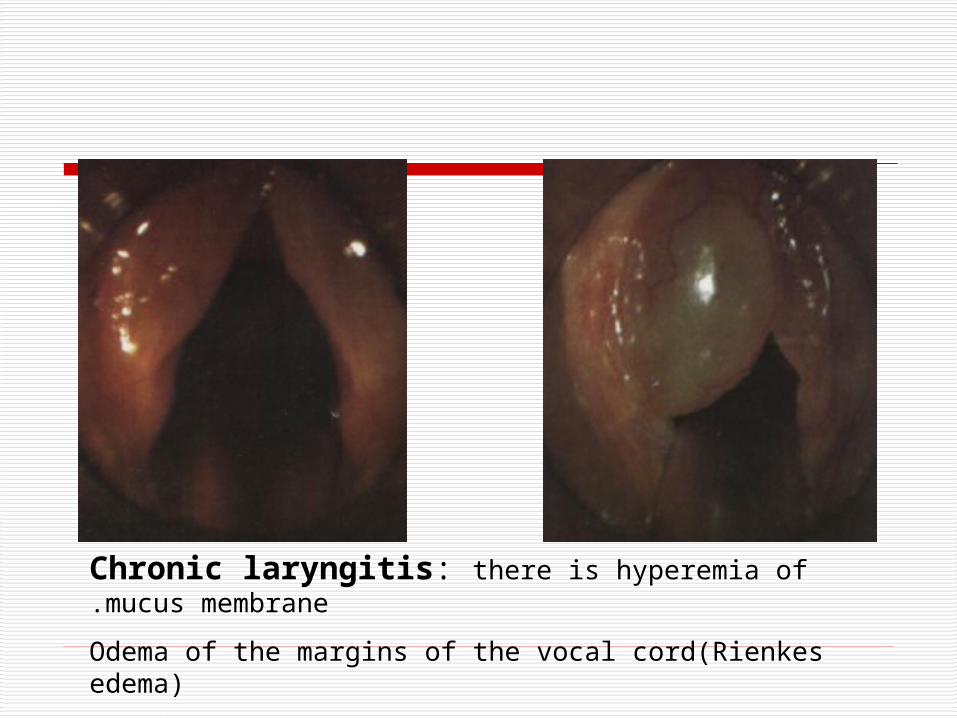
Chronic laryngitis: there is hyperemia of mucus membrane.
Odema of the margins of the vocal cord(Rienkes edema)
Laryngeal appearanceThree types:1. Hyperaemic.2. Hypertrophic.3. Edematous.In all types the larynx is always affected
bilaterally & symmetrically.
Treatment:Vocal restElimination of irritating factors such as
dust and smoking. Systemic antibiotics.Carbocisteine ( a mucolytic ) when
secretion are thick.Stripping of the vocal cords is performed
endoscopically in persistent cases.
Chronic Nonspecific laryngitisChronic Nonspecific laryngitisHyperkeratois of the larynx (leukoplakia)Hyperkeratois of the larynx (leukoplakia)
Definition: A localized form of epithelial hyperplasia characterized by leukoplakic raised patches on the vocal cord.
Pathology: There is hyperplastic change in the epithelium, together with extension of the papillae into the cornium; and basement membrane remains intact.
Clinical features:Hoarseness ( gradual onset).Examination :there is a white raised patch on one
or both vocal cords the anterior and middle thirds are usually involved. Mobility of cords is not impaired.
Treatment: -Infection in the mouth, throat and nose treated. -Stripping of the cords can sometimes be done through a
direct laryngoscope but recurrence is usualConstant supervision is essential to detect early malignant
change demanding radical removal or radiotherapy. -Biopsy is mandatory in suspicious cases and may require
repetition.
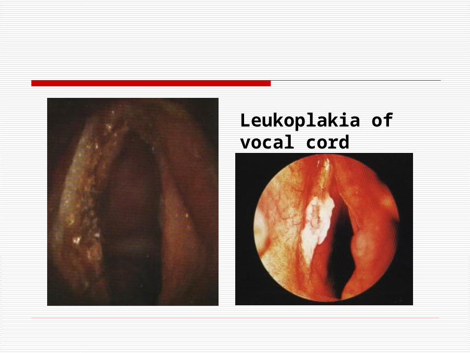
Leukoplakia of vocal cord
Chronic Nonspecific laryngitisChronic Nonspecific laryngitisPachydermia LaryngisPachydermia Laryngis::Definition:
A form of chronic hypertrophic laryngitis affecting the epithelium and subepithelium of the posterior part of the larynx.
Etiology:-A rare condition more common in men.
-The cause usually unknown but aggravating factors includes excessive alcohol and tobacco.
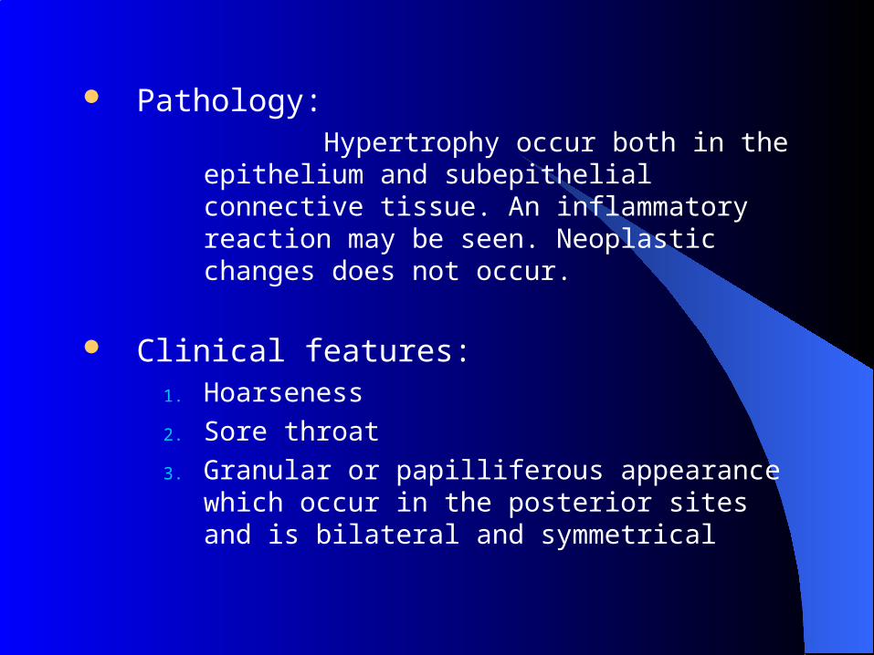
Pathology: Hypertrophy occur both in the epithelium and
subepithelial connective tissue. An inflammatory reaction may be seen. Neoplastic changes does not occur.
Clinical features:1. Hoarseness
2. Sore throat
3. Granular or papilliferous appearance which occur in the posterior sites and is bilateral and symmetrical
Treatment: Similar to that for simple chronic laryngitis.
surgical removal and diathermy of the masses give little relief and are inadvisable.
Chronic Nonspecific laryngitisChronic Nonspecific laryngitisContact GranulomaContact Granuloma:: Unilateral. Situated medially or superiorly on the vocal process of
the arytenoid cartilage. Confused with contact ulcer. The granuloma has a typical polypoid appearance which
is a local reaction to trauma. Granulation tissue can develop if the perichondrium is
damaged either vocal trauma or through trauma from an endotracheal tube.
Slight hoarseness, with history of previous surgery or usage of excessive voice.
Treated by simple removal by microlaryngoscopy but local recurrence are common.
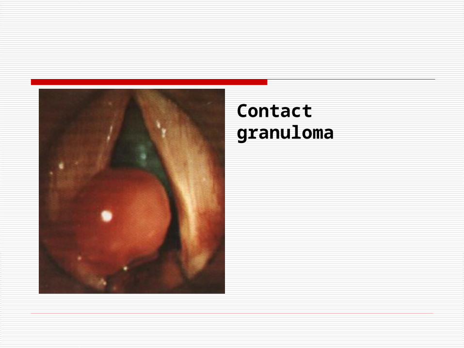
Contact granuloma
Chronic Nonspecific laryngitisChronic Nonspecific laryngitisAtrophic LaryngitisAtrophic Laryngitis:: Uncommon . usually associated with atrophic
rhinitis and pharyngitis. Aggravating factors : dusty atmosphere, industry
fumes and chronic infection of the paranasal sinuses.
Hoarseness and sore throat both of which are improved temporarily by hawking and coughing up the crust.
Sometimes dyspnea. On examination :the mucosa will be dry and
atrophic, crusts different sizes lie over the mucosa which may be excoriated when they are removed.
Treatment : 1. Treatment of infection any where2. Change of atmospheric conditions3. Removal of crust will give some local relief. This is aided by: Inhalation of menthol Carbocisteine by mouth. Hormones (results are uncertain) Laryngeal spray e.g. Benadryl, or 0.5%
solutions of sodium bicarbonate.
Chronic Specific LaryngitisChronic Specific LaryngitisTuberculous LaryngitisTuberculous Laryngitis::
Acute miliary tuberculosis of larynx: the laryngeal lesion are accompanied by
lesion in the pharynx.Tubercles appear on the swollen mucosa of
the epiglottis and arytenoids, these break down and form greyish ulcer.
Severe pain is usually present.Treatment is that of general infection.
Chronic tuberculosis of the larynx (laryngeal phthisis):
EtiologyIt is almost always secondary to the pulmonary lesion. It
may be: Sputogenic. Or hematogenic Or carried by lymph stream.
Pathology: with sputogenic type of infection the tubercle
bacillus can infect the intact laryngeal mucosa, the submucosal layer become infected and small round cell infiltration occur.
One or more surface nodules soon appear which caseate and leads to ulceration and later on there will be formation of granulation tissue and cellular swellings which is called
pseudo-edema.
Clinical features:
Weakness of voice with periods of aphonia.HoarsenessCough is a prominent symptom.Pain on swallowing if the laryngeal inlet is
involved.Referred otalgia is common.Dyspnea rare.Localized tenderness is rare unless
perichondritis is present.
Laryngoscopic appearance:
1. Slight impairment of adduction.2. Marked injection of one vocal cord may involve the whole of
the cord or the posterior part of it.3. Ulceration of the edge of the cord (mouse-nibbled)4. Granulation in the interarytenoid region or on the vocal
process of the arytenoid cartilage.5. Edema of the mucosa of the ventricle.6. Pseudo-edema of the epiglottis and arytenoids (turban larynx)
of a pale sausage-like appearance, with occasional small bluish superficial ulcers.
7. Vocal cord paralysis may occur from apical pulmonary disease, this affect the right side more commonly than the left.
: Diagnosis:
1. CXR must always be taken when there are persistant laryngeal symptoms to exclude TB of the lungs.
2. Sputum will usually contain tubercle bacilli.
3. Biopsy when any doubt exist. The lesions most often confused with TB laryngitis are Carcinoma, ulcerative type of syphilis, lupus, pachydermia and chronic simple laryngitis.
Treatment: is that of primary lung TB.
Lupus of the larynx:
Rare and secondary to diseases in the nose and pharynx. Young females are more commonly affected. The epiglottis is involved first, then the aryepiglottic
fold. The vocal cords may be involved much later. Reddish granulation tissue are seen on a pale mucosa,
area of ulceration and scarring coexist. Asymptomatic and discovered on routine examination of
the larynx. There may be dyspnea and hoarseness which are late symptoms.
Prognosis is good.
Treatment:1. Antituberculous chemotherapy.
2. Calciferol ( vit. D2)150,000 i.u daily for 3-6 months, 2 pints of milk per day should be taken with calciferol.
3. Tracheostomy in cases with marked stenosis.