Chorioallantoic Membrane (CAM) Assay of Different Extracts ...
6
S246 Indian Journal of Pharmaceutical Education and Research | Vol 52 | Issue 4s [Suppl 2] | Oct-Dec, 2018 Research Arcle www.ijper.org Chorioallantoic Membrane (CAM) Assay of Different Extracts of Rhizome and Inflorescence of Heliconia rostrata Moonmun Dhara * , Lopamudra Adhikari, Raja Majumder School of Pharmaceutical Sciences, Siksha ‘O’ Anusandhan University, Kalinga Nagar, Ghatikia, Bhubaneswar-751003, Odisha, INDIA. ABSTRACT Background: Rhizomes of Heliconia rostrata (Heliconiaceae) has antiophidic, in vitro antioxidant and antibacterial activity. Aim: Extracts of rhizome and inflorescence of H. rostrata were studied to test their angiogenesis and anti-angiogenesis effect using ex-ovo chorioallantoic membrane (CAM) assay. Methods: Dried defatted rhizomes of H. rostrata were macerated using ethanol, methanol and water to obtain rhizome ethanol extract (REE), rhizome methanol extract (RME) and rhizome aqueous extract (RAE), respectively. Similarly, defatted dried inflorescence of H. rostrata was macerated with water to obtain inflorescence aqueous extract (FAE). Dried extracts were used in 0.50, 1.00, 1.50, 2.00 and 2.50 mg dose applied in 3mm diameter filter paper for the CAM study. Pictorial as well as a semi-quantitative method was used to evaluate the anti- angiogenic effect of the extracts in different doses. Changes before at 0h and after treatment at 4 h were compared. Results: The CAM treated with RAE and FAE showed anti-angiogenic effect at 2.50 mg dose per paper. While, CAM treated with REE and RME showed anti-angiogenic effect at 1.50 mg dose per paper. Semi-quantitative evaluation done by five different evaluators (blindly) suggested RME to be the strongest among the extracts in terms of the anti-angiogenic effect. No angiogenic effect is seen. Conclusion: But the higher doses at which the anti-angiogenic effect is seen suggests that Heliconia rostrata may be considered neither angiogenic nor anti-angiogenic. Key words: Rhizome, Inflorescence, Chorioallantoic, Angiogenesis, Anti-angiogenesis. DOI: 10.5530/ijper.52.4s.104 Correspondence: Miss. Moonmun Dhara, School of Pharmaceutical Sciences, Siksha ‘O’ Anusandhan University, Kalinga Nagar, Ghatikia, Bhubaneswar-751003, Odisha, INDIA. Phone: +91 9040593741 E-mail: moonmundhara@ gmail.com INTRODUCTION According to the Botanical Drug devel- opment Guidance, the knowledge of the carcinogenicity of a botanical drug product is one of the key steps in its development as a drug for clinical use. 1 Many in vivo studies are present that allow the preliminary investigation of carcinogenic potential of a new molecule or drug prod- uct without being directly entering into the studies on rodents. One of them is Chick embryo chorioallantoic membrane (CAM) assay. CAM assay has been widely used as an in vivo model to study angiogenic effect, tumor cell invasion and metastasis of many new molecules and herbal preparations. 2 CAM assay is an in vivo pre-clinical model used for testing the vascular and anti-vascular Submission Date: 02-02-2018; Revision Date: 17-05-2018; Accepted Date: 14-08-2018 effects of health care agents. This technique of studying therapeutic agents is not only easy but also is economical in terms of cost and time. It has high reproducibility and gives tissue responses parallel to mammalian models. 3 Sequencing of chicken genome revealed that it possesses approximately same number of genes as human beings, also with high level of sequence conservation. 4 CAM assay is considered to be suitable alternative model for the analysis of acute toxicity of a drug before going for a full scale inves- tigation on the rodents in development of an anticancer agent. 1 CAM assay not only allows toxicological analysis, but also allows the study of tissue graft, tumour growth,
Transcript of Chorioallantoic Membrane (CAM) Assay of Different Extracts ...

S246 Indian Journal of Pharmaceutical Education and Research | Vol 52 | Issue 4s [Suppl 2] | Oct-Dec, 2018
Research Article
www.ijper.org
Chorioallantoic Membrane (CAM) Assay of Different Extracts of Rhizome and Inflorescence of Heliconia rostrata
Moonmun Dhara*, Lopamudra Adhikari, Raja Majumder
School of Pharmaceutical Sciences, Siksha ‘O’ Anusandhan University, Kalinga Nagar, Ghatikia, Bhubaneswar-751003, Odisha, INDIA.
ABSTRACTBackground: Rhizomes of Heliconia rostrata (Heliconiaceae) has antiophidic, in vitro antioxidant and antibacterial activity. Aim: Extracts of rhizome and inflorescence of H. rostrata were studied to test their angiogenesis and anti-angiogenesis effect using ex-ovo chorioallantoic membrane (CAM) assay. Methods: Dried defatted rhizomes of H. rostrata were macerated using ethanol, methanol and water to obtain rhizome ethanol extract (REE), rhizome methanol extract (RME) and rhizome aqueous extract (RAE), respectively. Similarly, defatted dried inflorescence of H. rostrata was macerated with water to obtain inflorescence aqueous extract (FAE). Dried extracts were used in 0.50, 1.00, 1.50, 2.00 and 2.50 mg dose applied in 3mm diameter filter paper for the CAM study. Pictorial as well as a semi-quantitative method was used to evaluate the anti-angiogenic effect of the extracts in different doses. Changes before at 0h and after treatment at 4 h were compared. Results: The CAM treated with RAE and FAE showed anti-angiogenic effect at 2.50 mg dose per paper. While, CAM treated with REE and RME showed anti-angiogenic effect at 1.50 mg dose per paper. Semi-quantitative evaluation done by five different evaluators (blindly) suggested RME to be the strongest among the extracts in terms of the anti-angiogenic effect. No angiogenic effect is seen. Conclusion: But the higher doses at which the anti-angiogenic effect is seen suggests that Heliconia rostrata may be considered neither angiogenic nor anti-angiogenic.
Key words: Rhizome, Inflorescence, Chorioallantoic, Angiogenesis, Anti-angiogenesis.
DOI: 10.5530/ijper.52.4s.104Correspondence:Miss. Moonmun Dhara,School of Pharmaceutical Sciences, Siksha ‘O’ Anusandhan University, Kalinga Nagar, Ghatikia, Bhubaneswar-751003, Odisha, INDIA.Phone: +91 9040593741E-mail: [email protected]
INTRODUCTIONAccording to the Botanical Drug devel-opment Guidance, the knowledge of the carcinogenicity of a botanical drug product is one of the key steps in its development as a drug for clinical use.1 Many in vivo studies are present that allow the preliminary investigation of carcinogenic potential of a new molecule or drug prod-uct without being directly entering into the studies on rodents. One of them is Chick embryo chorioallantoic membrane (CAM) assay. CAM assay has been widely used as an in vivo model to study angiogenic effect, tumor cell invasion and metastasis of many new molecules and herbal preparations.2 CAM assay is an in vivo pre-clinical model used for testing the vascular and anti-vascular
Submission Date: 02-02-2018;Revision Date: 17-05-2018;Accepted Date: 14-08-2018effects of health care agents. This technique
of studying therapeutic agents is not only easy but also is economical in terms of cost and time. It has high reproducibility and gives tissue responses parallel to mammalian models.3 Sequencing of chicken genome revealed that it possesses approximately same number of genes as human beings, also with high level of sequence conservation.4 CAM assay is considered to be suitable alternative model for the analysis of acute toxicity of a drug before going for a full scale inves-tigation on the rodents in development of an anticancer agent.1 CAM assay not only allows toxicological analysis, but also allows the study of tissue graft, tumour growth,
Dhara, et al.: Comment 2. CAM Assay on H. rostrata
Indian Journal of Pharmaceutical Education and Research |Vol 52 | Issue 4s [Suppl 2] | Oct-Dec, 2018 S247
metastasis, wound healing, drug delivery, angiogenesis and anti-angiogenesis of a substance.5
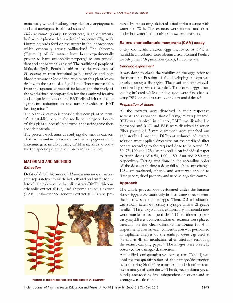
Heliconia rostrata (family: Heliconiaceae) is an ornamental herbaceous plant with attractive inflorescence (Figure 1). Humming birds feed on the nectar in the inflorescence which eventually causes pollination.6 The rhizomes (Figure 1) of H. rostrata have been experimentally proven to have antiophidic property,7 in vitro antioxi-dant and antibacterial activity.8 The traditional people of Malaysia (Ipoh, Perak) is said to use the rhizomes of H. rostrata to treat intestinal pain, jaundice and high blood pressure.9 One of the studies on this plant leaves dealt with the synthesis of gold and silver nanoparticles from the aqueous extract of its leaves and the study of the synthesized nantoparticles for their antiproliferative and apoptoic activity on the EAT cells which resulted in significant reduction in the tumor burden in EAT bearing mice.10
The plant H. rostrata is considerably new plant in terms of its establishment in the medicinal category. Leaves of this plant successfully showed anticarcinogenic ther-apeutic potential.10 The present work aims at studying the various extracts of rhizome and inflorescence for their angiogenesis and anti-angiogenesis effect using CAM assay so as to prove the therapeutic potential of this plant as a whole.
MATERIALS AND METHODSExtraction
Defatted dried rhizomes of Heliconia rostrata was macer-ated separately with methanol, ethanol and water for 72 h to obtain rhizome methanolic extract (RME), rhizome ethanolic extract (REE) and rhizome aqueous extract (RAE). Inflorescence aqueous extract (FAE) was pre-
pared by macerating defatted dried inflorescence with water for 72 h. The extracts were filtered and dried under hot water bath to obtain powdered extracts.
Ex-ovo chorioallantoic membrane (CAM) assay
5 day old fertile chicken eggs incubated at 37oC in humidified incubator were obtained from Central Poultry Development Organization (E.R.), Bhubaneswar.
Candling experiment
It was done to check the viability of the eggs prior to the treatment. Position of the developing embryo was checked using a flashlight. The dead and underdevel-oped embryos were discarded. To prevent eggs from getting infected while opening, eggs were first cleaned using 70% ethanol to remove the dirt and debris.11
Preparation of doses
All the extracts were dissolved in their respective solvents and a concentration of 20mg/ml was prepared. REE was dissolved in ethanol; RME was dissolved in methanol and RAE and FAE were dissolved in water. Filter papers of 3 mm diameter12 were punched out and sterilized properly. Different volumes of extract solution were applied drop wise on the sterilized filter papers according to the required dose to be tested. 25, 50, 75, 100 and 125µl were applied on individual paper to attain doses of 0.50, 1.00, 1.50, 2.00 and 2.50 mg, respectively. Testing was done in the ascending order of the doses each time a dose fail to show any change. 125µl of methanol, ethanol and water was applied to filter papers, dried properly and used as negative control.
Approach
The whole process was performed under the laminar flow.12 Eggs were cautiously broken using forceps from the narrow side of the eggs. Then, 2-3 ml albumin was slowly taken out using a syringe with a 21-gauge needle.12 The embryo and its extra embryonic membranes were transferred to a petri dish5. Dried filtered papers carrying different concentration of extracts were placed carefully on the chorioallantoic membrane for 4 h. Experimentation on each concentration was performed in triplicate. Images of the embryo were captured at 0h and at 4h of incubation after carefully removing the extract carrying paper.13 The images were carefully observed for damage/destruction. A modified semi quantitative score system (Table 1) was used for the quantification of the damage/destruction by comparing 0h (before treatment) and 4h (after treat-ment) images of each dose.12 The degree of damage was blindly recorded by five independent observers and an average was calculated.Figure 1: Inflorescence and rhizome of H. rostrata.
Dhara, et al.: Comment 2. CAM Assay on H. rostrata
S248 Indian Journal of Pharmaceutical Education and Research | Vol 52 | Issue 4s [Suppl 2] | Oct-Dec, 2018
RESULTS AND DISCUSSIONAngiogenesis is an important step in the tissue expansion which when get enhanced leads to the formation of tumors and haematological malignancies.14 Anti-angio-genesis therapy is one of the standard care therapies for various types of tumors which act by blocking the formation of new blood vessels in tumor cells. Synthetic drugs like Bevacizumab, Ramucirumab, Axitinib, Sorafenib acts by this mechanism and helps in the treatment of metastatic colorectal cancer, gastroesophageal junction adenocarcinoma, renal cell carcinoma, hepatocellular carcinoma, respectively.15
CAM assay is widely used for proangiogenic and antian-giogenic evaluation of different materials.16
Allantois of the chick embryo which appears in 3-5 days of incubation pushes out the body of embryo to extra embryonic coelam. From day 4-10, the allantoic vesicle enlarges rapidly and mesodermal layer of the allantois fuses with mesodermal layer of chorion to form chorio-allantoic membrane (CAM).4 The CAM assay fits in comfortably in the “three R’s” strategy of “Principles of Humane Experimental Tech-nique” which was established to reduce the use of animals for the investigation processes. The first “R” refers to the replacement of the animal models with other possible in vitro models. But the use of in vitro models is often not ideal because of the discrepancy between the systemic environments of the systems like vascular system. But, CAM is the outermost extra-embryonic membrane that is highly vascularized for gaseous exchange and calcium transportation between the embryo and environment. Thus, this problem of discrepancy is overcome with CAM assay. Though CAM assay is an in vivo model, prior approval from any ethical committee is not necessary for experimentation process. This is because of the fact that CAM assay is performed on the chick embryo which are 4 to 5 days old and the chick embryos younger than day 15 are assumed to experience no pain. This not only simplifies the planning process but also makes this assay less time
consuming. This covers the second “R” which refers to refinement. The third “R” is for the reduction of use of animal. Since the prior information of safe dose range is gathered from the CAM assay, wastage of animal life is minimized and the set dose can easily be confirmed with the use of less number of animals.3
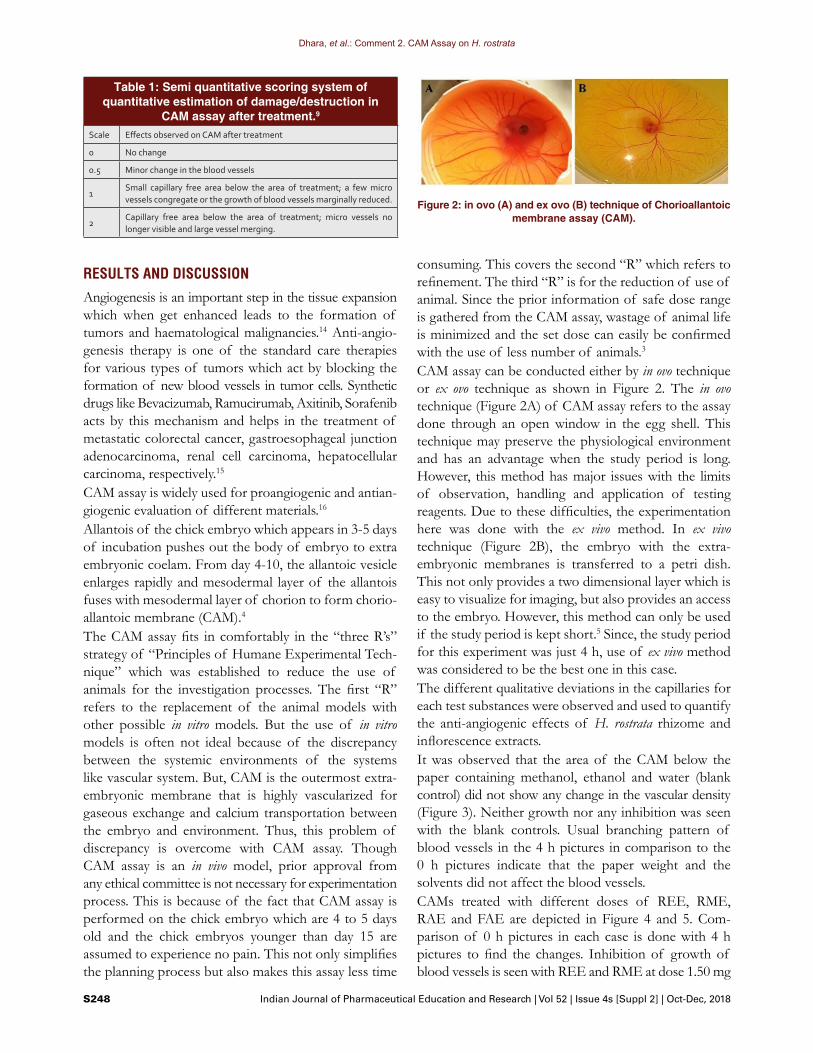
CAM assay can be conducted either by in ovo technique or ex ovo technique as shown in Figure 2. The in ovo technique (Figure 2A) of CAM assay refers to the assay done through an open window in the egg shell. This technique may preserve the physiological environment and has an advantage when the study period is long. However, this method has major issues with the limits of observation, handling and application of testing reagents. Due to these difficulties, the experimentation here was done with the ex vivo method. In ex vivo technique (Figure 2B), the embryo with the extra-embryonic membranes is transferred to a petri dish. This not only provides a two dimensional layer which is easy to visualize for imaging, but also provides an access to the embryo. However, this method can only be used if the study period is kept short.5 Since, the study period for this experiment was just 4 h, use of ex vivo method was considered to be the best one in this case. The different qualitative deviations in the capillaries for each test substances were observed and used to quantify the anti-angiogenic effects of H. rostrata rhizome and inflorescence extracts. It was observed that the area of the CAM below the paper containing methanol, ethanol and water (blank control) did not show any change in the vascular density (Figure 3). Neither growth nor any inhibition was seen with the blank controls. Usual branching pattern of blood vessels in the 4 h pictures in comparison to the 0 h pictures indicate that the paper weight and the solvents did not affect the blood vessels. CAMs treated with different doses of REE, RME, RAE and FAE are depicted in Figure 4 and 5. Com-parison of 0 h pictures in each case is done with 4 h pictures to find the changes. Inhibition of growth of blood vessels is seen with REE and RME at dose 1.50 mg
Table 1: Semi quantitative scoring system of quantitative estimation of damage/destruction in
CAM assay after treatment.9
Scale Effects observed on CAM after treatment
0 No change
0.5 Minor change in the blood vessels
1Small capillary free area below the area of treatment; a few micro vessels congregate or the growth of blood vessels marginally reduced.
2Capillary free area below the area of treatment; micro vessels no longer visible and large vessel merging.
Figure 2: in ovo (A) and ex ovo (B) technique of Chorioallantoic membrane assay (CAM).

Dhara, et al.: Comment 2. CAM Assay on H. rostrata
Indian Journal of Pharmaceutical Education and Research |Vol 52 | Issue 4s [Suppl 2] | Oct-Dec, 2018 S249
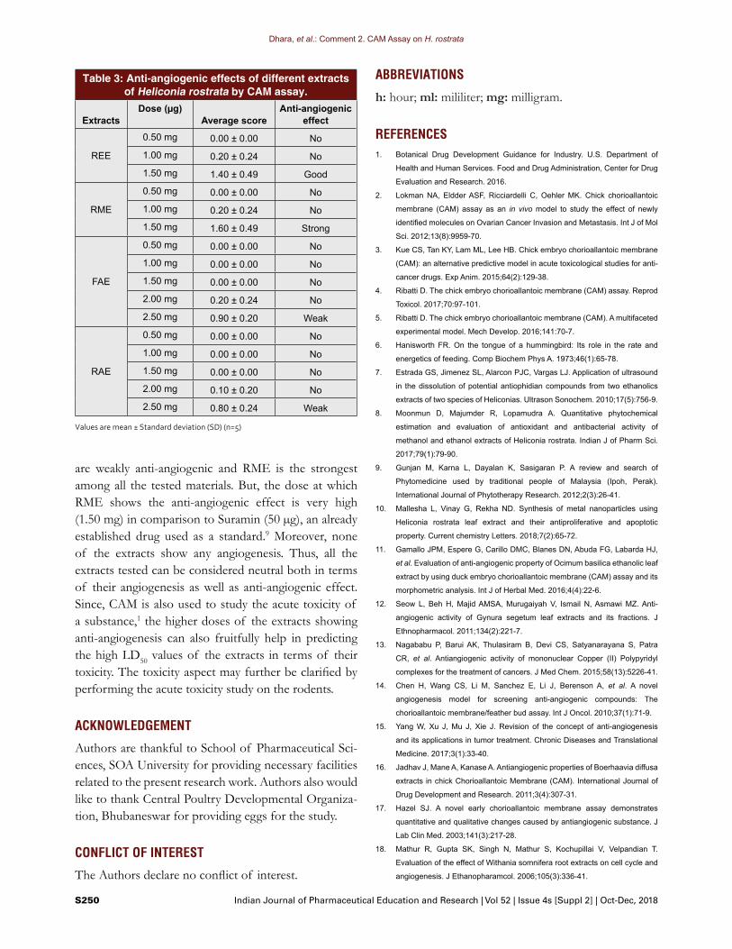
(Figure 4). No change is observed with the lower doses, 1.00mg and 0.50mg of REE and RME. In case of RAE and FAE, damaged or destroyed blood vessel is seen at dose 2.50 mg (Figure 5). Lower doses, 2.00 mg, 1.50 mg, 1.00 mg and 0.50 mg show no change. The branching pattern of blood vessels and the pre-existing vessels was reduced, with dose 1.50 mg for REE and RME (Figure 4) and 2.50 mg for FAE and RAE (Figure 5). Almost capillary free area is observed with the RME, 1.50 mg (Figure 4). The degree of damage was evaluated by average scores calculated using the pictorial observations. This semi-quantitative method was based on the observations made by five different observers having no idea of doses and extracts during observations and scores. The scoring was done according to the score pattern in Table 1 ranging from 0 to 2. Table 3 depicts the average score and the interpretation thus made according to Table 2. RME shows strong anti-angiogenic effect than REE at dose 1.5 mg per disc which shows good anti-angiogenic effect while at 2.5 mg dose both RAE and FAE show weak anti-angiogenic effect. Angiogenesis refers to the development of new blood vessels from the predating vasculature,12 whereas anti-angiogenesis refers to the inhibition of growth or death of the blood vessels. Both angiogenesis and anti- angio-genesis have their merits as well as demerits. Angiogenesis plays an important role in growth and metastasis of tumors and thus, cancer.17 Here, anti-angiogenic activity is considered to be a promising approach for treatment of cancer.18
CONCLUSIONThe Chorioallantoic membrane assay of REE, RME, RAE and FAE at different doses was performed using ex-ovo method. The method was adopted to check for the anti-angiogenic effect of the extracts so as to establish the fact that whether or not said extracts of H. rostrata be used for the treatment of cancer. Taking in consideration the observations made from the pictorial study and average scores (Table 2), both RAE and FAE
Figure 3: CAM of methanol, ethanol and water, the solvents used to dissolve the extracts.
Figure 4: CAM assay of REE and RME at different doses. Damaged/destroyed blood vessels are marked in circle.
Figure 5: CAM assay of FAE and RAE at different doses. Dam-aged/destroyed blood vessels are marked in circle.
Table 2: Interpretation of anti-angiogenic effect to be drawn from the average scores of semi-quantitative
scoring systems.Average
score Conclusions
< 0.5 No anti-angiogenic effect
≥ 0.5 and ≤ 1 Weak anti-angiogenic effect
> 1 and < 1.5 Good anti-angiogenic effect
≥ 1.5 strong anti-angiogenic effect
Dhara, et al.: Comment 2. CAM Assay on H. rostrata
S250 Indian Journal of Pharmaceutical Education and Research | Vol 52 | Issue 4s [Suppl 2] | Oct-Dec, 2018
ABBREVIATIONS
h: hour; ml: mililiter; mg: milligram.
REFERENCES1. Botanical Drug Development Guidance for Industry. U.S. Department of
Health and Human Services. Food and Drug Administration, Center for Drug
Evaluation and Research. 2016.
2. Lokman NA, Eldder ASF, Ricciardelli C, Oehler MK. Chick chorioallantoic
membrane (CAM) assay as an in vivo model to study the effect of newly
identified molecules on Ovarian Cancer Invasion and Metastasis. Int J of Mol
Sci. 2012;13(8):9959-70.
3. Kue CS, Tan KY, Lam ML, Lee HB. Chick embryo chorioallantoic membrane
(CAM): an alternative predictive model in acute toxicological studies for anti-
cancer drugs. Exp Anim. 2015;64(2):129-38.
4. Ribatti D. The chick embryo chorioallantoic membrane (CAM) assay. Reprod
Toxicol. 2017;70:97-101.
5. Ribatti D. The chick embryo chorioallantoic membrane (CAM). A multifaceted
experimental model. Mech Develop. 2016;141:70-7.
6. Hanisworth FR. On the tongue of a hummingbird: Its role in the rate and
energetics of feeding. Comp Biochem Phys A. 1973;46(1):65-78.
7. Estrada GS, Jimenez SL, Alarcon PJC, Vargas LJ. Application of ultrasound
in the dissolution of potential antiophidian compounds from two ethanolics
extracts of two species of Heliconias. Ultrason Sonochem. 2010;17(5):756-9.
8. Moonmun D, Majumder R, Lopamudra A. Quantitative phytochemical
estimation and evaluation of antioxidant and antibacterial activity of
methanol and ethanol extracts of Heliconia rostrata. Indian J of Pharm Sci.
2017;79(1):79-90.
9. Gunjan M, Karna L, Dayalan K, Sasigaran P. A review and search of
Phytomedicine used by traditional people of Malaysia (Ipoh, Perak).
International Journal of Phytotherapy Research. 2012;2(3):26-41.
10. Mallesha L, Vinay G, Rekha ND. Synthesis of metal nanoparticles using
Heliconia rostrata leaf extract and their antiproliferative and apoptotic
property. Current chemistry Letters. 2018;7(2):65-72.
11. Gamallo JPM, Espere G, Carillo DMC, Blanes DN, Abuda FG, Labarda HJ,
et al. Evaluation of anti-angiogenic property of Ocimum basilica ethanolic leaf
extract by using duck embryo chorioallantoic membrane (CAM) assay and its
morphometric analysis. Int J of Herbal Med. 2016;4(4):22-6.
12. Seow L, Beh H, Majid AMSA, Murugaiyah V, Ismail N, Asmawi MZ. Anti-
angiogenic activity of Gynura segetum leaf extracts and its fractions. J
Ethnopharmacol. 2011;134(2):221-7.
13. Nagababu P, Barui AK, Thulasiram B, Devi CS, Satyanarayana S, Patra
CR, et al. Antiangiogenic activity of mononuclear Copper (II) Polypyridyl
complexes for the treatment of cancers. J Med Chem. 2015;58(13):5226-41.
14. Chen H, Wang CS, Li M, Sanchez E, Li J, Berenson A, et al. A novel
angiogenesis model for screening anti-angiogenic compounds: The
chorioallantoic membrane/feather bud assay. Int J Oncol. 2010;37(1):71-9.
15. Yang W, Xu J, Mu J, Xie J. Revision of the concept of anti-angiogenesis
and its applications in tumor treatment. Chronic Diseases and Translational
Medicine. 2017;3(1):33-40.
16. Jadhav J, Mane A, Kanase A. Antiangiogenic properties of Boerhaavia diffusa
extracts in chick Chorioallantoic Membrane (CAM). International Journal of
Drug Development and Research. 2011;3(4):307-31.
17. Hazel SJ. A novel early chorioallantoic membrane assay demonstrates
quantitative and qualitative changes caused by antiangiogenic substance. J
Lab Clin Med. 2003;141(3):217-28.
18. Mathur R, Gupta SK, Singh N, Mathur S, Kochupillai V, Velpandian T.
Evaluation of the effect of Withania somnifera root extracts on cell cycle and
angiogenesis. J Ethanopharamcol. 2006;105(3):336-41.
are weakly anti-angiogenic and RME is the strongest among all the tested materials. But, the dose at which RME shows the anti-angiogenic effect is very high (1.50 mg) in comparison to Suramin (50 µg), an already established drug used as a standard.9 Moreover, none of the extracts show any angiogenesis. Thus, all the extracts tested can be considered neutral both in terms of their angiogenesis as well as anti-angiogenic effect. Since, CAM is also used to study the acute toxicity of a substance,1 the higher doses of the extracts showing anti-angiogenesis can also fruitfully help in predicting the high LD50 values of the extracts in terms of their toxicity. The toxicity aspect may further be clarified by performing the acute toxicity study on the rodents.
ACKNOWLEDGEMENT
Authors are thankful to School of Pharmaceutical Sci-ences, SOA University for providing necessary facilities related to the present research work. Authors also would like to thank Central Poultry Developmental Organiza-tion, Bhubaneswar for providing eggs for the study.
CONFLICT OF INTEREST
The Authors declare no conflict of interest.
Table 3: Anti-angiogenic effects of different extracts of Heliconia rostrata by CAM assay.
ExtractsDose (µg)
Average scoreAnti-angiogenic
effect
REE
0.50 mg 0.00 ± 0.00 No
1.00 mg 0.20 ± 0.24 No
1.50 mg 1.40 ± 0.49 Good
RME
0.50 mg 0.00 ± 0.00 No
1.00 mg 0.20 ± 0.24 No
1.50 mg 1.60 ± 0.49 Strong
FAE
0.50 mg 0.00 ± 0.00 No
1.00 mg 0.00 ± 0.00 No
1.50 mg 0.00 ± 0.00 No
2.00 mg 0.20 ± 0.24 No
2.50 mg 0.90 ± 0.20 Weak
RAE
0.50 mg 0.00 ± 0.00 No
1.00 mg 0.00 ± 0.00 No
1.50 mg 0.00 ± 0.00 No
2.00 mg 0.10 ± 0.20 No
2.50 mg 0.80 ± 0.24 Weak
Values are mean ± Standard deviation (SD) (n=5)

Dhara, et al.: Comment 2. CAM Assay on H. rostrata
Indian Journal of Pharmaceutical Education and Research |Vol 52 | Issue 4s [Suppl 2] | Oct-Dec, 2018 S251
SUMMARY
Chick embrryo chorioalltoic membrane (CAM) assay was performed on four different extracts of rhizome (REE, RME, RAE) and inflorescence (FAE) of Heli-conia rostrata at different doses ranging from 0 to 2.50 mg. CAM assay is an in vivo assay that provides per-fect environment for a safe, effective and economic angiogenesis and antiangiogenesis study before mov-ing on to study on rodents. In the present study, dif-ferent doses of the extracts were applied on the CAM using sterilized filter paper. The degree of change in the CAM was noted and demonstrated in pictorial fashion and by a semi-quantitative score system rang-ing between 0-2 with the increasing damage to the CAM. Both REE and RME were found to show anti-angiogenic effect at 1.50 mg dose level, while FAE and RAE showed some antiangiogenic effect at dose level of 2.50 mg. RME was found to have strongest anti-angiogenic effect among the four extracts. Yet, RME was not considered to be completely antiangiogenic because of the very high dose at which this effect is observed.
Dr. Lopamudra Adhikari: Associate Professor, Department of Pharmaceutical Analysis and Quality Assurance, School of Pharmaceutical Sciences, Siksha O Anusandhan (Deemed to be University), Bhubaneswar, Odisha, India.
About Authors
PICTORIAL ABSTRACT
Cite this article: Dhara M, Adhikari L, Majumder R. Chorioallantoic Membrane (CAM) Assay of Different Extracts of Rhizome and Inflorescence of Heliconia rostrata. Indian J of Pharmaceutical Education and Research. 2018;52(4 Suppl 2):s246-s251
Moonmun Dhara: Research Scholar, School of Pharmaceutical Sciences, Siksha O Anusandhan (Deemed to be University), Bhubaneswar, Odisha, India
Mr. Raja Majumder: Research Scholar, School of Pharmaceutical Sciences, Siksha O Anusandhan (Deemed to be University), Bhubaneswar, Odisha, India