Chloroplast DNA synthesis in light and dark grown cultured Nicotiana tabacum cells as determined by...
5
Plant Cell Reports (1985) 4:41-45 Plant Cell Reports © Springer-Verlag 1985 Chloroplast DNA synthesis in light and dark grown cultured Nicotiana tabacum cells as determined by molecular hybridization Gordon Cannon, Sabine Hcinhorst, Janusz Siedlecki, and Arthur Weissbach Department of Cell Biology, Roche Institute of Molecular Biology, Roche Research Center, Nutley, NJ 07110, USA Received December 14, 1984- Communicated by O. L. Gamborg Abstract A simple method using molecular hybridization was devised to quantitatively measure chloroplast DNA synthesis in rive. Total cellular DNA isolated from ~icotiana tabacum suspension cells, labeled with H-thymidine, was hybridized to nitrocellulose membrane-bound cloned chloroplast DNA (ct DNA) fragments. Colorless, dark grown N. tabacum cells were found to contain approximately 3300-4800 chloroplast genome copies per cell, whereas light grown, green cells contain about 9500-12000 chloro- plast genomes per cell. This difference in ct DNA levels suggests that the chloroplast genome is somewhat amplified during growth of the cells in the light. The hybridization technique was also used to measure the efficiency of hybridization between cloned spinach ct DNA and tobacco ct DNA. The two DNAs were found to cross-hybridize with an efficiency of 69-75%. Introduction The structure of the chloroplast genome is well established. It exists as a circular double stranded DNA molecule of approximately i0 daltons (Bedbrook and Kolodner 1979). Each chloroplast contains multiple copies of the genome and copy numbers from 8-200 per chloroplast have been reported for different species (Herrmann and Possingham 3980). It has also been demonstrated in numerous higher plants that cellular chloroplast DNA (ct DNA) content is dependent on age, tissue type and developmental stage of the cell containing the chloroplast (Scott and Possingham 1983). Little is known about at DNA synthesis or how it is regulated and relatively few studies have addressed the question of ct DNA synthesis in vivo. A major obstacle to these studies has been the lack of a method for easily quantitating ct DNA synthesis. Resolution of nuclear, mitochondrial and ct genomes on density gradients has been successful for some algae (Blamire et el. 1974) but this method is seldom suitable for separation of organellar genomes of higher plants (Lamppa and Bendich 1979). Likewise, isolation of intact chloroplasts after an in rive labeling period is difficult to use for quantitation because of varying yields from experiment to experiment and because contamination by nuclear and mitochondrial DNAs is often observed. To avoid these problems we have devised a method, based on molecular hybridization of total radio- labeled cellular DNA to cloned ct DNA, that permits us to specifically follow et DNA synthesis in growing suspension cultures of plant cells. Using this method we have been able to measure the chloroplast Offprint requests to: A. Weissbach genome copy number in cultured Nicotiana tabacum cells grown in the dark (colorless) or in the light (green). Also this paper reports the relative cross-hybridization efficiencies of tobacco and spinach ct DNAs, so that the ct DNA of either species can be used for accurate determination of chloroplast DNA by molecular hybridization on nitrocellulose membrane filters. Haterials and Methods Plant }~terial and Growth Conditions Nicotiana tabacum var. Wisconsin 38 suspension cultures were grown on Murashige and 8koog media (Hurashige and Skoog 1962) supplemented with 800 mg/l casein hydrolysate, 0.5 mg/l folic acid, 0.5 mg/l biotin, i0 mg/l thiamine, 0.2 mg/l benzyladenine (BA), 0.6 mg/l a-naphthaleneacetic acid and 3 g/l sucrose. The cultures were grown in the dark in 500 ml erlenmeyer flasks on a rotary shaker (150 rpm). Nicotiana tabacum var. Samsun (referred to as the Yamada line) was a gift from F. Sate and Y. Yamada of Kyoto University and was grown as suspension cultures on modified Linsmaier-Skoog media (Nishida et al. 1980) containing 3% sucrose. The light grown suspension cultures were maintained on a rotary shaker (150 rpm) under I0000 lux of light provided by wide spectrum fluorescent lamps (Verilux). Dark grown suspension cultures were maintained on a similar shaker in total darkness. Autotrophic cultures were maintained under 5% CO 2 on similar media lacking sucrose. Determination of Nuclear DNA Content Nuclei were isolated from cultured N. tabacum cells by the method of Willmitzer and Wagner (1981). DNA was estimated by the fluorometric assay described by Vytasek (1982). No significant difference in DNA quantities per nucleus was observed in light or dark grown cells. P_rej~aration of Cloned Ct DNA Fragments Recombinant plasmids containing Pstl endonuclease restriction fragments of spinach (Spinacia oleraceae) ct DNA cloned into pBR322 were obtained from J. Palmer (Palmer and Thompson 1981). Plasmids containing tobacco (Nicotia~a tabacum) ct DNA fragments in the PstI site of pBR322 were obtained from R. Fluhr (Fluhr et al. 1983). The recombinant plasmid pNT-32 that contains the psbA gene coding for the tobacco chloroplast 32000 dalton protein was obtained from L. Bogorad (personal communia~tSon). Isolation of spinach ct DNA fragments was accom- plished by digesting a mixture (20 vg) of plasmids that contained an equimolar amount of the following PstI spinach ct fragments: 17.3 Kbp, 13.5 Kbp, 12.3 Kbp, 8.9 Kbp, 8~i Kbp, 7.7 Khp, 2.7 Kbp and 1.9 Kbp with Pstl (see Figure i). The tobacco ct DNA fragment mixture was prepared by digesting
-
Upload
gordon-cannon -
Category
Documents
-
view
212 -
download
0
Transcript of Chloroplast DNA synthesis in light and dark grown cultured Nicotiana tabacum cells as determined by...
Plant Cell Reports (1985) 4:41-45
Plant Cell Reports © Springer-Verlag 1985
Chloroplast DNA synthesis in light and dark grown cultured Nicotiana tabacum cells as determined by molecular hybridization
Gordon Cannon, Sabine Hcinhorst, Janusz Siedlecki, and Arthur Weissbach
Department of Cell Biology, Roche Institute of Molecular Biology, Roche Research Center, Nutley, NJ 07110, USA
Received December 14, 1984- Communicated by O. L. Gamborg
Abstract
A simple method using molecular hybridization was devised to quantitatively measure chloroplast DNA synthesis in rive. Total cellular DNA isolated from ~icotiana tabacum suspension cells, labeled with H-thymidine, was hybridized to nitrocellulose
membrane-bound cloned chloroplast DNA (ct DNA) fragments. Colorless, dark grown N. tabacum cells were found to contain approximately 3300-4800 chloroplast genome copies per cell, whereas light grown, green cells contain about 9500-12000 chloro- plast genomes per cell. This difference in ct DNA levels suggests that the chloroplast genome is somewhat amplified during growth of the cells in the light. The hybridization technique was also used to measure the efficiency of hybridization between cloned spinach ct DNA and tobacco ct DNA. The two DNAs were found to cross-hybridize with an efficiency of 69-75%.
Introduction
The structure of the chloroplast genome is well established. It exists as a circular double stranded DNA molecule of approximately i0 daltons (Bedbrook and Kolodner 1979). Each chloroplast contains multiple copies of the genome and copy numbers from 8-200 per chloroplast have been reported for different species (Herrmann and Possingham 3980). It has also been demonstrated in numerous higher plants that cellular chloroplast DNA (ct DNA) content is dependent on age, tissue type and developmental stage of the cell containing the chloroplast (Scott and Possingham 1983). Little is known about at DNA synthesis or how it is regulated and relatively few studies have addressed the question of ct DNA synthesis in vivo. A major obstacle to these studies has been the lack of a method for easily quantitating ct DNA synthesis. Resolution of nuclear, mitochondrial and ct genomes on density gradients has been successful for some algae (Blamire et el. 1974) but this method is seldom suitable for separation of organellar genomes of higher plants (Lamppa and Bendich 1979). Likewise, isolation of intact chloroplasts after an in rive labeling period is difficult to use for quantitation because of varying yields from experiment to experiment and because contamination by nuclear and mitochondrial DNAs is often observed. To avoid these problems we have devised a method, based on molecular hybridization of total radio- labeled cellular DNA to cloned ct DNA, that permits us to specifically follow et DNA synthesis in growing suspension cultures of plant cells. Using this method we have been able to measure the chloroplast
Offprint requests to: A. Weissbach
genome copy number in cultured Nicotiana tabacum cells grown in the dark (colorless) or in the light (green). Also this paper reports the relative cross-hybridization efficiencies of tobacco and spinach ct DNAs, so that the ct DNA of either species can be used for accurate determination of chloroplast DNA by molecular hybridization on nitrocellulose membrane filters.
Haterials and Methods
Plant }~terial and Growth Conditions Nicotiana tabacum var. Wisconsin 38 suspension cultures were grown on Murashige and 8koog media (Hurashige and Skoog 1962) supplemented with 800 mg/l casein hydrolysate, 0.5 mg/l folic acid, 0.5 mg/l biotin, i0 mg/l thiamine, 0.2 mg/l benzyladenine (BA), 0.6 mg/l a-naphthaleneacetic acid and 3 g/l sucrose. The cultures were grown in the dark in 500 ml erlenmeyer flasks on a rotary shaker (150 rpm). Nicotiana tabacum var. Samsun (referred to as the Yamada line) was a gift from F. Sate and Y. Yamada of Kyoto University and was grown as suspension cultures on modified Linsmaier-Skoog media (Nishida et al. 1980) containing 3% sucrose. The light grown suspension cultures were maintained on a rotary shaker (150 rpm) under I0000 lux of light provided by wide spectrum fluorescent lamps (Verilux). Dark grown suspension cultures were maintained on a similar shaker in total darkness. Autotrophic cultures were maintained under 5% CO 2 on similar media lacking sucrose.
Determination of Nuclear DNA Content Nuclei were isolated from cultured N. tabacum cells by the method of Willmitzer and Wagner (1981). DNA was estimated by the fluorometric assay described by Vytasek (1982). No significant difference in DNA quantities per nucleus was observed in light or dark grown cells.
P_rej~aration of Cloned Ct DNA Fragments Recombinant plasmids containing Pstl endonuclease restriction fragments of spinach (Spinacia oleraceae) ct DNA cloned into pBR322 were obtained from J. Palmer (Palmer and Thompson 1981). Plasmids containing tobacco (Nicotia~a tabacum) ct DNA fragments in the PstI site of pBR322 were obtained from R. Fluhr (Fluhr et al. 1983). The recombinant plasmid pNT-32 that contains the psbA gene coding for the tobacco chloroplast 32000 dalton protein was obtained from L. Bogorad (personal communia~tSon).
Isolation of spinach ct DNA fragments was accom- plished by digesting a mixture (20 vg) of plasmids that contained an equimolar amount of the following PstI spinach ct fragments: 17.3 Kbp, 13.5 Kbp, 12.3 Kbp, 8.9 Kbp, 8~i Kbp, 7.7 Khp, 2.7 Kbp and 1.9 Kbp with Pstl (see Figure i). The tobacco ct DNA fragment mixture was prepared by digesting

42
a mixture (28 ~g) of plasmids containing equimolar amounts of the following tobacco PstI ct DNA frag- ments: 20.2 Kbp, 19.2 Kbp, 8.2 Kbp and 6.9 Kbp (Figure i). The ct DNA fragments of either mixture were then freed of the vector DNA by preparative electrophoresis on a 0.6% agarose column in a continuous elution electrophoresis device (PREP-GEL, Bethesda Research Laboratories).
The 1 Kbp ct DNA fragment from within the coding region of the Nicotiana tabacum psbA gene was pre- pared by digestion of pNT32 (200 ~g) with EcoRI and subsequent isolation of the 3.0 Kbp insert by electrophoresis on low temperature gelling agarose (Sea-Plaque - FMC Corporation) following the method of Schmitt and Cohen (1983). The 3.0 Khp fragment so obtained was further digested with restriction endonucleases fnuDII and XbaI to yield the desired 1 Kbp fragment (as based on sequence data in Zurawski et al. 1982),-which was then isolated by electrophoresis on low melting agarose as above.
Preparation of Nitrocellulose Membranes Containing Ct DNA
Mixtures of either tobacco or spinach cloned ct DNA were prepared by mixing the plasmids containing the proper ct DNA fragments (see above) in equimolar amounts. After brief alkali denaturation, the DNA was diluted to 1 ~g/ml with 6 x SSC (i x SSC consists of 0.15 M NaCI~ 0.015 M Na-Citrate) and adsorbed onto nitrocellulose membranes (Sartorius type SM PI2; 2~5 cm dia.; 0.45 ~m pore size) according to Gillespie and Spiegelman (1965). Each membrane filter had the equivalent of 5 ~g of ct DNA adsorbed to it.
Radio-labeling of Ct DNA Fragments Equimolar mixtures of purified spinach or tobacco ct DNA fragments (see ~ove) free of pBR322 DNA were radio-labeled with ~ P-deoxycytidine triphosphate by random primer extension using DNA polymerase Klenow fragment (Eoehringer Mannheim) as described ~ Summers (1975). Specific radioactivity of_the -P-labeled ct DNA was routinely 0.5-1.2 x i08 cpm/~g
of DNA. The average length of the labeled et DNA fragments was approximately 500 bp as determined by electrophoresis on 2% agarose gels.
Extraction and Purification of N. tabacum Cellular DNA Isolation of cellular DNA from N. tabacum suspension cultures was carried out by a modification of the procedure reported by Murray and Thompson (1980). Cells (typically 0.5 - 1.5 g fresh weight) were washed on course sintered glass filters with two volumes of breaking buffer [50 mM Tris-HCl (pH 8.0), 20 mM EDTA, 1 M NaCI], resuspended in 5 ml of the same buffer and passed through a French pressure cell at 20000 psi. The resulting extract was clarified by centrifugation at 20000 x g for 15 minutes. Two volumes of cold ethanol were added to the supernatant and the pre- cipitate collected by centrifugation. The pellet was resuspended in a minimal volume of 50 mM Tris-HCl (pH 8.0) containing 2 mM EDTA and 0.5% sodium dodecylsulfate (SDS). Proteinase K (E. Merck, Darmstadt) was added to a final concentration of 150 ~/ml and the solution was incubated at 37°C for two hours. It was then extracted with phenol:chloro- form (i:I vol/vol) saturated with i0 mM Tris-HC! (pH 8.0) containing 1 mM EDTA and the aqueous phase was precipitated with two volumes of cold ethanol. The precipitate was resuspended in 2-3 ml of cetyltrimethylammonium bromide (CTAB) buffer con- taining 1% CTAB, 50 mM Tris-HCI (pH 8.0), i mM EDTA, 0.7 M NaCI and 1 mM B-mercaptoethanol. After extrac- tion with chloroform:isoamyl alcohol (24:1 vol/vol), the aqueous phase was removed and precipitated with CTAB precipitation buffer [1% CTAB, 50 m~ Tris-HCl (pH 8.0), 2 mM EDTA, 1 mM B-mercaptoethanol]. The DNA obtained by this proCedure was collected by
centrifugation, resuspended into ELUTIP-d high salt buffer [i M NaCI, 20 mM Tris-HCl (pH 7.4), 1 m~ EDTA]
and reprecipitated with 2 volumes of cold ethanol. The resulting precipitate was resuspended into ELUTIP-d low salt buffer [0.2 M NaCI, 20 mM Tris-HCl (pH 7.4), 1 mM EDTA] and passed over an ELUTIP-d column (Schleicher and Schuell). After washing the column
with 2-3 volumes of low salt buffer, the DNA was eluted with 0.4 ml of ELUTIP-d high salt buffer and precipitated by addition of 2 volumes cold ethanol. The resulting pellet accounted for 20-40% of the DNA in the initial cell free extract. To rid the DNA of any possible contaminating RNA and to shear the DNA, the DNA pellet was resuspended into 0,22 ml of 0.3 M Na0H in a 1.5 ml Eppendorf tube (Brinkmann Instrument, Inc.) and placed in boiling water for 3 minutes. It was then cooled in an ice bath and neutralized by addition of 0.012 ml Tris-HCl (pH 8.0) and 0.02 ml of 3.3 M HCI. The final pH was between 7.0 and 7.5~ Electrophoresis on 2% agarose gels demonstrated the length of the DNA to range from 400-800 nucleotides long.
Hybridization Nitrocellulose membranes containing a mixture of plasmids equivalent to 5 vg of ct DNA were pre- hybridized in 20 ml glass scintillation vials with sealable tops. Two ml of prehybridization fluid [50% formamide, 3 x SSC, i x Denhardt solution, 0.1% SDS, 1 mM EDTA (Denhardt 1966)] was added to each vial containing a membrane on which ct DNA had been bound and a blank membrane containing no DNA. The vials were then placed into a 37°C water bath for 4 hours. The prehybridization fluid was removed by aspiration and replaced with hybridization fluid (prehybridization fluid containing 50 ~g/ml denatured calf thymus DNA). For hybridizations between two
~ oned ct DNAs, 0.i pg of the appropriate denatured -P-labeled ct DNA (approximately i0000 cpm) was
added to,the scintillation vial. For hybridization between ~H-labeled total cell~lar DNA and cloned ct DNA, 0.7 ~g of the denatured H-cellular DNA was added tonthe vial. In addition, 0.01 pg (about i000 cpm) of B2P-labeled ct DNA fragments identical to those bound on the membrane was added to act as an internal control. The vials were resealed and placed at 37°C for 44 hours after which the membrane filters were removed and washed twice in 2 x SSC containing 0pl% SDS at room temperature followed by two washes with 0.2 x SSC containing 0.1% SDS at 37°C. The membrane filters were blotted and dried under an incandescent lamp and counted in Beckman RediSolv-NA scintillation fluid. The control hybridizations between 100% homologous DNAs usually showed a hybridi- zation efficiency of about 60-70% and the observed experimental hybridizations were corrected according- ly. Observed hybridization values were also corrected for the percentage of the chloroplast genome for which no equivalent fragment existed on the nitrocellulose membrane. Corrections were carried out as described by the following equation:
Corrected Hybridization =
(%)
Observed Hybridization of Experimental DNA (%)
~Fraction of] Fraction of the |Internal | IChl°r°plast Genome |Control | X Present in Membrane 132p-DNA 1 [Bound DNA [Hybridized J
In all hybridizations, the quantity of input chloro- plast DNA was never greater than one-tenth of chloro- plast DNA bound to the membrane. The stringency of the hybridization conditions was calculated to be equivalent to 72°C in 100% aqueous media containing 0.5 ~! NaCI. The reproducibility of the hybridization was determined to be + 10%.

43
Results and Discussion
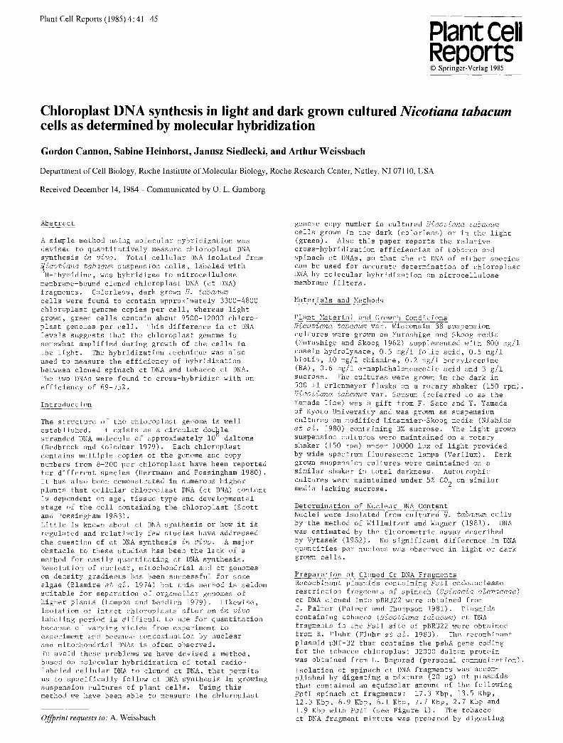
Use of Molecular Hybridization to Determine Cross-Hybridization of Tobacco and >pinach Cloned Ct DNA Fluhr and Edelman (1981) have shown considerable con- servation of nucleotide sequence arrangement between the chloroplast genomes of Niootiana tabacum and Spinaoia oleraoeae. However, small scale rearrange- ments can be a frequent occurence within the genomes of chloroplasts (Palmer and Thompson 1981). Such rearrangements or base substitutions could signifi- cantly effect cross-hybridization between the two chloroplast genomes. Since some of our earlier work required hybridization between tobacco total cellular DNA and cloned spinach ct DNA, we decided to evaluate the extent of cross-hybridization between positionally similar cloned fragments of the tobacco and spinach chloroplast genomes. Figure 1 contains the PstI endonuclease restriction site maps of the tobacco and spinach chloroplast genomes. Cloned DNA fragments from each of the chloroplast genomes (as indicated in the legend to Figure i) were combined to make an
Sp, nach
PstI RESTRICTION MAP OF
CHLOROPLAST GENOME
Fig. i. PstI restriction endonuclease cleavage site maps o~ spinach (Crouse et al. 1978) and tobacco (Fluhr et al. 1983) chloroplast genomes. The tobacco ct DNA fragment mixture referred to in the text con- sisted of the following fragments: 20.2 Kbp, 19.2 Kbp, 9.3 Kbp, 8.2 Kbp and 6.9 Kbp. The spinach ct DNA fragment mixture consisted of the following: 17.3 Kbp, 13.5 Kbp, 12.3 Kbp, 8.9 Kbp, 8.1 Kbp, 7.7 Kbp, 2.7 Kbp and 1.9 Kbp fragments. The inverted repeat regions (IR) are denoted by the solid black arcs and genes coding for the ribosomal RNA species are indicated within the repeated regions. ......................................................
equimolar mixture of tobacco ct DNA fragments repre- senting 46.0% of the tobacco chloroplast genome and an equimolar mixture of spinach ct DNA fragments representing 47.6% of the spinach chloroplast genome. The two cloned ct DNA mixtures have in common frag- ments that represent about 41.5% of the total chloro- plast genome, most of which are located in the large single copy region of the genome (Figure i). The cloned ~bacco ct DNA mixture was then used to pre- pare a ~ P-labeled probe that was subsequently hybri- dized to the plasmid mixture containing spinach ct DNA bound to a nitrocellulose membrane. The estimated percent of probe in common with the membrane bound
spinach ct DNA was 89.7 as determined by comparison of published maps of the two ct genomes. Conversely, ~ cloned spinach ct DNA mixture was labeled wfth
and used as a probe for hybridization with the plasmid mixture containing tobacco ct DNA immobilized on nitrocellulose. The estimated percent of probe in common with the membrane bound tobacco DNA was 87.3.
Cont~@l hybridizations were conducted between each of the ~-P-labeled DNA mixtures and its unlabeled plasmid counterpart bound to a nitrocellulose membrane. An average direct value of 60-70% hybridization between identical DNAs (data not shown) was observed using the hybridization conditions detailed in "~terials and Methods." As seen in Table 1 the tobacco ct DNA mixture and the spinach ct DNA mixture cross-hybridized to each other with about 75% efficiency (74.6% for the spinach ct DNA to tobacco ct DNA and 78.8% for the tobacco ct DNA to spinach ct DNA). This hybridization ......................................................
Table i. Cross-hybridization of spinach and tobacco ct DNA.
Corrected DNA Bound to Efficiency of b Nitrocellulose Hybridization
Probe Membrane (%) Plasmids containing
Cloned tobacco cloned spinach • a • a
ct DNA mlxture ct DNA mlxture 78.8 Plasmids containing
Cloned spinach cloned tobacco • a . a
ct DNA mlxture ct DNA mlxture 74.6
1 Kbp tobacco ct DNA fragment from within the coding region for the 32Kd protein
Plasmid with 8.9 Kbp spinach ct DNA fragment containing the entire coding region for the
32Kd protein 95.4
asee "Materials and }iethods" for composition of mixtures anJ hybridization conditions. The probes (0.i ng~ i0 cpm) did not contain the vector pBR322
~NA. Calculated as described in "Materials and Methods."
.................... - ................................
value lends support to the suggestion by Fluhr and Edelman (1981) that numerous small scale rearrange- ments have occurred in the tobacco and spinach chloro- plast genome and reflects a significant difference in the nucleotide sequence of two chloroplast genomes.
Cross-Hybridization of the Chloroplast psbA Gene Codinz for the 32000 Dalton Protein To further test the reliability of the hybridization procedure, the 1 Kbp DNA fragment from within the cQding region of the N. tabacum psbA gene%~oding for th~ 32000 dalton chloroplast protein was --P-labeled and hybridized to a nitrocellulose bound 8.9 Kbp spinach at DNA fragment that contains the entire coding region for the 32000 dalton protein (Zurawski et al. 1982). The coding sequence of the chloroplast gene (psbA) coding for the chloroplast 32000 dalton protein of photosystem II is highly conserved in higher plants. The nucleotide sequence for this gene in Nicotiana debneyi is 95.7% homologous to that in spinach (Zurawski et al. 1982). Since the restriction endonuclease pattern with EooRI, Fn~DII, XbaI and PstI of the M. tabacum psbA gene is identical to that of N~ debneyi (unpublished data) it is likely that the psbA gene of N. tabacum also shares the same homology to the corresponding gene in the spinach chloroplast genome. As seen in Table i, the tobacco probe hybridizes to the membrane bound spinach ct DNA fragment with 95% efficiency (corrected value), indicating the good correlation between sequence homology and hybridization efficiency observed under

44
these conditions.
H~bridization of Total Cellular Nicotiana*abacmm Wisconsin 38 DNA to Membrane Bound Cloned Ct DNA from Tobacco or Spinach The use of nitrocellulose bound plasmids containing cloned spinach ct DNA fragments to measure ct DNA levels in tobacco cells ~as evaluated by extracting total cellular DNA from ~H-thymidine labeled suspen- sion cultures of Niootiana tabaoum Wisconsin 38 and hybridizing it to both spinach and tobacco cloned ct DNA bound to nitrocellulose membranes. Table 2 shows that 8.3% (corrected value) of the total
.......................... ~ ............. -- ...........
able 2. Hybridization of H-labeled total N. tabacum DNA (Wisconsin 38) to cloned spinach or tobacco ct DNA fragments.
Nitrocellulose % of Chloroplast Genome Corrected b Membrane Bound Represented by Membrane Hybridization Ct DNA Mixture Bound Ct DNA Mixture .... (%)
Tobacco ~ 46.0 8.3 Spinach a 47.6 5.7
~See "Materials and Methods" for composition of mix- tures and hybridization conditions. The membrane ~ound ct DNA was in the plasmid form. Calculated as described In Materials and Methods." ....................................................
H-labeled tobacco cellular DNA hybridized to the cloned tobacco ct D~A, whereas only 5.7% (corrected value) of the same ~H-labeled tobacco cellular DNA hy- bridized to the membrane bound cloned spinach ct DNA. From these data, it can be estimated that the tobacco ct DNA hybridizes to spinach ct DNA with an efficiency of 69% under the conditions used in these studies. This is in good agreement with the approximately 75% cross-hybridization observed between cloned spinach and tobacco ct DNA. It is, therefore, feasible to use cloned spinach ct DNA bound on nitrocellulose mem- branes to accurately measure levels of ct DNA in tobacco cellular DNA provided the experimentally observed values are appropriately corrected for the 75% (74.6-78.8%) cross-hybridization between ct DNA of spinach and tobacco.
C hhloroplast Copy_Number in Light and Dark Grown Cells The N. tabacum line (Yamada line) utilized for these studies is capable of greening when grown in the light (Yamada and Sato 1978). Green, light grown cells (36 Dg chlorophyll/ml packed cells) and colorless, dark grown cells (0 zg chlorophyll/ml packed cells) were continuously radio-labeled for 6 days by d~ily addi- tion of H-thymidine to the culture. The -H-labeled total cellular DNA was isolated from aliquots removed on days 5 and 6 and each was hybridized to the tobac- co ct DNA fragment mixture. The average percentage hybridization to the ct DNA for the dark grown Yamada DNA was found to be 5.0%, 8.3% for DNA from dark grown Wisconsin 38, and 15% for DNA from light grown Yamada cells. Since there are 11.2 pg of DNA per nucleus in the Yamada line and i0 pg of DNA per nucleus in the Wisconsin 38, as directly determined in this labora- tory, these hybridization values correspond to 3300 chloroplast genome copies per cell for the dark grown Yamada cells, 4800 copies for dark grown Wisconsin 38 cells, and 100Q0 chloroplast genome copies per light grown Yamada cells. Similarl~, DNA from green Yamada cells was radio-labeled with -H-thymidine while growing in media with or without sucrose in a 5% CO 2 atmosphere. The DNA was isolated and hybridized to cloned tobacco ct DNA. The percent of hybridization for the autotrophically grown cells (no sucrose) was 14.5% and 18.3% for the photoheterotrophic cells (sucrose), corresponding to 9500 and 12000 chloroplast genome copies per cell, respectively.
Table 3. Chloroplast genome copy number in tobacco £ells. --
Cells Wisconsin 38 Yamada Yamada Yamada Yamada
Plant Tissue
Growth Conditions Ct Genome/Cell Dark, + Sucrose, Air 4800 Dark, + Sucrose, Air 3300 Light, + Sucrose, Air i0000 Light, - Sucrose, 5% CO 2 9500
Light, + Sucrose, 5% CO 2 12000
Leaf 2600a-5800 b Roots <600 a
~ Siegel-1974 Zimmerman and Goldberg-1976
.....................................................
The value of 3300 genome copies per dark grown Yamada cell was close to that obtained with dark grown Wisconsin 38 cells as shown in Table 3. Chloroplast genome copy numbers in this range agree well with the reported values for tobacco leaves (Zimmerman and Goldberg 1977) and are some 5-6 fold higher than those reported for tobacco root tissue (Siegel 1974). Since only part of the ct genome was used in determining the cellular level of ct DNA, we cannot rule out the possibility that some untested parts of the genome may exist in higher or lower proportions than reported here, however, we feel this is unlikely. We conclude that the 2.8~3.6 fold increase in chloroplast copy number found in light grown N. tabacum suspension cells over that in dark grown cells represents a regulatory amplification of the chloroplast genome during growth in the light.
References
Bedbrook J R, Kolodner R (1979) Ann Rev Plant Physiol 30: 593~620
Blamire J, Flechtner V R, Sager R (1974) Proc Natl Acad Sci USA 71: 2867-2871
Crouse E J, Schmitt J M, Bohnert H J, Gordon K, Hermann, R G (1978) In: Akoynoglou G, Argyroudi- Akoynoglou J H (Eds) Chloroplast Development, Elsevier/North-Holland Biomedical Press, Amsterdam, pp 565-569
Denhardt D (1966) Biochem Biophys Res Commun 23: 641-646
Fluhr R, Edelman M (1981) Nucl Acids Res 9: 6841- 6853
Fluhr R, Fromm H, Edelman ~ (1983) Gene 25: 271-280
Gillespie D, Spiegelman S (1965) J Mol Biol 12: 814-842
Herman R G, Possingham J V (1980) In: Reinert J (Ed) Results and Problems in Cell Differentiation, Vol i0, pp 45-85
Lamppa G K, Bendich A J (1979) Plant Physiol 64: 126-130
Murashige T, Skoog F (1962) Physiol Plant 15: 473-497
Murray M G, Thompson W F (1980) Nucl Acids Res 8: 4321-4325
Nishida K, Sato F, Yamada Y (1980) Plant Cell Physiol 21: 47-54
Palmer J D, Thompson W F (1981) Gene 15: 21-26
Schmitt J J, Cohen, B N (1983) Anal Biochem 462: 464
Scott N S, Possingham J V (1983) J Exptl Botany 34: 1756-1767
Siegel A (1974) In: Makken R, Davies D R, Hapwood DA Homes R V (eds) Modification of the Information

Content of Plant Cells Proc Sec John Innes Symp North Holland/Elsevier, pp 15-26
Summers J (1975) J Virol 15: 946-953
Vytasek R (1982) Anal Biochem 120: 243-248
Willmitzer L, Wagner K G (1981) Exp Cell Res 135: 69-77
45
Yamada Y, Sato F (1978) Plant Ceil Physiol 19: 691- 702
Zimmerman L, Goldberg R B (1977) Chromosomal 59: 227-252
Zurawski G, Bohnert H J, Whitfeld P R, Bottomley W (1982) Proc Natl Acad Sci USA 79: 7699-7703