CHLOROPHYLL D, A GREEN PIGMENT OF RED ALGAE · CHLOROPHYLL D, A GREEN PIGMENT OF RED ALGAE BY...
20
CHLOROPHYLL D, A GREEN PIGMENT OF RED ALGAE BY WINSTON M. MANNING AND HAROLD H. STRAIT\; (From the Carnegie Institution of Washington, Division of Plant Biology, Stanford University, California) (Received for publication, July 1,1943) Recent investigations have emphasized t,he diversity of chlorophylls in plants (l-3). Higher plants and green algae contain chlorophyll a and chlorophyll b. By contrast, diatoms, dinoflagellates, and brown algae do not contain chlorophyll b but do contain, in addition to chloro- phyll a, a characteristic green pigment, chlorophyll c (I, 2). Red algae (Rhodophyta) are sharply distinguished from all other plants with respect to anatomy, life history, and the occurrence of certain pro- teinaceous pigments. In order to determine whether or not this distinc- t,iveness is reflected by the occurrence of unique green pigments, we have investigated the chlorophylls of several marine speciesof red algae. Earlier work has shown that chlorophyll a is the principal chlorophyll of red algae, and that chlorophyll b is absent, or present only in traces (4-7). The occurrence of chlorofucine (chlorophyll c) in red algae, re- ported 70 years ago by Sorby (4), has not been verified (2, 5, 7). Until now, no other chlorophyll-like pigment has been reported for red algae. EXPERIMENTAL Material and Methods The red algae were collected at low tide near Moss Beach north of Half AIoon Bay, California. We are indebted to Dr. Gilbert M. Smith of Stanford University for identification of these algae. In most cases the material was used within 24 hours after collection. When not used within this time, the algae were moistened with sea water and stored at 6” in a loosely covered container. The methods employed for extraction and purification of the pigments, and for measurement of spectral absorption, were essentially those de- scribed in preceding papers (1, 2). When modification or extension of these methods was necessary, the procedures are described in the sections pertaining to the preparation of individual compounds. For most species of red algae, chlorophyll extraction required several hours when the fresh thalli were treated directly with methanol. For one species, Erythrophyllum delesserioides, extraction with methanol was nearly complete in 20 to 30 minutes. When the algae were killed by by guest on April 22, 2020 http://www.jbc.org/ Downloaded from
Transcript of CHLOROPHYLL D, A GREEN PIGMENT OF RED ALGAE · CHLOROPHYLL D, A GREEN PIGMENT OF RED ALGAE BY...
CHLOROPHYLL D, A GREEN PIGMENT OF RED ALGAE
BY WINSTON M. MANNING AND HAROLD H. STRAIT\;
(From the Carnegie Institution of Washington, Division of Plant Biology, Stanford University, California)
(Received for publication, July 1,1943)
Recent investigations have emphasized t,he diversity of chlorophylls in plants (l-3). Higher plants and green algae contain chlorophyll a and chlorophyll b. By contrast, diatoms, dinoflagellates, and brown algae do not contain chlorophyll b but do contain, in addition to chloro- phyll a, a characteristic green pigment, chlorophyll c (I, 2).
Red algae (Rhodophyta) are sharply distinguished from all other plants with respect to anatomy, life history, and the occurrence of certain pro- teinaceous pigments. In order to determine whether or not this distinc- t,iveness is reflected by the occurrence of unique green pigments, we have investigated the chlorophylls of several marine species of red algae.
Earlier work has shown that chlorophyll a is the principal chlorophyll of red algae, and that chlorophyll b is absent, or present only in traces (4-7). The occurrence of chlorofucine (chlorophyll c) in red algae, re- ported 70 years ago by Sorby (4), has not been verified (2, 5, 7). Until now, no other chlorophyll-like pigment has been reported for red algae.
EXPERIMENTAL
Material and Methods
The red algae were collected at low tide near Moss Beach north of Half AIoon Bay, California. We are indebted to Dr. Gilbert M. Smith of Stanford University for identification of these algae. In most cases the material was used within 24 hours after collection. When not used within this time, the algae were moistened with sea water and stored at 6” in a loosely covered container.
The methods employed for extraction and purification of the pigments, and for measurement of spectral absorption, were essentially those de- scribed in preceding papers (1, 2). When modification or extension of these methods was necessary, the procedures are described in the sections pertaining to the preparation of individual compounds.
For most species of red algae, chlorophyll extraction required several hours when the fresh thalli were treated directly with methanol. For one species, Erythrophyllum delesserioides, extraction with methanol was nearly complete in 20 to 30 minutes. When the algae were killed by
by guest on April 22, 2020
http://ww
w.jbc.org/
Dow
nloaded from
2 CHLOROPHYLL D OF RED ALGAE
immersion for 1 minute in boiling water, subsequent extraction of chloro- phylls with methanol was much more rapid, being complete, in from 10 to 30 minutes for all species. Extraction of the chlorophylls with ethanol, with pyridine, or with acetone-water (4: 1 by volume) was less rapid than with methanol.
None of these solvents extracted appreciable quantities of phycoe- rythrin, the characteristic red pigment of red algae (8,9). After exhaustive extraction lvith methanol, the algal material was always bright red or pink, even for species which were originally green, brown, or purple. For the inexperienced collector, the residual red color, which appears within a few minutes upon extraction of the heat-killed organism, affords a simple and rapid means of recognizing as red algae (Rhodophyta) certain species which otherwise might be mistaken for brown or green algae.
Spectral absorpt,ion and fluorescence measurements of extracted pig- ments were made with a photoelectric spectrophotometer, constructed by Smith (10). Most of the absorption spectra shown in this paper are plotted as log log (lo/l) ~C~SUS wave-length. The lability of the pigments and the presence of colorless concomitants precluded determination of the specific absorption coefficients.
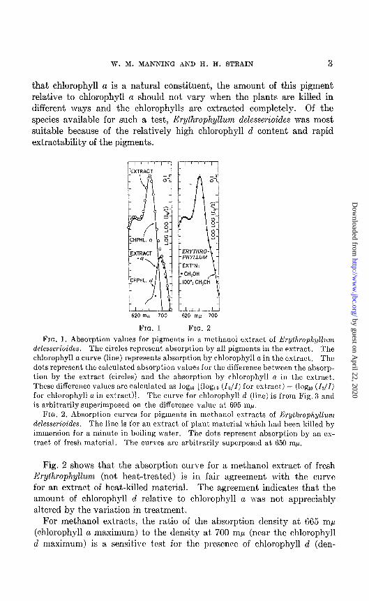
Natural Occurrence of Chlorophyll d-In Fig. 1 are presented the char- acteristic spectral absorption values for a methanol extract of Erythrophyl- lum delesserioides, killed by immersion for 1 minute in boiling water. The curve for chlorophyll a (1) is superposed arbitrarily on the absorption values for the extract. Coincidence of the circles with the curve in the regions of maximum absorption indicates t)hat the extract contains a large proportion of chlorophyll a. Between 680 and 720 m,u, absorption values for the extract diverge a great deal from the chlorophyll a curve. This divergence demonstrates the presence of another pigment (or pigments) that absorbs strongly at the longer wave-lengths. The relative spectral absorption by this second pigment in the extracts of Erythrophyllum was estimated by subtraction of the fractional absorption due to chlorophyll a from the total absorption, as indicated by the formula in the caption for Fig. 1. The values obtained in this may, shown in Fig. 1, correspond with the characteristic absorption curve for a green pigment prepared by chromatographic adsorption from red algae (see Fig. 3). Superposabil- ity of the absorption curve on the difference values indicates that the absorption in the region 680 to 720 rnp, over and above that due to chloro- phyll a, is primarily or solely due to this second green pigment. For this pigment, we propose the name chlorophyll d.
If chlorophyll d is a natural constituent of red algae, in the same sense
by guest on April 22, 2020
http://ww
w.jbc.org/
Dow
nloaded from
W. M. MANNING AND H. H. STRAIN 3
that chlorophyll a is a natural constituent, the amount of this pigment relative to chlorophyll a should not vary when the plants are killed in different ways and the chlorophylls are extracted completely. Of the species available for such a test, Erythrophyllum delesserioides was most suitable because of the relatively high chlorophyll d content and rapid extractability of the pigments.
620 rnfi 700 t
620 m,, 700
FIG. 1 FIG. 2 FIG. 1. Absorption values for pigments in :I methanol extract of E’rythrophyllunt
delesserioides. The circles represent absorption by all pigments in the extract. The chlorophyll a curve (line) represents absorption by chlorophyll a in the extract. The c1ot.s represent the calculated absorption values for the ditfercnce between the absorp- tion by the extract. (circles) and the absorption by chlorophyll a in the extract. These difference values arc calculated as log,, [(log,, (lo/Z) for extract) - (log,, (lo/Z) for chlorophyll a in extract)]. The curve for chlorophyll cl (line) is from Fig. 3 and is arbitrarily superimposed on the difference value at 695 mp.
FIG. 2. Absorption curves for pigments in methanol extracts of Erythrophyllum delesserioides. The line is for an extract of plant material which had been killed by immersion for a minute in boiling water. The dots represent absorption by an cx- tract of fresh material. The curves arc arbitrarily superposed at 650 rnp.
Fig. 2 shows that the absorption curve for a methanol extract of fresh Erythrophyllum (not heat-treated) is in fair agreement with the curve for an extract of heat-killed mat.erial. The agreement indicates that the amount of chlorophyll d relative to chlorophyll a was not appreciably altered by the variation in treatment.
For methanol extracts, the ratio of the absorption density at 665 rnp (chlorophyll a maximum) to the density at 700 rnp (near the chlorophyll d maximum) is a sensitive test for the presence of chlorophyll d (den-
by guest on April 22, 2020
http://ww
w.jbc.org/
Dow
nloaded from

4 CHLOROPHYLL D OF RED ALGAE
sity = log,, (IO/Z)). For pure chlorophyll a in methanol, this ratio is ap- proximately 90. With increasing amounts of chlorophyll d, the ratio rapidly decreases until for pure d it is 0.25. The numerical values of this ratio for methanol extracts of several species of red algae are shown in Table I. The presence, even in small amounts, of light-scattering material or of various impurities, would appreciably lower the observed values for the ratio; so that values in Table I greater than 45 or 50 cannot be regarded as conclusive evidence for the presence of chlorophyll d. More- over, pigment extraction was incomplete for some of the species listed in Table I; hence, these values should be regarded as only approximate indications of the relative amounts of chlorophylls a and d.
TABLE I
Ratios of Absorption Density at 665 Mp to Absorption Density at 700 Mp for Methanol Solutions of Pigments Extracted from Red Algae
Density = log,, (lo/Z).
Species
Erythrophyllum delesserioides Rhodomela larix. .............. Endocladia muricata ........... Botryoglossum jarlowianum .... Cryptopleura violacea .......... Gigartina papillata ................ Calliarthron setchelliae ............ Zridophycus jlaccidum., .......... Gelidium purpurascens ........... Gigartina agardhii .............
~-__ Ratio
.-
15
28 32 36
i 36 / 36 / 38
39 42 42
Species Ratio
Plocamium paci$cum ............ Prionitis lanceolatu .............. Callithamnion pikeanum ......... Microcladia coulteri. ............ Gigartina ealijornica ............. Zialosaccion glandijorme ....... Hymenena jlabelligera ........... Odonihulia jloccosa ........... Chlorophyll d ...................
<‘ a ...................
46 49 55 55 50 62 63 65 0 25
90’
DiIerential Extractability of Chlorophylls a and d---Extraction of certain fresh, unheated algae with methanol removed chlorophyll d more rapidly than chlorophyll a. As a result, the pigment mixtures removed by brief extraction contained a much higher proportion of chlorophyll d than did those obtained by longer, more nearly complete extraction. For example, a 5 gm. sample of Endocladia muricata, after extraction for 2 minutes with 100 ml. of met,hanol, yielded a solution with an absorption ratio (665 mp: 700 mp) of 21; after extraction for 1 hour the solution contained over 3 times as much total pigment but exhibited an absorption ratio of 32, indicating more chlorophyll a and therefore relatively less chlorophyll d. In a similar experiment with Gigartina agardhii, exkaction for 2 minutes yielded a solution very rich in chlorophyll d, with a ratio of only 9.8, whereas extraction for 5.5 hours yielded a solution with nearly 10 times the total pigment concentration, but with much more chlorophyll a in pro-
by guest on April 22, 2020
http://ww
w.jbc.org/
Dow
nloaded from
W. M. MANNING AND II. H. STRAIN 5
portion to chlorophyll d, as shown by an absorption ratio of 38. Another sample from the same collection of Gigartina, placed in boiling water for 1 minute, was then extracted quickly and completely, yielding a solution with an absorption ratio of 46. By contrast, the closely related species Gigartina papillata showed no evidence of differential extractability, extraction being equally slow for chlorophylls a and d.
Differential extraction of chlorophyll is not confined to red algae, having been observed at low t’emperature for chlorophylls a and c in the diatom Isthmia nervosa (2). In this case, chlorophyll c was extracted more rapidly than chlorophyll a.
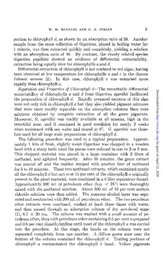
Xepmation and Propertics of Chlorophyll d-The remarkable differential extractability of chlorophylls a and d from Gigartina agardhii facilitated the preparation of chlorophyll d. Rapidly prepared extracts of this alga were not only rich in chlorophyll d but they also yielded pigment mixtures that were more readily separable on the adsorption columns than were mixtures obtained by complete ext,raction of all the green pigments. Moreover, G. agardhii was readily available at all seasons, high in the intertidal zone, and it remained in good condition for nearly 2 weeks when moistened with sea water and stored at 6”. G. agardhii was there- fore used for all large scale preparations of chlorophyll d.
The following procedure was used in a typical preparation. Approxi- mately 1 kilo of fresh, slightly moist Gigartina was chopped in a wooden bowl with a sharp knife until the pieces were reduced in size to 2 or 3 mm. This chopped material was placed in a large container with 2 liters of methanol, and agitated frequently. After 30 minutes, the green extract was poured off and the residue steeped with another liter of methanol for 5 to 10 minutes. These two methanol extracts, which contained nearly all the chlorophyll d but not over 15 per cent of the chlorophyll a originally present in the plant material, were combined in a 6 liter separatory funnel. Approximately 300 ml. of petroleum ether (b.p. < 75”) were thoroughly mixed with the methanol solution. About 500 ml. of 10 per cent sodium chloride solution were then added. The aqueous alcohol layer was sepa- rated and reextracted with 200 ml. of petroleum ether. The two petroleum ether extracts mere combined, I\-ashed at least three times with water, and then passed through an adsorption column of dry powdered sugar (l), 6.7 X 20 cm. The column was mashed with a small amount, of pe- troleum ether, then with petroleum ether containing 0.5 per cent n-propanol and 0.5 per cent dimethylaniline until most of the chlorophyll a was carried into the percolate. At this stage, the bands on the column were not separated completely from one another. A diffuse green zone near the bottom of the column contained the chlorophyll d. Trailing portions of chlorophyll a contaminated t’he chlorophyll d band. Yellow pigments
by guest on April 22, 2020
http://ww
w.jbc.org/
Dow
nloaded from
6 CHLOROPHYLL D OF RED ALGAE
were adsorbed below, in, and sometimes above the chlorophyll d band. Still higher on the column, there usually occurred a diffuse green zone which contained small amounts of chlorophyll b. This pigment probably came principally from traces of lXva, a green alga which frequently grows attached to Gigartina.
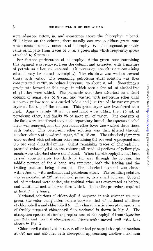
For further purification of chlorophyll d the green zone containing this pigment was removed from the column and extracted with a mixture of petroleum ether and ethanol. (If necessary, the elutriate containing et’hanol may be stored overnight.) The elutriate was washed several times with water. The remaining petroleum ether solution was then concentrated at 20”, at reduced pressure, to about 40 ml. Sometimes a precipitate formed at this stage, in which case a few ml. of alcohol-free ethyl ether were added. The pigments were then adsorbed on a short column of sugar, 4.7 X 8 cm., and washed with petroleum ether until a narrow yellow zone was carried below and just free of the narrow green layer at the top of the column. This green layer was transferred to a flask. Approximately 10 ml. of methanol were added, then 75 ml. of petroleum ether, and finally 25 or more ml. of water. The contents of the flask were transferred to a small separatory funnel, the aqueous alcohol layer was removed, and the pet,roleum ether layer was washed three times with water. This petroleum ether solution was then filtered through another column of powdered sugar, 4.7 X 18 cm. The adsorbed pigments were washed with petroleum ether containing 0.5 per cent n-propanol and 0.5 per cent dimethylaniline. Slight remaining traces of chlorophyll a preceded chlorophyll d on the column; all residual portions of yellow pig- ments were adsorbed above the d band. When the chlorophyll d had been carried approximately two-thirds of the way through the column, the middle portion of the d band was removed, both the leading and the trailing portions being discarded. The adsorbed pigment was eluted with ether, or with methanol and petroleum ether. The resulting solut.ion was evaporated at 20”, at reduced pressure, to a small volume. Several ml. of methanol were added, the residual ether was evaporated as before, and addit,ional methanol was then added. The entire procedure required at least 7 or 8 hours.
Methanol solutions of chlorophyll d prepared in this manner are pure green, the color being intermedia,te between that, of methanol solutions of chlorophyll a and chlorophyll b. The characteristic absorption spectrum of freshly prepared chlorophyll d in methanol is shown in Fig. 3. The absorption spectra of similar preparations of chlorophyll d from Gigartina papillata and from h’rythrophyllwm delesserioides agreed well with that shown in Fig. 3.
Chlorophyll d dissolved in u. s. P. ether had principal absorption maxima at 686 rnp and 445 ml*, with absorption approaching another maximum
by guest on April 22, 2020
http://ww
w.jbc.org/
Dow
nloaded from
W. M. MANNING AND H. H. STRAIN 7
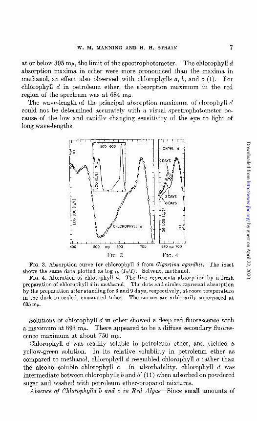
at or below 395 ml.c, the limit of the spectrophotometer. The chlorophyll d absorption maxima in ether were more pronounced than the maxima in methanol, an effect also observed with chlorophylls a, b, and c (1). For chlorophyll d in petroleum ether, the absorption maximum in the red region of the spectrum was at 684 rnp.
The wave-length of the principal absorpt.ion maximum of clorophyll d could not be determined accurately with a visual spectrophotometer be- cause of the low and rapidly changing sensitivity of the eye to light of long wave-lengths.
HLOROPHYLL d
400 500 mp 600 700
FIG. 3
r-7 CHPHL. d
640 rnfi 700
FIG. 4
FIG. 3. absorption curve for chlorophyll d from Gigartina agardhii. The inset shows the same data plotted as log 1~ (lo/Z). Solvent, methanol.
FIG. 4. Alteration of chlorophyll d. The line represent.s absorption by a fresh preparation of chlorophyll d in methanol. The dots and circles represent absorption by the preparation after standing for 3 and 9 days, respect,ively, at room temperature in the dark in sealed, evacuated tubes. The curves are arbitrarily superposed at 695 rnp.
Solutions of chlorophyll d in ether showed a deep red fluorescence with a maximum at 693 mp. There appeared to be a diffuse secondary fluores- cence maximum at about 750 rnp.
Chlorophyll d was readily soluble in petroleum ether, and yielded a yellow-green solution. In its relative solubility in petroleum et,her as compared to methanol, chlorophyll d resembled chlorophyll a rather than the alcohol-soluble chlorophyll c. In adsorbability, chlorophyll d was intermediate between chlorophylls b and 6’ (11) when adsorbed on powdered sugar and washed wit,h petroleum ether-propanol mixtures.
Absence of Chlorophylls b and c in Red Algae--Since small amounts of
by guest on April 22, 2020
http://ww
w.jbc.org/
Dow
nloaded from
8 CHLOROPHYLL D OF RED ALGAE
chlorophyll b (probably from contamination by the green alga, Ulva) were often observed on adsorption columns during large scale preparations of chlorophyll d from Gigartina agardhii, a more critical test was made for the presence of chlorophyll b in Gigartina agardhii and in Hymenena jlabelligera.
A 10 gm. sample of Gigartina agardhii was carefully selected on the basis of freedom from appreciable contamination by other algae, then killed by heating at 100” for 1 minute. The pigments were extracted with methanol and an aliquot was removed for spectrophotometric determination of the amount of chlorophyll a. The pigments in the remaining solution were quantitatively transferred to pet,roleum ether and adsorbed and washed on a column of powdered sugar (3 X 20 cm.). No band of chlorophyll b was observed on the column. Three contiguous zones of the column, including the entire region where traces of chlorophyll b could have been adsorbed, were removed. The pigments from each zone were eluted separately, dissolved in methanol, and examined spectrophotometrically. Small amount’s of alteration products of chlorophyll a (2) were observed in these zones but there was no positive evidence for the presence of chlorophyll b. At most, the amount. of chlorophyll b could not have exceeded 0.3 per cent of the amount of chlorophyll a present in the original extract. A nearly identical procedure was used for Hymenena jlabelligera, with similar results.
Chlorophyll c was not found by spectrophotometric examination of the extracts of red algae, nor by adsorption of the pigments on columns of sugar.
From these experiments it is evident that chlorophylls b and c either are absent from the red algae which were examined, or are present only in traces.
Metal Constituent of Chlorophyll d-On two occasions fresh preparations of chlorophyll d, each from 2 kilos of Gigartina agardhii, were transferred to petroleum ether and washed thoroughly with water to remove possible water-soluble impurities. The petroleum ether solutions were evaporated nearly to dryness, below room temperature at reduced pressure. III
each case approximately 2 ml. of ether were added to the residue. The ether solutions were transferred to small weighed platinum crucibles and evaporated to dryness. The residues were then ignited at red heat for approximately 20 minutes. The residual ash was white and in each case weighed 0.12 f 0.008 mg. Water failed to dissolve the ash. A little hydrochloric acid was added and the resulting solutions were bransferred to volumetric flasks and made up to 15 ml.
In order to determine whether or not the ash from chlorophyll d con- tained magnesium, the solutions were analyzed calorimetrically for
by guest on April 22, 2020
http://ww
w.jbc.org/
Dow
nloaded from
W. M. MANNING AND H. H. STRAIN 9
magnesium, with Titan yellow or p-nitrobenzeneazo-a+naphthol as reagents (12). With both reagents, sodium cyanide was used to reduce possible color quenching by other metals. In every case the blank, the st.andard, and the test solutions were made up to 10 ml. and measured spectrophoto- met.rically (at 535 rnp for Titan yellow and at 650 rnp for p-nitrobenzeneazo- a-naphthol). Four aliquots from the solution prepared from one sample of ash were analyzed with Titan yellow and gave values of 0.115, 0.095, 0.110, and 0.102 mg., respectively (average 0.106 mg.), for the magnesium oxide content of the ash. For the other preparation of ash, three determin- ations with Titan yellow gave values of 0.097, 0.119, and 0.113 mg., re- spectively; two determinations with p-nitrobenzeneazo-a-naphthol gave ITalues of 0.109 and 0.105 mg. The average for these last five determina- tions was 0.109 mg. Considering the very small quantities involved, these arerage values for magnesia content are in satisfactory agreement wit,h the total weights of the ash (0.12 f 0.008 mg. in each case).
Tjecause of the specificity of Titan yellow, and because the two colori- metric reagents gave results agreeing with each other and also with the weights of ash, it may be concluded that the met,al const,ituent of the chlorophyll d molecule is magnesium.
Isomerization of Chlorophyll d-Chlorophyll d dissolved in methanol and allowed to stand in the dark, either in the presence or absence of oxygen, undergoes alteration, with an absorption band at 661 rnp gradually be- coming prominent (see Fig. 4). This change in spectrum has been found to result from isomerization of chlorophyll d.
In a typical experiment,, a chlorophyll d solution, after standing for 9 days, was transferred to petroleum ether, adsorbed on sugar, and washed with petroleum ether containing 0.5 per cent n-propanol and 0.5 per cent dimethylaniline. Instead of the original single zone, at least three OI
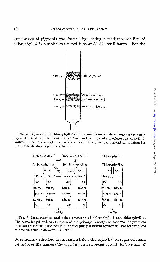
four bands were visible on the column (see Fig. 5). Topmost except for traces of one or two strongly adsorbed pigments, was the green band of chlorophyll d. Below this and well separated from the d band were two adjacent bands incompletely separated from each other, the upper yellowgreen zone merging into a lower blue-green zone. Fart’her down on the column was a single, pale, blue-green band.
After the pigments from the two lower bands were eluted separately and heated, or allowed to stand in methanol for a few days, each was converted into a mixture of t.he four pigments previously observed to arise from chlorophyll d. The second green band (second from t’he top of t,he column, Fig. 5) was not obtained in sufficient purity for a critical test, but probably it too was converted into a similar mixture. This behavior, as well as evidence presented in subsequent sections, indicates that t,hese four pigments probably are interconvertible isomers. The
by guest on April 22, 2020
http://ww
w.jbc.org/
Dow
nloaded from
10 CHLOROPHYLL D OF RED ALQAE
same series of pigments was formed by heating a methanol solution of chlorophyll d in a sealed evacuated tube at 80-82” for 2 hours. For the
yellow-green
blue-green
blue-green
FIG. 5. Separation of chlorophyll d and its isomers on powdered sugar after wash- ing with petroleum ether containing 0.5 per cent n-propanol and 0.5 per cent dimethyl- aniline. The wave-length values are those of the principal absorption maxima for the pigments dissolved in methanol.
Chlorophyll
ChlOrC!:hylI
d' lsochlorophyll d’
d Isochlo!phyll d
KOH I
Km I
658 rnp 636 rnp
I I C”,COOH CH,COM C”..Ccm
I C”,COO”
I 672 mp 691 m,u 660 mp 672 rnp
690 rnp
Chlorophyll a’
Chlor:;hyll C.I
“a cH,M*I IT
Pheophytin u I
667’mg
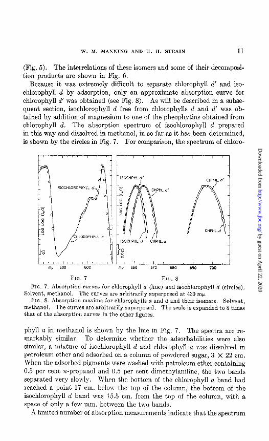
FIG. 6. Isomerization and other reactions of chlorophyll d and chlorophyll a. The wave-length values are those of the principal absorption maxima for products of alkali treatment dissolved in methanol plus potassium hydroxide, and for products of acid treatment dissolved in ether.
three isomers adsorbed in succession below chlorophyll d on sugar columns, we propose the names chlorophyll d’, isoch.lorophyll d, and isochlorophyll d’
by guest on April 22, 2020
http://ww
w.jbc.org/
Dow
nloaded from
TV. M. MANNING AND H. H. STRAIN 11
(Fig. 5). The interrelations of these isomers and some of their decomposi- tion products are shown in Fig. 6.
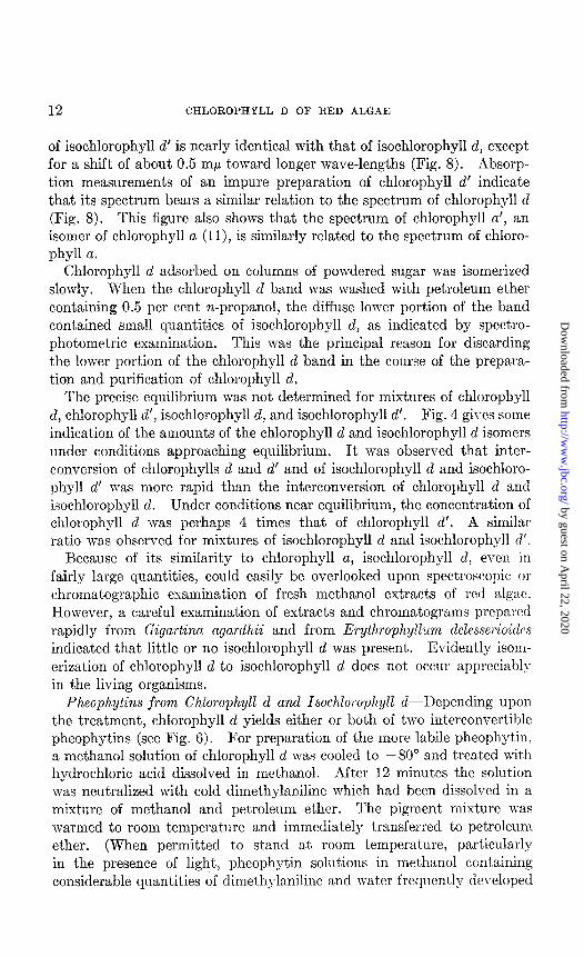
Recause it was extremely difficult to separate chlorophyll d’ and iso- chlorophyll d by adsorption, only an approximate absorption curve for chlorophyll d’ was obtained (see Fig. 8). As will be described in a subse- quent section, isochlorophyll d free from chlorophylls d and d’ was ob- tained by addition of magnesium to one of the pheophytins obtained from chlorophyll d. The absorption spectrum of isochlorophyll d prepared in this way and dissolved in methanol, in so far as it has been determined, is shown by the circles in Fig. 7. For comparison, the spectrum of chloro-
r
ISOCHLOROPHYL
w 500 600
FIG. 7
1 IS0CHPHL.d’ CHPHL. d’
ISOCHPHL. d CHPHL.n
w ’ ! ’ ’ ‘U 660 670
FIG. 8 FIG. 7. Absorption curves for chlorophyll a (line) and isochlorophyll d (circles).
Solvent, met,hanol. The curves are arbitrarily superposed at 430 rnp. FIG. 8. Absorption maxima for chlorophylls a and d and their isomers. Solvent,
methanol. The curves are arbitrarily superposed. The scale is expanded to 8 times that of the absorption curves in the other figures.
phyll a in methanol is shown by the line in Fig. 7. The spectra are re- markably similar. To determine whether t,he adsorbabilities were also similar, a mixture of isochlorophyll d and chlorophyll a was dissolved in petroleum ether and adsorbed on a column of powdered sugar, 3 X 22 cm. When the adsorbed pigments were washed with petroleum ether containing 0.5 per cent n-propanol and 0.5 per cent dimethylaniline, the two bands separated very slowly. When the bottom of the chlorophyll a band had reached a point 17 cm. below the top of the column, the bottom of the isochlorophyll d band was 15.5 cm. from the top of the column, with a space of only a few mm. between the two bands.
A limited number of absorption measurements indicate that the spectrum
by guest on April 22, 2020
http://ww
w.jbc.org/
Dow
nloaded from
12 CHLOROPHYLL D OF RED ALGAE
of isochlorophyll d’ is nearly identical with that of isochlorophyll d, except for a shift of about 0.5 rnp toward longer wave-lengths (Fig. 8). hbsorp- tion measurements of an impure preparation of chlorophyll d’ indicate that its spectrum bears a similar relation to the spectrum of chlorophyll d (Fig. 8). This figure also shows that the spectrum of chlorophyll a’, an isomer of chlorophyll a (ll), is similarly related to the spectrum of chloro- phyll a.
Chlorophyll d adsorbed on columns of powdered sugar was isomerized slowly. When the chlorophyll d band was washed with petroleum ether containing 0.5 per cent n-propanol, the diffuse lower portion of the band contained small quantities of isochlorophyll d, as indicated by spectro- photometric examination. This was the principal reason for discarding the lower portion of the chlorophyll d band in the course of the prepara- tion and purification of chlorophyll d.
The precise equilibrium was not determined for mixtures of chlorophyll d, chlorophyll d’, isochlorophyll d, and isochlorophyll d’. Fig. 4 gives some indication of the amounts of the chlorophyll d and isochlorophyll d isomers under conditions approaching equilibrium. It was observed that inter- conversion of chlorophylls d and d’ and of isochlorophyll d and isochloro- phyll d’ was more rapid than the interconversion of chlorophyll d and isochlorophyll d. Under conditions near equilibrium, the concentration of chlorophyll d was perhaps 4 times that of chlorophyll d’. A similar ratio was observed for mixtures of isochlorophyll d and isochlorophyll d’.
Because of its similarity to chlorophyll a, isochlorophyll d, eve11 in fairly large quantities, could easily be overlooked upon spectroscopic or chromatographic examination of fresh methanol extracts of red algae. However, a careful examination of extracts and chromatograms prepared rapidly from Gigurtinu ugurdhii and from Erythrophyllum delesscrioidrs indicated that little or no isochlorophyll d was present. Evidently isom- erization of chlorophyll d to isochlorophyll d does not occur appreciably in the living organisms.
Pheophytins from Chlorophyll d and Isochlorophyll d-Depending upon the treatment, chlorophyll d yields either or both of two interconvertible pheophytins (see Fig. 6). For preparation of the more labile pheophytin, a methanol solution of chlorophyll d was cooled t,o -80” and treated with hydrochloric acid dissolved in methanol. After 12 minutes the solution was neutralized with cold dimethylaniline which had been dissolved in a mixture of methanol and petroleum ether. The pigment mixture was warmed to room temperature and immediately transferred to petroleum ether. (When permitted to stand at room temperat’ure, particularly in the presence of light, pheophytin solutions in methanol containing considerable quantities of climethylanilinc and wat’er frequently developed
by guest on April 22, 2020
http://ww
w.jbc.org/
Dow
nloaded from
a deep purple-red color, due presumably to alteration of the dimethyl- aniline.) The petroleum ether solution of pheophytin was mashed with water and then adsorbed on powdered sugar. After development of the chromatogram with petroleum ether containing 0.1 per cent n-propanol and 0.5 per cent dimet,hylaniline the principal band consisted of a yellow- brown pigment, pheophytin d, which was less adsorbed than the original chlorophyll d. Below this band of pheophytin d there were traces of a gray band which resembled pheophytin a. Identical results were obtained in another preparation in which all steps prior to adsorption were carried out in an atmosphere of hydrogen. The absorption spectrum of pheophy- tin d in methanol is shown in Fig. 9. Since the pigment was somewhat unstable, the absorption curve is probably not highly accurate.
Chlorophyll d, treated at room temperature with hydrochloric acid in methanol and then neutralized with dimethylaniline, was transformed principally into the gray pigment which had been produced in traces by the low temperature treatment. In this case, only traces of the brown pheophytin cl were observed. We propose the name isopheophytin d for this gray pigment. The absorption spectrum of isopheophytin d in methanol is shown in Fig. 9. Isopheophytin d, like pheophytin a, forms a purple-gray solution in methanol and a deep blue solution in methanol containing hydrochloric acid.
Isochlorophyll d treated with acid, either at 20” or at -8O”, was con- verted almost entirely into the gray isopheophytin d.
Pheophytin d in methanol, when reacidified at room temperature and subsequently neutralized with dimethylaniline, was transformed almost completely into the gray isopheophytin d. Isopheophytin d was not re- converted into pheophytin d when allowed to stand in acid solution at -80” for 90 minutes.
Adsorption of a dried preparation of isopheophytin d which had stood in the dark in an evacuated desiccator for 2 weeks showed that the pig- ment had been t,ransformed in part to the brown pheophytin d. A faint gray band, presumably isopheophytin d’, was also observed just below the principal gray band of isopheophytin d.
Preparations of pheophytin d were much less stable than those of iso- pheophytin d. Pheophytin d was readily transformed, upon st,anding, into a misture containing isopheophytin d. A pheophytin corresponding to chlorophyll d’ was not observed, but limit.ed material precluded an adequate search for the hypothetical pheophytin df.
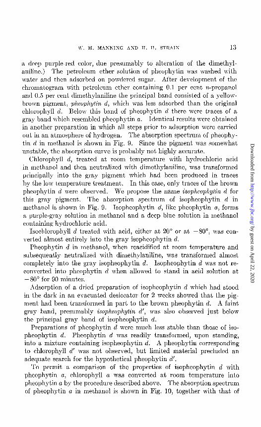
To permit a comparison of the properties of isopheophytin d with pheophytin a, chlorophyll a was converted at room temperature into pheophytin a by the procedure described above. The absorption spectrum of pheophytin a in methanol is shown in Fig. 10, together with that of
by guest on April 22, 2020
http://ww
w.jbc.org/
Dow
nloaded from
14 CHLOROPHYLL D OF RED ALGAE
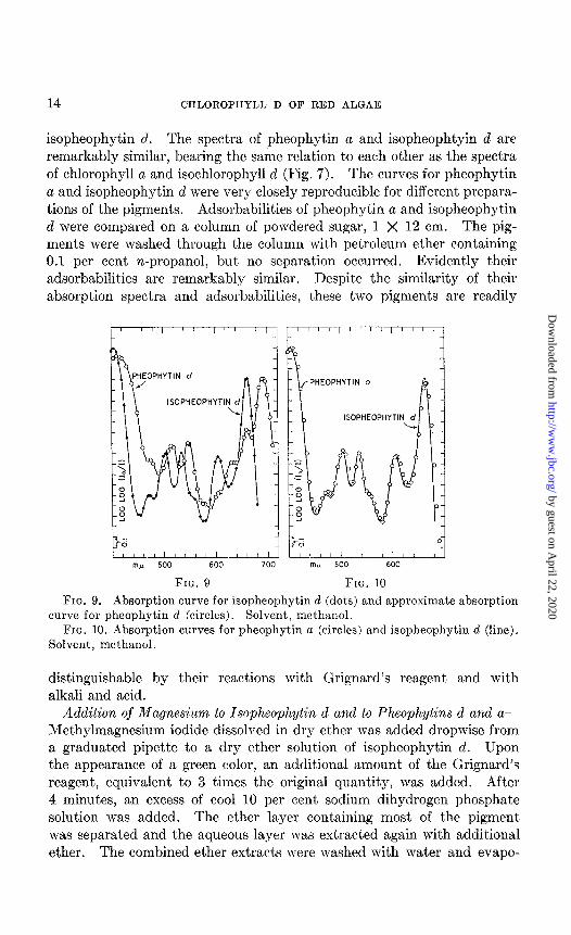
isopheophytin d. The spectra of pheophytin a and isopheophtyin d are remarkably similar, bearing the same relation to each other as the spectra of chlorophyll a and isochlorophyll d (Fig. 7). The curves for pheophytin a and isopheophytin d were very closely reproducible for different prepara- tions of the pigments. Adsorbabilities of pheophytin a and isopheophytin d were compared on a column of powdered sugar, 1 X 12 cm. The pig- ments were washed through the column with petroleum ether containing 0.1 per cent n-propanol, but no separat,ion occurred. Evidently their adsorbabilities are remarkably similar. Despite the similarity of their absorption spectra and adsorbabilities, these two pigments are readily
ISOPHEOPHYTI
HEOPHYTIN a
ISOPHEOPHYTI
FIG. 9 FIG. 10
FIG. 9. Absorption curve for isopheophytin d (dots) and approximate absorption curve for pheophytin d (circles). Solvent, methanol.
FIG. 10. Absorption curves for pheophytin a (circles) and isopheophytin d (line). Solvent, methanol.
distinguishable by their reactions with Grignard’s reagent and with alkali and acid.
Addition of Magnesium to Isopheophytin d and to Pheophytins d and a- Methylmagnesium iodide dissolved in dry ether was added dropwise from a graduated pipette to a dry ether solution of isopheophytin d. Upon the appearance of a green color, an additional amount of the Grignard’s reagent, equivalent to 3 times the original quantity, was added. After 4 minutes, an excess of cool 10 per cent sodium dihydrogen phosphate solution was added. The ether layer containing most of the pigment was separated and the aqueous layer was extracted again with additional ether. The combined ether extracts were washed with water and evapo-
by guest on April 22, 2020
http://ww
w.jbc.org/
Dow
nloaded from
W. M. MANNING AND Il. H. STRAIN 15
rated nearly to dryness below room temperature at reduced pressure. Petroleum ether was then added and the resulting solution adsorbed on powdered sugar and washed with petroleum ether containing 0.5 per cent n-propanol and 0.5 per cent dimethylaniline. Six or more green or blue- green bands were visible on the column. The principal band was iso- chlorophyll d. The absorption spectrum for such a preparation is shown in Fig. 7. Little or no chlorophyll d or d’ was formed directly from iso- pheophytin d, but both pigments were produced slowly by isomerization of the regenerated isochlorophyll d.
Pheophytin d treated in the same way with Grignard’s reagent gave only very small yields of green or blue-green pigments. Little or no chlorophyll d or isochlorophyll d was formed. Considerable quantities of isopheophytin d were formed. A much more strongly adsorbed gray pigment was also formed (X,,,., 659 rnp in methanol). This pigment, recovered and treated with Grignard’s reagent, yielded a strongly adsorbed green pigment (X,,,., 658 rnl.c in methanol) which was also found among the products formed by action of Grignard’s reagent on isopheophytin d and on pheophytin d. Treatment of this strongly adsorbed green pig- ment with hydrochloric acid in methanol regenerated t,he strongly adsorbed gray pheophytin. The relation of this pheophytin to the other pheophytins described here is not clear.
Pheophytin a reacted with Grignard’s reagent in a manner very similar to isopheophytin d, at least six or seven blue-green bands being observed on adsorption columns. Chlorophyll a was the principal product but constituted less than half of the total pigment mixture. Most of the products were adsorbed above chlorophyll a, which they resembled with respect to the wave-lengths of their absorption maxima. Formation of several chlorophyll u-like products, with a comparatively low yield of chlorophyll a, is in contrast with the report by earlier investigators of a yield of 0.8 gm. of chlorophyll a from 1.0 gm. of pheophytin a (13).
E$ect of Alkali on Chlorophyll and Pheophytin Isomers-Fig. 6 gives the position of the principal absorption maximum in the red region of the spectrum for products formed by alkali treatment and subsequent acid treatment of chlorophyll d, isochlorophyll d, pheophytin d, and isopheo- phytin d. For comparison, data for chlorophyll a and pheophytin a are also included in Fig. 6. To avoid confusion, secondary maxima are not recorded.
For each pigment. the following procedure was used. A solution of the chlorophyll or pheophytin in methanol was treated with 2 gm. of potassium hydroxide dissolved in methanol. The wave-length of the principal absorption maximum in the red region of the spectrum for the resulting solution was determined with a Bausch and Lomb visual spectrophotometer
by guest on April 22, 2020
http://ww
w.jbc.org/
Dow
nloaded from
16 CHLOROPHYLL D OF RED ALGAE
(see Fig. 6). This solution was then treated with an excess of glacial acetic acid. Ether was added and the pigments transferred to the ether by addition of 10 per cent sodium chloride solution. The ether layer was separated, washed several times with mater, and the position of the principal absorption maximum again observed. 1 or 2 ml. of concentrated HCl were then added to the ether and thoroughly mixed. This treatment removed most of the pigment from the ether layer. After about 5 minutes the pigment was retransferred to the ether by the addition of large quanti- ties of sodium chloride solution. The ether solution was then mashed thoroughly with water and the position of the principal absorption maxi- mum again observed. It should be noted that in the last two cases the positions of the absorption maxima listed in Fig. 6 were determined in ether, while the determination for the first product of the alkali treatment was made in methanol solution containing potassium hydroxide.
From Fig. 6 it may be seen that, although the intermediate products were different in each case, the final product of this series of treatments appeared to be the same for chlorophyll d and its derivatives, so far as the position of the absorption maximum may be regarded as an indication of identity. None of these products was examined chromatographically. By contrast, the difference between the isochlorophyll d series and the chlorophyll a series was accentuated, rather than diminished, in the final products formed by alkali and acid.
DISCUSSION
In na,ming chlorophyll d and its derivatives, we have endeavored to use a system in harmony with the accepted nomenclature of the chlorophylls. The structure of chlorophyll d is obviously related to that of chlorophyll a. Extension of the term chlorophyll to such related green pigments appears logical and desirable.
In naming the isomers of chlorophyll d, it appeared desirable to retain the letter d as part of the designation. Numerals, either as subscripts 01 suffixes, were avoided, since they have been employed, in the case of chlorophylls a and b, to identify other types of products (14, 15). The use of the prime designation (chlorophyll d’), for an isomer with an absorp- tion spectrum only slightly different from that of the parent substance, is in harmony with the designat,ion for the corresponding isomers of chloro- phylls a and b (11). For an isomer with an absorption spectrum greatly different from that of the parent substance, the prefix iso (isochlorophyll d) serves to give a distinguishing name while indicating the isomeric relation to the original pigment. Moreover, the prime designation combined with the prefix (isochlorophyll d’) then denotes a compound apparently related t,o isochlorophyll d in a manner corresponding to the relation of chlorophyll d’ to chlorophyll d.
by guest on April 22, 2020
http://ww
w.jbc.org/
Dow
nloaded from
W. M. MANNING AND H. H. STRAIN 17
Extension of this system of nomenclature to the pheophytins of chloro- phyll d and its isomers provides a convenient means of indicating analogous relationships between the isomeric pheophytins.
The striking similarities between isochlorophyll cl and chlorophyll a, and between isopheophytin d and pheophytin a, both in physical properties and in chemical behavior, indicate that the molecular structures are very similar. So far as the chromophoric groups are concerned, the a and iso-d series of compounds must resemble each other much more closely than either resembles the b series. The adsorbabilities of chlorophyll a and isochlorophyll d are also very similar. Nevertheless, the difference in structure is sufficient so that the iso-d compounds are readily and reversibly converted to another series (the d series) which, at least superficially,
0 100 w 500 600 700
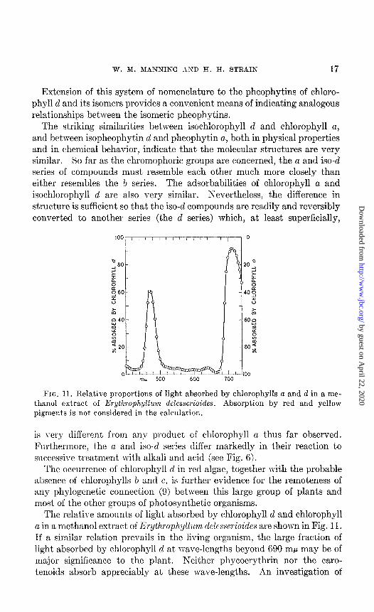
FIG. 11. Relative proportions of light absorbed by chlorophylls a and CE in a me- thanol extract of Erythrophyllum delesserioides. Absorption by red and yellow pigments is not considered in the calculation.
is very different from any product of chlorophyll a thus far observed. Furthermore, the CI and iso-d series differ markedly in their reaction to successive treatment wit’h alkali and acid (see Fig. 6).
The occllrrence of chlorophyll d in red algae, t.ogether I\-ith the probable absence of chlorophylls b and c, is furt.her evidence for the remoteness of any phylogeneCc connect,ion (9) between this large group of plants and most, of the ot.her groups of phot*osynthet,ic organisms.
The relative amounts of light absorbed by chlorophyll d and chlorophyll a in a methanol extract of E~ythrophyllum delesserioides are shown in Fig. 11. If a similar relation prevails in the living organism, the large fraction of light absorbed by chlorophyll d at] wave-lengt,hs beyond 690 rnp may be of major significance to the plant. Neither phycoerythrin nor the caro- tenoids absorb appreciably at these wave-lengths. An investigation of
by guest on April 22, 2020
http://ww
w.jbc.org/
Dow
nloaded from
18 CHLOROPHYLL D OF RED ALGAE
photosynthesis in Erythrophyllum in monochromatic red light should yield a clear cut answer regarding the photosynthetic effectiveness of chlorophyll d. If the light of long wave-lengths which is absorbed by this pigment can be utilized in photosynthesis, the presence of chlorophyll d must extend by at least 30 rnp the range of light utilized by plants in this fundamental process. The minimum energy per photon required to bring about the photochemical reaction (or reactions) in photosynthesis would accordingly be reduced by about 4 per cent.
Since water absorbs red light much more intensely than light of shorter wave-lengths, chlorophyll d in red algae can contribute little to absorption of red light except for plants growing near the surface. Brown algae (and other plants) are relatively transparent to light absorbed readily by chlorophyll d. Hence, for many red algae which grow on reefs in shallow water and which are shaded to a considerable extent by brown algae, light absorbed by chlorophyll d may amount to an appreciable fraction of the total energy available for photosynthesis.
SUMMARY
Various species of red algae contain, in addition to chlorophyll CL, a second, green, magnesium-containing pigment, chlorophyll d. Neither chlorophyll b nor chlorophyll c was found in these algae.
Chlorophyll d was most easily prepared by adsorption of the pigments obtained by partial extraction of Gigartina agardhii. Maximum light absorption by chlorophyll d occurs at a wave-length longer than that of the maximum of chlorophyll a; in methanol, maximum absorption for chlorophyll d is at 696 rnp, compared with 665 mp for chlorophyll a. Ab- sorption at long wave-lengths by chlorophyll d may extend by 30 rnp the range of light utilized in photosynthesis.
Chlorophyll d was converted, rapidly upon heating or slowly at room temperature, into a mixture containing three isomers in addition to un- altered chlorophyll d. One of these isomers, chlorophyll d’, has an ab- sorption spectrum very similar to that of chlorophyll d, whereas t,he other two isomers, isochlorophyll d and isochlorophyll d’, have spectra resembling the spectrum of chlorophyll a. The isomers were found to be reconvertible to chlorophyll d.
Treatment of chlorophyll d with acid removed the magnesium and formed a mixture of two interconvertible pheophytins. At -8O”, treat- ment with acid produced principally the labile yellow-brown pheophytin d. At room temperature, gray isopheophytin d was the principal product. Pheophytin d was rapidly converted to isopheophytin d when treated with acid at room temperature. Treatment of isochlorophyll d with acid, either at room temperature or at -8O”, produced isopheophytin d. Iso-
by guest on April 22, 2020
http://ww
w.jbc.org/
Dow
nloaded from
W. M. MANNING AND H. H. STRAIN 19
pheophytin d is remarkably similar to pheophytin a in its absorption spectrum and in its adsorbability on powdered sugar. Treatment of isopheophytin d with Grignard’s reagent produced isochlorophyll d but litt,le or no chlorophyll cl. Neither chlorophyll d nor isochlorophyll d was formed by treatment of pheophytin d with Grignard’s reagent.
When chlorophyll d and its isomers were treated successively with alkali and acid, the same final product was formed in each case. Chloro- phyll a treated in the same manner yielded a product distinctly different from the chlorophyll d product.
We are indebted to the other members of the Division of Plant Biology for invaluable discussion and advice.
BIBLIOGRAPHY
I. Strain, H. H., and Manning, W. M., J. Biol. Chem., 144, 625 (1942). 2. Strain, H. H., Manning, W. M., and Hardin, G., J. Biol. Chem., 148,655 (1943). 3. Strain, H. H., and Manning, W. M., Carnegie Inst. Washington Yr. Bk., 41, 118
(1942). 4. Sorby, H. C., Proc. Roy. Sot. London, 21, 442 (1873). 5. Kylin, II., Z. physiol. Chem., 166, 39 (1927). 6. Fischer, H., and Breitner, S., Ann. Chem., 622, 151 (1936). 7. Seybold, A., Egle, K., and Htilsbruch, W., Bot. Arch., 42, 239 (1941). 8. Kylin, II., Z. physiol. Chem., 197, 1 (1931). 9. Smith, G. M., Cryptogamic botany, New York, 1 (1938).
10. Smith, J. H. C., J. Am. Chem. Sot., 68, 247 (1936). 11. Strain, H. H., and Manning, W. M., J. Biol. Chena., 146, 275 (1942). 12. Staff of the research laboratory of Hopkin and Williams, Ltd., Organic reagents
for metals, London (1938). 13. Willst~atter, R., and For&n, L., Ann. Chem., 396, 180 (1913); cf. Willstatter, It.,
and Stoll, A., Untersuchungen tiber Chlorophyll, Berlin, 329 (1913). 14. Conant, J. B., and Dietz, E. M., J. Am. Chem. Sot., 66, 839 (1933). 15. Mackinney, G., Annual review of biochemistry, Stanford University, 9, 459
(1940).
by guest on April 22, 2020
http://ww
w.jbc.org/
Dow
nloaded from
Winston M. Manning and Harold H. StrainOF RED ALGAE
CHLOROPHYLL D, A GREEN PIGMENT
1943, 151:1-19.J. Biol. Chem.
http://www.jbc.org/content/151/1/1.citation
Access the most updated version of this article at
Alerts:
When a correction for this article is posted•
When this article is cited•
alerts to choose from all of JBC's e-mailClick here
#ref-list-1
http://www.jbc.org/content/151/1/1.citation.full.htmlaccessed free atThis article cites 0 references, 0 of which can be
by guest on April 22, 2020
http://ww
w.jbc.org/
Dow
nloaded from