1 Analytical Chemistry Introduction Analytical chemistry ...
CHINESE JOURNAL OF ANALYTICAL CHEMISTRY Volume 44, Issue 4, April 2016 Online English edition of the Chinese language journal
Cite this article as: Chin J Anal Chem, 2016, 44(4), 633–639.
________________________ Received 16 January 2016; accepted 3 March 2016 *Corresponding author. Email: [email protected] This work was supported by the Major Technology Project of Dongguan, China (No. 2014215102) and the SEM Technology Innovation Fund of China (No. 14C26214402622). Copyright © 2016, Changchun Institute of Applied Chemistry, Chinese Academy of Sciences. Published by Elsevier Limited. All rights reserved. DOI: 10.1016/S1872-2040(16)60926-X
RESEARCH PAPER
A Microfluidic Fluorescence Immunoassay Test Card for Rapid Detection of Heart‐type Fatty Acid Binding Protein MENG Fan-Da1,2, HUO Wei-Song1, HE Mei-Lin1,2, LI Hao1,2, LIAN Jie3, SHI Xi-Zeng4, GAO Yun-Hua1,* 1 Key Laboratory of Photochemical Conversion and Optoelectronic Materials, Technical Institute of Physics and Chemistry, Chinese Academy of Sciences, Beijing 100190, China
2 University of Chinese Academy of Sciences, Beijing 100049, China 3 Department of Forensic Science, People’s Public Security University of China, Beijing 100038, China 4 Dongguan Bosh Biotechnologies, LTD, Dongguan 523808, China
Abstract: A kind of easily assembly self-driving test card was developed for the rapid detection and quantification of heart-type fatty acid binding protein (H-FABP) utilizing time-resolved fluorescence microspheres as signal probes. It was easy to make microfluidic channel structures with double-sided adhesive and cut out of test card substrate and cover based on polymethyl methacrylate (PMMA) material by the laser cutting technique. The membrane of polystyrene containing maleic anhydride functional groups was coated on PMMA plate surface by dip-coating method. Furthermore, the capture antibody was immobilized effectively on PMMA surface by covalent binding with maleic anhydride groups. The cover was treated with plasma treatment to improve the hydrophilic ability, so that the liquid could flow smoothly in the microchannel. The whole analysis process could be completed within 10 min. There was a very good linear correlation between response and H-FABP concentration in the range of 0.5‒100 ng mL–1 (R2 = 0.9966), and the detection limit was 0.1 ng mL–1 (S/N = 3). The intra-assay precision was less than 10% and the inter-assay precision was less than 15%. This detection method has the advantages such as high sensitivity, fast detection and good accuracy. Key Words: Heart-type fatty acid binding protein; Detection; Microfluidic system; Time-resolved fluorescence
1 Introduction
The development of microfluidics provides a novel platform for the fast and high-throughput of biomarker detection[1–6]. In comparison with traditional methods, microfluidic detection has several excellent advantages such as efficient, fast detection, reagent consumption, operate- automated and so on. At present, the materials of microfluidic substrate are silicon, amorphous silicon, glass, quartz and polymers. Polymers mainly include epoxy resin, polyurea, polyurethane and polystyrene. Polymethyl methacrylate methyl (PMMA) and polydimethyl siloxane (PDMS)[7–10] have become the preferred materials for fabrication microfluidic
devices due to its advantages such as inexpensive material and easy molding compared with inorganic material. Among all polymers, PMMA has been widely used in the field of in-vitro diagnostics due to its excellent optical properties and biological affinity[11]. However, the strong hydrophobicity and high background fluorescence limit its application in the microfluidic devices. In addition, various shapes of microfluidic microchannels can be realized with the development of technology, but the low-cost microchannel manufacturing with high efficiency and precision is still one of the main difficulties in the microfluidic detection field.
Heart-type fatty acid binding protein (H-FABP) is a low molecular weight protein (15 kDa)[12], which mainly
MENG Fan-Da et al. / Chinese Journal of Analytical Chemistry, 2016, 44(4): 633–639
responsible for combining with fatty acids and regulating cell metabolism. As the specific protein presented in the cytoplasm of myocardial cells, H-FABP can be released into the blood in the early myocardial injury. Thus it can be used as a new biomarker of early diagnosis myocardial injury for acute myocardial infarction (AMI). Current methods of detecting H-FABP[13–16] were ELISA, immunoturbidimetric assay, immuno-colloidal golden method, latex agglutination test and so on. However, these methods have disadvantages such as time-consuming, tedious operation, low sensitivity, poor accuracy, and over-reliance on large-scale detection equipment or other shortcomings, which limit its application in point-of-care test (POCT).
Time-resolved fluorescence immunoassay (TRFIA) is established based on the traditional fluorescence immunoassay and it is a new non-radioactive immunoassay technique utilizing lanthanide chelates as the label[17–19]. The time- resolved fluorescence (TRF) method can effectively reduce the background fluorescence in detection, leading to high assay sensitivity.
In this article, we introduced an easy assembly self-driving test card based on microfluidic technique and time-resolved fluorescence immunoassay technique. The laser cutting technology provided a quick method to cut the PMMA substrate and the microchannel based on double-sided adhesive. The plasma technology was used to improve the hydrophilic ability of PMMA. Finally, we realized one-step rapid detection of H-FABP in serum. The method had a potential prospect because of its high sensitivity, simple operation, easy processability and high throughput test for multi-biomarker. 2 Experimental 2.1 Instruments and reagents
EnSpire multi-mode microplate reader was purchased from
PerkinElmer Germany. Laser System was from Universal, USA. Refrigerated centrifuge was purchased from Sigma, Germany. EASY pure LF ultrapure water system was from Barnstead, USA. Microarray spotter was from GeSim, Germany. Mouse anti-human H-FABP monoclonal antibody pair (FA03, FA01) and human H-FABP were purchased from Hytest Industries International, Finland. High degree PMMA
(about 1 mm thickness) was from Hongnian Metallic Material Shenzhen, LTD. Double-sided adhesive (about 100 μm thickness) was from 3 M USA. Carboxyl-modified Europium containing microspheres (1%, w/V, 200 nm) was from Microdetection Shanghai, LTD. Polystyrene (PS) graft maleic anhydride and toluene was purchased from Lanyihuagong Beijing, LTD. Boric acid, Borax, Sodium chloride, Anhydrous sodium carbonate, sodium hydroxide and 1-(3- dimethylaminopropyl)-3-ethylcarbodiimide (EDC) were purchased from Sigma-Aldrich. Other reagents were all analytical grade. 2.2 Design and assembly of fluorescence test card
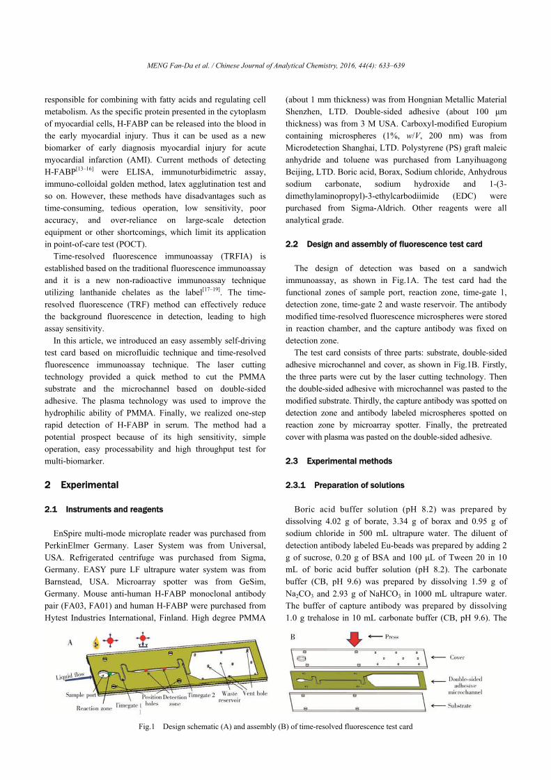
The design of detection was based on a sandwich
immunoassay, as shown in Fig.1A. The test card had the functional zones of sample port, reaction zone, time-gate 1, detection zone, time-gate 2 and waste reservoir. The antibody modified time-resolved fluorescence microspheres were stored in reaction chamber, and the capture antibody was fixed on detection zone.
The test card consists of three parts: substrate, double-sided adhesive microchannel and cover, as shown in Fig.1B. Firstly, the three parts were cut by the laser cutting technology. Then the double-sided adhesive with microchannel was pasted to the modified substrate. Thirdly, the capture antibody was spotted on detection zone and antibody labeled microspheres spotted on reaction zone by microarray spotter. Finally, the pretreated cover with plasma was pasted on the double-sided adhesive.
2.3 Experimental methods 2.3.1 Preparation of solutions
Boric acid buffer solution (pH 8.2) was prepared by
dissolving 4.02 g of borate, 3.34 g of borax and 0.95 g of sodium chloride in 500 mL ultrapure water. The diluent of detection antibody labeled Eu-beads was prepared by adding 2 g of sucrose, 0.20 g of BSA and 100 μL of Tween 20 in 10 mL of boric acid buffer solution (pH 8.2). The carbonate buffer (CB, pH 9.6) was prepared by dissolving 1.59 g of Na2CO3 and 2.93 g of NaHCO3 in 1000 mL ultrapure water. The buffer of capture antibody was prepared by dissolving 1.0 g trehalose in 10 mL carbonate buffer (CB, pH 9.6). The
Fig.1 Design schematic (A) and assembly (B) of time-resolved fluorescence test card

MENG Fan-Da et al. / Chinese Journal of Analytical Chemistry, 2016, 44(4): 633–639
standard solution of H-FABP was prepared by adding a certain amount of antigens of H-FABP in 100% fetal bovine serum. H-FABP sample was prepared by adding a certain amount of antigens of H-FABP in 100% healthy human serum. Moreover, these solutions were stored at 4 °C until use. 2.3.2 Preparation of detection antibody labeled
europium beads 100 μL of europium bead suspension was diluted to 500 μL
by borate buffer solution (pH 8.20), and then 50 μL EDC (10 mg mL–1) solution was added. After shaking for 15 min at room temperature, the supernatant was removed by centrifugation at 10000 rpm and the residue was suspended in 500 μL of borate buffer. 100 μg of H-FABP antibody FA01 was then added into the solution and reacted at 4 °C for 12 h, and then 50 μL of borate buffer containing 10% BSA was added and reacted at 4 °C for 2 h. Finally, the supernatant was removed by centrifugation at 10000 rpm, and the detection antibody labeled beads was suspended in borate buffer containing 0.1% BSA and stored at 4 °C. Figure 2A is the labeling schematic.
2.3.3 Immobilization of capture antibody
PMMA surface cannot effectively immobilize the capture
antibody. In this study, maleic anhydride film was introduced on PMMA surface by the dip-coating method with polystyrene graft maleic anhydride solution. Since the PMMA with high polymerization degree was poorly soluble in cold toluene solution, the film of polystyrene graft maleic anhydride on PMMA surface could be formed by dip-coating method. The solution of capture antibody was printed onto the specified position of the substrate surface by Nano-Plotter NP 2.1 (GeSiM) and incubated at 37 °C and 70% humidity for 10 min, and then the test card was assembled. Figure 2B shows the principle of the antibody covalent to the PMMA surface. 2.3.4 Process of immune analysis
An 80 μL aliquot of serum sample was pipette into the
sample port of test card. The target antigen (H-FABP) in the sample was firstly reacted with the detection antibody- modified europium beads stored in reaction zone to form
antigen-bead complexes, and then the antigen-bead complexes would form capture antibody-antigen-bead complexes by reaction with the capture antibody in the detection zone. Finally, the sample was driven into the waste reservoir by the capillary force. The fluorescence intensity was tested by multi-mode microplate reader with the detection parameters: excitation wavelength of 340 nm, emission wavelength of 615 nm and delay time of 600 μs. The whole process could be completed within 10 min. In addition, the detection did not require any additional washing step. 3 Results and discussion 3.1 Optimization of detection conditions
The spectra of excitation and emission of 200 nm europium
beads were tested. As shown in Fig.3A, the best excitation wavelength and maximum emission wavelength of europium beads were 340 and 615 nm, respectively.
The effect of detection delay time on fluorescence intensity was observed at different delay time from 0 to 900 μs with the blank test card filled with human serum. As shown in Fig.3C, the fluorescence intensity at 615 nm decreased with the increasing delay times, and the background fluorescence was very low when the delay time reaches 600 μs. Consequently, the delay time 600 μs was set as the final detection condition. 3.2 Optimization of surface modification conditions 3.2.1 Optimization of surface modification method of
PMMA The polystyrene graft maleic anhydride film was coated on
PMMA substrate surface by dip-coating method. The active maleic anhydride groups in film could effectively solve the modification of capture antibody on PMMA surface. Figure 4A is IR absorption spectrum of polystyrene graft maleic anhydride, and the anhydride absorption peak is clearly observed at 1860 cm–1. Figure 4B shows the different immobilizing abilities on the surface of PMMA and modified PMMA. The immobilizing ability of capture antibody is improved greatly by modification, and thereby the detection sensitivity can be improved.
Fig.2 Principle of the antibody labeling to beads (A) and principle of the antibody covalent to PMMA surface (B)

MENG Fan-Da et al. / Chinese Journal of Analytical Chemistry, 2016, 44(4): 633–639
Fig.3 Excitation and emission spectra of TRF beads (A), effect of delay-time on detection (B) and partial enlarged view (C)
Fig.4 IR absorption spectrum of polystyrene graft maleic anhydride (A) and antibody immobilizing ability of different surface (B) 3.2.2 Optimization of surface modification solution
concentration Polystyrene graft maleic anhydride toluene solutions at
concentration levels of 0, 0.5, 1.0, 1.5 2.0 and 2.5% (w/V) were used to modify PMMA. The influence of concentration of polystyrene graft maleic anhydride on the immune reaction was observed in the presence of 0, 7 and 50 ng mL–1 of H-FABP, respectively. Figure 5A shows that the fluorescence intensity increased slowly as the concentration of polystyrene graft maleic anhydride was more than 1.0%. Consequently, 2% polystyrene graft maleic anhydride solution was used in
this assay. 3.2.3 Immobilizing condition of capture antibody
The effect of humidity on the film stability of polystyrene
graft maleic anhydride was observed by the immune reaction at H-FABP concentration of 10 ng mL–1. As shown in Fig.5B, the antibody immobilizing ability of modified PMMA surface significantly decreased as the time more than 2 days under condition of any humidity. However, the substrate surface could remain high immobilizing ability to antibody when it had been stored for two days at 10% humidity.
Fig.5 Effect of polystyrene graft maleic anhydride concentration on detection (A) and antibody immobilizing ability of polystyrene graft maleic anhydride surface in different relative humidity (B)

MENG Fan-Da et al. / Chinese Journal of Analytical Chemistry, 2016, 44(4): 633–639
3.2.4 Optimization of concentration of capture antibody
The influence of concentration of capture antibody on the
immune reaction was observed in the presence of 0, 7 and 50 ng mL–1 H-FABP. Figure 6A shows that the fluorescence intensity increased slowly as the concentration of capture antibody was more than 50 μg mL–1. Consequently, 100 μg mL–1 of capture antibody was used in this assay. 3.2.5 Stability of capture antibody on substrate surface
The stability of the capture antibody spotted on the
substrate surface could affect the detection precision and accuracy. The stability of capture antibody was observed under the capture antibody concentration of 100 μg mL–1 and H-FABP concentration of 10 ng mL–1. Figure 6B shows that the capture antibody still had high reactivity after 6 days at 37 °C. 3.3 Analytical characteristics of assay 3.3.1 Standard curve of H-FABP detection
Under the optimal system parameters, there was a good
linearity between the fluorescence intensity and H-FABP concentration in the range of 0.5–100 ng mL–1 (correlation coefficients are more than 0.99), as shown in Fig.7A. The result showed that the curve was well fitted and could be applied to the quantification of H-FABP. The limit of detection (LOD) was defined as 3 times of the standard deviation of the blank. Finally, 0.1 ng mL–1 was confirmed as the LOD of this assay. 3.3.2 Specificity of assay
The specificity of the test card was observed at capture
antibody concentration of 100 μg mL–1 and H-FABP concentration of 10 ng mL–1. As shown in Fig.7B, the test card had good specificity while 10 μg mL–1 CRP (C-reactive protein) or 50 ng mL–1 cTnT (cardiac troponin T) was added. 3.3.3 Relative standard deviation and precision
The intra- and inter-assay RSD at the concentration of
cutoff value (7 ng mL–1 H-FABP) and its adjacent concentration was observed. Table 1 showed that the average recovery was between 97.3% and 102.1%, all the inter-assay RSD was less than 10%, and all the intra-assay RSD was less than 15%. This could meet the requirement of clinical application.
Fig.6 Effect of capture antibody concentration on detection (A) and stability of immobilized capture antibody (B)
Fig.7 Standard curve for H-FABP (A) and specificity detection of the test card (B)

MENG Fan-Da et al. / Chinese Journal of Analytical Chemistry, 2016, 44(4): 633–639
Table 1 Recovery and precision of H-FABP detection at different concentration levels (n = 4)
3.4 Comparison of detection results
Thirty healthy human serum samples were ixed and the
concentration of H-FABP was measured to be 1.88 ng mL–1 by the test card. The mixed healthy human serum was divided into nine parts, and each part was respectively spiked with 0.5, 1.0, 5.0, 10, 20, 40, 60, 80 and 100 ng mL–1 H-FABP antigen. The serum samples were measured 4 times by the test card. As shown in Fig.8 and Table 2, the correlation coefficient was 0.99, the average recovery was about 95% and the inter-assay RSD was less than 15%. The developed microfluidic test card could meet the requirement of clinical application, and the test card could be developed to detect most proteins.
Fig.8 Correlation of addition concentration and measured results
Table 2 Recovery and precision of H-FABP detection at different
concentration levels (n = 4) Added
concentration of H-FABP (ng mL–1)
True concentration of H-FABP (ng mL–1)
Detected concentration of H-FABP (ng mL–1)
Average recovery
(%)
Assay precision (RSD, %)
10 11.9 11.0 92.8 10.3 40 41.9 40.8 97.4 5.3
References [1] Mohammed M I, Desmulliez M P. Lab Chip, 2011, 11(4):
569–595 [2] Wang W X, Liu W P, Wu B, Liang G T, Liu D Y. Chinese J.
Anal. Chem., 2015, 43(5): 637–642 [3] Melin J, Rundstrom G, Peterson C, Bakker J, MacCraith B D,
Read M, Ohman O, Jonsson C. Anal. Biochem., 2011, 409 (1): 7–13
[4] Gao Y Z, Zhang L, Huo W S, Shi X Z, Lian J, Gao Y H. Chinese J. Anal. Chem., 2015, 43(6): 802–807
[5] Su W T, Feng K, Qin J H. Chinese J. Anal. Chem., 2015, 43(10): 1490–1498
[6] Kim Y G, Moon S, Kuritzkes D R, Demirci U. Biosens. Bioelectron., 2009, 25(1): 253–258
[7] Gubala V, Harris L F, Ricco A J, Tan M X, Williams D E. Anal. Chim., 2012, 84(2): 487–515
[8] Bange A, Halsall H B, Heineman W R. Biosens. Bioelectron., 2005, 20(12): 2488–2503
[9] Darain F, Yager P, Gan K L, Tjin S C. Biosens. Bioelectron., 2009, 24 (6): 1744–1750
[10] Ng A H C, Uddayasankar U, Wheeler A R. Ana. Bio. Chem., 2010, 397(3): 991–1007
[11] Toossi A, Moghadas H, Daneshmand M, Sameoto D. J. Micromech. Microeng., 2015, 25(8): 085008
[12] Zimmermann-Ivol C G, Burkhard P R, Le Floch-Rohr J, Allard L, Hochstrasser D F, Sanchez J C. Mol. Cell Proteomics., 2004, 3(1): 66–72
[13] Mihailescu C M, Stan D, Iosub R, Moldovan C, Savin M. Talanta, 2015, 132: 37–43
[14] Ecollan P, Collet J P, Boon G, Tanguy M L, Fievet M L, Haas R, Bertho N, Siami S, Hubert J C, Coriat P, Montalescot G. Int. J. Cardiol., 2007, 119(3): 349–354
[15] Chan C P Y, Sum K W, Cheung K Y, Glatz J F C, Sanderson J E, Hempel A, Lehmann M, Renneberg I, Renneberg R. J. Immunol. Methods, 2003, 279(1-2): 91–100
[16] Chan C P, Wan T S, Watkins K L, Pelsers M M, van der Voort D, Tang F P, Lam K H, Mill J, Yuan Y, Lehmann M, Hempel A, Sanderson J E, Glatz J F, Renneberg R. Biosens. Bioelectron., 2005, 20(12): 2566–2580
[17] Song X, Knotts M. Anal. Chim. Acta, 2008, 626(2): 186–192 [18] Chen M J, Wu Y S, Lin G F, Hou J Y, Li M, Liu T C. Anal.
Chim. Acta, 2012, 741: 100–105 [19] Blomberg K R, Mukkala V M, Hakala H H, Makinen P H,
Suonpaa M U, Hemmila I A. Anal. Bioanal. Chem., 2011, 399(4): 1677–1682
Added concentration of H-FABP (ng mL–1)
Average recovery (%)
Intra-assay precision (RSD, %)
Inter-assay precision (RSD, %)
1 100.5 1.8 12.8 7 97.3 3.8 9.0
20 102.1 3.5 5.3 50 97.5 6.8 11.1

本文献由“学霸图书馆-文献云下载”收集自网络,仅供学习交流使用。
学霸图书馆(www.xuebalib.com)是一个“整合众多图书馆数据库资源,
提供一站式文献检索和下载服务”的24 小时在线不限IP
图书馆。
图书馆致力于便利、促进学习与科研,提供最强文献下载服务。
图书馆导航:
图书馆首页 文献云下载 图书馆入口 外文数据库大全 疑难文献辅助工具