Chinese Herbal Medicines (CHM)
8
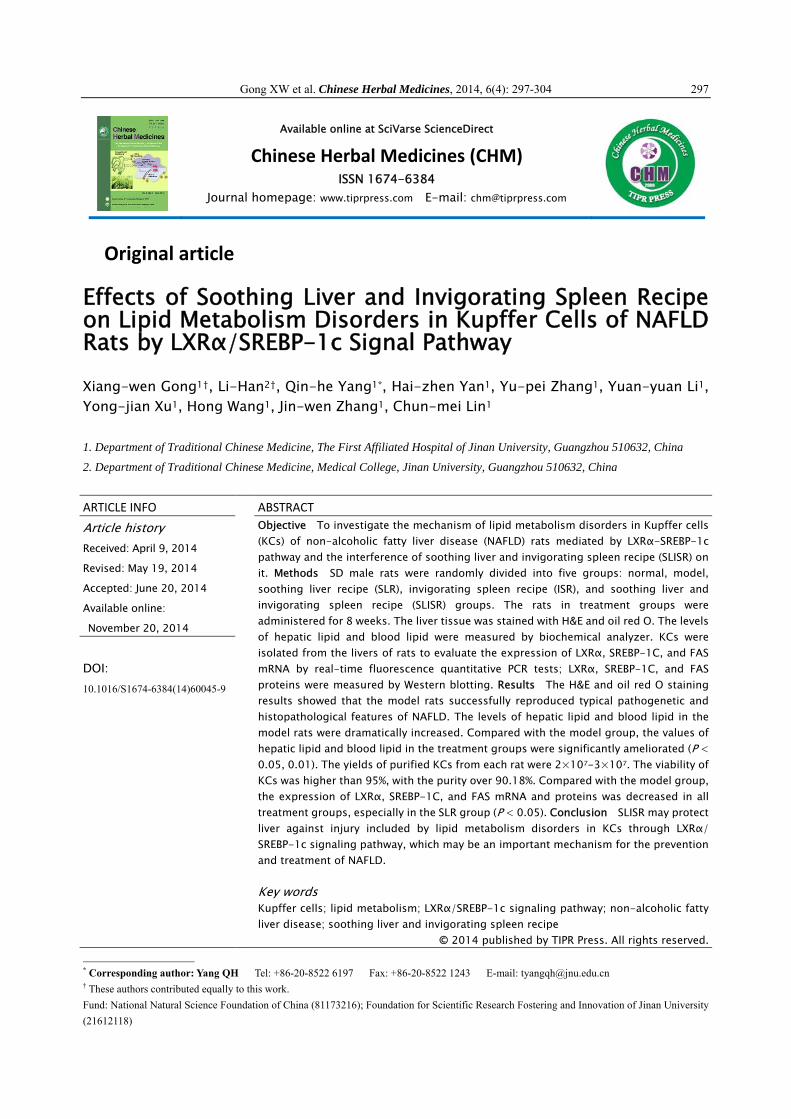
Gong XW et al. Chinese Herbal Medicines, 2014, 6(4): 297-304 297 Available online at SciVarse ScienceDirect Chinese Herbal Medicines (CHM) ISSN 1674-6384 Journal homepage: www.tiprpress.com E-mail: [email protected] Original article Effects of Soothing Liver and Invigorating Spleen Recipe on Lipid Metabolism Disorders in Kupffer Cells of NAFLD Rats by LXRα/SREBP-1c Signal Pathway Xiang-wen Gong 1† , Li-Han 2† , Qin-he Yang 1* , Hai-zhen Yan 1 , Yu-pei Zhang 1 , Yuan-yuan Li 1 , Yong-jian Xu 1 , Hong Wang 1 , Jin-wen Zhang 1 , Chun-mei Lin 1 1. Department of Traditional Chinese Medicine, The First Affiliated Hospital of Jinan University, Guangzhou 510632, China 2. Department of Traditional Chinese Medicine, Medical College, Jinan University, Guangzhou 510632, China ARTICLE INFO ABSTRACT Article history Received: April 9, 2014 Revised: May 19, 2014 Accepted: June 20, 2014 Available online: November 20, 2014 DOI: 10.1016/S1674-6384(14)60045-9 Objective To investigate the mechanism of lipid metabolism disorders in Kupffer cells (KCs) of non-alcoholic fatty liver disease (NAFLD) rats mediated by LXRα-SREBP-1c pathway and the interference of soothing liver and invigorating spleen recipe (SLISR) on it. Methods SD male rats were randomly divided into five groups: normal, model, soothing liver recipe (SLR), invigorating spleen recipe (ISR), and soothing liver and invigorating spleen recipe (SLISR) groups. The rats in treatment groups were administered for 8 weeks. The liver tissue was stained with H&E and oil red O. The levels of hepatic lipid and blood lipid were measured by biochemical analyzer. KCs were isolated from the livers of rats to evaluate the expression of LXRα, SREBP-1C, and FAS mRNA by real-time fluorescence quantitative PCR tests; LXRα, SREBP-1C, and FAS proteins were measured by Western blotting. Results The H&E and oil red O staining results showed that the model rats successfully reproduced typical pathogenetic and histopathological features of NAFLD. The levels of hepatic lipid and blood lipid in the model rats were dramatically increased. Compared with the model group, the values of hepatic lipid and blood lipid in the treatment groups were significantly ameliorated (P < 0.05, 0.01). The yields of purified KCs from each rat were 2×10 7 -3×10 7 . The viability of KCs was higher than 95%, with the purity over 90.18%. Compared with the model group, the expression of LXRα, SREBP-1C, and FAS mRNA and proteins was decreased in all treatment groups, especially in the SLR group (P < 0.05). Conclusion SLISR may protect liver against injury included by lipid metabolism disorders in KCs through LXRα/ SREBP-1c signaling pathway, which may be an important mechanism for the prevention and treatment of NAFLD. Key words Kupffer cells; lipid metabolism; LXRα/SREBP-1c signaling pathway; non-alcoholic fatty liver disease; soothing liver and invigorating spleen recipe © 2014 published by TIPR Press. All rights reserved. * Corresponding author: Yang QH Tel: +86-20-8522 6197 Fax: +86-20-8522 1243 E-mail: [email protected] † These authors contributed equally to this work. Fund: National Natural Science Foundation of China (81173216); Foundation for Scientific Research Fostering and Innovation of Jinan University (21612118)
Transcript of Chinese Herbal Medicines (CHM)
Gong XW et al. Chinese Herbal Medicines, 2014, 6(4): 297-304 297
Available online at SciVarse ScienceDirect
Chinese Herbal Medicines (CHM) ISSN 1674-6384
Journal homepage: www.tiprpress.com E-mail: [email protected]
Original article
Effects of Soothing Liver and Invigorating Spleen Recipe on Lipid Metabolism Disorders in Kupffer Cells of NAFLD Rats by LXRα/SREBP-1c Signal Pathway
Xiang-wen Gong1†, Li-Han2†, Qin-he Yang1*, Hai-zhen Yan1, Yu-pei Zhang1, Yuan-yuan Li1, Yong-jian Xu1, Hong Wang1, Jin-wen Zhang1, Chun-mei Lin1
1. Department of Traditional Chinese Medicine, The First Affiliated Hospital of Jinan University, Guangzhou 510632, China
2. Department of Traditional Chinese Medicine, Medical College, Jinan University, Guangzhou 510632, China
ARTICLE INFO ABSTRACT
Article history Received: April 9, 2014
Revised: May 19, 2014
Accepted: June 20, 2014
Available online:
November 20, 2014
DOI: 10.1016/S1674-6384(14)60045-9
Objective To investigate the mechanism of lipid metabolism disorders in Kupffer cells(KCs) of non-alcoholic fatty liver disease (NAFLD) rats mediated by LXRα-SREBP-1c pathway and the interference of soothing liver and invigorating spleen recipe (SLISR) on it. Methods SD male rats were randomly divided into five groups: normal, model,soothing liver recipe (SLR), invigorating spleen recipe (ISR), and soothing liver and invigorating spleen recipe (SLISR) groups. The rats in treatment groups wereadministered for 8 weeks. The liver tissue was stained with H&E and oil red O. The levels of hepatic lipid and blood lipid were measured by biochemical analyzer. KCs were isolated from the livers of rats to evaluate the expression of LXRα, SREBP-1C, and FAS mRNA by real-time fluorescence quantitative PCR tests; LXRα, SREBP-1C, and FAS proteins were measured by Western blotting. Results The H&E and oil red O staining results showed that the model rats successfully reproduced typical pathogenetic and histopathological features of NAFLD. The levels of hepatic lipid and blood lipid in the model rats were dramatically increased. Compared with the model group, the values of hepatic lipid and blood lipid in the treatment groups were significantly ameliorated (P <0.05, 0.01). The yields of purified KCs from each rat were 2×10⁷-3×10⁷. The viability ofKCs was higher than 95%, with the purity over 90.18%. Compared with the model group, the expression of LXRα, SREBP-1C, and FAS mRNA and proteins was decreased in all treatment groups, especially in the SLR group (P < 0.05). Conclusion SLISR may protect liver against injury included by lipid metabolism disorders in KCs through LXRα/SREBP-1c signaling pathway, which may be an important mechanism for the preventionand treatment of NAFLD. Key words Kupffer cells; lipid metabolism; LXRα/SREBP-1c signaling pathway; non-alcoholic fatty liver disease; soothing liver and invigorating spleen recipe
© 2014 published by TIPR Press. All rights reserved.
* Corresponding author: Yang QH Tel: +86-20-8522 6197 Fax: +86-20-8522 1243 E-mail: [email protected] † These authors contributed equally to this work. Fund: National Natural Science Foundation of China (81173216); Foundation for Scientific Research Fostering and Innovation of Jinan University (21612118)
Gong XW et al. Chinese Herbal Medicines, 2014, 6(4): 297-304 298
1. Introduction
Non-alcoholic fatty liver disease (NAFLD) represents a spectrum starting from fatty liver, and non fat alcoholic hepatitis (NASH) with fatty liver and inflammation as the evidence of damage to hepatocytes can progress to cirrhosis or in the most extreme form of NAFLD, which is hepatocellular carcinoma or liver failure (Lăţea et al, 2013).
Epidemiological studies suggest that NAFLD has become a major global health problem with the prevalence rates ranging from 6% to 35% worldwide, and the rising incidence of NAFLD in China was 20%. The most worrying thing is that more and more children are falling into NAFLD (Cohen et al, 2011; Vernon et al, 2011; Pirgon et al, 2013). Thus, it is important to study the prevention of NAFLD.
Because the mechanism of NAFLD is not clear at present (Musso et al, 2012), there are some different studies explaining the pathogenesis of NAFLD from different reports, but several studies have shown that Kupffer cells (KCs) play a key role in NAFLD (Smith, 2013; Stienstra et al, 2010). KCs, insulin resistance, oxidative stress, inflammatory markers, and iron overload are closely related to NAFLD (Zhu et al, 2009). Our research team has discovered the inflammatory signaling pathways involved in the pathogenesis of NAFLD such as NF-κB/IKKβ, JNK, and MARK (Han et al, 2013; Yang et al, 2012). At the same time, lipid metabolism disorders were mediated by LXRα/SREBP- 1c signal pathway in the liver tissue of NAFLD rats (Yang et al, 2011; He et al, 2011; Xu et al, 2013). To our knowledge, few reports have assessed the influence of LXRα/SREBP-1c signal pathway on lipid metabolism of KCs. In recent years, the relationship between KCs and the lipid metabolism- related genes is gradually becoming a hot spot in the pathogenesis of NAFLD (Maher et al, 2008). Consequently, this research provides an experimental basis for the study of significant correlation between KCs and the lipid metabolism-related genes.
Because the underlying pathogenesis of NAFLD is far from clear, there is a lack of consensus regarding the most effective and appropriate pharmacological therapy. (Tomeno et al, 2013) The systematic review and meta-analysis have shown that Chinese materia medica (CMM) was efficacious to treat NAFLD (Shi et al, 2012). Our previously clinical studies and animal experiments have shown that insulin resistance, lipid metabolism, and inflammatory cytokines were important targets for the prevention and therapy of NAFLD through the use of soothing liver and invigorating spleen recipe (SLISR). LXRα/SREBP-1c signal pathway can influence the lipid metabolism disorder; Therefore, it also has become an important target for CMM to cure NAFLD (Wang et al, 2013). Our research team has reported that SLISR had the beneficial effect on LXR, SREBP-1c mRNA and protein in the liver tissue of NAFLD rats (Yang et al, 2011; He et al, 2011; Xu et al, 2013). Hence, we investigated whether SLISR could regulate the lipid metabolism disorder through LXRα/SREBP-1c signaling pathway on KCs, and the current study provided more evidence for which the anti-NAFLD
efficacy on regulating lipid metabolism disorder was balanced by the use of SLISR. 2. Materials and methods 2.1 Animals
Seventy-five adult male SD rats, weighing (200 ± 20) g, were obtained from Experimental Animal Science Center of Guangzhou University of Chinese Medicine (China, Animal License Key No. 0107792; License No. SCXK(Yue)2008- 0020). The use of animals in this study was approved by the Ethics Committee of Medical College of Jinan University. The rats were separately housed in Animal Administration Laboratory, Jinan University. The rats were housed under the conditions of controlled temperature of (24 ± 2) oC and humidity of (70% ± 10%) in a 12-12 h light-dark cycle (lights on from 8:00 am to 8:00 pm), with free access to diet and water. 2.2 Animal grouping, modeling, and drugs
After 7 d adaptive breeding, the rats were randomly divided into five groups (n = 15, nine rats for liver samples collection, six rats for isolation of KCs, such as normal group, model group, soothing liver recipe (SLR) group, invigorating spleen recipe (ISR) group, and soothing liver and invigorating spleen recipe group (SLISR, the combination of SLR and ISR). The rat model of NAFLD was duplicated according to the method previously reported (Yang et al, 2012) with some minor modifications. The animal model of NAFLD was established by high fat diet (HFD, composed of 88% regular chow, 10% axungia porci, 1.5% cholesterol, and 0.5% bile salt), except for the normal group with normal chow diet. All rats in the treatment groups were ig administered with decoction (10 mL/kg body weight) (Shi, 2000), while the rats in the normal and model groups were fed with the same dose of distilled water. The course of treatment was 8 weeks.
SLR (Chaihu Shugan Powder) is composed of seven CMM: Bupleuri Radix, Citri Reticulatae Pericarpium, Chuanxiong Rhizoma, Cyperi Rhizoma, Bitter Orange Aurantii Fructus, Paeoniae Alba Radix, and Glycyrrhizae Radix with a conventional dose ratio of 6:6:5:5:5:5:3. ISR (Shenling Baizhu Powder) includes Lablab Album Semen, Atractylodis Macrocephalae Folium, Poria Rubra, Glycyrrhizae Radix, Platycodi Radix, Sulphur, Ginseng Radix, Amomi Flos et Pedunculus, Dioscorea Rhizoma, and Coicis Semen in a ratio of 5:5:5:5:4:3:3:3:2:2. The SLISR contains the mixture of Chaihu Shugan Powder and Shenling Baizhu Powder at a ratio of 1:1. All the CMM were formula granules purchased from Shenzhen Sanjiu Medical Co., Ltd. (1005001S). The formula granules were put into the solvent of distilled water and preserved at −4 oC in refrigerator. 2.3 Reagents
Nycodenz kit was purchased from Axis-Shield Co., Ltd. (Switzerland), type IV collagenase was obtained from Gibco
Gong XW et al. Chinese Herbal Medicines, 2014, 6(4): 297-304 299
Co., Ltd. (USA). Pronase E was purchased from Roche Co., Ltd. (Switzerland). Hepes was obtained from Amresco Co., Ltd. (USA). DNase I and EGTA were obtained from Sigma Co., Ltd. (USA). Trozol nucleic acid extraction reagent was obtained from Invitrogen Co., Ltd. (USA). Reverse Transcription Kit was obtained from Toyobo Co., Ltd. (Japan). EpiQuik Nuclear Extraction Kit and Bradford Protein Assay Kit were obtained from Shanghai Beyotime Institute of Biotechnology (China). Anti-LXRα (No. GR9889-9), Anti- SREBP-1c (No. GR111569-1), and Anti-FAS (No. YI 092905CS) were purchased from Abcam Co., Ltd. (USA). Quantscript RT Kit (No. DRR047A) and Golden HS SYBR Green qPCR Mix (No. DRR820A) were purchased from Takara Co., Ltd. (Japan). 2.4 Histopathological examination of liver
After the treatment, by macroscopic observation on general changes of rat livers in the model group, the livers were performed paraffin imbedding and H&E staining, and steatosis of liver was observed under light microscope. Frozen sections were prepared and oil red O staining was used to observe the distribution situation of lipid droplet in liver cells.
2.5 Biochemical test in liver and abdominal aorta serum
After rats were anesthetized by ip injection of 3% pentobarbital (2 mL/kg body weight), livers were taken out quickly. Liver tissues were put into isopropanol. Homogenates were manufactured using a Tissue Lyser-II Homogenizer (Qiagen, Germany), centrifuged at 3000 × g for 10 min at 4 oC, and then clear supernatants were collected. Total cholesterol (TC) and triglyceride (TG) in the liver tissue were determined with automatic biochemical analyzer (Olympus, Japan). Blood sample (5 mL) was drawn from the abdominal aorta into a tube, and was centrifuged at 10 000 × g for 10 min at 4 oC. The supernatant was then used to measure the contents of TC, TG, high-density lipoprotein cholesterol (HDL-C), and low-density lipoprotein cholesterol (LDL-C) in serum by automatic biochemical analyzer. 2.6 Separation and identification of KCs
KCs were isolated and identified from six rats in each group as previously described with some modifications (Feng et al, 2012). After rats were anesthetized with 3% pentobarbital (1 mL/kg body weight), the portal vein and inferior caval vein were dissociated, the liver was perfused in situ with 200 mL ethylene glycol tetraacetic acid (EGTA, 0.5 mmol/L) in D-Hanks solution at 20 mL/min, at 37 oC until the colour of liver changed into amber. Then the liver was transferred to a culture dish and perfused ex situ with 0.03% collagenase IV in Hanks solution, which contained 5 mmol/L calcium ion and should be preheated to 37 oC, at 20 mL/min in a recirculating fashion for 15 min. The liver was then placed into 10 mL RPMI-1640 culture medium containing 10% fetal calf serum (FBS), the capsule and fibrous tissue
were removed, and the remaining tissue was cut into small pieces. After the obtained liver homogenate was filtered through 200 and 300 μm nylon mesh, the cell suspension was centrifuged at 50 × g for 3 min at 4 oC and the clear supernatant was collected in another tube, centrifuged at 400 × g for 10 min at 4 oC. The cell pellet was subsequently resuspended in RPMI-1640 medium containing 10% FBS.
Then some 15 mL centrifuge tubes were carefully laid into 2.5 mL Nycodez working solution (24%) in the bottom, 2.5 mL Nycodez working solution (11%) in the middle layer and 2.5 mL cell suspension in the top. Then it was centrifuged at 800 × g for 15 min at 4 oC. KCs with a clouding appearance between 11% and 24% Nycodez layers were collected to another 15 mL tube and resuspended in GBSS, then centrifuged at 400 × g for 15 min at 4 oC twice. The cell pellet was then resuspended and seeded on culture dish at a density of 2 × 10⁶ – 5 × 10⁶ cells/mL with RPMI-1640 medium containing 10% FBS, and incubated in a 5% CO2 atmosphere for 30 min at 37 oC. By further using adhesion purification, the purity of KCs was improved, and the cell viability was tested by trypan blue dye exclusion. 2.7 Determination of LXRα, SREBP‐1, and FAS mRNA expression in KCs
Total RNA was extracted from KCs using Trizol reagent, the integrity of each RNA sample was evaluated by agarose gel electrophoresis, and its purity and concentration were assayed, then the total RNA was reversely transcribed to cDNA. According to the gene order of LXRα, SREBP-1C, FAS, and glyceraldehyde-3-phosphate dehydrogenase (GAPDH) supplied by Genebank, the primer was designed and synthesized by Shanghai Generay Biological Engineering Co., Ltd. GAPDH was considered as the reference gene (Table 1). The reaction conditions were as follows: a: predenaturation for 1 min at 95 oC; b: denaturation for 10 s at 95 oC; c: GAPDH at 57.5 oC, LXRα at 54 oC, SREBP-1c at 58 oC, FAS at 54 oC, renaturation for 20 s; d: extension for 30 s at 68 oC; b−d repeated for 39 times; e: analysis of solubility curve, 72 oC to 95 oC for 5−10 s. After the reaction was finished, the results were analyzed using Opticon Monitor 3.1 software, and the formula 2− Ct△△ was used for relative quantification. 2.8 Western blotting
Western blotting was used to determine the proteins of KCs toll like LXRα, SREBP-1c, FAS, and GAPDH. GAPDH was used as an internal control. KCs were split in RIPA lysis buffer and centrifuged at 8000 × g for 5 min at 4 oC, and the supernatants were collected. The concentration of supernatant protein was determined by BCA protein assay. Protein (60 mg) was resolved by 10% sodium dodecyl sulfatepoly-acrylamide gel electrophoresis (SDS-PAGE) and preceded with transmembrane. The polyvinylidene difluoride (PVDF) membrane was blocked with 5% skim milk in tris-buffered saline tween-20 (TBST), shaken for 1 h at room temperature, and

Gong XW et al. Chinese Herbal Medicines, 2014, 6(4): 297-304 300
Table 1 PCR primers
Genes Sequences Amplified fragment lengths / bp
Upstream: 5' AGAAACTGAAGCGTCAAGAAGAGG 3' 148 LXRα
Downstream: 5' GGCAGCCACCAACTTCTCAA 3' Upstream: 5' GCTCACAAAAGCAAATCACTGAAAG 3' 103 SREBP-1c Downstream: 5' GCGTTTCTACCACTTCAGGTTTCA 3' Upstream:5' ATGGCTGTCCTGCCTCTGGT 3' 193 FAS Downstream:5' CACGAACGCTCCTCTTCAACTC 3' Upstream:5‘GATCCCGCTAACATCAAATG 3’ 134 GAPDH Downstream:5‘GAGGGAGTTGTCATATTTCTC 3’
then incubated overnight at 4 oC with specific primary antibodies. Then horseradish peroxidase (HRP) conjugated goat-anti-rabbit antibody was added and incubated at room temperature for 1 h. After being washed for three times in TBST, the PVDF membrane was put into developer and exposed to X-ray film. The films were scanned and analyzed by gel image processing system. 2.9 Statistical analysis
Data were expressed as sx ± and analyzed using SPSS 13.0 Software (Chicago, USA). One-way analysis of variance (ANOVA) and Tukey’s test was used to determine the statistical significance of the differences. P < 0.05 was considered statistically significant. 3. Results 3.1 Observation on morphology of liver tissue in NAFLD rats
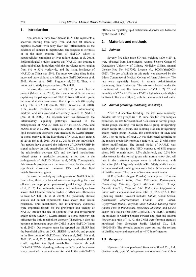
According to the results of H&E staining, for the normal group, the hepatic lobule outline was clear, central vein was located in the middle of the hepatic lobule, hepatic sinusoidal structure was normal, liver rope to central vein axis arranged radically, hepatocytes of the rats were polygonal, and the nuclear had a clear structure and located in the centre of the cell. The cytoplasm was dyed rich in red and well-distributed. No lipid droplet deposition was found. Hepatocytes from rats in the model group were swelling. The lipid droplets inside the cytoplasm were in different sizes. The lipid droplets were mainly macro-vesicular ones with the nuclear pressed to the edge. The hepatocytes were ballooned extensively and spotty necrosis can be seen. The above pathological changes indicated the successful establishment of NAFLD rats. The levels of the lipid droplet deposition decreased in all the intervention groups. Compared with the model group, the conditions of the infiltration of inflammatory cells in the treatment groups were obviously alleviated in different degrees, especially for the SLR group (Figure 1). According to the results of oil red O staining, for the normal group, hepatocyte nucleus were dyed blue, and no obvious orange lipid droplets were seen on hepatocyte cytoplasm. Hepatocytes from rats in the model group were swelling, hepatocyte nucleus were deep blue, and a large number of orange lipid droplets could be seen
in hepatocytes. A few orange lipid droplets were merged into one bigger orange lipid droplets. The levels of the lipid droplets deposition decreased in all the intervention groups. Compared with the model group, the conditions of the infiltration of inflammatory cells in the intervened groups were alleviated in different degrees, especially in the SLR group (Figure 2). 3.2 Levels of TC and TG in liver tissue and effects of SLR
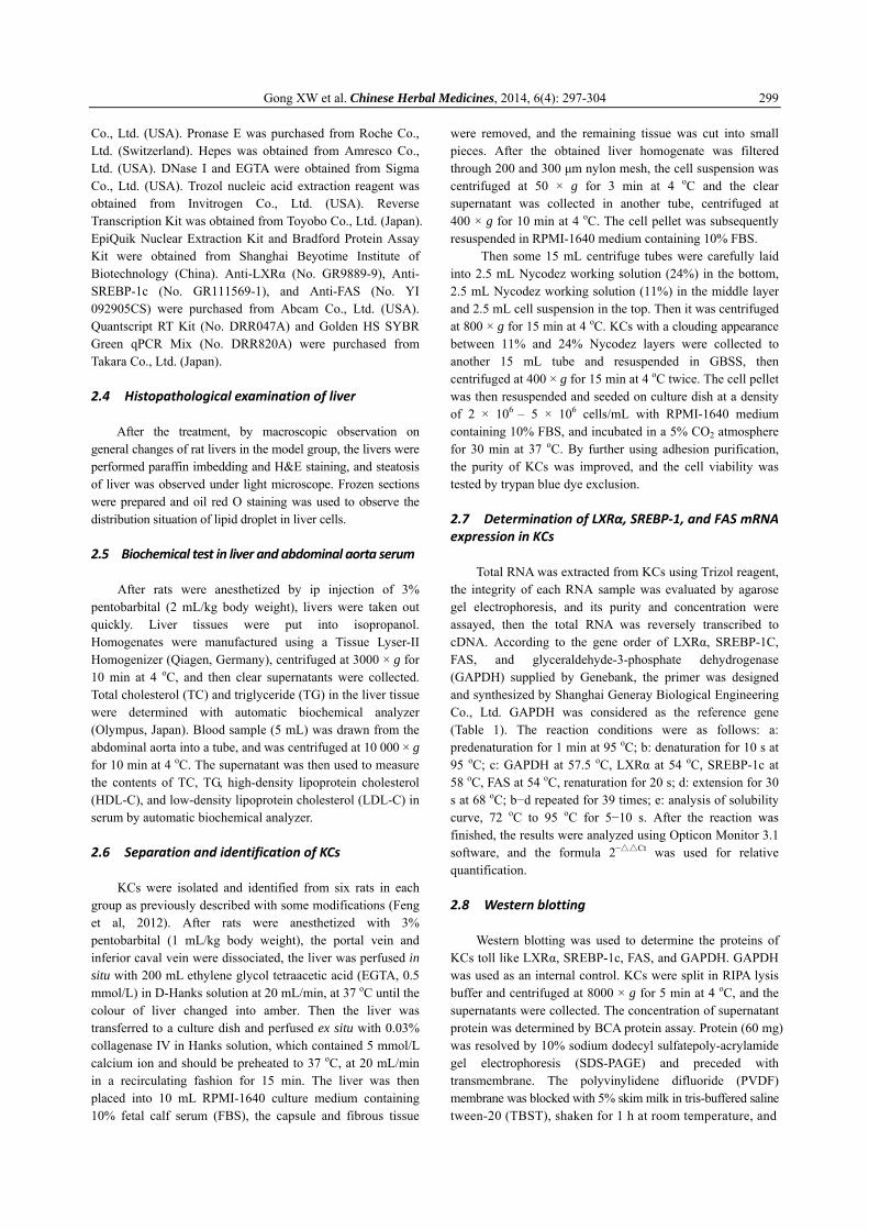
Compared with the normal group, the TC and TG values in rat liver tissue of the model group increased with a significant difference (P < 0.01). The TC and TG values in each treatment group were reduced to different degrees compared with the model group, especially for SLR group (P < 0.05, Figure 3).
3.3 Levels of TC, TG, HDL‐C, and LDL‐C in abdominal aorta serum and effects of SLR
Compared with the normal group, the TC, TG, and
LDL-C values in abdominal aorta serum of the model group increased with a significant difference (P < 0.01), and the HDL-C value in the model group decreased with a significant difference (P < 0.01). The TC, TG, and LDL-C values in each treatment group were significantly lower than those in the model group (P < 0.01, 0.05), the HDL-C values in each treatment group were significantly higher than those in the model group (P < 0.01, 0.05, Figure 4). 3.4 Population, purity, and viability of KCs
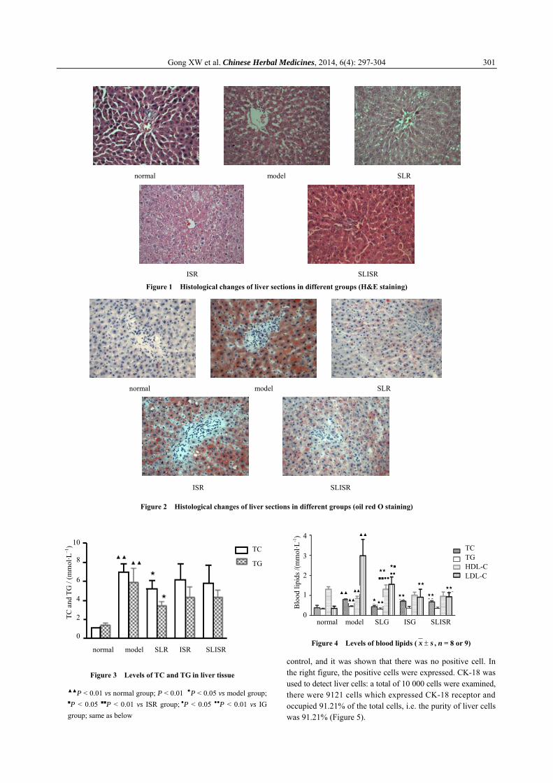
The yields of purified cell of KCs in each rat were 2.0 × 10⁷−3.0 × 10⁷. The viability of KCs isolated was higher than 95%, with the purity over 91.21%. The number and purity degree of KCs complied with the requirement of the follow-up testing. The KCs population after purified was 2.0 × 10⁷−3.0 × 10⁷ in each NAFLD rats. A small quantity of cells were chosen, and after trypsin digestion, they were stained with Typan blue. Cell counting was carried out on blood count plate using inverted phase contrast microscope to determine the activity over 95%.
KCs were identified by FCM, and the parallel lines were made. Considering M1 as initiation point, the left side of the parallel line presented negative cells, and the right side presented positive cells. In the left figure, rabbit IgG was chosen as the
Gong XW et al. Chinese Herbal Medicines, 2014, 6(4): 297-304 301
Figure 1 Histological changes of liver sections in different groups (H&E staining)
Figure 2 Histological changes of liver sections in different groups (oil red O staining)
Figure 3 Levels of TC and TG in liver tissue
▲▲P < 0.01 vs normal group; P < 0.01 ★P < 0.05 vs model group;
■P < 0.05 ■■P < 0.01 vs ISR group; ●P < 0.05 ●●P < 0.01 vs IG group; same as below
Figure 4 Levels of blood lipids ( ± sx , n = 8 or 9)
control, and it was shown that there was no positive cell. In the right figure, the positive cells were expressed. CK-18 was used to detect liver cells: a total of 10 000 cells were examined, there were 9121 cells which expressed CK-18 receptor and occupied 91.21% of the total cells, i.e. the purity of liver cells was 91.21% (Figure 5).
normal model SLR ISR SLISR
10
8
6
4
2
0
TC
TG
TC a
nd T
G /
(mm
ol·L
−1) TC
TG HDL-CLDL-C
normal model SLG ISG SLISR
4
3
2
1
0
Blo
od li
pids
/(m
mol
·L-1
)
▲▲
▲▲
▲▲
▲▲
★★★
★★
■■●●
★■ ●●
★★
★★
★★
★★
normal model SLR
ISR SLISR
normal model SLR
ISR SLISR
Gong XW et al. Chinese Herbal Medicines, 2014, 6(4): 297-304 302
Figure 5 Purity of KCs identified by FCM
3.5 LXRα, SREBP‐1c, and FAS mRNA expression in KCs of NAFLD rats and impact of SLR
Compared with the normal group, the LXRα, SREBP-1c, and FAS mRNA values of the model group increased by 4.66,
6.80, and 7.62 folds (P < 0.01). Among the treatment groups, the LXRα, SREBP-1c, and FAS mRNA values were significantly lower than those in the model group, especially in the SLR group (P < 0.01, Table 2). 3.6 LXRα, SREBP‐1c, and FAS protein expression in KCs of NAFLD rats and impact of SLR
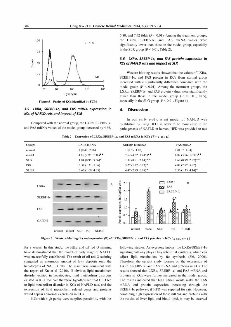
Western blotting results showed that the values of LXRα, SREBP-1c, and FAS protein in KCs from normal group increased with a significantly difference compared with the model group (P < 0.01). Among the treatment groups, the LXRα, SREBP-1c, and FAS protein values were significantly lower than those in the model group (P < 0.01, 0.05), especially in the SLG group (P < 0.01, Figure 6). 4. Discussion
In our early study, a rat model of NAFLD was established by using HFD, in order to be more close to the pathogenesis of NAFLD in human, HFD was provided to rats
Table 2 Expression of LRXα, SREBP-1c, and FAS mRNA in KCs ( 6=± n , sx )
Groups LXRα mRNA SREBP-1c mRNA FAS mRNA
normal 1 (0.49−2.06) 1 (0.55−1.82) 1 (0.57−1.74) model 4.66 (2.95−7.36)▲▲ 7.62 (4.32−13.45)▲▲ 6.82 (3.76−12.38)▲▲ SLG 1.84 (0.95−3.56)★ 1.32 (0.81−2.14)★★ 1.68 (0.99−2.87)★★ ISG 2.58 (1.31−5.06) 3.27 (1.72−6.23)★ 4.08 (2.87−5.82) SLISR 2.60 (1.68−4.03) 4.47 (2.99−6.68)★ 2.36 (1.35−4.14)★
Figure 6 Western blotting (A) and expression (B) of LXRα, SREBP-1c, and FAS proteins in KCs ( 6=± n , sx )
for 8 weeks. In this study, the H&E and oil red O staining have demonstrated that the model of early stage of NAFLD was successfully established. The result of oil red O staining suggested an enormous amount of fatty deposits onto the hepatocytes of NAFLD rats. The result was consistent with the report of Xu et al (2010). If obvious lipid metabolism disorder existed in hepatocytes, lipid metabolism disorders existed in KCs too. We therefore hypothesized that HFD led to lipid metabolism disorder in KCs of NAFLD rats, and the expression of lipid metabolism related genes and proteins would appear abnormal expression in KCs.
KCs with high purity were supplied possibility with the
following studies. As everyone knows, the LXRα/SREBP-1c signaling pathway plays a key role in fat synthesis, which can adjust lipid metabolism by fat synthesis (Shi, 2000). Therefore, the current study focuses on the expression of LXRα, SREBP-1c, and FAS mRNA and proteins in KCs. The results showed that LXRα, SREBP-1c, and FAS mRNA and proteins in KCs were further increased in the model group. The results indicated that high LXRα would make the FAS mRNA and protein expression increasing through the SREBP-1c pathway, if HFD was supplied for rats. However, combining high expression of those mRNA and proteins with the results of liver lipid and blood lipid, it may be asserted
100 101 102 103 104
100
75
50
25
0
Even
ts
91.21%
M1
Lysozyme
normal model SLR ISR SLISR
2.5
2.0
1.5
1.0
0.5
0.0 Prot
ein
expr
essi
on in
Kup
ffer c
ells
LXR α FAS SREBP-1c
B LXRα
SREBP-1c
FAS
GAPDH
normal model SLR ISR SLISR
A
Gong XW et al. Chinese Herbal Medicines, 2014, 6(4): 297-304 303
that the lipid metabolism disorder has existed in KCs, which is the important mechanism of NAFLD. Several reasons should be considered as following.
1) LXRα is the gene producing fatty acids, and the downstream genes include FAS. LXR not only directly combined with FAS promoter, but also indirectly adjusted the expression of FAS gene through SREBP-1c pathway. The excessive transcription of FAS gene resulted in the increase of fatty acid synthesis, which was the important mechanism of NAFLD (Higuchi et al, 2008). Beyond that, in LXRα- deficient mice, the basic standard of SREBP-1c mRNA expression was decreased in liver tissue, similarly, we found no reduction in FAS mRNA expression due to directly by SREBP-1c mRNA (Quinet et al, 2006).
2) The results of oil red O staining and electron microscopy showed that an enormous amount of fat deposited onto KCs and endothelial cells of NAFLD rabbits (Nenseter et al, 1992). Abnormal blood lipid deposited in KCs cytoplasm membrane might change the lipid structure and interfere with cell surface receptor function (Lee and Hwang, 2006).
3) Once lipid metabolism disorder existed in hepatocytes, which would affect the lipid metabolism disorder in KCs, high blood lipid might lead to fatty liver cells release danger signal which made an adverse influence on KCs (Novgorodtseva et al, 2013; Tiniakos et al, 2010; Zhu et al, 2012).
4) The changes of lipid composition might also influence the intrinsic function of cell membrane, such as mitochondria load free cholesterol.
Currently, medicinal herbs such as CMM have been received increasing attention as an alterative source of the treatment for a variety of diseases (Luo et al, 2013). In traditional Chinese medicine (TCM) theory, NAFLD belongs to the Gan-Pi and Gan-Zhu, and we previously demonstrated that the stagnation of liver qi, and spleen deficiency was the basic pathogenesis of NAFLD. SLISR should be throughout the whole disease (Qiao et al, 2008; Yang et al, 2007). However, based on the TCM theory, the same disease has different syndromes in whole disease. Hence, in this study, the effects of soothing liver recipe were more superior than those of invigorating spleen recipe and SLISR, which might be due to the stagnation of liver qi, is the most important syndrome in the early stage of NAFLD. Epidemiological researches showed that the syndrome of stagnation of liver qi and spleen deficiency was one of the most common syndromes of NAFLD in China, and the proportion was 34.7% (Wei et al, 2002). This study suggested the clinical characteristics of Chinese NAFLD population in contemporary. Therefore, the syndrome of stagnation of liver qi and spleen deficiency has become the most important syndrome in expert consensus document of NAFLD (Zhang et al, 2002). Beyond that, the evidence-based medicine has shown that CMM treated NAFLD with not only better clinical therapeutic effect but also less adverse reactions than Western medicine (Li et al, 2011). Bupleuri Radix Liver Powder and Shenling Baizhu Powder were classical formula in CMM.
SLISR includes Bupleuri Radix Liver Powder and Shenling Baizhu Powder. Ten effective components including saikosaponin A, paeoniflorin, hesperidin, ferulic acid, paeoniflorin, liquorice glycosides, naringin, hesperidin, new hesperidin, and ginsenosides in SLISR were determined by HPLC/DAD (Li et al, 2011; Ding, 2012; Wang et al, 2010; 2012). We may speculate that SLISR has anti-NAFLD effect due to above components. This conclusion should be demonstrated via further studies. In this study, SLISR might improve the fatty change of liver tissues and reduce the value of liver lipid and blood lipid in NAFLD rats, which also reduced the expression of LXRα, SREBP-1c, and FAS mRNA and proteins in KCs. Therefore, the results showed that SLISR could mediate the lipid metabolism disorders through LXRα/SREBP-1c signal pathway in KCs of NAFLD rats. Combined with early studies, we can know that the insulin resistance, lipid metabolism, and inflammatory markers have become targets of SLISR (Stienstra et al, 2010; Zhu et al, 2009; Tao et al, 2013). LXRα/SREBP-1c signal pathway could mediate the lipid metabolism in KCs of NAFLD rats, which may be an important target of SLISR used to treat NAFLD. References Cohen JC, Horton JD, Hobbs HH, 2011. Human fatty liver disease:
Old questions and new insights. Science 332: 1519-1523. Ding T, 2012. Shenlingbaizhu powder of ginseng saponin content
determination. Chin J Basic Med Tradit Chin Med 10: 1134-1135. Feng GF, Yang QH, Wang WJ, He XM, Zhang YP, Ji GY, Li GQ,
Zhao YF, Shen CH, Yang XL, 2012. Simultaneously isolation and identification of hepatocytes and Kupffer cells from nonalcoholic steatohepatitis rat. Guangdong Med J 33: 40-43.
Han L, Yang QH, Yang XS, Hu CF, Zhang YP, Fen GF, Wang WJ, He XM, Wang YP, Cheng SB, Jin L, Yan HZ, Huang J, 2013. Effects of Shu-gan Jian-pi prescriptions on expression of NF-kB p65 and IKKβ at mRNA and protein levels in Kupffer cells from rats with non-alcoholic steatohepatitis. Chin J Pathophysiol 29: 641-646.
He XM, Yang QH, Li PF, Chen Z, Fen GF, Wang WJ, Liu HT, Zhang YP, Ji GY, 2011. Effect of soothing liver and invigorating spleen recipe on SREBP-1c mRNA and protein expression in hepatic tissues of rats with non-alcoholic fatty liver disease. J Chin Med Mater 34(6): 931-937.
Higuchi N, Kato M, Shundo Y, 2008. Liver X receptor in with nonalcoholic fatty liver disease. Hepatol Res 38: 1122-1129.
Lăţea L, Negrea Ş, Bolboaca S 2013. Primary non-alcoholic fatty liver disease in hypertensive patients. Australas Med J 6(6): 325-330.
Lee JY, Hwang DH, 2006. The modulation of inflammatory gene expression by lipids: Mediation through Toll-like receptors. Mol Cells 21(2): 174-185.
Li L, Su DM, Han HX, Li JX,2011. Traditional Chinese medicine for non-alcoholic steatohepatitis: A systematic review. Chin J Evid Base Med 11: 195-203.
Luo JK, Zhou HJ, Wu J, Tang T, Liang QH, 2013. Electroacupuncture at Zusanli (ST36) accelerates intracerebral hemorrhage-induced angiogenesis in rats. Chin J Integr Med 19: 367-373.
Maher JJ, Leon P, Ryan JC, 2008. Beyond insulin resistance: Innate immunity in nonalcoholic steatohepatitis. Hepatology 48: 670-678.
Musso G, Gambino R, Cassader M, 2012. Cholesterol metabolism and the pathogenesis of non-alcoholic steatohepatitis. Prog Lipid Res 52: 175-191.
Gong XW et al. Chinese Herbal Medicines, 2014, 6(4): 297-304 304
Nenseter M, Gudmundsen O, Roos N, Maelandsmo G, Drevon C, Berg T, 1992. Role of liver endothelial and Kupffer cells in clearing low density lipoprotein from blood in hyper- cholesterolemic rabbits. J Lipid Res 33: 867-877.
Novgorodtseva TP, Karaman IuK, Zhukova NV, 2013. Modification of fatty acid composition of polar and neutral lipids of blood and liver in rat in conditions of prolonged high-fat diet. Biomed Khim 59: 644-654.
Pirgon Ö, Bilgin H, Çekmez F, Kurku H, Dündar BN, 2013. Association between insulin resistance and oxidative stress parameters in obese adolescents with non-alcoholic fatty liver disease. J Clin Res Pediatr Endocrinol 5: 33-39.
Qiao NL, Yang QH, JI GY, 2008. Discuss liver qi and spleen deficiency is the basic pathogensis of non-fatty liver disease. Lishizhen Med Mater Med Res 153: 1238-1239.
Quinet EM, Savio DA, Halpern AR, Chen L, Schuster GU, Gustafsson JA, Basso MD, Nambi P,2006. Liver X receptor (LXR)-β regulation in LXRα-deficient mice: Implications for therapeutic targeting. Mol Pharmacology 70: 1340-1349.
Shi KQ, Fan YC, Liu WY, Li LF, Chen YP, Zheng MH,2012. Traditional Chinese medicines benefit to nonalcoholic fatty liver disease: A systematic review and meta-analysis. Mol Biol Rep 39: 9715-9722.
Shi X, 2000. Experimental Zoology of Modern Medicine. People’s Military Medical Publishing House; Beijing.
Smith K, 2013. Liver disease: Kupffer cells regulate the progression of ALD and NAFLD. Nat Rev Gastroenterol Hepatol 10(9): 503.
Stienstra R, Saudale F, Duval C, Keshtkar S, Groener JE, van Rooijen N, Staels B, Kersten S, Müller M, 2010. Kupffer cells promote hepatic steatosis via interleukin-1beta-dependent suppression of peroxisome proliferator-activated receptor alpha activity. Hepatology 51: 511-522.
Tao W, Xu X, Wang X, Li B, Wang Y, Li Y, Yang L, 2013. Network pharmacology-based prediction of the active ingredients and potential targets of Chinese herbal Radix Curcumae formula for application to cardiovascular disease. J Ethnopharmacol 145: 1-10.
Tiniakos DG, Vos MB, Brunt EM, 2010. Nonalcoholic fatty liver disease: Pathology and pathogenesis. Ann Rev Pathol 5: 145-171.
Tomeno W, Yoneda M, Imajo K, Ogawa Y, Kessoku T, Saito S, Eguchi Y, Nakajima A, 2013. Emerging drugs for non-alcoholic steatohepatitis. Expert Opin Emerg Drugs 18: 1-12.
Vernon G, Baranova A, Younossi ZM, 2011. Systematic review: The
epidemiology and natural history of non-alcoholic fatty liver disease and non-alcoholic steatohepatitis in adults. Aliment Pharrmacol Ther 34: 274-285.
Wang M, Sun S, Wu T, Zhang L, Song H, Hao W, Zheng P, Xing L, Ji G, 2013. Inhibition of LXRalpha/SREBP-1c-mediated hepatic steatosis by Jiang-Zhi Granule. Evid Based Complement Alternat Med 584634. doi: 10.1155/2013/584634.
Wang CY Zhang DS, Zhang WM, Guo CY, Xue GP, 2010. Determination of four effective components in Chaihu Shugan Powder by HPLC/DAD. Chin Tradit Pat Med 32: 422-425.
Wang SL, Jia W, Gao XL, 2012. HPLC characteristics of aqueous extract of Chaihu Shugan Powder and determination of markers. Chin Tradit Pat Med 34: 2384-2388.
Wei HF, Xin LJ, 2002. Preliminary research on nonalcoholic fatty liver syndrome classification. Liaoning J Tradit Chin Med 29: 655-656.
Xu L, Kim JK, Bai Q, Zhang X, Kakiyama G, Min HK, Sanyal AJ, Pandak WM, Ren S, 2013. 5-Cholesten-3β,25-diol 3-sulfate decreases lipid accumulation in diet-induced nonalcoholic fatty liver disease mouse model. Mol Pharmacol 83: 648-658.
Xu ZJ, Fan JG, Ding XD, Qiao L, Wang GL, 2010. Characterization of high-fat, diet-induced, non-alcoholic steatohepatitis with fibrosis in rats. Dig Dis Sci 55(4): 931-940.
Yang QH, Hu SP, Zhang YP, 2012. Effects of different therapeutic methods and typical recipes of Chinese medicine on activation of c-Jun N-terminal kinase in Kupffer cells of rats with fatty liver disease. Chin J Integr Med 18: 769-774.
Yang QH, Wang WJ, Fen GF, He XH, Zhang YP, Ji GY, 2011. Effects of soothing liver, invigorating spleen recipes on expression of LXRα mRNA and protein in hepatic tissue of rats with non-alcoholic fatty liver disease. Chin J Gerontol 31: 4371-4375.
Yang QH, Lin JS, Ping HH, 2007. Thinking and countermeasures of prevention and treatment of nonalcoholic fatty liver disease in traditional Chinese medicine. J Tradit Chin Med 48: 746-748.
Zhang SS, Li JX, 2011. Diagnosis and treatment of traditional Chinese Medicine Consensus on Nonalcoholic fatty liver disease. Beijing J Tradit Chin Med 29: 655-656.
Zhu M, Zhao L, Gong JP, 2009. Functions of Kupffer cells in non-alcoholic fatty liver disease. Int J Dig Dis7 29: 100-102.
Zhu M, Shi J, 2012. Roles of kupffer cells activation in pathogenesis and progression of non-alcoholic fatty liver disease. Chin J Hepatol 20: 718-720.