Chimeric gag-V3 virus-neutralizing · Proc. Nati. Acad. Sci. USA Vol. 89, pp. 10527-10531,...
5
Proc. Nati. Acad. Sci. USA Vol. 89, pp. 10527-10531, November 1992 Medical Sciences Chimeric gag-V3 virus-like particles of human immunodeficiency virus induce virus-neutralizing antibodies (AIDS vaccine/chimeric proteins/baculovirus expression system) LIZHONG Luo*, YAN LI*, PAULA M. CANNONt, SUNYOUNG KIMt, AND C. YONG KANG*t *Faculty of Science, Western Science Centre, The University of Western Ontario, London, ON, N6A 5B7 Canada; and tHarvard Medical School, New England Deaconess Hospital, Boston, MA 02215 Communicated by Max D. Summers, June 24, 1992 (received for review May 11, 1992) ABSTRACT A 41-kDa unprocessed human immunodefi- ciency virus 2 (HIV-2) gag precursor protein that has a deletion of a portion of the viral protease assembles as virus-like particles by budding through the cytoplasmic membrane of recombinant baculovirus-infected insect cells. We have con- structed six different combinations of chimeric genes by cou- pling the truncated HIV-2 gag gene to the neutralizing domain (V3) or the neutralizing and the CD4 binding dons (V3+CD4BD) of gpl20 env gene sequences from HIV-1 or HIV-2. The env gene sequences were inserted either into the middle of the gag gene or at the 3' terminus of the gag gene. Virus-like particles were formed by chimeric gene products only when the env gene sequences were linked to the 3' terminus of the gag gene. Insertion of env gene sequence in the middle of the gag gene resulted in high-level chimeric gene expression but without the formation of virus-like particles. Three different chimeric genes [gag gene with HIV-1 V3 (1V3), gag gene with HIV-2 V3 (2V3), and gag gene with HIV-2 V3+CD4BD (2V3+CD4BD)] formed virus-like particles that were secreted into the cell culture medium. In contrast, the HIV-1 V3+CD4BD/HIV-2 gag construct did not form virus-like particles. The chimeric gag-env particles had spherical mor- phology and the size was lighdy larger than that of the gag particles, but the chimeric particles were similar to the mature HIV particles. Western blot analysis showed that the gag-env chimeric proteins were recognized by antibodies in HIV- positive human serum and rabbit anti-gpl20 serum. Rabbit anti-gag 1V3 and anti-gag 2V3 sera reacted with authentic gpl20 of HIV-1 and HIV-2, respectively, and neutralized homologous HIV infectivity. Our results show that precursor gag protein has potential as a carrier for the presentation of foreign epitopes in good immunologial context. The gag protein is highly immunogenic and has the ability to carry large foreign inserts; as such, it offers an attractive approach for HIV vaccine development. The human immunodeficiency viruses (HIVs) are the etio- logic agents for acquired immunodeficiency syndrome (AIDS). The envelope glycoprotein gpl20 of HIV has been the major target for developing a candidate vaccine against AIDS (1, 2). gpl20 recognizes the cellular receptor (CD4) on helper T lymphocytes (3) and carries the V3 loop domain that induces neutralizing antibodies (4, 5). The third hypervari- able loop region (V3) of HIV-1 gpl20 (amino acid residues 308-331) contains not only a major immunodominant neu- tralizing epitope but also the epitopes for antigen-dependent cellular cytotoxicity (6) and cytotoxic T-lymphocyte recog- nition (7, 8). Although the majority of the amino acids in the V3 loop are variable among different strains of HIV, a G-P-G-R motif at the tip of the loop is conserved (9). Huang et al. (10) and Bjorling et al. (11) demonstrated that the principal neutralization domain of the envelope glycoprotein of HIV-2 is also located in the region corresponding to the hypervariable motif in the V3 loop of HIV-1 gpl20. The CD4 binding region, which is located within the C-terminal third of HIV-1 gpl20 (amino acid residues 397-439), plays an essen- tial role in infectivity of HIV (12). This region also seems to be weakly immunogenic because it forms a pocket that is not accessible to the immune system; thus high-titer neutralizing antibody against this region is not presently available (13). To overcome these obstacles, we chose HIV-2 gag particles as a carrier for the presentation of the V3 loop or the V3 loop plus the CD4 binding domain. The gag protein has the unique ability to form particles in the absence of all other compo- nents of the virus (14), and the chimeric gag particles are devoid of genomic RNA. Formation of chimeric gag particles containing the major neutralizing epitope (V3) and/or the CD4 binding domain (CD4BD) of gp120 may allow efficient generation of HIV-neutralizing antibodies; antigens pre- sented in a particulate form may enhance immunogenicity of the epitopes and multiple copies of specific epitopes can be presented. Furthermore, secreted chimeric particles can be safely and easily collected and purified from cell culture medium by centrifugation. In this report, we show construction of six different chimeric gag genes containing either the V3 loop (V3) or the V3 loop plus the CD4 binding domain (V3+CD4BD) of gp120 from HIV-1 or HIV-2. These constructs were expressed in insect cells using a baculovirus expression vector. Our data show that certain combinations of these fusion proteins are expressed, assembled as virus-like particles, and retain an- tigenicity and immunogenicity of gag and env epitopes. MATERIALS AND METHODS Cells and Viruses. Autographa californica nuclear polyhe- drosis virus (AcNPV) and recombinant AcNPV were grown and assayed in Spodopterafrugiperda (SF9) cell monolayers using complete TNM-FH medium containing 10%o fetal bo- vine serum at 270C as described (14-16). Wild-type AcNPV DNA was purified by the method of Smith and Summers (17). Clones. Plasmids pHXB-2D and p1BM containing the entire HIV-lHXB2D and HIV-2NIHz genomes, respectively, were obtained from R. Gallo (National Institutes of Health, Bethesda, MD). The recombinant plasmids pUC19-gpl2O- NSS, pUC18-gpl20, and pUC19-GAG containing full-length cDNA copies of the HIV-1 env, HIV-2 env, and a truncated gag gene of HIV-2, respectively, were subcloned from pHXB-2D and p1BM as described elsewhere (ref. 14; Y.L., L.L., D. Thomas, and C.Y.K., unpublished data). Abbreviations: HIV, human immunodeficiency virus; AcNPV, Au- tographa californica nuclear polyhedrosis virus; TCIDso, tissue culture 50%o infective dose; RT, reverse transcriptase; p.i., postin- fection. tTo whom reprint requests should be addressed. 10527 The publication costs of this article were defrayed in part by page charge payment. This article must therefore be hereby marked "advertisement" in accordance with 18 U.S.C. §1734 solely to indicate this fact. Downloaded by guest on May 31, 2020
Transcript of Chimeric gag-V3 virus-neutralizing · Proc. Nati. Acad. Sci. USA Vol. 89, pp. 10527-10531,...

Proc. Nati. Acad. Sci. USAVol. 89, pp. 10527-10531, November 1992Medical Sciences
Chimeric gag-V3 virus-like particles of human immunodeficiencyvirus induce virus-neutralizing antibodies
(AIDS vaccine/chimeric proteins/baculovirus expression system)
LIZHONG Luo*, YAN LI*, PAULA M. CANNONt, SUNYOUNG KIMt, AND C. YONG KANG*t*Faculty of Science, Western Science Centre, The University of Western Ontario, London, ON, N6A 5B7 Canada; and tHarvard Medical School, NewEngland Deaconess Hospital, Boston, MA 02215
Communicated by Max D. Summers, June 24, 1992 (receivedfor review May 11, 1992)
ABSTRACT A 41-kDa unprocessed human immunodefi-ciency virus 2 (HIV-2) gag precursor protein that has a deletionof a portion of the viral protease assembles as virus-likeparticles by budding through the cytoplasmic membrane ofrecombinant baculovirus-infected insect cells. We have con-structed six different combinations of chimeric genes by cou-pling the truncated HIV-2 gag gene to the neutralizing domain(V3) or the neutralizing and the CD4 binding dons(V3+CD4BD) of gpl20 env gene sequences from HIV-1 orHIV-2. The env gene sequences were inserted either into themiddle of the gag gene or at the 3' terminus of the gag gene.Virus-like particles were formed by chimeric gene productsonly when the env gene sequences were linked to the 3' terminusof the gag gene. Insertion of env gene sequence in the middle ofthegag gene resulted in high-level chimeric gene expression butwithout the formation of virus-like particles. Three differentchimeric genes [gag gene with HIV-1 V3 (1V3), gag gene withHIV-2 V3 (2V3), and gag gene with HIV-2 V3+CD4BD(2V3+CD4BD)] formed virus-like particles that were secretedinto the cell culture medium. In contrast, the HIV-1V3+CD4BD/HIV-2 gag construct did not form virus-likeparticles. The chimeric gag-env particles had spherical mor-phology and the size was lighdy larger than that of the gagparticles, but the chimeric particles were similar to the matureHIV particles. Western blot analysis showed that the gag-envchimeric proteins were recognized by antibodies in HIV-positive human serum and rabbit anti-gpl20 serum. Rabbitanti-gag 1V3 and anti-gag 2V3 sera reacted with authenticgpl20 of HIV-1 and HIV-2, respectively, and neutralizedhomologous HIV infectivity. Our results show that precursorgag protein has potential as a carrier for the presentation offoreign epitopes in good immunologial context. The gagprotein is highly immunogenic and has the ability to carry largeforeign inserts; as such, it offers an attractive approach forHIVvaccine development.
The human immunodeficiency viruses (HIVs) are the etio-logic agents for acquired immunodeficiency syndrome(AIDS). The envelope glycoprotein gpl20 of HIV has beenthe major target for developing a candidate vaccine againstAIDS (1, 2). gpl20 recognizes the cellular receptor (CD4) onhelper T lymphocytes (3) and carries the V3 loop domain thatinduces neutralizing antibodies (4, 5). The third hypervari-able loop region (V3) of HIV-1 gpl20 (amino acid residues308-331) contains not only a major immunodominant neu-tralizing epitope but also the epitopes for antigen-dependentcellular cytotoxicity (6) and cytotoxic T-lymphocyte recog-nition (7, 8). Although the majority of the amino acids in theV3 loop are variable among different strains of HIV, aG-P-G-R motif at the tip of the loop is conserved (9). Huanget al. (10) and Bjorling et al. (11) demonstrated that the
principal neutralization domain of the envelope glycoproteinof HIV-2 is also located in the region corresponding to thehypervariable motif in the V3 loop of HIV-1 gpl20. The CD4binding region, which is located within the C-terminal third ofHIV-1 gpl20 (amino acid residues 397-439), plays an essen-tial role in infectivity of HIV (12). This region also seems tobe weakly immunogenic because it forms a pocket that is notaccessible to the immune system; thus high-titer neutralizingantibody against this region is not presently available (13). Toovercome these obstacles, we chose HIV-2 gag particles asa carrier for the presentation of the V3 loop or the V3 loopplus the CD4 binding domain. The gag protein has the uniqueability to form particles in the absence of all other compo-nents of the virus (14), and the chimeric gag particles aredevoid ofgenomic RNA. Formation ofchimeric gag particlescontaining the major neutralizing epitope (V3) and/or theCD4 binding domain (CD4BD) of gp120 may allow efficientgeneration of HIV-neutralizing antibodies; antigens pre-sented in a particulate form may enhance immunogenicity ofthe epitopes and multiple copies of specific epitopes can bepresented. Furthermore, secreted chimeric particles can besafely and easily collected and purified from cell culturemedium by centrifugation.
In this report, we show construction of six differentchimeric gag genes containing either the V3 loop (V3) or theV3 loop plus the CD4 binding domain (V3+CD4BD) ofgp120from HIV-1 or HIV-2. These constructs were expressed ininsect cells using a baculovirus expression vector. Our datashow that certain combinations of these fusion proteins areexpressed, assembled as virus-like particles, and retain an-tigenicity and immunogenicity of gag and env epitopes.
MATERIALS AND METHODSCells and Viruses. Autographa californica nuclear polyhe-
drosis virus (AcNPV) and recombinant AcNPV were grownand assayed in Spodopterafrugiperda (SF9) cell monolayersusing complete TNM-FH medium containing 10%o fetal bo-vine serum at 270C as described (14-16). Wild-type AcNPVDNA was purified by the method ofSmith and Summers (17).
Clones. Plasmids pHXB-2D and p1BM containing theentire HIV-lHXB2D and HIV-2NIHz genomes, respectively,were obtained from R. Gallo (National Institutes of Health,Bethesda, MD). The recombinant plasmids pUC19-gpl2O-NSS, pUC18-gpl20, and pUC19-GAG containing full-lengthcDNA copies of the HIV-1 env, HIV-2 env, and a truncatedgag gene of HIV-2, respectively, were subcloned frompHXB-2D and p1BM as described elsewhere (ref. 14; Y.L.,L.L., D. Thomas, and C.Y.K., unpublished data).
Abbreviations: HIV, human immunodeficiency virus; AcNPV, Au-tographa californica nuclear polyhedrosis virus; TCIDso, tissueculture 50%o infective dose; RT, reverse transcriptase; p.i., postin-fection.tTo whom reprint requests should be addressed.
10527
The publication costs of this article were defrayed in part by page chargepayment. This article must therefore be hereby marked "advertisement"in accordance with 18 U.S.C. §1734 solely to indicate this fact.
Dow
nloa
ded
by g
uest
on
May
31,
202
0
10528 Medical Sciences: Luo et al.
Neutralization Assay. Virus was prepared from H9 cellsinfected with HIV-111IB or HIV-2ROD. The infectivity [tissueculture 50% infective dose (TCID50)] of the viral stocks wasdetermined by measuring syncytium formation using thehuman T-lymphoid line C8166 (18). Virus neutralizations byimmune rabbit sera were carried out by the standard methodsfor detection of reverse transcriptase (RT) (19) and gagproteins.
RESULTSConstruction and Expression of Chimeric Genes. To deter-
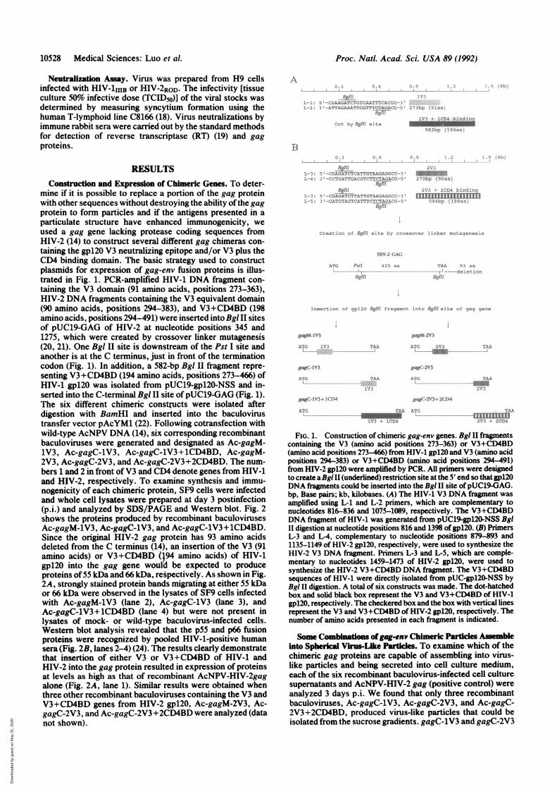
mine if it is possible to replace a portion of the gag proteinwith other sequences without destroying the ability of the gagprotein to form particles and if the antigens presented in aparticulate structure have enhanced immunogenicity, weused a gag gene lacking protease coding sequences fromHIV-2 (14) to construct several different gag chimeras con-taining the gpl20 V3 neutralizing epitope and/or V3 plus theCD4 binding domain. The basic strategy used to constructplasmids for expression of gag-env fusion proteins is illus-trated in Fig. 1. PCR-amplified HIV-1 DNA fragment con-taining the V3 domain (91 amino acids, positions 273-363),HIV-2 DNA fragments containing the V3 equivalent domain(90 amino acids, positions 294-383), and V3+CD4BD (198amino acids, positions 294-491) were inserted into Bgl II sitesof pUC19-GAG of HIV-2 at nucleotide positions 345 and1275, which were created by crossover linker mutagenesis(20, 21). One Bgi II site is downstream of the Pst I site andanother is at the C terminus, just' in front of the terminationcodon (Fig. 1). In addition, a 582-bp Bgl II fragment repre-senting V3+CD4BD (194 amino acids, positions 273-466) ofHIV-1 gpl20 was isolated from pUC19-gpl20-NSS and in-serted into the C-terminal Bgl II site of pUC19-GAG (Fig. 1).The six different chimeric constructs were isolated afterdigestion with BamHI and inserted into the baculovirustransfer vector pAcYM1 (22). Following cotransfection withwild-type AcNPV DNA (14), six corresponding recombinantbaculoviruses were generated and designated as Ac-gagM-1V3, Ac-gagC-1V3, Ac-gagC-lV3 + 1CD4BD, Ac-gagM-2V3, Ac-gagC-2V3, and Ac-gagC-2V3+2CD4BD. The num-bers 1 and 2 in front of V3 and CD4 denote genes from HIV-1and HIV-2, respectively. To examine synthesis and immu-nogenicity of each chimeric protein, SF9 cells were infectedand whole cell lysates were prepared at day 3 postinfection(p.i.) and analyzed by SDS/PAGE and' Western blot. Fig. 2shows the proteins produced by recombinant baculovirusesAc-gagM-1V3, Ac-gagC-lV3, and Ac-gagC-1V3+ 1CD4BD.Since the original HIV-2 gag protein has 93 amino acidsdeleted from the C terminus (14), an insertion of the V3 (91amino acids) or V3+CD4BD (194 amino acids) of HIV-1gpl20 into the gag gene would be expected to produceproteins of55 kDa and 66 kDa, respectively. As shown in Fig.2A, strongly stained protein bands migrating at either 55 kDaor 66 kDa were observed' in the lysates of SF9 cells infectedwith Ac-gagM-lV3 (lane 2), Ac-gagC-1V3 (lane 3), andAc-gagC-1V3+ 1CD4BD (lane 4) but were not present inlysates of mock- or wild-type baculovirus-infected cells.Western blot analysis revealed that the p55 and p66 fusionproteins were recognized by pooled HIV-1-positive humansera (Fig. 2B, lanes 2-4) (24). The results clearly demonstratethat insertion of either V3 or V3+CD4BD of HIV-1 andHIV-2 into the gag protein resulted in expression of proteinsat levels as high as that of recombinant AcNPV-HIV-2gagalone (Fig. 2A, lane 1). Similar results were obtained whenthree other recombinant baculoviruses containing the V3 andV3+CD4BD genes from HIV-2 gp120, Ac-gagM-2V3, Ac-gagC-2V3, and Ac-gagC-2V3+2CD4BD were analyzed (datanot shown).
A
_ l 'tGA.AGATCThGtCAAr A -I:'2-ATAtAAAT'FC ,TT'TCT.A-Ai-.
/ :;rIl L- v eiV.3 1CC4 L1C 1'
rxA 2'c- r, ; el
B
Bgll 2'vI _ --CGAgATCTCATTGTkAAuiic -
-4: -C'CTIIATT(,A.GT(.-TTCTAGAC- G-7(i---5 2brfzig/I!
fggll/I]-:'- CGAGATCT'l A -'rGTAAUA22ic<.:...
,AtGTAGTCA-TTCTCTAcAC'-figl
(. I edt -; 4/g/ll } ;.¾ S , " e; ':'¾. -: p ... ..-:¾.e c.
HIIV 4A.
Arctl;-:'A-A
liwr
gugM
MVIgagM- IV3
ATGi'C :I.I..
gaC:- IVT 3
ATG -ZAA
gagk AA :.
AL.,
A.l-'Am,
2's''n
oD
FIG. 1. Construction of chimeric gag-env genes. Bgi II fragmentscontaining the V3 (amino acid positions 273-363) or V3+CD4BD(amino acid positions 273-466) from HIV-1 gpl20 and V3 (amino acidpositions 294-383) or V3+CD4BD (amino acid positions 294-491)from HIV-2 gpl20 were amplified by PCR. AU primers were designedto create aBglII (underlined) restriction site at the 5' end so thatgpl20DNA fragments could be inserted into the BgI II site of pUC19-GAG.bp, Base pairs; kb, kilobases. (A) The HIV-1 V3 DNA fragment wasamplified using L-1 and L-2 primers, which are complementary tonucleotides 816-836 and 1075-1089, respectively. The V3+CD4BDDNA fragment of HIV-1 was generated from pUC19-gpl2O-NSS BgiII digestion at nucleotide positions 816 and 1398 ofgp120. (B) PrimersL-3 and L-4, complementary to nucleotide positions 879-893 and1135-1149 of HIV-2 gp120, respectively, were used to synthesize theHIV-2 V3 DNA fragment. Primers L-3 and L-5, which are comple-mentary to nucleotides 1459-1473 of HIV-2 gpl20, were used tosynthesize the HIV-2 V3+CD4BD DNA fragment. The V3+CD4BDsequences of HIV-1 were directly isolated from pUC-gpl2O-NSS byBgl II digestion. A total of six constructs was made. The dot-hatchedbox and solid black box represent the V3 and V3+CD4BD of HIV-1gp120, respectively. The checkered box and the box with vertical linesrepresent the V3 and V3+CD4BD of HIV-2 gp120, respectively. Thenumber of amino acids presented in each fragment is indicated.
Some Combinations ofgag-env Chimeric Particles Assembleinto Spherical Virus-Like Particles. To examine which of thechimeric gag proteins are capable of assembling into virus-like particles and being secreted into cell culture medium,each of the six recombinant baculovirus-infected cell culturesupernatants and AcNPV-HIV-2 gag (positive control) wereanalyzed 3 days p.i. We found that only three recombinantbaculoviruses, Ac-gagC-1V3, Ac-gagC-2V3, and Ac-gagC-2V3+2CD4BD, produced virus-like particles that could beisolated from the sucrose gradients. gagC-1V3 and gagC-2V3
Proc. NatL Acad. Sci. USA 89 (1992)
11 :-I:2 :-.
rrrrrtTTrrrrrmTrmI q 4 !:.,. -. q,
1'!"A!
nsert: o;- 1)1,'!' fr A.,. me.- tj f; , ':
gagC- IVA + 1C,D4
AIG
Dow
nloa
ded
by g
uest
on
May
31,
202
0

Proc. Natl. Acad. Sci. USA 89 (1992) 10529
M 1 2 3 4 C Wt M 1 2 3 4 C Wt
0|L'God--as-F 4iL L
D
M 1 2 3 4 Wt C
IW,
- 66-554339
FIG. 2. Expression of chimeric gag-env proteins in SF9 cellsinfected by recombinant baculoviruses. Recombinant baculovirus-infected SF9 cell lysates were prepared by resuspending the cellpellet in water and adding an equal volume of 2x dissociation buffer(10%o 2-mercaptoethanol/10%o SDS/25% glycerol/100 mM Tris-HCl,pH 7.0/0.04% bromophenol blue). (A) Cell lysates were analyzed byelectrophoresis in 12% polyacrylamide gels containing SDS (SDS/PAGE) and the protein bands were visualized by staining withCoomassie blue (23). (B) Western blot analyses were then performedusing human HIV-1 immunoglobulin obtained from the AcquiredImmunodeficiency Syndrome Research and Reference Reagent Pro-gram, Division of Acquired Immunodeficiency Syndrome, NationalInstitute of Allergy and Infectious Diseases (24). Lanes 1-4, recom-binant viruses AcNPV-HIV-2gag, Ac-gagM-1V3, Ac-gagC-1V3,and Ac-gagC-1V3+1CD4. The chimeric gag-env particles releasedinto the culture supernatant were purified by centrifugation in20-60% discontinuous sucrose density gradients, subjected to SDS/PAGE, and detected by Coomassie blue staining (C) and Westernblot (D) using human HIV-1 immunoglobulin (24). Lanes 1, purifiedgag particles; lanes 2-4, purified gagC-1V3, gagC-2V3, and gagC-2V3+2CD4 chimeric particles, respectively; lanes M, marker pro-teins; lanes C, uninfected cell control; lanes Wt, wild-type AcNPV-infected cells. The major fusion proteins p55 and p66 are indicated byarrows and possible cleavage products are indicated by open arrows.
particles were recovered from the 50% sucrose cushion,whereas gagC-2V3+2CD4BD chimeric particles banded ontop of the 60% sucrose cushion. These partially purifiedchimeric gag particles were analyzed by SDS/PAGE (Fig. 2C and D). The major fusion protein bands of 55 kDa and 66kDa were visualized by Coomassie blue staining (Fig. 2C)and by Western blot using HIV-1-positive human sera (Fig.2D). We examined the morphology of sucrose gradient-purified particles by transmission electron microscopy usinguranyl acetate staining (Fig. 3). HIV-2 gag particles werespherical (Fig. 3A), had diameters of -100 nm, and weresimilar to those of mature HIV-1 particles budding fromHIV-1-infected cells (25, 26). The chimeric particles gagC-1V3 (Fig. 3B), gagC-2V3 (Fig. 3C), and gagC-2V3+2CD4BD(Fig. 3D) exhibited morphologies similar to that of the gagparticles. The size of chimeric gag particles was slightlylarger than that of the gag particle alone, with approximatediameters of 130 nm, similar to the diameter ofmature HIV-1particles (27, 28).No virus-like particles were detected after infection with
Ac-gagM-1V3, Ac-gagM-2V3, or Ac-gagC-1V3+1CD4BDrecombinant baculoviruses, although the level of expressionwas high (data not shown), as previously reported (29). Ourresults demonstrate that insertion ofV3 into the middle ofthe
FIG. 3. Electron micrographs of sucrose gradient-purified gag-env chimeric particles. (A) HIV-2 gag particles produced by SF9cells infected with recombinant AcNPV-HIV-2 gag. (B) Chimericgag-env particles produced by recombinant Ac-gagC-1V3. (C) Chi-meric gag particles produced by recombinant Ac-gagC-2V3. (D)Chimeric gag particles produced by recombinant Ac-gagC-2V3+2CD4. Samples were stained with uranyl acetate. (Bars = 100nm.)
gag gene destroys the ability to form particles. This providesevidence that a large undisrupted N-terminal portion of thegag protein is essential for particle formation. Furthermore,there are likely to be size and sequence restrictions on theforeign protein that can be packaged into virus-like gagparticles (30).
Antigencity and Imuogenicity of Chimeric gag Partcles.Antigenicity of chimeric gag particles was investigated byimmunoblot analysis. Purified chimeric gag particles weresubjected to SDS/PAGE and analyzed by Western blottingwith rabbit antisera directed against HIV-1 and HIV-2 gp120and 1251-labeled protein A. As expected, the HIV-2 gagprotein was not recognized by anti-gp120 sera (Fig. 4, lane 1),whereas the nonglycosylated forms of gp120 of HIV-1 andHIV-2 showed strong reactivity with their correspondingantisera (Fig. 4, lanes 2). The 55-kDa and 66-kDa fusionproteins were specifically recognized by rabbit antiseraagainst HIV-1 or HIV-2 gpl2Os (Fig. 4, lanes 3 and 4). Theintensity of the band was proportional to the size of insertedheterologous polypeptide. Our results clearly demonstratethat the chimeric proteins can be detected by antiserum
A2 3 4 C W
B2 3 4 C W
rqua
36b --
*sl4w .;.r x
FIG. 4. Immunoblot analysis of chimeric gag-env proteins. Thechimeric gag-env proteins and gpl20 of HIV-1 and HIV-2 weresubjected to SDS/PAGE and electrotransferred to nitrocellulosefilters. Filters were incubated with rabbit antisera specific for HIV-1gp120 (A) and HIV-2 gp120 (B) and with 17-I-labeled protein A.,J.anes1, HIV-2 gag protein; lanes 2, gpl20 protein; lanes 3, chimericgagC-1V3 protein; lanes 4, chimeric gagC-1V3+1CD4 protein; lanesC, cell control; lanes W, wild-type AcNPV-infected cell control. Therabbit antisera against HIV-1 and HIV-2 gp12O will be describedelsewhere (Y.L., L.L., D. Thomas, and C.Y.K., unpublished data).
A B
97-66-42- _
31-
21- _
14-
CM 1 2 3 4 Wt C
97-66-
42- _ -_
31-
21- _
14- _
-M
r 11
4i.
I
Medical Sciences: Luo et al.
no
III- i 0*;r* 1, we
.7".7 -
.4imm
LN-l-,AA *
ask
Dow
nloa
ded
by g
uest
on
May
31,
202
0

10530 Medical Sciences: Luo et al.
specific for gp120 and reaffirm that inserted sequences ofV3or V3+CD4BD are antigenic.We also examined the capacity of particles to induce
antibodies to gag and env proteins ofHIV. The antisera madeagainst gagC-1V3 and gagC-2V3 chimeric particles in rabbitsrecognize not only carrier HIV-2 gag protein but also non-glycosylated gp120 of HIV-1 and HIV-2 (data not shown).These results clearly demonstrate that the V3 loop domain inchimeric gagC-1V3 and gagC-2V3 particles retains antigenicand immunogenic properties.Iinuune Sera Against Chimerlcgag-V3 Partles of HIV-l or
HIV-2 Neutralize Virus Infectivity in Vitro. Rabbit antiseradirected against chimeric particles were used to neutralize theinfectivity of HIV as assayed by RT activity and gag p24production. Rabbit anti-gagC-lV3 and anti-gagC-2V3 serawere mixed with either 5000TCID50 units ofHIV-lme or 8000TCID50 units of HIV-2RoD, respectively, and the mixtureswere used to infect H9 cells. The amount of p24 gag proteinand RT activity in the culture medium were assayed asquantitation of virus production on different days after in-fection (days 1-16), and the levels were compared with thoseof control samples in which virus was incubated with pre-immune serum or rabbit anti-gp120 serum.
Fig. 5 shows that rabbit anti-gagC-lV3 and anti-gagC-2V3sera contained antibodies capable of neutralizing HIV infec-tion of H9 cells. By day 9 p.i., antisera to gagC-1V3 andgagC-2V3 chimeric particles completely blocked the produc-tion of HIV-1 and HIV-2, respectively (Fig. 5). However, atday 12 p.i., a small amount ofHIV-1 was detected in culturestreated with anti-gagC-lV3 serum (Fig. 5 A and B). Incontrast, the antisera to gagC-2V3 chimeric particles com-pletely neutralized HIV-2 infectivity (Fig. 5 C and D). Rabbitanti-gpl20 sera of HIV-2 showed stronger neutralizing activ-
AP.1. AntiC(days) sera
1 2
13 2
6 2
3
19 2
12 231
BP.I. Anti(days) sera
11 2
313 2
3
16 2
3
19 2
112 2
3
RT Activity x 103 cpm/ml
2000 4000 6000 8000 10000
000
#1*Anti-lgpl2OAS9.7 #2 CAnti-gagC-1V30 #3 *Pre-immune5.2
400I178
6670
84 1
6 1 0 0
p24 Antigen (ng/ml.)
0 1000 2000
000
000
Proc. Natl. Acad. Sci. USA 89 (1992)
ity of HIV-2RoD than rabbit anti-gpl20 sera of HIV-1 againstHIV-1B (Fig. 5).
Papsidero et al. (1989) reported that the p17 gag protein ofHIV-1 contained several virus-neutralizing epitopes (31).Although this observation has not been confirmed by others,we tested the neutralization activity of rabbit antiserumdirected against HIV-2 gag particles alone. Our result clearlyshowed that the antisera to HIV-2 gag alone did not blockHIV-2 infectivity (data not shown). We conclude that theneutralizing activity induced by chineric gagC-V3 particlesis directed against the V3 domain but not to gag p17.
DISCUSSIONThe hypervariable V3 region ofHIV gpl20 induces the majorneutralizing antibodies (32-34) and stimulates helper andcytotoxic T-lymphocyte response in humans and mice (7, 8,35). Thus, this domain of gpl20 is an attractive candidate asa major component of an AIDS vaccine. Antigenic presen-tation of V3 region carried by virus-like gag particles couldbe advantageous for development ofa vaccine against AIDS.
In this study, we chose to use the truncated HIV-2 gagprotein to carry heterologous epitopes representing V3 loop(V3) or V3 plus CD4 binding domain (CD4BD) ofgpl20 fromHIV-1 and HIV-2. We found 91 amino acids representingHIV-1 V3 alone and 90 amino acids representing HIV-2 V3alone were efficiently packaged into virus-like particles, andvirus-neutralizing antibodies were generated in rabbits bythese particles. Furthermore, 198 amino acids representingHIV-2 V3+CD4BD were also packaged into the chimeric gagparticles. In contrast, 194 amino acids representing theV3+CD4BD of HIV-1 failed to be packaged into the gagparticles. Our results suggested that the homotypic proteinscan interact with each other to form particles, whereas
C PT Acti va t-v x pv:- -) : ,:
P.1 Anti 0 1000 20 C CO 4'::(days) sera -- ---- - -
4 0
6
#4 Anti-2gpiIA4 C #5 Anti-gagC-2V
6 5 #6 Pre-immune
4~C6
4 ..,
9 5 06 24
4
4 p3316 5 o
6 4:'.
Dp2 e Antigqer. noa
96 30
3050 P. I.days)
#1 f Anti-lgpl2OAS#2 Anti-gagC-1V,#3 U Pre-immune
00.070.08
21. 80.2
415.7
2104
1 2 659
Anti u i;vO C.!seraL~ -..
5
I
-.".
..
,.)..
-P4 §fi Ant .-< gp . :s i Anti -qaqC -;
* Pre- mmunc
L....
50
18.20O
12 56
416 5
6
FicG 5. Neutralization of HIV-1i1iB and HIV-2ROD infection with immune rabbit sera. Antisera against gagC-1V3 (HIV-1) and gagC-2V3(HIV-2) chimeric particles were prepared by the procedure of Luo et al. (14) and tested for neutralization of virus using RT and viral p24 orp26 assays. (A and B) HIV-1. (C and D) HIV-2. The neutralizing activities of anti-gagC-1V3 and anti-gagC-2V3 sera were determined byincubation of serum4:5 dilution) with a stock virus preparation of HIV-111IB (5000 TCID50 units) or HIV-2ROD (8000 TCID5o units) at 370C for1 hr before infecting H9 cells. Viral infection was monitored by RT activity (A and C) and the production of HIV-1 p24 (B) or HIV-2 p26 (D)gag proteins at 1-16 days p.i.
J.A
I
Dow
nloa
ded
by g
uest
on
May
31,
202
0

Proc. NatL Acad. Sci. USA 89 (1992) 10531
oversized heterotypic proteins cannot interact properly toform a particle. Insertion ofV3 sequences in the middle ofthegag gene prevented particle assembly, which suggests that anuninterrupted gag protein is necessary for particle assembly.The chimeric gag particles elicit neutralizing antibodies in
rabbits that completely block HIV infection but gag particlesalone do not. This result demonstrates that linear epitopes ofV3 are properly presented by the chimeric particles. Due tothe low yield of particle production, we did not prepareantiserum against the gagC-2V3+2CD4BD particles to de-termine whether or not these particles can induce neutralizingantibodies and/or blocking antibodies against CD4 binding.Because the conserved domain of the envelope proteins of
HIV-1 and HIV-2 are weakly immunogenic in native envelopeconfiguration (36) and viral antigens presented as componentsof membrane structures are more immunogenic than theirsoluble counterparts (37), chimeric particles of viral or subvi-ral components of polio virus, adenovirus, human hepatitis Bvirus, etc., containing HIV epitopes, have been evaluated.The major disadvantage of these approaches is the limitationof maximal acceptable size of inserted epitopes, the level ofexpression, and the complexity of purification method. In ourstudy, although we do not know yet the maximum sizelimitation ofpolypeptide extension that can be accommodatedin the virus-like gag particles, the largest of them, gagC-2V3+2CD4, has 198 amino acids attached to the C terminus,which makes a total of 623 amino acids as compared with 518amino acids of the normal gag precursor polypeptide. So far,this represents the largest size offoreign polypeptide packagedinto virus-like particles compared with other systems in whichthe capacity was limited to about 20-100 foreign amino acids.
Considering possible immune enhancement phenomena byantibodies against various epitopes of HIV envelope glyco-proteins (38, 39) and the superantigen and autoimmunitytheories to explain the depletion of uninfected CD4-positiveT lymphocytes (40, 41), the chimeric gag virus-like particlesprovide an attractive option to develop a recombinant subunitvaccine using defined epitopes that can induce humoral andcell-mediated immunity against HIV infection.
We thank Drs. K. Dimock and K. Wright for their critical andconstructive review of this manuscript and R. Nicholls for electronmicroscopy. We thank N. Delcellier and D. McLean for theirtechnical support. This work was supported by grants from theMedical Research Council of Canada, Korea Green Cross Corpora-tion, and Ontario Ministry of Colleges and Universities.
1. Berman, P. W., Gregory, T. J., Riddle, L., Nakamura, G. R.,Champe, M. A., Porter, J. P., Wurm, F. M., Hershberg, R. D.,Cobb, E. K. & Eichberg, J. W. (1990) Nature (London) 345, 622-625.
2. Girard, M., Kieny, M.-P., Pinter, A., Barre-Sinoussi, F., Nara, P.,Kolbe, H., Kusumi, K., Chaput, A., Reinhart, T., Muchmore, E.,Ronco, J., Kaczorek, M., Gomord, E., Gluckman, J.-C. & Fultz,P. N. (1991) Proc. Nati. Acad. Sci. USA 88, 542-546.
3. McDougal, J. S., Nicholson, J. K. A., Cross, G. D., Cort, S. P.,Kennedy, M. S. & Mawle, A. C. (1986) J. Immunol. 137, 2937-2944.
4. Putney, S. D., Matthews, T. J., Robey, W. G., Lynn, D. L., Rob-ert-Guroff, M., Mueller, W. T., Langlois, A. J., Ghrayeb, J., Pet-teway, S. R., Weinhold, K. J., Fischinger, P. J., Wong-Staal, F.,Gallo, R. C. & Bolognesi, D. P. (1986) Science 234, 1392-1395.
5. Robey, W. G., Arthur, L. O., Matthews, T. J., Langlois, A., Cope-land, T. D., Lerche, N. W., Oroszlan, S., Bolognesi, D. P., Gilden,R. V. & Fishinger, P. J. (1986) Proc. Nati. Acad. Sci. USA 83,7023-7027.
6. Broliden, P. A., Ljunggren, K., Hinkula, J., Norrby, E., Akerblom,L. & Wahren, B. (1990) J. Virol. 64, 936-940.
7. Takahashi, H., Cohen, J., Hosmalin, A., Cease, K. B., Houghten,R., Cornette, J., Delisi, C., Moss, B., Germain, R. N. & Berzofsky,J. A. (1988) Proc. Nati. Acad. Sci. USA 85, 3105-3109.
8. Takahashi, H., Nakagawa, Y., Pendleton, C. D., Houghten, R. A.,
Yokomuro, K., Germain, R. N. & Berzofsky, J. A. (1992) Science255, 333-336.
9. LaRosa, G. J., Davide, J. P., Weinhold, K., Waterbury, J. A.,Profy, A. T., Lewis, J. A., Langlois, A. J., Dreesman, G. R.,Boswell, R. N., Shadduck, P., Holley, L. H., Karpuls, M., Bo-lognesi, D. P., Matthews, T. J., Emini, E. A. & Putney, S. D.(1990) Science 249, 932-935.
10. Huang, M. L., Essex, M. & Lee, T.-H. (1991)J. Virol. 65,5073-5079.11. Bjorling, E., Broliden, K., Bernardi, D., Utter, G., Thorstensson,
R., Chiodi, F. & Norrby, E. (1991) Proc. Natl. Acad. Sci. USA 88,6082-6086.
12. Lasky, L. A., Nakamura, G., Smith, D. H., Fennie, C., Shimasaki,C., Patzer, E., Berman, P., Gregory, T. & Capon, D. J. (1987) Cell50, 975-985.
13. Wong-Staal, F. (1990) in Fields Virology (Raven, New York), 2ndEd., pp. P1529-1543.
14. Luo, L., Li, Y. & Kang, C. Y. (1990) Virology 179, 874-880.15. Kang, C. Y. (1988) Adv. Virus Res. 35, 177-192.16. Summers, M. D. & Smith, G. E. (1987) Tex. Agric. Exp. Stn. Bull.
1555.17. Smith, G. E. & Summers, M. D. (1978) Virology 89, 517-527.18. Nara, P. L., Hatch, W. C., Dunlop, N. M., Robey, W. G. &
Fischinger, P. J. (1987) AIDS Res. Hum. Retroviruses 3, 283-303.19. Poiesz, B. J., Ruscetti, F. W., Gazdar, A. F., Bunn, P. A., Minna,
J. D. & Gallo, R. C. (1980) Proc. NatI. Acad. Sci. USA 77,7415-7419.
20. Garson, K., Percival, H. & Kang, C. Y. (1990) Virology 177,106-115.
21. Sung, W. L., Zahab, D. M., MacDonald, C. A. & Tam, C. S. (1986)Gene 47, 261-267.
22. Matsuura, Y., Possee, R. D., Overton, H. A. & Bishop, D. H. L.(1987) J. Gen. Virol. 68, 1233-1250.
23. Laemmli, U. K. (1970) Nature (London) 227, 680-685.24. Prince, A. M., Harowitz, B., Baker, L., Shulman, R. W., Ralph,
H., Valinsky, J., Cundell, A., Brotman, B., Boehle, W., Roy, F.,Piet, M., Reesink, H., Lelie, N., Termette, M., Miedema, F.,Barbosa, L., Nemo, G., Nastala, C. L., Allan, J. S., Lee, D. R. &Eichberg, J. W. (1988) Proc. Nall. Acad. Sci. USA 85, 6944-6948.
25. Gelderblom, H. R., Hausmann, E. H. S., Ozel, M., Pauli, G. &Koch, M. A. (1987) Virology 156, 171-176.
26. Gelderblom, H. R., Ozel, M., Hausmann, E. H. S., Winkel, T.,Pauli, G. & Koch, M. A. (1988) Micron Microsc. Acta 19, 41-60.
27. Schupbach, J., Popovic, M., Gilden, R. V., Gonda, M. A., Sarn-gadharan, M. G. & Gallo, R. C. (1984) Science 224, 503-505.
28. Chrystie, I. L. & Almeida, J. D. (1988) J. Med. Virol. 25, 281-288.29. Morikawa, S., Booth, T. F. & Bishop, D. H. L. (1991) Virology
183, 288-297.30. Weldon, R. A., Jr., Erdie, C. R., Oliver, M. G. & Wills, J. W.
(1990) J. Virol. 64, 4169-4179.31. Papsidero, L. D., Sheu, M. & Ruscetti, F. W. (1989) J. Virol. 63,
267-272.32. Rusche, J. R., Javaherian, K., McDanal, C., Petro, J., Lynn, D. L.,
Grimaila, R., Langlois, A., Gall, R. C., Arthur, L. O., Fischinger,P. J., Bolognesi, D. P., Putney, S. D. & Matthews, T. J. (1988)Proc. Natl. Acad. Sci. USA 85, 3198-3202.
33. Palker, T. J., Clark, M. E., Langlois, A. J., Matthews, T. J., Wein-hold, K. J., Randall, R. R., Bolognesi, D. P. & Haynes, B. F.(1988) Proc. Natl. Acad. Sci. USA 85, 1932-1936.
34. Goudsmit, J., Debouck, C., Meloen, R. H., Smit, L., Bakker, M.,Asher, D. M., Wolff, A. V., Gibbs, C. J., Jr., & Gajdusek, D. C.(1988) Proc. Natl. Acad. Sci. USA 85, 4478 4482.
35. Orentas, R. J., Hildreth, J. E. K., Obah, E., Polydefkis, M., Smith,G. E., Clements, M. L. & Siliciano, R. F. (1990) Science 248,1234-1237.
36. Guyader, M., Emerman, M., Sonigo, P., Clavel, F., Montagnier, L.& Alizon, M. (1987) Nature (London) 326, 662-669.
37. Ho, R. J. Y., Burke, R.-L. & Merigan, T. C. (1989) J. Virol. 63,2951-2958.
38. Homsy, J., Meyer, M., Tateno, M., Clarkson, S. & Levy, J. A.(1989) Science 244, 1357-1360.
39. Robinson, W. E., Jr., Kawamura, T., Gorny, M. K., Lake, D., Xu,J. Y., Matsumoto, Y., Sugano, T., Masuho, Y., Mitchell, W. M.,Hersch, E. & Zolla-Pazner, S. (1990) Proc. Natl. Acad. Sci. USA87, 3185-3189.
40. Imberti, L., Scottiri, A., Bettinardi, A., Pouti, M. & Primi, D. (1991)Science 254, 860-862.
41. Kion, T. A. & Hoffmann, G. W. (1991) Science 253, 1138-1140.
Medical Sciences: Luo et al.
Dow
nloa
ded
by g
uest
on
May
31,
202
0