Chest Film Primer Images and Material courtesy of: David S. Feigin, M.D. Colonel (Ret), USA, MC...
37
Chest Film Primer Chest Film Primer Images and Material Images and Material courtesy of: courtesy of: David S. Feigin, M.D. David S. Feigin, M.D. Colonel (Ret), USA, MC Colonel (Ret), USA, MC Professor of Radiology, Professor of Radiology, Uniformed Services University of the Health Uniformed Services University of the Health Sciences Sciences “ “ A Systematic Approach To Abnormal Chest Images: A Systematic Approach To Abnormal Chest Images: Radiographs And Computed Tomograms” Radiographs And Computed Tomograms” Updates and compilation: Updates and compilation: Les Folio, DO, MPH Les Folio, DO, MPH Col, USAF, MC, SFS Col, USAF, MC, SFS Associate Professor of Radiology, Associate Professor of Radiology, Uniformed Services University of the Health Uniformed Services University of the Health Sciences Sciences
-
Upload
willow-treadaway -
Category
Documents
-
view
214 -
download
1
Transcript of Chest Film Primer Images and Material courtesy of: David S. Feigin, M.D. Colonel (Ret), USA, MC...

Chest Film PrimerChest Film Primer Images and Material Images and Material
courtesy of:courtesy of:David S. Feigin, M.D. David S. Feigin, M.D. Colonel (Ret), USA, MCColonel (Ret), USA, MCProfessor of Radiology, Professor of Radiology,
Uniformed Services University of the Health SciencesUniformed Services University of the Health Sciences““A Systematic Approach To Abnormal Chest A Systematic Approach To Abnormal Chest
Images:Images:Radiographs And Computed Tomograms”Radiographs And Computed Tomograms”
Updates and compilation:Updates and compilation:Les Folio, DO, MPHLes Folio, DO, MPHCol, USAF, MC, SFSCol, USAF, MC, SFS
Associate Professor of Radiology, Associate Professor of Radiology, Uniformed Services University of the Health SciencesUniformed Services University of the Health Sciences

Main MenuMain Menu5 PATTERNS OF
PATHOLOGY
1. Mass
2. Consolidative
3. Interstitial- Linear- Nodular
4. Vascular
5. Airway- Wall-Thickened- Obstructive
EXIT
NORMAL ANATOMY
1. A-P Chest Radiograph
2. Lateral Chest Radiograph
THE SEARCH PATTERN
1. A-P Chest Radiograph
2. Lateral Chest Radiograph
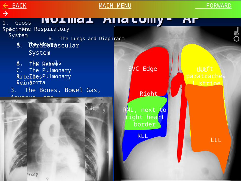
Normal Anatomy- APNormal Anatomy- AP BACK MAIN MENU FORWARD
1. Gross Specimen
3. Cardiovascular System
A. The CavalsB. The HeartC. The Pulmonary ArteriesE. Aorta
2. The Respiratory System
A. The Airway B. The Lungs and Diaphragm
D. The Pulmonary Veins
3. The Bones, Bowel Gas, Azygous, etc.
RUL, next to minor fissure
RLL
SVC Edge Left paratracheal
stripe
Right Atrium
Left Ventricle
LLL
LUL
RML, next to right heart
border

Normal Anatomy- LateralNormal Anatomy- Lateral BACK MAIN MENU FORWARD
2. Cardiovascular System
1. Respiratory System
A. The AirwayB. The Lungs and Diaphragm
A. The VesselsB. The Heart
3. The Bones (no link yet)
Trachea
Left main bronchus
RLL, with major fissure
RML, with minor fissure
RUL
Right Pulmonary
Artery
Left Pulmonary
Artery
Inferior Vena Cava
Left Ventricle
Right Ventricle
Aorta

1.1. SVC EdgeSVC Edge2.2. Rt Paratracheal LineRt Paratracheal Line3.3. Lt Paratracheal StripeLt Paratracheal Stripe
(both red and black lines)(both red and black lines)
4.4. Aortic ArchAortic Arch5.5. Descending AortaDescending Aorta
(only left edge seen, and not (only left edge seen, and not always)always)
6.6. Rt AtriumRt Atrium7.7. Azygoesophageal edgeAzygoesophageal edge8.8. Lt VentricleLt Ventricle9.9. Main Pulmonary ArteryMain Pulmonary Artery
AKA: trunk, middle mogulAKA: trunk, middle mogul
Mediastinum Mid
Quiz yourself: Mediastinum Lines, Edges
1
2
3
4
56
78
9
Recommendation: Test yourself before advancing to the answers

Left VentricleLeft Ventricle
(curved line)(curved line)
Lateral
IVC (arrows)
Right Pulmonary Artery Right Pulmonary Artery (red) (red)
Left Pulmonary Artery (green)
TracheaTrachea
Lt MSB on endLt MSB on end
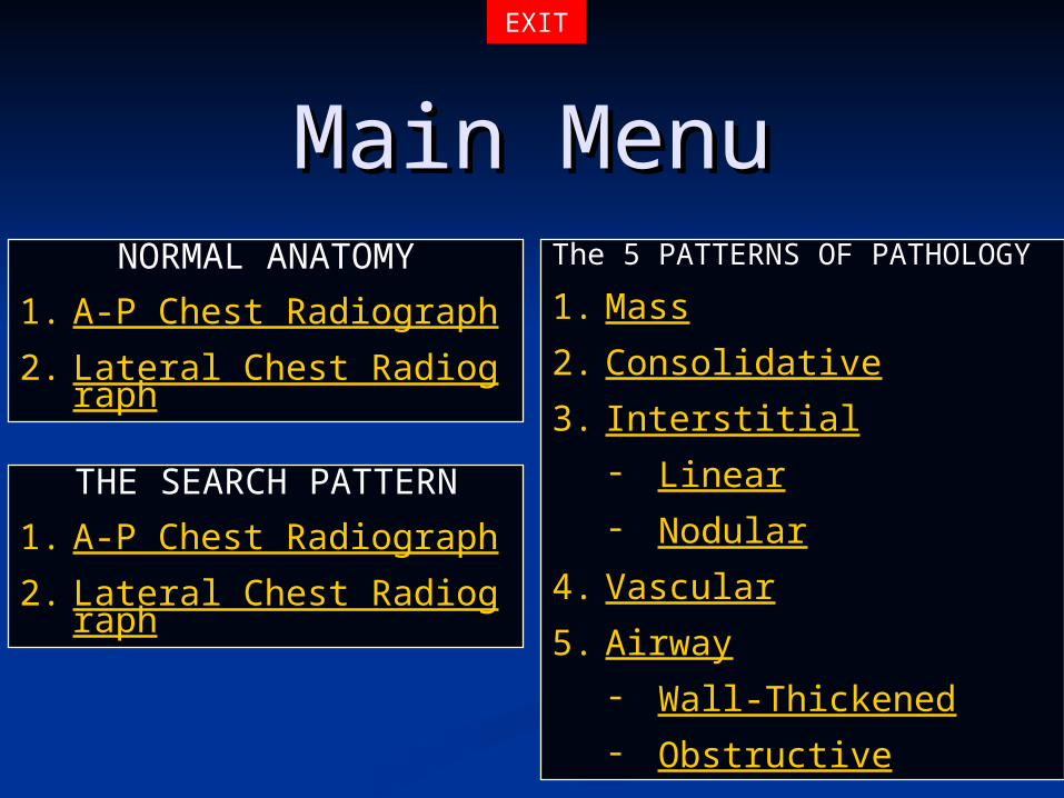
Main MenuMain MenuThe 5 PATTERNS OF
PATHOLOGY
1. Mass
2. Consolidative
3. Interstitial- Linear- Nodular
4. Vascular
5. Airway- Wall-Thickened- Obstructive
EXIT
NORMAL ANATOMY
1. A-P Chest Radiograph
2. Lateral Chest Radiograph
THE SEARCH PATTERN
1. A-P Chest Radiograph
2. Lateral Chest Radiograph
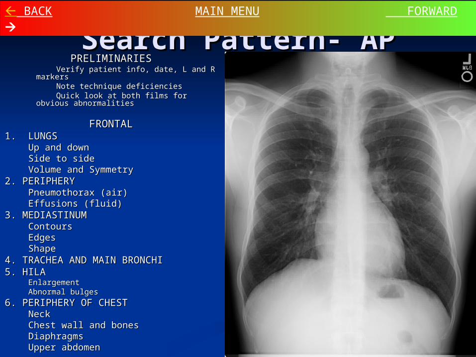
Search Pattern- APSearch Pattern- AP BACK MAIN MENU FORWARD
PRELIMINARIESPRELIMINARIES Verify patient info, date, L and R markersVerify patient info, date, L and R markers Note technique deficienciesNote technique deficiencies Quick look at both films for obvious Quick look at both films for obvious
abnormalitiesabnormalities
FRONTALFRONTAL1. LUNGS1. LUNGS
Up and downUp and downSide to sideSide to sideVolume and SymmetryVolume and Symmetry
2. PERIPHERY2. PERIPHERYPneumothorax (air)Pneumothorax (air)Effusions (fluid)Effusions (fluid)
3. MEDIASTINUM3. MEDIASTINUMContoursContoursEdgesEdgesShapeShape
4. TRACHEA AND MAIN BRONCHI4. TRACHEA AND MAIN BRONCHI5. HILA5. HILA
EnlargementEnlargementAbnormal bulgesAbnormal bulges
6. PERIPHERY OF CHEST6. PERIPHERY OF CHESTNeckNeckChest wall and bonesChest wall and bonesDiaphragmsDiaphragmsUpper abdomenUpper abdomen
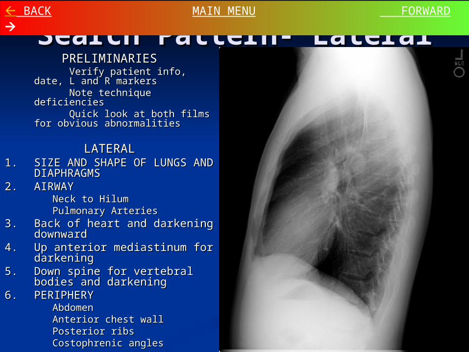
Search Pattern- LateralSearch Pattern- Lateral BACK MAIN MENU FORWARD
PRELIMINARIESPRELIMINARIES Verify patient info, date, L and R Verify patient info, date, L and R
markersmarkers Note technique deficienciesNote technique deficiencies Quick look at both films for Quick look at both films for
obvious abnormalitiesobvious abnormalities
LATERALLATERAL1.1. SIZE AND SHAPE OF SIZE AND SHAPE OF
LUNGS AND DIAPHRAGMSLUNGS AND DIAPHRAGMS2.2. AIRWAYAIRWAY
Neck to HilumNeck to HilumPulmonary ArteriesPulmonary Arteries
3.3. Back of heart and darkening Back of heart and darkening downwarddownward
4.4. Up anterior mediastinum for Up anterior mediastinum for darkeningdarkening
5.5. Down spine for vertebral Down spine for vertebral bodies and darkeningbodies and darkening
6.6. PERIPHERYPERIPHERYAbdomenAbdomenAnterior chest wallAnterior chest wallPosterior ribsPosterior ribsCostophrenic anglesCostophrenic angles

Main MenuMain Menu5 PATTERNS OF
PATHOLOGY
1. Mass
2. Consolidative
3. Interstitial- Linear- Nodular
4. Vascular
5. Airway- Wall-Thickened- Obstructive
EXIT
NORMAL ANATOMY
1. A-P Chest Radiograph
2. Lateral Chest Radiograph
THE SEARCH PATTERN
1. A-P Chest Radiograph
2. Lateral Chest Radiograph
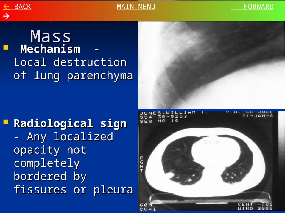
1. Mass1. Mass MechanismMechanism - Local - Local
destruction of lung destruction of lung parenchymaparenchyma
Radiological signRadiological sign - - Any localized Any localized opacity not opacity not completely bordered completely bordered by by fissures or pleurafissures or pleura
BACK MAIN MENU FORWARD

1. Mass1. Mass Differential DiagnosisDifferential Diagnosis
MalignancyMalignancy - Primary or secondary - Primary or secondary Granulomatous diseaseGranulomatous disease - Infectious or - Infectious or
noninfectious, active or inactive noninfectious, active or inactive Other Other inflammation, including pneumonia and inflammation, including pneumonia and
abscess, Benign neoplasm, Congenital abnormalityabscess, Benign neoplasm, Congenital abnormality
Crucial appearance characteristics for Crucial appearance characteristics for inactivityinactivity Calcification – central, lamellarCalcification – central, lamellar Evolution – 2-year stability or regressionEvolution – 2-year stability or regression
BACK MAIN MENU FORWARD
2. Consolidative (Alveolar) 2. Consolidative (Alveolar) PatternPattern
MechanismMechanism Produced in pure form and by Produced in pure form and by ALVEOLAR ALVEOLAR
FILLINGFILLING May be mimicked by alveolar collapse, as in airway May be mimicked by alveolar collapse, as in airway
obstructionobstruction Rarely, confluent interstitial thickeningRarely, confluent interstitial thickening
Radiological signsRadiological signs Fluffy, cloud-like, coalescent opacitiesFluffy, cloud-like, coalescent opacities Sharp edges when limited by fissures or pleuraSharp edges when limited by fissures or pleura Complete air bronchograms through the cloudsComplete air bronchograms through the clouds
BACK MAIN MENU FORWARD
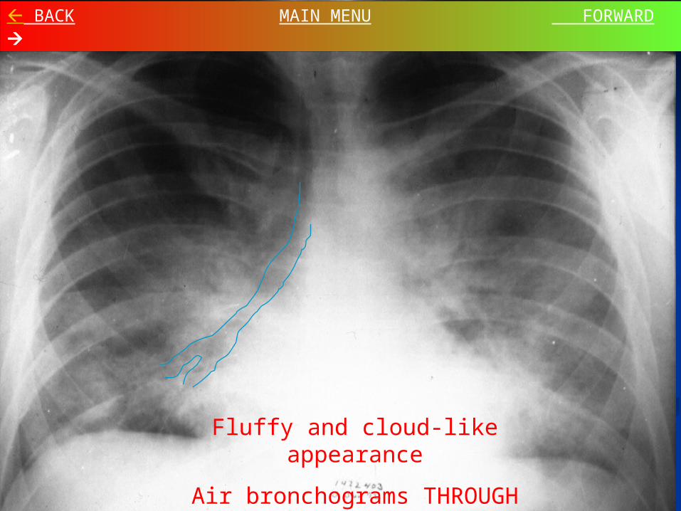
Fluffy and cloud-like appearance
Air bronchograms THROUGH clouds
BACK MAIN MENU FORWARD
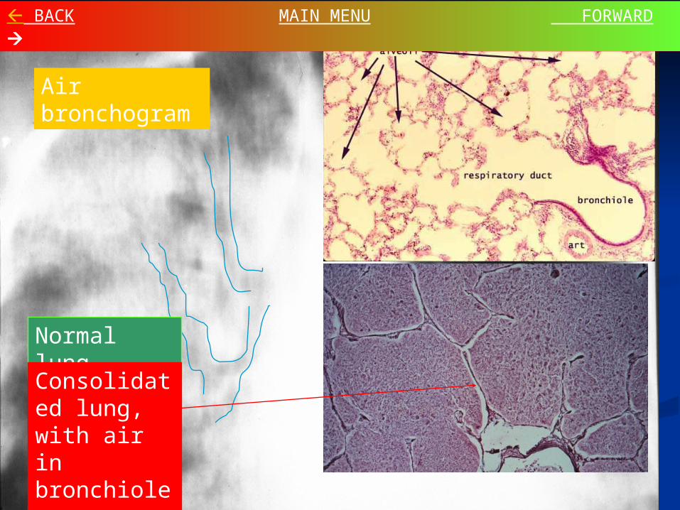
Air bronchogram
Normal lung
Consolidated lung, with air in bronchioles
BACK MAIN MENU FORWARD

2. Consolidative (alveolar) 2. Consolidative (alveolar) PatternPattern
Differential Diagnosis (5 general)Differential Diagnosis (5 general) HemorrhageHemorrhage - - BLOODBLOOD - embolism, trauma - embolism, trauma ExudateExudate - - PUSPUS - pneumonia, pneumonitis - pneumonia, pneumonitis TransudateTransudate - - WATERWATER - congestion, ARDS - congestion, ARDS SecretionsSecretions - - PROTEINPROTEIN - Mucous plugging, - Mucous plugging,
Alveolar proteinosisAlveolar proteinosis MalignancyMalignancy - - CELLSCELLS - Alveolar cell - Alveolar cell
carcinoma, Lymphomacarcinoma, Lymphoma
BACK MAIN MENU FORWARD

3. Interstitial Pattern3. Interstitial Pattern
Composition of pulmonary interstitium:Composition of pulmonary interstitium: Alveolar walls, septiAlveolar walls, septi Connective tissue surrounding bronchi Connective tissue surrounding bronchi
and vessels (peribronchial and and vessels (peribronchial and perivascular spaces) perivascular spaces)
MechanismMechanism Thickening of lung intersticesThickening of lung interstices Architectural destruction of interstitium Architectural destruction of interstitium
(honeycomb or “end stage” lung)(honeycomb or “end stage” lung)
BACK MAIN MENU FORWARD

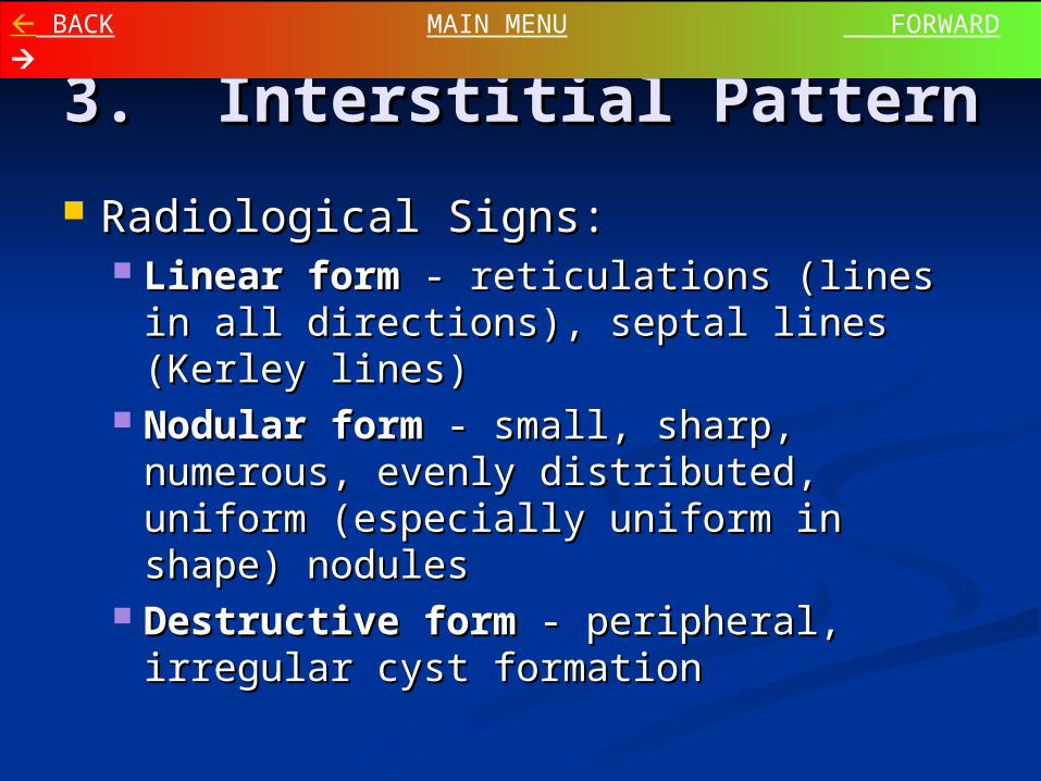
3. Interstitial Pattern3. Interstitial Pattern
Radiological Signs:Radiological Signs: Linear formLinear form - reticulations (lines in all - reticulations (lines in all
directions), septal lines (Kerley lines)directions), septal lines (Kerley lines) Nodular formNodular form - small, sharp, - small, sharp,
numerous, evenly distributed, uniform numerous, evenly distributed, uniform (especially uniform in shape) nodules(especially uniform in shape) nodules
Destructive formDestructive form - peripheral, - peripheral, irregular cyst formationirregular cyst formation
BACK MAIN MENU FORWARD
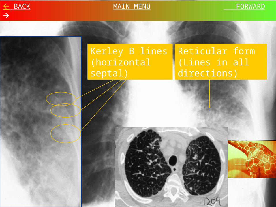
Reticular form (Lines in all directions)
Kerley B lines (horizontal septal)
BACK MAIN MENU FORWARD
3. Interstitial Pattern – 3. Interstitial Pattern – Linear FormLinear Form
Differential Diagnosis: The “LIFE Differential Diagnosis: The “LIFE Lines”Lines” Lymphangitic spread of malignancyLymphangitic spread of malignancy InflammationInflammation FibrosisFibrosis EdemaEdema
BACK MAIN MENU FORWARD

3. Interstitial Pattern3. Interstitial Pattern
Radiological Signs:Radiological Signs: Linear formLinear form - reticulations (lines in all - reticulations (lines in all
directions), septal lines (Kerley lines)directions), septal lines (Kerley lines) Nodular formNodular form - small, sharp, - small, sharp,
numerous, evenly distributed, uniform numerous, evenly distributed, uniform (especially uniform in shape) nodules(especially uniform in shape) nodules
Destructive formDestructive form - peripheral, - peripheral, irregular cyst formationirregular cyst formation
BACK MAIN MENU FORWARD

Multiple small nodules, uniform in shape and distribution
BACK MAIN MENU FORWARD
3. Interstitial Pattern- 3. Interstitial Pattern- Nodular FormNodular Form
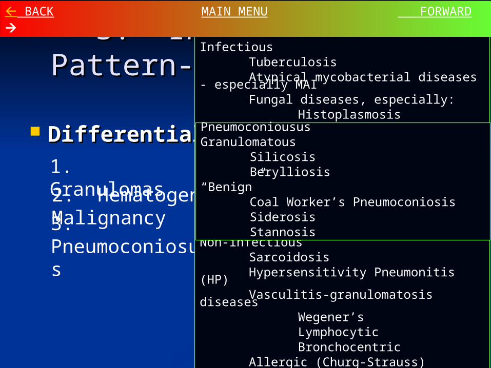
Differential DiagnosisDifferential Diagnosis1. Granulomas
2. Hematogenous Spread of Malignancy3. Pneumoconiosus
Granulomatous Diseases:Infectious
TuberculosisAtypical mycobacterial diseases -
especially MAIFungal diseases, especially:
HistoplasmosisCoccidioidomycosisBlastomycosis (N. A. and S.
A.)CryptococcosisSporotrichosis
Bacterial diseases, especially:NocardiosisActinomycosis
Non-infectiousSarcoidosisHypersensitivity Pneumonitis (HP)Vasculitis-granulomatosis diseases
Wegener’sLymphocyticBronchocentric
Allergic (Churg-Strauss)Langerhans Granulomatosis
(eosinophilic granuloma, histiocytosis)
(LCG)
PneumoconioususGranulomatous
SilicosisBerylliosis
“Benign”Coal Worker’s PneumoconiosisSiderosisStannosis
BACK MAIN MENU FORWARD
3. Interstitial Pattern3. Interstitial Pattern
Radiological Signs:Radiological Signs: Linear formLinear form - reticulations (lines in all - reticulations (lines in all
directions), septal lines (Kerley lines)directions), septal lines (Kerley lines) Nodular formNodular form - small, sharp, - small, sharp,
numerous, evenly distributed, uniform numerous, evenly distributed, uniform (especially uniform in shape) nodules(especially uniform in shape) nodules
Destructive formDestructive form - peripheral, - peripheral, irregular cyst formationirregular cyst formation
BACK MAIN MENU FORWARD

Peripheral cyst formation, ‘Honeycomb’ lung
Early findings are non-specific. The
peripheral cyst formation (“End-Stage Lung”) is a
late finding.
BACK MAIN MENU FORWARD
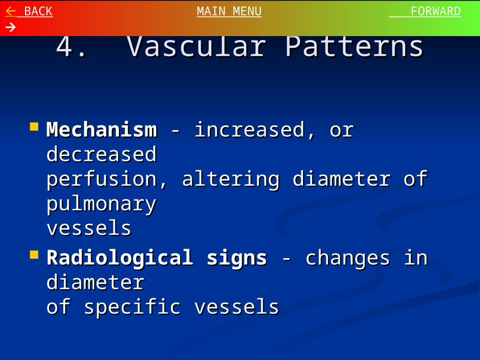
4. Vascular Patterns4. Vascular Patterns
MechanismMechanism - increased, or - increased, or decreaseddecreasedperfusion, altering diameter of perfusion, altering diameter of pulmonarypulmonaryvesselsvessels
Radiological signs Radiological signs - changes in - changes in diameterdiameterof specific vesselsof specific vessels
BACK MAIN MENU FORWARD

4. Vascular Patterns4. Vascular Patterns
Common examplesCommon examples CongestionCongestion - engorged veins, especially upper lungs- engorged veins, especially upper lungs EmphysemaEmphysema - diminished vessels - diminished vessels Shunt vascularityShunt vascularity - all vessels enlarged - all vessels enlarged Lymphangitic carcinomaLymphangitic carcinoma - irregular infiltration - irregular infiltration
around vessels may resemble vessel enlargementaround vessels may resemble vessel enlargement Arterial hypertensionArterial hypertension - large central arteries with - large central arteries with
peripheral taperingperipheral tapering ThromboembolismThromboembolism - locally diminished vessels with - locally diminished vessels with
possible vessel mass centrally locatedpossible vessel mass centrally located Bronchial circulationBronchial circulation - irregular vessels in unusual - irregular vessels in unusual
directionsdirections
BACK MAIN MENU FORWARD

Congested vasculature
Engorged vessels, especially upper lungs
BACK MAIN MENU FORWARD

Emphysematous changes
Diminished vasculature
BACK MAIN MENU FORWARD
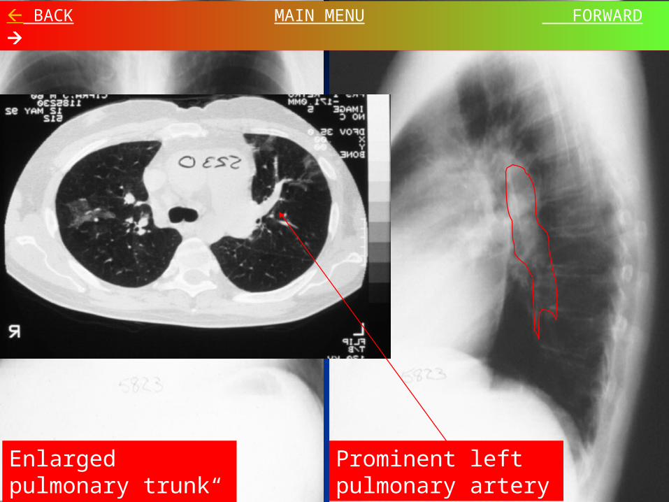
Enlarged pulmonary trunk (“middle mogul”)
Prominent left pulmonary artery
BACK MAIN MENU FORWARD

5. Airway (Bronchial) 5. Airway (Bronchial) PatternsPatterns
MechanismMechanism Complete or partial obstruction of airwaysComplete or partial obstruction of airways Thickening of airway wallsThickening of airway walls
FormsForms Complete airway obstructionComplete airway obstruction - opacity and - opacity and
decreased volumedecreased volume Partial obstructionPartial obstruction - lucency and increased - lucency and increased
volumevolume Wall thickeningWall thickening - tram tracks, central cystic - tram tracks, central cystic
spaces or circlesspaces or circles
BACK MAIN MENU FORWARD

Bronchial wall thickening (circles and “tram tracks”)
Flattened diaphragms on lateral
BACK MAIN MENU FORWARD

Differential diagnosisDifferential diagnosis OpacitiesOpacities - endobronchial malignancies, - endobronchial malignancies,
granulomas, inflammatory, benign or granulomas, inflammatory, benign or congenital masses, mucous plugs, foreign congenital masses, mucous plugs, foreign bodiesbodies
LucenciesLucencies - COPD, cysts, blebs, - COPD, cysts, blebs, pneumatocelespneumatoceles
ThickeningThickening - bronchiectasis, chronic - bronchiectasis, chronic bronchitisbronchitis
BACK MAIN MENU FORWARD 5. Airway (Bronchial) 5. Airway (Bronchial)
PatternsPatterns

5. Airway (Bronchial) 5. Airway (Bronchial) PatternsPatterns
Lobar atelectasis (collapse)Lobar atelectasis (collapse) Primary SignsPrimary Signs
Vessel number assymetryVessel number assymetry Fissure as edgeFissure as edge
Secondary signsSecondary signs Volume lossVolume loss Elevation of diaphragmElevation of diaphragm Shift of mediastinum and ribsShift of mediastinum and ribs
BACK MAIN MENU FORWARD

Right Left
Upper
Lower
Atelectasis Atelectasis PatternsPatterns
RLL
RUL
LLL
LUL
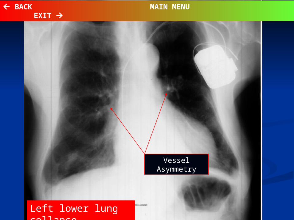
Left lower lung collapse
Vessel Asymmetry
BACK MAIN MENU EXIT