Preparation and Characterization of Liposomal Everolimus ...
Tumor metastasis occurs through such steps as dissocia-tion of tumor cells from the primary site, their adhesion tothe extracellular matrix in the target organ, and invasion ofthe cells to make metastatic colonization. The malignancy oftumor cells is one of the factors to decide metastatic poten-tial, but the level of host defense is another one because a de-crease in host defense may produce an appropriate environ-ment for tumor metastasis. We previously demonstrated thatthe intravenous injection of a lower number of metastatictumor cells failed to form metastatic colonies in the targetorgan, whereas a larger number completed tumor metasta-sis.1) In addition, pre-treatment of 2-chloroadenosine, whichweakened the mouse’s immune strength, impaired the initialstage of tumor metastasis fewer tumor cells were injected.2)
These findings indicate that immune surveillance in the hostplays an important role in the initial phase of tumor metasta-sis. Thus, increasing host defense may prevent tumor cellsfrom metastasizing.
Phytosterol is a constituent component of plant cells. It hasbeen estimated that the Asian diet contains 300—400 mg ofphytosterol per day.3) Although phytosterol comes in manydifferent forms, the most abundant phytosterols is b-sito-sterol (Sito). On the other hand, recent research on phyto-sterols has reported that they lower plasma cholesterol con-centration.4,5) They also have various biological functionssuch as prevention of prostatauxe,6) amelioration of diabetes7)
and anti-cancer effects.8—13) However, since phytosterol areinsoluble or poorly soluble in water, problems exist regardingthe development of administration methods and carriers. Liposomes are spherical vesicles whose membranes consistof phospholipids and/or sterols, and are good candidate forformulating Sito to overcome the problem.
Liposomes can be utilized as drug carriers for a variety ofsubstances such as small molecular drugs, proteins, nu-cleotides and plasmids. Some studies have already demon-strated that liposomalization of therapeutic and cosmeticagents can enhance their activity by improving their stability
and permeability as well as, giving targeting ability and con-trolling the release of entrapped agents. Some proteins, suchas insulin,14,15) calsitonin,16,17) and erythropoietin,18) whichare can be digested and have low permeability through intes-tinal membrane, have been shown to improve their pharma-cological effects through oral administration by various for-mulations of liposomes. We previously showed that oral ad-ministration of liposomal lactoferrin enhanced host immuneability and anti-inflammatory effects.19,20)
In this study, Sito was used as a constituent of liposomewhich is prepared for the purpose of the oral delivery of Sito.We examined adhesive behavior of the liposome to mucosaand its absorption into plasma. Furthermore, we confirmedthat oral administration of liposomal Sito induced IL-18 pro-duction in the small intestine, which may modulate immuno-logic function, and enhanced NK activity. Additionally, lipo-somal Sito intake caused the suppression of lung metastaticcolonization in an experimental metastasis model.
MATERIALS AND METHODS
Materials Egg phosphatidylcholine (EPC, Sigma, St.Louis, MO, U.S.A.), b-sitosterol (Sito, Tama Seikagaku,Tokyo, Japan), cholesterol (Chol, Sigma), 1,1�-dioctadecyl-3,3,3�,3�-tetramethylindo carbocyanine perchlorate (DiI,Lambda Probes & Diagnostics, Graz, Austria) and otherchemical materials were analytical grades and purchasedfrom local supplier and used as received. Radio labeled [4-14C] b-sitosterol and [4-14C] cholesterol were purchasedfrom American Radiolabeled Chemicals Inc. (St. Louis, MO,U.S.A.).
Preparation of Liposomes Liposomes composed ofEPC and Sito or Chol in a molar ratio of 2 : 1 were preparedby a thin lipid layer method.21) In brief, EPC and Sito or Cholwere dissolved in a small amount of chloroform, and the sol-vent was rotary evaporated at 40 °C to obtain a thin lipidfilm. The thin lipid film was dried in a vacuum oven
400 Vol. 31, No. 3
Chemoprevention of Tumor Metastasis by Liposomal bb-Sitosterol Intake
Hiromichi IMANAKA,a,b Hiroyuki KOIDE,a Kosuke SHIMIZU,a Tomohiro ASAI,a
Naomi KINOUCHI SHIMIZU,a Atsushi ISHIKADO,b Taketoshi MAKINO,b and Naoto OKU*,a,c
a Department of Medical Biochemistry, Graduate School of Pharmaceutical Sciences, University of Shizuoka; c GlobalCOE Program, University of Shizuoka; 52–1 Yada, Suruga-ku, Shizuoka 422–8526, Japan: and b R&D Division, SunstarInc.; 3–1 Asahi-machi, Takatsuki, Osaka 569–1195, Japan.Received October 22, 2007; accepted December 10, 2007; published online December 14, 2007
To investigate chemopreventive effect of liposomal bb-sitosterol on tumor metastasis, we prepared liposomalbb-sitosterol composed of egg yolk phosphatidylcholine for oral delivery. Although orally administered bb-sito-sterol (4 mmmol as bb-sitosterol/mouse) was not absorbed into plasma, the amount of immune response cytokinessuch as IL-12 and IL-18 was increased in the small intestine after the liposome intake. Moreover, after daily oraladministration of the liposome for 7 d, natural killer (NK) cell activity in the mice was increased, suggesting thatthe immune surveillance activity of mice was enhanced by the liposomal bb-sitosterol intake. Thus, we examinedmetastatic potential of B16BL6 melanoma cells, which were intravenously injected into mice after sequential ad-ministration of liposomal bb-sitosterol for 7 d. The number of metastatic colonies in the lungs was significantlyless than that of control group two weeks after the injections of the cells. These results suggest that daily liposo-mal bb-sitosterol intake prevents tumor metastasis may be due to enhancement of gut immune surveillance sys-tems.
Key words liposome; sitosterol; oral delivery; tumor metastasis; chemoprevention
Biol. Pharm. Bull. 31(3) 400—404 (2008)
© 2008 Pharmaceutical Society of Japan∗ To whom correspondence should be addressed. e-mail: [email protected]

overnight to ensure complete removal of the solvent, andthen hydrated by phosphate-buffered saline (PBS, pH 7.0) byvoltexing at 50 °C. The obtained liposomal solution was sub-jected to 5 freeze and thaw cycles by liquid N2 and warmwater bath, respectively. Then liposomal suspension wassized at about 100 nm by extrusion. The particle size of theliposome was confirmed by dynamic light scattering analysiswith Zetasizer Nano (Malvern instruments, Malvern, U.K.).Fluorescence reagent: DiI and [14C]-labeled Sito or Cholwere added to each liposome as needed.
Absorption and Distribution Assay of bb-SitosterolFive-week-old C57BL/6 male mice which had fasted 24 h be-fore administration were orally administered liposomal Sito(LS) or liposomal Chol (LC) (4 mmol as sterols/mouse). Theselected organs were excised from the mice sacrificed 1.5, 3and 6 h after administration of the liposomes and thenwashed with a large amount of saline solution. The amountof [14C]-labeled Sito and Chol in the organs was measured bya liquid scintillation counter.
Observation of Mucoadhesive Behavior of LiposomesThe experimental design followed the method of Takeuchi etal.,22) Liposomes containing DiI were administered intragas-trically into five-week-old male C57BL/6 mice that hadfasted 24 h before administration. The small intestine was ex-cised from the mice sacrificed 1.5 h after administration andthen washed with a large amount of saline solution. Eachpart of the intestine was sliced with Cryostat, and each sam-ple was placed on a confocal laser scanning microscope andobserved at an excitation wavelength of 550 nm and an emis-sion wavelength of 570 nm.
Observation of IL-18 Expression in Small IntestineFive-week-old C57BL/6 male mice that had fasted 24 h be-fore administration were orally administered with LS or LC(4 mmol/mouse). The small intestines were excised from themice sacrificed 1.5 h after administration and washed with alarge amount of saline solution. For immunohistochemicalanalysis, the small intestine was sliced with Cryostat. IL-18producing cells were immunostained using rabbit anti-mouseIL-18 monoclonal antibodies. Immunoreactivity was de-tected using a Zenon rabbit IgG labeling kit (MolecularProbes, Inc., Eugene, OR, U.S.A.). Samples were observed atan excitation wavelength of 490 nm and an emission wave-length of 520 nm.
Measurement of IL-12 and IL-18 Production in SmallIntestine For detection of IL-12 and IL-18 in the intestinalepithelium, LS, LC or PBS was administrated orally to 5-week-old male C57BL/6 mice. The small intestine was re-moved 1.5 and 24 h after administration. The intestinal ep-ithelium was homogenized in a lysis buffer (T-PERTM tissueprotein extraction reagent (Pierce Biotechnology, Inc., Rock-ford, IL, U.S.A.) containing 1% protease inhibitor cocktail(Roche, Indianapolis, IN, U.S.A.)). Homogenates were cen-trifuged at 10000 g for 5 min to collect the supernatants. IL-12 and IL-18 levels were determined using a specific ELISAkit (Endogen® Mouse IL-12 ELISA Kit, Pierce Biotechnol-ogy, Inc., Rockford, IL, U.S.A. and Mouse IL-18 ELISA Kit,MBL, Nagoya, Japan). The same measurement was subjectedafter 7 d of administration. Five-week-old C57BL/6 malemice were orally administered with LS, LC or PBS for 7 d.The small intestine was removed 24 h after the last adminis-tration. The amount of IL-12 and IL-18 was measured by the
same method as described above. The amount of IL-12 andIL-18 in serum was also measured at the same time duringthis experiment.
NK Cell Activity Assay Five-week-old C57BL/6 malemice were orally administered PBS, LS or LC (4 mmol/mouse) for 7 d and then sacrificed and the splenocytes (effector cells) of each mouse were prepared. YAC-1 cells(target cells) sensitive to NK cell activity were also labeledwith Na2
51CrO4 (3.7 MBq/106 cells). Both effector and target cells were co-cultured for 4 h at 37 °C in a 5% CO2
atmosphere and the radioactivity released into the condi-tioned medium was measured by a g-counter. NK cell activ-ity was calculated as the percentage of the experimental re-lease of 51Cr into the medium where the maximum releasewas assumed as 100%. Percent of specific 51Cr-release wascalculated by using the following formula;
% lysis�[(experimental release)�(spontaneous release)
/(total release)�(spontaneous release)]�100
Chemoprevention of Experimental Lung Metastasis bybb-Sitosterol Liposome Intake B16BL6 melanoma cells(3�104/0.2 ml/animal) were intravenously injected into micepreviously administered with each sample seven times everyday. After two weeks, the lungs of each mouse were dissectedand the number of metastatic colonies was counted.
RESULTS AND DISCUSSION
Characterization and in Vivo Distribution of Liposo-mal bb-Sitosterol To evaluate the effect of orally adminis-tered liposomes, liposomes were prepared by a thin lipidlayer method and sized by extrusion. The mean diameter ofLS or LC was 117 nm and 121 nm, respectively, as shown inTable 1. The application of Sito had been difficult because itis insoluble or poorly soluble in water. The liposomalizationof Sito overcomes the problem and administrable suspensionSito was prepared.
The tissue levels of Sito and Chol were determined at 1.5,3 and 6 h after administration of radiolabeled LS and LC.Figure 1A shows the tissue distribution pattern of Sito andChol after administration of [14C]LS and [14C]LC. Chol accu-mulation in liver, lungs, plasma, small intestine and spleenwas much higher than that of Sito. The plasma level of Cholwas increased time dependently, suggesting that Chol wastaken up in the body through intestine. On the contrary, theplasma level of Sito did not increase after administration ofLS (Fig. 1B). This result suggests that Sito in liposome waspoorly absorbed into small intestinal epithelium. Free Sito isknown to hardly be absorbed into plasma through small in-testine.23) Present study indicated that the liposomalization ofSito could not improve the poor absorption of Sito. Most ofadministered Sito might transit to colon and feces. Next toinvestigate the reason of poor absorption of Sito through in-testine, we determined the localization of SL at intestine.
March 2008 401
Table 1. Physical Properties of Liposomal b-Sitosterol and Cholesterol
Liposome Lipid (mol/mol) Mean diameter (nm�S.D.)
LS EPC/b-sitosterol�2/1 117�41LC EPC/cholesterol�2/1 121�45
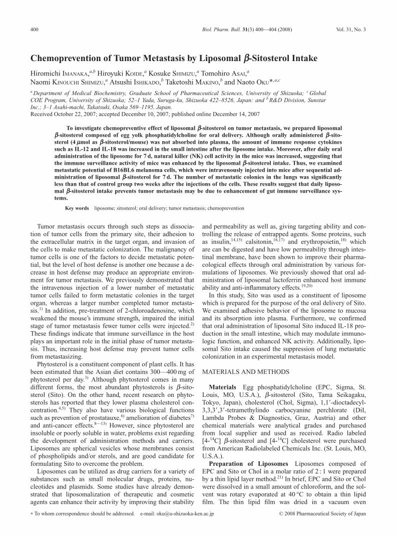
DiI-labeled liposomes were administered intragastrically tothe mice, and the small intestine was removed 1.5 h after ad-ministration. The mucoadhesive profiles of the liposomes inthe small intestinal tube were evaluated by observing theresidual liposomes on the mucosa with conforcal laser scan-ning microscopy. As a result, LS and LC showed quite simi-lar adhesiveness and penetrative behavior to the small intes-tinal mucosa. DiI in both liposomes penetrated the intestinalmucosa in the same manner (Fig. 2A). The mucoadhesive
property of both liposomes was measured for all parts of theintestinal tract. Looking at the mucous layer side, consider-able amounts of liposomes were observed through the intes-tinal tract from the upper to the lower part of the small intes-tine. Liposomes were also observed in the basolateral side ofthe intestinal membrane. Since the absorption of Chol intoplasma was quite high compared with that of Sito, we con-sidered that Chol might be separated from liposomes afterdegraded them in the small intestine and absorbed into
402 Vol. 31, No. 3
Fig. 1. Absorption and Distribution of Orally Administered Liposomal Sito and Chol
C57BL/6 male mice were orally administered LS or LC (4 mmol as sterol/mouse). Then, Sito and Chol uptake was measured by [14C]-labeled compounds. Distribution of Sitoand Chol in selectede organs 3 h after oral administration of liposomes are shown in (A). Closed and open bars show data for Sito and Chol, respectively. Data representmean�S.D. (n�5). Time dependent uptake of Sito and Chol in plasma by oral administration of these liposomes is shown in (B). Uptake quantities of Chol in plasma of mice pre-viously administered with LC increased. On the contrary, Sito in the plasma of those previously administered with LS did not increase. Closed and open circles show data for Sitoand Chol, respecively. Data represent mean�S.D. (n�5).
Fig. 2. Confocal Laser Scanning Microscopy Photographs of Small Intestine 1.5 h after Intragastric Administration of Liposomes Containing Sito or Chol
Five-week-old C57BL/6 male mice were orally administered LS or LC (4 mmol as sterol/mouse). Small intestines were excised from mice sacrificed 1.5 h after administrationand washed with large amount of saline solution. Photographs on the top show the lumen and mucosal sides. The bottom side show corresponds to the lateral side. Parts of smallintestine were indicated as following; (i): intestinal lumen, (ii): villi of intestine, (iii): muscle layer, (iv): serous membrane. DiI-labeled liposome with b-sitosterol (LS) or choles-terol (LC) penetrated the intestinal mucosa in the same manner (A). IL-18 production in small intestine was examined by immunostaining (B). The numbers of positively stainedintestinal epithelial cells were observed by LS administration.
plasma.Effect of Liposomal bb-Sitosterol Intake on the Host Im-
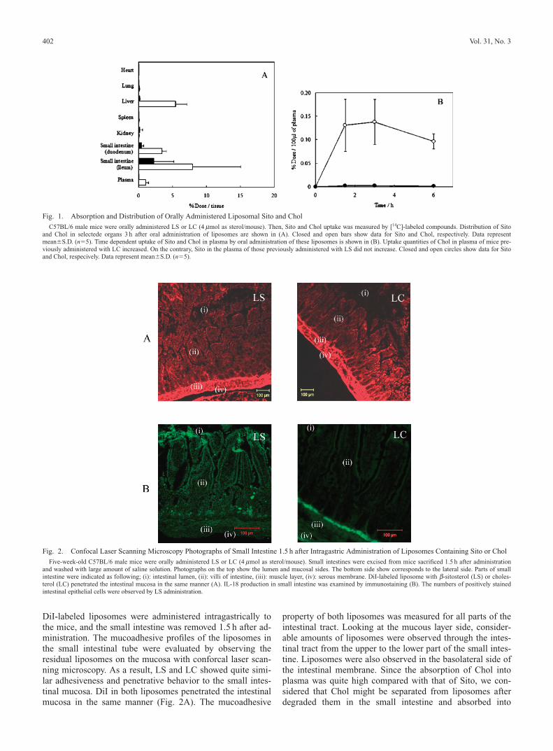
munity Recent investigation demonstrated that gut immu-nity plays an important role in the systemic immune system.For example, intestinal epithelium produces IL-18 for LPSstimulation. Therefore, we examined the influence of orallyadministered liposomes on IL-18 production in small intes-tine by immunostaining. As a result, the numbers of posi-tively stained intestinal epithelial cells increased after admin-istration of LS compared with those of LC treated mice (Fig.2B). We also quantified the production level of IL-12 and IL-18 in the intestinal epithelium by using a specific ELISA kit.Figure 3A shows the production of IL-12 in the small intes-tinal epithelium isolated from the liposomes treated mice.Significant increase in the IL-12 level of small intestine wasobserved at 1.5 and 24 h after administration of SL. Signifi-cant difference in the IL-12 level was also detected in miceorally administrated with samples once in a day for 7 d. Ineach case, the amount of IL-12 in the small intestine of theLS-treated mice was higher than the LC- and PBS-treatedmice. On the other hand, no significant difference was ob-served between the LC- and PBS-treated mice in all casestested.
The amount of IL-18 was also measured identically as theIL-12. The result was shown in Fig. 3B. The IL-18 level wasincreased in LS treated mice like as the production of IL-12.These results suggest that administration of LS led to IL-12and IL-18 production in the small intestine within 1.5 h afteradminstration, even though Sito did not penetrate the intes-tinal epithelium. Borghesi et al. (1999) have already reportedthe rapid immune response in small intestine.24) In theirstudy, the marked increase in the number of M cells was de-tected in the rabbit small intestine as early as 1 h after expo-sure to Streptococcus pneumoniae R36a. Therefore, we con-sidered that LS had the potential to stimulate gut immune atthe time point tested. Since the plasma levels of IL-12 andIL-18 were not changed (data not shown), these cytokinesproduction might be limited at the small intestine.
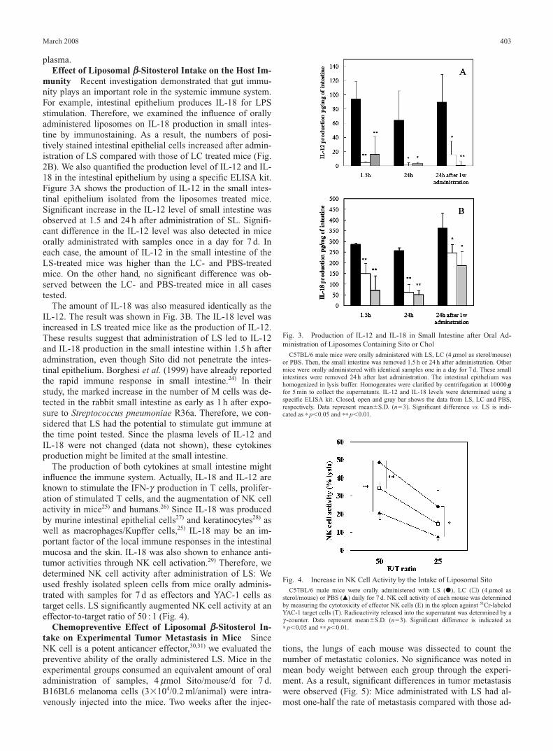
The production of both cytokines at small intestine mightinfluence the immune system. Actually, IL-18 and IL-12 areknown to stimulate the IFN-g production in T cells, prolifer-ation of stimulated T cells, and the augmentation of NK cellactivity in mice25) and humans.26) Since IL-18 was producedby murine intestinal epithelial cells27) and keratinocytes28) aswell as macrophages/Kupffer cells,25) IL-18 may be an im-portant factor of the local immune responses in the intestinalmucosa and the skin. IL-18 was also shown to enhance anti-tumor activities through NK cell activation.29) Therefore, wedetermined NK cell activity after administration of LS: Weused freshly isolated spleen cells from mice orally adminis-trated with samples for 7 d as effectors and YAC-1 cells astarget cells. LS significantly augmented NK cell activity at aneffector-to-target ratio of 50 : 1 (Fig. 4).
Chemopreventive Effect of Liposomal bb-Sitosterol In-take on Experimental Tumor Metastasis in Mice SinceNK cell is a potent anticancer effector,30,31) we evaluated thepreventive ability of the orally administered LS. Mice in theexperimental groups consumed an equivalent amount of oraladministration of samples, 4 mmol Sito/mouse/d for 7 d.B16BL6 melanoma cells (3�104/0.2 ml/animal) were intra-venously injected into the mice. Two weeks after the injec-
tions, the lungs of each mouse was dissected to count thenumber of metastatic colonies. No significance was noted inmean body weight between each group through the experi-ment. As a result, significant differences in tumor metastasiswere observed (Fig. 5): Mice administrated with LS had al-most one-half the rate of metastasis compared with those ad-
March 2008 403
Fig. 3. Production of IL-12 and IL-18 in Small Intestine after Oral Ad-ministration of Liposomes Containing Sito or Chol
C57BL/6 male mice were orally administered with LS, LC (4 mmol as sterol/mouse)or PBS. Then, the small intestine was removed 1.5 h or 24 h after administration. Othermice were orally administered with identical samples one in a day for 7 d. These smallintestines were removed 24 h after last administration. The intestinal epithelium washomogenized in lysis buffer. Homogenates were clarified by centrifugation at 10000 gfor 5 min to collect the supernatants. IL-12 and IL-18 levels were determined using aspecific ELISA kit. Closed, open and gray bar shows the data from LS, LC and PBS,respectively. Data represent mean�S.D. (n�3). Significant difference vs. LS is indi-cated as ∗ p�0.05 and ∗∗ p�0.01.
Fig. 4. Increase in NK Cell Activity by the Intake of Liposomal Sito
C57BL/6 male mice were orally administered with LS (�), LC (�) (4 mmol assterol/mouse) or PBS (�) daily for 7 d. NK cell activity of each mouse was determinedby measuring the cytotoxicity of effector NK cells (E) in the spleen against 51Cr-labeledYAC-1 target cells (T). Radioactivity released into the supernatant was determined by ag-counter. Data represent mean�S.D. (n�3). Significant difference is indicated as∗ p�0.05 and ∗∗ p�0.01.

ministrated with LC or PBS.We could not directly demonstrate a relation of the inhibi-
tion of lung colonization and the enhanced production of IL-12 and IL-18 in the intestinal epithelium. However, we con-firmed greater NK cell activity in the LS administered mice.Therefore, it is possible that experimental lung metastasismodel is sensitive to the effects of IL-18 and IL-12 either di-rectly or indirectly, and the production of IL-12 and IL-18may be important for LS to inhibit metastasis.
There have been many studies, in which phytosterol inhib-ited the growth of metastatic tumor cell. Awad et al. (2000)have already reported that Sito effectively inhibited PC-3human prostate cancer cell growth in in vitro and in vivostdies.32) They speculated that the inhibitory mechanism ofSito involves signal transduction. In their in vitro study, Sitoeffectively inhibited the invasion of PC-3 cells to the base-ment membrane, Matrigel. The data obtained on cell adhe-sion to extracellular matrix proteins known to be involved inmetastasis33) indicated that Sito treatment influenced bindingto fibronectin and laminin. They suggested that Sito mightinfluence the expression of integrin receptors on the tumorcells. However, our study clearly indicates that Sito is barelyabsorbed into plasma and LS was administered before thecancer injection. Therefore, imaging that LS or Sito directlyinfluences tumor cells is difficult, when Sito is administeredthrough oral route. Considering our observations, orally ad-ministered LS stimulated mucosal immunity. Intestinal ep-ithelium produced IL-12 and IL-18, which continuously en-hanced NK cell activity. In the immune suppression results,B16BL6 metastasis on lung was inhibited.
CONCLUSION
In the present study, we prepared liposomal b-sitosterol(LS) and investigated the advantages of this formulation to abody. The results indicated that the oral administration of LSshowed chemopreventive effects on tumor metastasis due tothe upregulation of host defense against metastatic tumorcells. The results of the present study suggest that the oraladministration of LS may enhance mucosal immunity, andpossibly the enhancement of NK cell activity by IL-18 and
IL-12 induction. Therefore, LS might serve as an effectivemeans for preventing metastasis. Thus, the oral administra-tion of LS may be useful for strengthen immune surveillancesystem in a body.
REFERENCES
1) Kikkawa H., Imafuku H., Tsukada H., Oku N., FEBS Lett., 467, 211—216 (2000).
2) Kikkawa H., Tsukada H., Oku N., Cancer, 89, 1626—1633 (2000).3) Nakajima K., Ikeda I., Kanno M., Fuchigami K., Shiroishi Y., Yasue
R., Matsumoto M., Jpn. J. Clin. Nutr., 58, 263—268 (1981).4) Noakes M., Clifton P., Ntanios F., Shrapnel W., Record I., McInerney
J., Am. J. Clin. Nutr., 75, 79—86 (2002).5) Mussner M. J., Parhofer K. G., Von Bergmann K., Schwandt P., Broedl
U., Otto C., Metabolism, 51, 189—194 (2002).6) Berges R. R., Windeler J., Trampisch H. J., Senge T., Lancet, 345,
1529—1532 (1995).7) Jenkins D. J., Kendall C. W., Marchie A., Jenkins A. L., Augustin L.
S., Ludwig D. S., Barnard N. D., Anderson J. W., Am. J. Clin. Nutr., 78,610s—616s (2003).
8) Awad A. B., Williams H., Fink C. S., J. Nutr. Biochem., 14, 111—119(2003).
9) Awad A. B., Roy R., Fink C. S., Oncol. Rep., 10, 497—500 (2003).10) De Jong A., Plat J., Mensink R. P., J. Nutr. Biochem., 14 362—369
(2003).11) Muti P., Awad A. B., Schunemann H., Fink C. S., Hovey K., Freuden-
heim J. L., Wu Y. W., Bellati C., Pala V., Berrino F., J. Nutr., 133,4252—4255 (2003).
12) Raicht R. F., Cohen B. I., Fazzini E. P., Sarwal A. N., Takahashi M.,Cancer Res., 40, 403—405 (1980).
13) Normen A. L., Brants H. A., Voorrips L. E., Andersson H. A., van denBrandt P. A., Goldbohm R. A., Am. J. Clin. Nutr., 74, 141—148(2001).
14) Spangler R. S., Diabetes Care, 13, 911—922 (1990).15) Takeuchi H., Yamamoto H., Niwa T., Hiro T., Kawashima Y., Pharm.
Res., 13, 896—901 (1996).16) Takeuchi H., Matsui Y., Yamamoto H., Kawashima Y., J. Controlled
Release, 86, 235—242 (2003).17) Fukunaga M., Miller M. M., Deftos L. J., Horm. Metab. Res., 23,
166—167 (1991).18) Maitani Y., Hazama M., Tojo Y., Shimoda N., Nagai T., J. Pharm. Sci.,
85, 440—445 (1996).19) Ishikado A., Imanaka H., Kotani M., Fujita A., Mitsuishi Y., Kane-
mitsu T., Tamura Y., Makino T., BioFactors, 21, 69—72 (2004).20) Ishikado A., Imanaka H., Takeuchi T., Harada E., Makino T., Biol.
Pharm. Bull., 28, 1717—1721 (2005).21) Oku N., Tokudome Y., Tsukada H., Kosugi T., Namba Y., Okada S.,
Biopharm. Drug Dispos., 17, 435—441 (1996).22) Takeuchi H., Matsui Y., Sugihara H., Yamamoto H., Kawashima Y.,
Int. J. Pharm., 303, 160—170 (2005).23) Hegele R. A., Robinson J. F., Curr. Drug Targets Cardiovas. Haemat.
Dis., 5, 31—37 (2005).24) Borghesi C., Taussig M. J., Nicoletti C., Lab. Invest., 79, 1393—1401
(1999).25) Okamura H., Tsutsui H., Komatsu T., Yutsudo M., Hakura A., Tani-
moto T., Torigoe K., Okura T., Nukada Y., Hattori K., Akita K.,Namba M., Tanabe F., Konishi K., Fukuda S., Kurimoto M., Nature(London), 378, 88—91 (1995).
26) Ushio S., Namba M., Okura T., Hattori K., Nukada Y., Akita K., Tana-be F., Konishi K., Micallef M., Fujii M., Torigoe K., Tanimoto T.,Fukuda S., Ikeda M., Okamura H., Kurimoto M., J. Immunol., 156,4274—4279 (1996).
27) Takeuchi M., Nishizaki Y., Sano O., Ohta T., Ikeda M., Kurimoto M.,Cell Tissue Res., 289, 499—503 (1997).
28) Stoll S., Muller G., Kurimoto M., Saloga J., Tanimoto T., YamauchiH., Okamura H., Knop J., Enk A. H., J. Immunol., 159, 298—302(1997).
29) Micallef M. J., Yoshida K., Kawai S., Hanaya T., Kohno K., Arai S.,Tanimoto T., Torigoe K., Fujii M., Ikeda M., Kurimoto M., Cancer Im-munol., 43, 361—367 (1997).
30) Herberman R. B., J. Invest. Dermatology, 83, 137s—140s (1984).31) Griffini P., Smorenburg S. M., Vogels I. M. C., Tigchelaar V. W., Van
Noorden C. J. F., Clin. Exp. Metastasis, 14, 367—380 (1996).32) Awad A. B., Fink C. S., Williams H., Kim U., Eur. J. Cancer Prev., 10,
507—513 (2001).33) Liotta L. A., Steeg P. S., Stetler-Stevenson W. G., Cell, 64, 327—336
(1991).
404 Vol. 31, No. 3
Fig. 5. Preventive Effect of LS Intake on Tumor Metastasis
C57BL/6 male mice were orally administered with LS (�), LC (�), or PBS (�)daily for continuous 7 d. Next day, B16BL6 melanoma cells were intravenously injectedinto the mice by tail vein. At 14 d after the injection of the metastatic cells, lungs ofeach mouse were dissected and the number of metastatic colonies was measured. Datalines represent mean of colony number with S.D. bar. Significant difference is indicatedas ∗∗ p�0.01.