Chemoautotrophic Carbon Fixation Rates and Active ... · Chemoautotrophic Carbon Fixation Rates and...
12
Chemoautotrophic Carbon Fixation Rates and Active Bacterial Communities in Intertidal Marine Sediments Henricus T. S. Boschker 1 *, Diana Vasquez-Cardenas 1 , Henk Bolhuis 1 , Tanja W. C. Moerdijk-Poortvliet 1 , Leon Moodley 2 1 Department of Marine Microbiology, Royal Netherlands Institute for Sea Research (NIOZ), Yerseke, The Netherlands, 2 Marine Environment Group, International Research Institute of Stavanger (IRIS), Randaberg, Norway Abstract Chemoautotrophy has been little studied in typical coastal marine sediments, but may be an important component of carbon recycling as intense anaerobic mineralization processes in these sediments lead to accumulation of high amounts of reduced compounds, such as sulfides and ammonium. We studied chemoautotrophy by measuring dark-fixation of 13 C- bicarbonate into phospholipid derived fatty acid (PLFA) biomarkers at two coastal sediment sites with contrasting sulfur chemistry in the Eastern Scheldt estuary, the Netherlands. At one site where free sulfide accumulated in the pore water right to the top of the sediment, PLFA labeling was restricted to compounds typically found in sulfur and ammonium oxidizing bacteria. At the other site, with no detectable free sulfide in the pore water, a very different PLFA labeling pattern was found with high amounts of label in branched i- and a-PLFA besides the typical compounds for sulfur and ammonium oxidizing bacteria. This suggests that other types of chemoautotrophic bacteria were also active, most likely Deltaproteobacteria related to sulfate reducers. Maximum rates of chemoautotrophy were detected in first 1 to 2 centimeters of both sediments and chemosynthetic biomass production was high ranging from 3 to 36 mmol C m 22 d 21 . Average dark carbon fixation to sediment oxygen uptake ratios were 0.2260.07 mol C (mol O 2 ) 21 , which is in the range of the maximum growth yields reported for sulfur oxidizing bacteria indicating highly efficient growth. Chemoautotrophic biomass production was similar to carbon mineralization rates in the top of the free sulfide site, suggesting that chemoautotrophic bacteria could play a crucial role in the microbial food web and labeling in eukaryotic poly-unsaturated PLFA was indeed detectable. Our study shows that dark carbon fixation by chemoautotrophic bacteria is a major process in the carbon cycle of coastal sediments, and should therefore receive more attention in future studies on sediment biogeochemistry and microbial ecology. Citation: Boschker HTS, Vasquez-Cardenas D, Bolhuis H, Moerdijk-Poortvliet TWC, Moodley L (2014) Chemoautotrophic Carbon Fixation Rates and Active Bacterial Communities in Intertidal Marine Sediments. PLoS ONE 9(7): e101443. doi:10.1371/journal.pone.0101443 Editor: Fabiano Thompson, Universidade Federal do Rio de Janeiro, Brazil Received February 17, 2014; Accepted June 6, 2014; Published July 8, 2014 Copyright: ß 2014 Boschker et al. This is an open-access article distributed under the terms of the Creative Commons Attribution License, which permits unrestricted use, distribution, and reproduction in any medium, provided the original author and source are credited. Funding: This work was supported by the following grants: Netherlands Organization for Scientific Research (NWO) VIDI grant number 864.04.009 to HTSB, Darwin Center for Biogeosciences grant number 142.16.3061 to HTSB, and ESF grant number 855.01.130. The funders had no role in study design, data collection and analysis, decision to publish, or preparation of the manuscript. Competing Interests: The authors have declared that no competing interests exist. * Email: [email protected] Introduction Reoxidation of reduced intermediates like sulfide and ammo- nium formed during anaerobic mineralization processes is an important process in coastal marine sediments. Oxygen is typically only found in the top millimeters of these sediments and along macrofauna burrows [1], and carbon mineralization proceeds in general by anaerobic processes primarily sulfate reduction. This results in the production and accumulation of large amounts of reduced compounds such as various forms of reduced sulfur and ammonium [2]. In typical coastal sediments, free sulfide in the porewater is however often only detected below a couple of centimeters as it quickly reacts with iron hydroxides forming iron sulfide (FeS) or pyrite (FeS 2 ) [3]. Only in very active sediments or sediments containing little reactive iron, free sulfide can be found near the oxic top layer [3]. Long term burial of reduced compounds is thought to be a minor process [3] and they are mostly transported to more oxidized horizons by either diffusion or bioturbation [4]. Oxygen is eventually the main oxidant of these reduced compounds although intermediate reoxidation steps by a variety of anaerobic pathways using nitrate or iron and manganese oxides may also be important [3]. It is estimated that reoxidation processes on average explain 70% of the sediment oxygen flux in shelf sediments [5] and this value is expected to be higher in active intertidal areas as anaerobic mineralization will be more important. Many of the known prokaryotes involved in reoxidation processes are chemo(litho)autotrophs that use the energy gained from inorganic reactions to grow by fixing inorganic carbon in the dark [6]. Chemoautotrophic carbon fixation has been shown to be an important process in, for instance, extreme marine ecosystems such as hydrothermal vents [7,8] and in the chemocline of anoxic marine basins [9,10]. The current consensus is however that chemoautotrophy is a relatively minor process in coastal sediments due to the relatively low growth yields of chemoautotrophic organisms and the competition with chemical oxidation reactions [3]. In addition, true chemoautotrophic bacteria have to compete with mixotrophic and heterotrophic bacteria that are able to oxidize reduced sulfur compounds [11], which could be relevant especially in active coastal sediments receiving large amounts of organic matter. Studies where chemoautotrophy was actually quantified by determining dark carbon fixation rates are rare for PLOS ONE | www.plosone.org 1 July 2014 | Volume 9 | Issue 7 | e101443
Transcript of Chemoautotrophic Carbon Fixation Rates and Active ... · Chemoautotrophic Carbon Fixation Rates and...

Chemoautotrophic Carbon Fixation Rates and ActiveBacterial Communities in Intertidal Marine SedimentsHenricus T. S. Boschker1*, Diana Vasquez-Cardenas1, Henk Bolhuis1, Tanja W. C. Moerdijk-Poortvliet1,
Leon Moodley2
1 Department of Marine Microbiology, Royal Netherlands Institute for Sea Research (NIOZ), Yerseke, The Netherlands, 2 Marine Environment Group, International Research
Institute of Stavanger (IRIS), Randaberg, Norway
Abstract
Chemoautotrophy has been little studied in typical coastal marine sediments, but may be an important component ofcarbon recycling as intense anaerobic mineralization processes in these sediments lead to accumulation of high amounts ofreduced compounds, such as sulfides and ammonium. We studied chemoautotrophy by measuring dark-fixation of 13C-bicarbonate into phospholipid derived fatty acid (PLFA) biomarkers at two coastal sediment sites with contrasting sulfurchemistry in the Eastern Scheldt estuary, the Netherlands. At one site where free sulfide accumulated in the pore water rightto the top of the sediment, PLFA labeling was restricted to compounds typically found in sulfur and ammonium oxidizingbacteria. At the other site, with no detectable free sulfide in the pore water, a very different PLFA labeling pattern was foundwith high amounts of label in branched i- and a-PLFA besides the typical compounds for sulfur and ammonium oxidizingbacteria. This suggests that other types of chemoautotrophic bacteria were also active, most likely Deltaproteobacteriarelated to sulfate reducers. Maximum rates of chemoautotrophy were detected in first 1 to 2 centimeters of both sedimentsand chemosynthetic biomass production was high ranging from 3 to 36 mmol C m22 d21. Average dark carbon fixation tosediment oxygen uptake ratios were 0.2260.07 mol C (mol O2)21, which is in the range of the maximum growth yieldsreported for sulfur oxidizing bacteria indicating highly efficient growth. Chemoautotrophic biomass production was similarto carbon mineralization rates in the top of the free sulfide site, suggesting that chemoautotrophic bacteria could play acrucial role in the microbial food web and labeling in eukaryotic poly-unsaturated PLFA was indeed detectable. Our studyshows that dark carbon fixation by chemoautotrophic bacteria is a major process in the carbon cycle of coastal sediments,and should therefore receive more attention in future studies on sediment biogeochemistry and microbial ecology.
Citation: Boschker HTS, Vasquez-Cardenas D, Bolhuis H, Moerdijk-Poortvliet TWC, Moodley L (2014) Chemoautotrophic Carbon Fixation Rates and ActiveBacterial Communities in Intertidal Marine Sediments. PLoS ONE 9(7): e101443. doi:10.1371/journal.pone.0101443
Editor: Fabiano Thompson, Universidade Federal do Rio de Janeiro, Brazil
Received February 17, 2014; Accepted June 6, 2014; Published July 8, 2014
Copyright: � 2014 Boschker et al. This is an open-access article distributed under the terms of the Creative Commons Attribution License, which permitsunrestricted use, distribution, and reproduction in any medium, provided the original author and source are credited.
Funding: This work was supported by the following grants: Netherlands Organization for Scientific Research (NWO) VIDI grant number 864.04.009 to HTSB,Darwin Center for Biogeosciences grant number 142.16.3061 to HTSB, and ESF grant number 855.01.130. The funders had no role in study design, data collectionand analysis, decision to publish, or preparation of the manuscript.
Competing Interests: The authors have declared that no competing interests exist.
* Email: [email protected]
Introduction
Reoxidation of reduced intermediates like sulfide and ammo-
nium formed during anaerobic mineralization processes is an
important process in coastal marine sediments. Oxygen is typically
only found in the top millimeters of these sediments and along
macrofauna burrows [1], and carbon mineralization proceeds in
general by anaerobic processes primarily sulfate reduction. This
results in the production and accumulation of large amounts of
reduced compounds such as various forms of reduced sulfur and
ammonium [2]. In typical coastal sediments, free sulfide in the
porewater is however often only detected below a couple of
centimeters as it quickly reacts with iron hydroxides forming iron
sulfide (FeS) or pyrite (FeS2) [3]. Only in very active sediments or
sediments containing little reactive iron, free sulfide can be found
near the oxic top layer [3]. Long term burial of reduced
compounds is thought to be a minor process [3] and they are
mostly transported to more oxidized horizons by either diffusion
or bioturbation [4]. Oxygen is eventually the main oxidant of these
reduced compounds although intermediate reoxidation steps by a
variety of anaerobic pathways using nitrate or iron and manganese
oxides may also be important [3]. It is estimated that reoxidation
processes on average explain 70% of the sediment oxygen flux in
shelf sediments [5] and this value is expected to be higher in active
intertidal areas as anaerobic mineralization will be more
important.
Many of the known prokaryotes involved in reoxidation
processes are chemo(litho)autotrophs that use the energy gained
from inorganic reactions to grow by fixing inorganic carbon in the
dark [6]. Chemoautotrophic carbon fixation has been shown to be
an important process in, for instance, extreme marine ecosystems
such as hydrothermal vents [7,8] and in the chemocline of anoxic
marine basins [9,10]. The current consensus is however that
chemoautotrophy is a relatively minor process in coastal sediments
due to the relatively low growth yields of chemoautotrophic
organisms and the competition with chemical oxidation reactions
[3]. In addition, true chemoautotrophic bacteria have to compete
with mixotrophic and heterotrophic bacteria that are able to
oxidize reduced sulfur compounds [11], which could be relevant
especially in active coastal sediments receiving large amounts of
organic matter. Studies where chemoautotrophy was actually
quantified by determining dark carbon fixation rates are rare for
PLOS ONE | www.plosone.org 1 July 2014 | Volume 9 | Issue 7 | e101443
typical coastal marine sediments and we have only been able to
locate four studies: two on shallow subtidal sediments from the
Baltic [12,13], one study on an intertidal sand flat in the German
Wadden Sea [14] and a recent study on three brackish coastal lake
sediments in Brazil [15]. However, recent estimates suggest that up
to 0.29 Pg C y21 could be potentially fixed by chemoautotrophic
microorganisms in near shore and shelf sediments worldwide
compared to 0.92 Pg C y21 of mineralization [16], suggesting a
major role in the sediment carbon cycle. Finally, the dominant
chemoautotrophic bacteria involved in sulfur oxidation are not
well known in coastal marine sediments. A recent study identified
an uncultered group of Gammaproteobacteria as important
players [14], but there may be many other groups involved in
the diversity of reoxidation processes that occur in marine
sediments.
We studied chemoautotrophy in two intertidal sites with
contrasting sulfur chemistry: a site where free sulfide was not
detected in the top few centimeters of the sediment and a very
active site where high concentrations of free sulfide were found
right to the top of the sediment. The main substrates driving
chemoautotrophy are therefore expected to be different at both
sites, namely free sulfide at the very active site versus iron sulfides
in the more typical coastal sediment. Chemoautotrophy rates were
determined by incubating sediment cores with stable isotope
labeled 13C-bicarbonate and measuring labeling in phospholipid
derived fatty acids (PLFA). This method both yields estimates of
total chemoautotrophy rates and provides an indication of the
active bacterial community [17–19]. The diversity of Rubisco
genes was studied to further indicate possible active chemoauto-
trophs that use the Calvin cycle for carbon fixation. Finally
chemoautotrophy rates were compared with diffusive oxygen
fluxes and carbon mineralization rates.
Materials and Methods
Description of field sitesTwo field sites in the Eastern Scheldt estuary (The Netherlands)
were selected, which were expected to show high mineralization
rates and have major differences in sulfur chemistry. The site in
the Zandkreek area (51u32’41’’N, 3u53’22’’E) was situated next to
a Pacific oyster (Crassostrea gigas) bed and was sampled in April
2005 (abbreviation ZK05) and October 2007 (ZK07). The Pacific
oyster is an invasive species in the area that was introduced in the
Eastern Scheldt around 1970. It stimulates sedimentation and
sediment carbon mineralization either by decreasing water
currents over the sediment or via pseudo-feces production and
biodeposition [20]. Sediments were non-sulfidic in the top 5
centimeters in 2005 and slightly sulfidic below 2 centimeter in
2007 (See Result).
The Rattekaai site (51u26’21’’N, 4u10’11’’E) was situated at the
entrance of a salt marsh creek where macroalgal debris (mainly
Ulva derived) accumulates and is buried during winter. The
sediment was highly sulfidic right to the top and samples were
taken from patches where the sediment was covered with a whitish
layer in April 2005 (RK05) and May 2006 (RK06). Based on
microscopy, typical Beggiatoa-like sulfur-oxidizing bacteria were
abundant in the top few millimeters of the Rattekaai sediment,
especially in 2005 and to a lesser degree in 2006.
Ethics statementThe Eastern Scheldt estuary is a Natura 2000 protected area
and research permits for both sites were granted by the
‘‘Vereeniging Natuurmonumenten’’ and the Provence of Zeeland.
Sediment samplingUndisturbed sediments were sampled with two sizes of
polycarbonate core liners. The smaller cores (internal diameter
4.6 cm) contained silicon-filled injection ports at every 0.5
centimeter and were used for measuring chemoautotrophy rates.
The larger cores (internal diameter 6 cm) were used for additional
measurements of porewater profiles and sediment characteristic,
and for measuring mineralization rates. Sediments were sampled
at low tide and therefore did not have overlying water. Cores were
processed the same day for chemoautotrophy rate measurements
and other analyses.
Chemoautotrophy ratesChemoautotrophy rate measurements were started by injecting
100 ml of 20 mM NaH13CO3 (99% 13C; Cambridge Isotope
Laboratories, Andover, MA, USA) horizontally into the sediment
cores at 0.5 cm depth intervals by using the line-injection method
[2]. The 13C-label was dissolved in artificial seawater lacking
calcium or magnesium in order to avoid precipitation [21]. The
label was made oxygen free by bubbling with nitrogen gas shortly
before injection. Sediment cores were incubated in the dark within
2uC of the in-situ temperature (see Table 1) for various periods of
up to 4 days, and were ventilated daily by removing the top
stopper for one minute (ZK) or incubated without top stoppers
(RK) to circumvent the development of suboxic condition in the
headspace. After incubation, sediment cores were sliced to a depth
of 5 cm and sediment slices were quickly centrifuged (4500 rpm,
5 min) to collect porewater for concentration and 13C analysis of
dissolved inorganic carbon (DIC). Sediments were subsequently
frozen at 220uC and lyophilized before further analysis.
Unlabelled, control cores were also processed.
PLFA analysis and calculation of chemoautotrophy ratesLyophilized sediments were analyzed for PLFA concentrations
and 13C-labeling as described before [22,23]. In short, PLFA were
extracted according to standard protocols and were analyzed by
gas chromatography – isotope ratio mass spectrometry (GC-
IRMS, Thermo, Bremen, Germany) on an a-polar analytical
column (HP5-MS, Agilent, Santa Clara, CA, USA). Stable carbon
isotope ratios are reported as d13C ratios on the VPDB scale.
Excess 13C in individual PLFA was calculated as in Boschker et al
[23] and divided by the atom percent excess 13C in the DIC pool
to calculate actual PLFA synthesis rates. Only very minor labeling
was found in poly-unsaturated PLFA typical for Eukarya (see
Results) suggesting that PLFA labeling was primarily by Bacteria.
We therefore used the labeling data for all common bacterial
PLFA in the 12:0 to 20:0 range in our calculations and not just the
specific bacterial biomarker PLFA [24]. Total bacterial chemo-
autotrophy rates were determined by summing synthesis rates in
all PLFA typically found in bacteria and converted to chemoau-
totrophic biomass production by dividing by the typical PLFA
content of aerobic bacteria (55 mmol PLFA-C (mol biomass C)21
[24,25]). To study the differences in active chemoautrophic
bacterial communities, we performed a principle component
analysis (PCA) on log-transformed PLFA 13C-labeling data (in
Mol%) using the Statistica software package (StatSoft, Tulsa,
USA).
Additional measurementsOxygen profiles were determined with oxygen microelectrodes
(Unisense Ox100, Aarhus, Denmark), which were lowered with a
micromanipulator into the sediment until no oxygen was detected.
Two profiles were recorded for each duplicate sediment core (four
Chemoautotrophy in Intertidal Marine Sediments
PLOS ONE | www.plosone.org 2 July 2014 | Volume 9 | Issue 7 | e101443

profiles total), which were kept within 2uC of the in-situ
temperature. Oxygen fluxes into the sediment were calculated as
described in Van Frausum et al. [26] with sediment tortuosity
estimated from sediment porosity as in Boudreau and Meysman
[27].
Sediment porewater was sampled by slicing duplicate sediment
cores in an anaerobic glove-box filled with 3% hydrogen in
nitrogen gas (Coy Laboratory Products, Ann Arbor, MI, USA)
and slices were centrifuged at 4500 rpm for 10 min at in situ
temperature. Samples for sulfide analysis were immediately fixed
in zinc acetate and analyzed according to Cline [28]. Samples for
ammonium and anion analysis were frozen, and analyzed on a
QuAAtro segmented flow analyzer (Seal Analytical, Norderstedt,
Germany) and suppressed high performance ion chromatography
on a Dionex Ionpac AS-14 column (Thermo, Sunnyvale, CA,
USA), respectively. Samples for 13C-DIC were added to headspace
vials (10 ml) and after acidification analyzed for DIC concentra-
tions and 13C-content by elemental analyzer - IRMS [29].
Sediment carbon mineralization rates were determined using
the jar method [30]. Sediment cores were sliced as above and were
incubated in completely filled centrifuge tubes. Centrifuge tubes
containing the sediment were sealed in air-tight incubation bags
filled with nitrogen gas to keep them strictly anaerobic and were
incubated within 2uC of the in situ temperature for up to 6 days.
Sediment porewater was collected and analyzed as described
above. Mineralization rates were calculated from DIC and
ammonium production with time and ammonium production
was converted to carbon mineralization rates by using the
sediment C/N ratio (Table 1).
Rubisco type IA clone librariesTo further study the diversity of chemoautotrophic bacteria that
utilize the Calvin cycle for carbon dioxide fixation, Rubisco clone
libraries were constructed for both sites in March 2008. Sediments
were sampled as described above, and the top 0.5 cm of the cores
showing the highest chemoautotrophy rates was collected and
immediately frozen at 280uC. Total community DNA was
extracted from 0.5 g of wet sediment using the MoBio UltraClean
Soil DNA Isolation kit according to protocol (MoBio, Carlsbad,
CA, USA).
We developed a new degenerative primer set to specifically
amplify Rubisco type IA as this group contains most of the true
chemoautotrophic bacteria involved in sulfur and ammonium
oxidation [31,32]. The new primer set also targets Beggiatoa like
Rubisco sequences [33], which was important as Beggiatoa-like
bateria were found at the RK site but were not covered by
previously published primer sets developed for chemoautotrophic
bacteria. The primer set also targets some of the lower branching
Type 1B sequences found in unicellular cyanobacteria, and
consists of forward primer 571 (GAYTTYACCAARGAYGAYG)
and reversed primer 898E (ACRCGGAARTGRATRCC). The
primer set was first tested against a positive control (Thioalk-
alimicrobium aerophilu kindly provided by Gerhard Muyzer, Delft
Technical University, The Netherlands) and PCR conditions were
subsequently optimized to specifically amplify the target sequences
from sediment DNA extracts.
The final PCR reaction mixture contained: 2.5 ml 10x standard
Taq reaction buffer (without Mg), 3.0 mmol L21 Mg2+, 0.2 mmol
L21 dNTPS, 0.2 mmol L21 of each primer (571 and 898E), 2 U of
NEB Taq DNA polymerase, 5% v/v DMSO, 0.2% w/v BSA and
16 ml autoclaved demi water. The PCR cycling intervals were
established as follows: preheating at 94uC for 5 minutes, followed
by 40 cycles of denaturation step at 94uC for 1 minute, annealing
step at 51uC for 30 seconds and extension at 72uC for 30 seconds.
The PCR reaction was finished with a final extension time of 7
minutes at 72uC. PCR products for each sample (RK and ZK)
were cloned into Escherichia coli Top10 cells using TOPO TA
cloning kit (Invitrogen, Carlsbad, CA, USA). Sequencing was
performed by a genetic analyzer (Applied Biosystems 3130
Genetic Analyzer, Carlsbad, CA, USA). Editing of the obtained
sequences was carried out using the BioEdit software package
(http://jwbrown.mbio.ncsu.edu/Bio-Edit/bioedit.html). Primer
sequences (T3, T7, 571, 898E) were removed from sequences,
then translated to protein sequences, and compared to known
sequences using BLAST. Protein sequence alignments and
phylogenetic analysis was done in MEGA V [34]. Sequences
have been deposited in the GenBank database under accession
numbers JQ659214 to JQ659253.
Results
In spring 2005, both sites (RK05 and ZK05) were studied in an
initial test to determine if chemoautotrophy rates could be
quantified by 13C-DIC labeling of PLFA in the dark. Sites were
sampled again in spring 2006 (RK06) and autumn 2007 (ZK07),
when a more extensive sampling program was executed.
Sediment biogeochemistryOxygen penetrated significantly deeper in ZK sediment (1–
2 mm) than RK sediment (0.2–0.5 mm, Table 1). At RK06, high
concentrations of free sulfide were found in the very top layer of
the sediment, whereas sulfide only started to accumulate below
2 cm sediment depth at ZK07 (Fig. 1). Sulfide and ammonium
concentrations were more than 10 times higher for RK06 than for
ZK07 throughout the sediment column (Fig. 1). In 2005,
porewater samples were also taken at both sites and analyzed for
sulfide, but samples were taken two weeks before chemoautotro-
phy measurements and not at exactly the same location, which is
Table 1. Sediment in-situ temperature, sediment characteristics, oxygen consumption rates, carbon mineralization rates,chemoautotrophy rates and yields (averages 6 standard deviations, N = 2) for the coastal marine sediments in this study.
Site/Year Temp. 6C POC1 (%) C/N1O2 penetrationdepth (mm)
O2 flux(mmol m22 d21)
C mineralization2
(mmol m22 d21)Chemoautotrophy2
(mmol C m22 d21)Yield C/O2
(mol C (mol O2)21)
RK05 14 - - 0.4560.10 17.263.0 - 5.561.9 0.3260.11
RK06 17 2.0 10.9 0.2360.06 192641 197636 36.364.8 0.1960.03
ZK05 14 - - 1.760.1 15.060.4 - 2.660.3 0.1760.02
ZK07 13 0.6 7.7 0.9560.06 15.561.6 105.9619.1 2.960.2 0.1860.01
1Data for 0–1 cm sediment depth. 2 Data integrated over 0–5 cm sediment depth.doi:10.1371/journal.pone.0101443.t001
Chemoautotrophy in Intertidal Marine Sediments
PLOS ONE | www.plosone.org 3 July 2014 | Volume 9 | Issue 7 | e101443
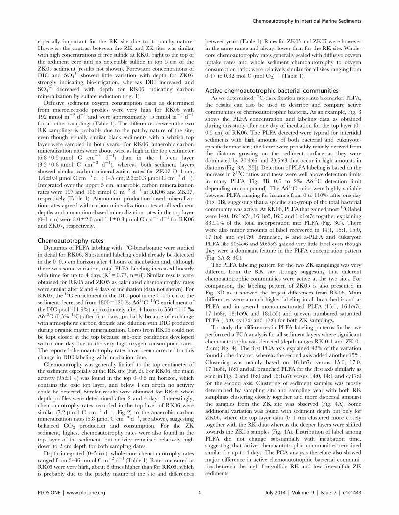
especially important for the RK site due to its patchy nature.
However, the contrast between the RK and ZK sites was similar
with high concentrations of free sulfide at RK05 right to the top of
the sediment core and no detectable sulfide in top 5 cm of the
ZK05 sediment (results not shown). Porewater concentrations of
DIC and SO42- showed little variation with depth for ZK07
strongly indicating bio-irrigation, whereas DIC increased and
SO42- decreased with depth for RK06 indicating carbon
mineralization by sulfate reduction (Fig. 1).
Diffusive sediment oxygen consumption rates as determined
from microelectrode profiles were very high for RK06 with
192 mmol m22 d21 and were approximately 15 mmol m22 d21
for all other samplings (Table 1). The difference between the two
RK samplings is probably due to the patchy nature of the site,
even though visually similar black sediments with a whitish top
layer were sampled in both years. For RK06, anaerobic carbon
mineralization rates were about twice as high in the top centimeter
(6.860.5 mmol C cm23 d21) than in the 1–5 cm layer
(3.260.8 mmol C cm23 d21), whereas both sediment layers
showed similar carbon mineralization rates for ZK07 (0–1 cm,
1.660.9 mmol C cm23 d21; 1–5 cm, 2.360.3 mmol C cm23 d21).
Integrated over the upper 5 cm, anaerobic carbon mineralization
rates were 197 and 106 mmol C m22 d21 at RK06 and ZK07,
respectively (Table 1). Ammonium production-based mineraliza-
tion rates agreed with carbon mineralization rates at all sediment
depths and ammonium-based mineralization rates in the top layer
(0–1 cm) were 8.062.0 and 1.160.3 mmol C cm23 d21 for RK06
and ZK07, respectively.
Chemoautotrophy ratesDynamics of PLFA labeling with 13C-bicarbonate were studied
in detail for RK06. Substantial labeling could already be detected
in the 0–0.5 cm horizon after 4 hours of incubation and, although
there was some variation, total PLFA labeling increased linearly
with time for up to 4 days (R2 = 0.77, n = 8). Similar results were
obtained for RK05 and ZK05 as calculated chemoautrophy rates
were similar after 2 and 4 days of incubation (data not shown). For
RK06, the 13C-enrichment in the DIC pool in the 0–0.5 cm of the
sediment decreased from 18006120 % Dd13C (13C enrichment of
the DIC pool of 1.9%) approximately after 4 hours to 5506110 %Dd13C (0.5% 13C) after four days, probably because of exchange
with atmospheric carbon dioxide and dilution with DIC produced
during organic matter mineralization. Cores from RK06 could not
be kept closed at the top because sub-oxic conditions developed
within one day due to the very high oxygen consumption rates.
The reported chemoautotrophy rates have been corrected for this
change in DIC labeling with incubation time.
Chemoautrophy was generally limited to the top centimeter of
the sediment especially at the RK site (Fig. 2). For RK06, the main
activity (9561%) was found in the top 0–0.5 cm horizon, which
contains the oxic top layer, and below 1 cm depth no activity
could be detected. Similar results were obtained for RK05 when
depth profiles were determined after 2 and 4 days. Interestingly,
chemoautotrophy rates recorded in the top layer of RK06 were
similar (7.2 mmol C cm23 d21, Fig 2) to the anaerobic carbon
mineralization rates (6.8 mmol C cm23 d21, see above), suggesting
balanced CO2 production and consumption. For the ZK
sediment, highest chemoautotrophy rates were also found in the
top layer of the sediment, but activity remained relatively high
down to 2 cm depth for both sampling dates.
Depth integrated (0–5 cm), whole-core chemoautotrophy rates
ranged from 3–36 mmol C m22 d21 (Table 1). Rates measured at
RK06 were very high, about 6 times higher than for RK05, which
is probably due to the patchy nature of the site and differences
between years (Table 1). Rates for ZK05 and ZK07 were however
in the same range and always lower than for the RK site. Whole-
core chemoautotrophy rates generally scaled with diffusive oxygen
uptake rates and whole sediment chemoautotrophy to oxygen
consumption ratios were relatively similar for all sites ranging from
0.17 to 0.32 mol C (mol O2)21 (Table 1).
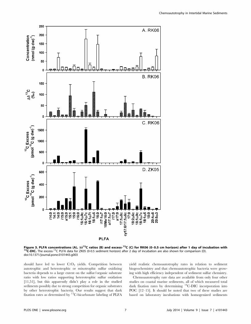
Active chemoautotrophic bacterial communitiesAs we determined 13C-dark fixation rates into biomarker PLFA,
the results can also be used to describe and compare active
communities of chemoautotrophic bacteria. As an example, Fig. 3
shows the PLFA concentration and labeling data as obtained
during this study after one day of incubation for the top layer (0–
0.5 cm) of RK06. The PLFA detected were typical for intertidal
sediments with high amounts of both bacterial and eukaryote-
specific biomarkers; the latter were probably mainly derived from
the diatoms growing on the sediment surface as they were
dominated by 20:4v6 and 20:5v3 that occur in high amounts in
diatoms (Fig. 3A; [35]). Detection of PLFA labeling is based on the
increase in d13C ratios and these were well above detection limits
in many PLFA (Fig. 3B; 0.6 to 2% Dd13C detection limit
depending on compound). The Dd13C ratios were highly variable
between PLFA ranging for instance from 0 to 110% after one day
(Fig. 3B), suggesting that a specific sub-group of the total bacterial
community was active. At RK06, PLFA that gained most 13C label
were 14:0, 16:1v7c, 16:1v5, 16:0 and 18:1v7c together explaining
8364% of the total incorporation into PLFA (Fig. 3C). There
were also minor amounts of label recovered in 14:1, 15:1, 15:0,
17:1v8 and cy17:0. Branched, i- and a-PLFA and eukaryote
PLFA like 20:4v6 and 20:5v3 gained very little label even though
they were a dominant feature in the PLFA concentration pattern
(Fig. 3A & 3C).
The PLFA labeling pattern for the two ZK samplings was very
different from the RK site strongly suggesting that different
chemoautotrophic communities were active at the two sites. For
comparison, the labeling pattern of ZK05 is also presented in
Fig. 3D as it showed the largest differences from RK06. Main
differences were a much higher labeling in all branched i- and a-
PLFA and in several mono-unsaturated PLFA (15:1, 16:1v7t,
17:1v8c, 18:1v9c and 18:1v5) and uneven numbered saturated
PLFA (15:0, cy17:0 and 17:0) for both ZK samplings.
To study the differences in PLFA labeling patterns further we
performed a PCA analysis for all sediment layers where significant
chemoautotrophy was detected (depth ranges RK 0-1 and ZK 0–
2 cm; Fig. 4). The first PCA axis explained 42% of the variation
found in the data set, whereas the second axis added another 15%.
Clustering was mainly based on 16:1v7c versus 15:0, 17:0,
17:1v8c, 18:0 and all branched PLFA for the first axis similarly as
seen in Fig. 3 and 16:0 and 16:1v7t versus 14:0, 14:1 and cy17:0
for the second axis. Clustering of sediment samples was mostly
determined by sampling site and sampling year with both RK
samplings clustering closely together and more dispersal amongst
the samples from the ZK site was observed (Fig. 4A). Some
additional variation was found with sediment depth but only for
ZK06, where the top layer data (0–1 cm) clustered more closely
together with the RK data whereas the deeper layers were shifted
towards the ZK05 samples (Fig. 4A). Distribution of label among
PLFA did not change substantially with incubation time,
suggesting that active chemoautotrophic communities remained
similar for up to 4 days. The PCA analysis therefore also showed
major difference in active chemoautotrophic bacterial communi-
ties between the high free-sulfide RK and low free-sulfide ZK
sediments.
Chemoautotrophy in Intertidal Marine Sediments
PLOS ONE | www.plosone.org 4 July 2014 | Volume 9 | Issue 7 | e101443
There was also indirect evidence of transfer of dark-fixed carbon
to fauna: some 13C-labelling was detected in bulk sediment PLFA
characteristic of Eukarya and therefore fauna, 18:2w6c for both
sites and 20:4v6 and 20:5v3 for the ZK site (Fig 3C and D).
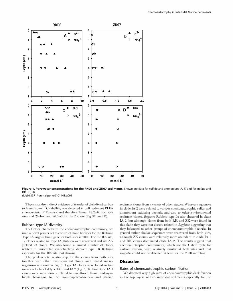
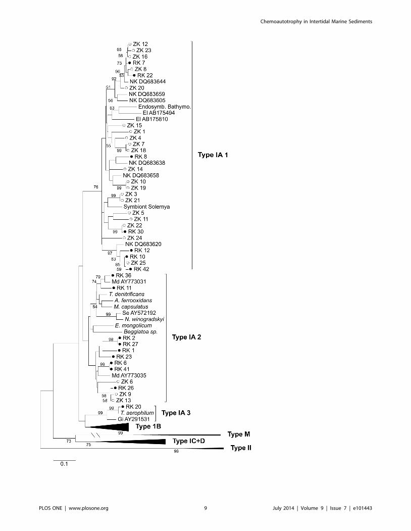
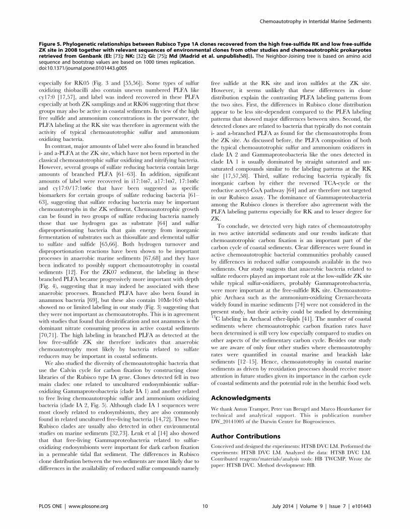
Rubisco type IA diversityTo further characterize the chemoautotrophic community, we
used a novel primer set to construct clone libraries for the Rubisco
Type IA large-subunit gene for both sites in 2008. For the RK site,
17 clones related to Type IA Rubisco were recovered and site ZK
yielded 23 clones. We also found a limited number of clones
related to unicellular cyanobacteria derived type IB Rubisco
especially for the RK site (not shown).
The phylogenetic relationship for the clones from both sites
together with other environmental clones and related micro-
organisms is shown in Fig. 5. Type IA clones were found in two
main clades labeled type IA 1 and IA 2 (Fig. 5). Rubisco type IA 1
clones were most closely related to uncultured faunal endosym-
bionts belonging to the Gammaproteobacteria and marine
sediment clones from a variety of other studies. Whereas sequences
in clade IA 2 were related to various chemoautotrophic sulfur and
ammonium oxidizing bacteria and also to other environmental
sediment clones. Beggiatoa Rubisco type IA also clustered in clade
IA 2, but although clones from both RK and ZK were found in
this clade they were not closely related to Beggiatoa suggesting that
they belonged to other groups of chemoautotrophic bacteria. In
general rather similar sequences were recovered from both sites,
although ZK clones were relatively more abundant in clade IA 1
and RK clones dominated clade IA 2. The results suggest that
chemoautotrophic communities, which use the Calvin cycle for
carbon fixation, were relatively similar at both sites and that
Beggiatoa could not be detected at least for the 2008 sampling.
Discussion
Rates of chemoautotrophic carbon fixationWe detected very high rates of chemoautotrophic dark fixation
in the top layers of two intertidal sediments especially for the
Figure 1. Porewater concentrations for the RK06 and ZK07 sediments. Shown are data for sulfide and ammonium (A, B) and for sulfate andDIC (C, D).doi:10.1371/journal.pone.0101443.g001
Chemoautotrophy in Intertidal Marine Sediments
PLOS ONE | www.plosone.org 5 July 2014 | Volume 9 | Issue 7 | e101443

RK06 sediment. Volumetric chemoautotrophy rates detected in
the top of the RK sediment were comparable to some of the
highest sulfate reduction rates detected in marine sediments [36].
These high rates are in part explained because reduced substrates
produced by mineralization processes are released throughout the
active sediment column whereas the reoxidation by chemoauto-
trophic bacteria is concentrated in the top of the sediment. Dark
fixation rates as measured in our study also includes anapleurotic
reactions by heterotrophic bacteria, which may account for up to 5
to 10% of the biomass produced by in all bacteria including
heterotrophs [37–39]. However, chemoautotrophy rates detected
in the top-layer of the RK06 sediment were actually similar to
carbon mineralization rates (Fig. 2 and see Results), suggesting
that the role of anapleurotic reactions was minimal explaining at
most about 5% of the measured dark-fixation rates if one assumes
a relatively high heterotrophic growth efficiency of 50% [40]. An
additional advantage of measuring dark fixation through 13C-
labeling of PLFA is that carbon fixed through anaplerotic
reactions is not directly utilized in the synthesis of lipids such as
fatty acids [39,41]. Based on oxygen consumption rates and
assuming a coupled system, chemoautotrophy explained between
18% (RK06) and 32% (RK05) of the sediment carbon cycling,
which would make chemoautotrophy the second or third most
important biological carbon cycling process after anaerobic carbon
mineralization and possibly photosynthesis by benthic diatoms in
these intertidal sediments.
Sulfur oxidizing bacteria are expected to be the main
chemoautotrophs in these intertidal sediments; the contribution
from ammonium oxidizing bacteria should be less important
because about 6 times more sulfide than ammonium is produced
during anaerobic carbon mineralization given the typical C:N
ratio for marine organic matter [42]. In addition, nitrifying
prokaryotes also tend to have lower growth yields per mol of
substrate oxidized than sulfur oxidizers [43]. The chemoautotro-
phy data from our study scaled well with measured diffusive
oxygen fluxes with an average whole system yield (6 SD) of
0.2260.07 mol C (mol O2)21 (Table 1), which is very similar to
the typically reported maximum growth yields for aerobic sulfur
oxidizing bacteria of 0.2360.11 mol C (mol O2)21 [44–50]. The
similarity in C:O2 yield between intertidal sediments and sulfur
oxidizing bacterial cultures can only be explained if most of the
sediment oxygen consumption was indeed used for reoxidation of
reduced sulfur and if reoxidation was predominantly performed by
obligate sulfur-oxidizing chemoautotrophic bacteria growing close
to their maximum reported yields. In addition, chemoautotrophic
bacteria should have effectively competed with chemical oxidation
processes such as the oxidation of free sulfide with oxygen, as has
been shown in gradient systems where oxygen and sulfide are
found in close proximity similar to the RK site [3]. Furthermore,
even though these coastal sediments receive very high organic
matter inputs, our data suggest that the activity by heterotrophic
and mixotrophic sulfur-oxidizing bacteria was also limited as this
Figure 2. Depth distribution of chemoautotrophy as estimated from 13C-DIC PLFA labeling in dark incubations for the RK and ZKsediments.doi:10.1371/journal.pone.0101443.g002
Chemoautotrophy in Intertidal Marine Sediments
PLOS ONE | www.plosone.org 6 July 2014 | Volume 9 | Issue 7 | e101443
should have led to lower C:O2 yields. Competition between
autotrophic and heterotrophic or mixotrophic sulfur oxidizing
bacteria depends to a large extent on the sulfur/organic substrate
ratio with low ratios supporting heterotrophic sulfur oxidation
[11,51], but this apparently didn’t play a role in the studied
sediments possibly due to strong competition for organic substrates
by other heterotrophic bacteria. Our results suggest that dark
fixation rates as determined by 13C-bicarbonate labeling of PLFA
yield realistic chemoautotrophy rates in relation to sediment
biogeochemistry and that chemoautotrophic bacteria were grow-
ing with high efficiency independent of sediment sulfur chemistry.
Chemoautotrophy rate data are available from only four other
studies on coastal marine sediments, all of which measured total
dark fixation rates by determining 14C-DIC incorporation into
POC [12–15]. It should be noted that two of these studies are
based on laboratory incubations with homogenized sediments
Figure 3. PLFA concentrations (A), Dd13C ratios (B) and excess-13C (C) for RK06 (0–0.5 cm horizon) after 1 day of incubation with13C-DIC. The excess-13C PLFA data for ZK05 (0-0.5 sediment horizon) after 2 day of incubation are also shown for comparison (D).doi:10.1371/journal.pone.0101443.g003
Chemoautotrophy in Intertidal Marine Sediments
PLOS ONE | www.plosone.org 7 July 2014 | Volume 9 | Issue 7 | e101443

[12,15]. Thomsen and Kristensen [12] reported maximum rates of
approximately 0.35 mmol C cm23 d21 (averaged over the same
depth as in the present study) similar to site ZK, whereas rates
found by Enoksson and Samuelsson [13] and Lenk et al [14] are
lower at about 0.12 mmol C cm23 d21. The data collected by
Thomsen and Kristensen [12] yield a C:O2 ratio of 0.24 mol C
(mol O2)21 similar to our data, whereas the chemoautrophy rates
reported by Enoksson and Samuelsson [13] are substantially
higher than expected from their oxygen consumption rates (C:O2
ratio about 1.2 mol C (mol O2)21). The C:O2 ratios from
Enoksson and Samuelsson [13] can not be readily explained in
relation to the reported growth yields of sulfur reducing bacteria
[44–50], suggesting that the chemoautotrophy rates for this
sediment may have been substantially overestimated possibly due
to the incomplete removal of 14C-DIC label. Santoro et al [15]
found much lower yields of about 0.025 mol C (mol O2)21 for
three brackish coastal lake sediments. Lenk et al [14] also reported14C-POC based chemoautotrophy rates of 4.1660.03 mmol C
m22 d21 for an intertidal flat consisting of permeable sands, which
related well to sediment sulfide fluxes [14,52]. Lenk et al [14] did
not report oxygen consumption rates. However, potential oxygen
fluxes of approximately 70 mmol O2 m22 d21 have been reported
for the same sand flat [53], suggesting a relatively low C:O2 yield
of about 0.06 mol C (mol O2)21. Oxygen fluxes may have been
overestimated because they are potential rates based on aerobic
incubations with sediments from different horizons that may not
always be oxic. The depth distributions of chemoautotrophy of
Enoksson and Samuelsson [13] and Lenk et al. [14] were different
from our study as they both found substantial rates deeper in the
sediment up to a depth of 10 cm. Lenk et al [14] studied a
permeable sediment where oxidants such as oxygen are trans-
ported deep into the sediment by advective porewater flows [53],
which may explain the high chemoautotrophy rates deeper in the
sediment. The chemoautotrophy rates in our study at the RK06
(3.2 to 6.8 mmol C cm23 d21) are the highest reported so far for
coastal marine sediments.
Furthermore, our results suggest that chemoautotrophically
fixed carbon may also be an important food source in the
microbial food web in these typical coastal sediments. Chemoau-
totrophy rates were similar to carbon mineralization rates in the
top layer of the RK06 sediment (Fig. 2 and Results). Net
consumption of DIC related to the chemoautotrophy has been
indicated in the top layer of active coastal sediments [12,54]. This
suggests that the production by chemoautotrophs dominated the
microbial food web and that heterotrophic bacterial secondary
production was less important (if one assumes a growth efficiency
of 50% for heterotrophic bacteria). Santoro et al [15] measured
growth of both chemoautotrophic and heterotrophic bacteria in
three brackish lakes and found that chemoautotrophy could
explain up to 50% of the heterotrophic bacterial growth.
Additionally, chemosynthetically produced biomass may poten-
tially be an important source of energy fueling the benthic food
web. We indeed detected limited labeling in eukaryotic fauna-
derived PLFA for both sediments (Fig 3C & D), which suggests
that sediment fauna may in part be feeding on chemoautotrophic
bacteria. Chemoautotrophic bacteria support the food web in
many extreme marine ecosystems like hydrothermal vents and
mud volcanoes with limited organic matter inputs [8,55]. Based on
our results, the role of chemoautotrophic carbon fixation in the
benthic food web of coastal marine sediments should receive more
attention.
Communities of chemoautotrophic bacteriaWe detected clear difference in PLFA labeling patterns for the
ZK and RK sediments suggesting that the active chemoautotro-
phic bacterial communities were substantially different (Fig. 3 and
4). The classical sulfur and ammonium oxidizing bacteria
predominantly contain even-numbered saturated and mono-
unsaturated PLFA such as 14:0, 16:1v7c, 16:0 and 18:1v7c
[17,55–60]. For the RK sediment, most of the label was indeed
recovered in these typical PLFA and the PLFA labeling pattern for
instance closely resembles the PLFA composition of Beggiatoa
Figure 4. Results of the PCA analysis of the 13C-labeling patterns in PLFA for all samples with detectable chemoautotrophy showingthe site scores (A) and variable scores (B) for the two PCA axes that explained most of the variance.doi:10.1371/journal.pone.0101443.g004
Chemoautotrophy in Intertidal Marine Sediments
PLOS ONE | www.plosone.org 8 July 2014 | Volume 9 | Issue 7 | e101443
Chemoautotrophy in Intertidal Marine Sediments
PLOS ONE | www.plosone.org 9 July 2014 | Volume 9 | Issue 7 | e101443
especially for RK05 (Fig. 3 and [55,56]). Some types of sulfur
oxidizing thiobacilli also contain uneven numbered PLFA like
cy17:0 [17,57], and label was indeed recovered in these PLFA
especially at both ZK samplings and at RK06 suggesting that these
groups may also be active in coastal sediments. In view of the high
free sulfide and ammonium concentrations in the porewater, the
PLFA labeling at the RK site was therefore in agreement with the
activity of typical chemoautotrophic sulfur and ammonium
oxidizing bacteria.
In contrast, major amounts of label were also found in branched
i- and a-PLFA at the ZK site, which have not been reported in the
classical chemoautotrophic sulfur oxidizing and nitrifying bacteria.
However, several groups of sulfate reducing bacteria contain large
amounts of branched PLFA [61–63]. In addition, significant
amounts of label were recovered in i17:1v7, a17:1v7, 17:1v8c
and cy17:0/17:1v6c that have been suggested as specific
biomarkers for certain groups of sulfate reducing bacteria [61–
63], suggesting that sulfate reducing bacteria may be important
chemoautotrophs in the ZK sediment. Chemoautotrophic growth
can be found in two groups of sulfate reducing bacteria namely
those that use hydrogen gas as substrate [64] and sulfur
disproportionating bacteria that gain energy from inorganic
fermentation of substrates such as thiosulfate and elemental sulfur
to sulfate and sulfide [65,66]. Both hydrogen turnover and
disproportionation reactions have been shown to be important
processes in anaerobic marine sediments [67,68] and they have
been indicated to possibly support chemoautotrophy in coastal
sediments [12]. For the ZK07 sediment, the labeling in these
branched PLFA became progressively more important with depth
(Fig. 4), suggesting that it may indeed be associated with these
anaerobic processes. Branched PLFA have also been found in
anammox bacteria [69], but these also contain 10Me16:0 which
showed no or limited labeling in our study (Fig. 3) suggesting that
they were not important as chemoautotrophs. This is in agreement
with studies that found that denitrification and not anammox is the
dominant nitrate consuming process in active coastal sediments
[70,71]. The high labeling in branched PLFA as detected at the
low free-sulfide ZK site therefore indicates that anaerobic
chemoautotrophy most likely by bacteria related to sulfate
reducers may be important in coastal sediments.
We also studied the diversity of chemoautotrophic bacteria that
use the Calvin cycle for carbon fixation by constructing clone
libraries of the Rubisco type IA gene. Clones detected fell in two
main clades: one related to uncultured endosymbiontic sulfur-
oxidizing Gammaproteobacteria (clade IA 1) and another related
to free living chemoautotrophic sulfur and ammonium oxidizing
bacteria (clade IA 2, Fig. 5). Although clade IA 1 sequences were
most closely related to endosymbionts, they are also commonly
found in related uncultured free-living bacteria [14,72]. These two
Rubisco clades are usually also detected in other environmental
studies on marine sediments [32,73]. Lenk et al [14] also showed
that that free-living Gammaproteobacteria related to sulfur-
oxidizing endosymbionts were important for dark carbon fixation
in a permeable tidal flat sediment. The differences in Rubisco
clone distribution between the two sediments are most likely due to
differences in the availability of reduced sulfur compounds namely
free sulfide at the RK site and iron sulfides at the ZK site.
However, it seems unlikely that these differences in clone
distribution explain the contrasting PLFA labeling patterns from
the two sites. First, the differences in Rubisco clone distribution
appear to be less site-dependent compared to the PLFA labeling
patterns that showed major differences between sites. Second, the
detected clones are related to bacteria that typically do not contain
i- and a-branched PLFA as found for the chemoautotrophs from
the ZK site. As discussed before, the PLFA composition of both
the typical chemoautotrophic sulfur and ammonium oxidizers in
clade IA 2 and Gammaproteobacteria like the ones detected in
clade IA 1 is usually dominated by straight saturated and un-
saturated compounds similar to the labeling patterns at the RK
site [17,57,58]. Third, sulfate reducing bacteria typically fix
inorganic carbon by either the reversed TCA-cycle or the
reductive acetyl-CoA pathway [64] and are therefore not targeted
in our Rubisco assay. The dominance of Gammaproteobacteria
among the Rubisco clones is therefore also agreement with the
PLFA labeling patterns especially for RK and to lesser degree for
ZK.
To conclude, we detected very high rates of chemoautotrophy
in two active intertidal sediments and our results indicate that
chemoautotrophic carbon fixation is an important part of the
carbon cycle of coastal sediments. Clear differences were found in
active chemoautotrophic bacterial communities probably caused
by differences in reduced sulfur compounds available in the two
sediments. Our study suggests that anaerobic bacteria related to
sulfate reducers played an important role at the low-sulfide ZK site
while typical sulfur-oxidizers, probably Gammaproteobacteria,
were more important at the free-sulfide RK site. Chemoautotro-
phic Archaea such as the ammonium-oxidizing Crenarcheoata
widely found in marine sediments [74] were not considered in the
present study, but their activity could be studied by determining13C labeling in Archaeal ether-lipids [41]. The number of coastal
sediments where chemoautotrophic carbon fixation rates have
been determined is still very low especially compared to studies on
other aspects of the sedimentary carbon cycle. Besides our study
we are aware of only four other studies where chemoautotrophy
rates were quantified in coastal marine and brackish lake
sediments [12–15]. Hence, chemoautotrophy in coastal marine
sediments as driven by reoxidation processes should receive more
attention in future studies given its importance in the carbon cycle
of coastal sediments and the potential role in the benthic food web.
Acknowledgments
We thank Anton Tramper, Peter van Breugel and Marco Houtekamer for
technical and analytical support. This is publication number
DW_20141005 of the Darwin Center for Biogeosciences.
Author Contributions
Conceived and designed the experiments: HTSB DVC LM. Performed the
experiments: HTSB DVC LM. Analyzed the data: HTSB DVC LM.
Contributed reagents/materials/analysis tools: HB TWCMP. Wrote the
paper: HTSB DVC. Method development: HB.
Figure 5. Phylogenetic relationships between Rubisco Type 1A clones recovered from the high free-sulfide RK and low free-sulfideZK site in 2008 together with relevant sequences of environmental clones from other studies and chemoautotrophic prokaryotesretrieved from Genbank (El: [73]; NK: [32]; Gi: [75]; Md (Madrid et al. unpublished)). The Neighbor-Joining tree is based on amino acidsequence and bootstrap values are based on 1000 times replication.doi:10.1371/journal.pone.0101443.g005
Chemoautotrophy in Intertidal Marine Sediments
PLOS ONE | www.plosone.org 10 July 2014 | Volume 9 | Issue 7 | e101443

References
1. Glud RN (2008) Oxygen dynamics of marine sediments. Marine BiologyResearch 4: 243–289.
2. Jørgensen BB (1978) Comparison of methods for the quantification of bacterial
sulfate reduction in coastal marine-sediments. 1. Measurement with radiotracertechniques. Geomicrobiology Journal 1: 11–27.
3. Jørgensen BB, Nelson DC (2004) Sulfide oxidation in marine sediments:
Geochemistry meets microbiology. Geological Society of America Special Paper379: 61–81.
4. Meysman FJ, Middelburg JJ, Heip CH (2006) Bioturbation: a fresh look at
Darwin’s last idea. Trends Ecol Evol 21: 688–695.
5. Soetaert K, Herman PMJ, Middelburg JJ (1996) A model of early diagenetic
processes from the shelf to abyssal depths. Geochimica et Cosmochimica Acta
60: 1019–1040.
6. Kelly DP, Wood AP (2006) The chemolithoautotrophic prokaryotes. In:
Dworkin M, Falkow S, Rosenberg E, Schleifer K-H, Stackebrandt E, editors.
The prokaryotes 2ed. 2 ed. Berlin: Springer pp. 441–456.
7. Jannasch HW, Wirsen CO (1979) Chemo-synthetic primary production at east
Pacific sea-floor spreading centers. Bioscience 29: 592–598.
8. Cavanaugh CM (1983) Symbiotic Chemoautotrophic Bacteria in Marine-Invertebrates from Sulfide-Rich Habitats. Nature 302: 58–61.
9. Tuttle JH, Jannasch HW (1979) Microbial dark assimilation of CO2 in the
Cariaco Trench. Limnology and Oceanography 24: 746–753.
10. Sorokin YI (1972) Bacterial populations and processes of hydrogen sulfideoxidation in the Black Sea. Journal Du Conseil 34: 423–454.
11. Robertson LA, Kuenen JG (2006) The colorless sulfur bacteria. In: Dworkin M,
Falkow S, Rosenberg E, Schleifer K-H, Stackebrandt E, editors. Theprokaryotes 2ed. Berlin: Springer. pp. 985–1011.
12. Thomsen U, Kristensen E (1997) Dynamics of SCO2 in a surficial sandy marine
sediment: The role of chemoautotrophy. Aquatic Microbial Ecology 12: 165–176.
13. Enoksson V, Samuelsson MO (1987) Nitrification and dissimilatory ammonium
production and their effects on nitrogen flux over the sediment-water interface inbioturbated coastal sediments. Marine Ecology-Progress Series 36: 181–189.
14. Lenk S, Arnds J, Zerjatke K, Musat N, Amann R, et al. (2011) Novel groups of
Gammaproteobacteria catalyse sulfur oxidation and carbon fixation in a coastal,intertidal sediment. Environmental Microbiology 13: 758–774.
15. Santoro AL, Bastviken D, Gudasz C, Tranvik L, Enrich-Prast A (2013) Dark
carbon fixation: An important process in lake sediments. PLoS One 8: e65813.
16. Middelburg JJ (2011) Chemoautotrophy in the ocean. Geophysical Research
Letters 38: DOI: 10.1029/2011GL049725.
17. Knief C, Altendorf K, Lipski A (2003) Linking autotrophic activity inenvironmental samples with specific bacterial taxa by detection of 13C-labelled
fatty acids. Environmental Microbiology 5: 1155–1167.
18. de Bie MJM, Starink M, Boschker HTS, Peene JJ, Laanbroek HJ (2002)Nitrification in the Schelde estuary: methodological aspects and factors
influencing its activity. Fems Microbiology Ecology 42: 99–107.
19. Glaubitz S, Lueders T, Abraham W-R, Jost G, Juergens K, et al. (2009) 13C-isotope analyses reveal that chemolithoautotrophic Gamma- and Epsilonpro-
teobacteria feed a microbial food web in a pelagic redoxcline of the central BalticSea. Environmental Microbiology 11: 326–337.
20. Smaal AC, Kater BJ, Wijsman J (2009) Introduction, establishment and
expansion of the Pacific oyster Crassostrea gigas in the Oosterschelde (SWNetherlands). Helgoland Marine Research 63: 75–83.
21. Kester DR, Duedall IW, Connors DN, Pytkowic.Rm (1967) Preparation of
artificial seawater. Limnology and Oceanography 12: 176–179.
22. Boschker HTS (2004) Linking microbial community structure and functioning:stable isotope (13C) labeling in combination with PLFA analysis. In: Kowalchuk
GA, de Bruijn FJ, Head IM, Akkermans AD, van Elsas JD, editors. MolecularMicrobial Ecology Manual II. Dordrecht, the Netherlands: Kluwer Academic
Publishers. pp. 1673–1688.
23. Boschker HTS, Nold SC, Wellsbury P, Bos D, de Graaf W, et al. (1998) Directlinking of microbial populations to specific biogeochemical processes by 13C-
labelling of biomarkers. Nature 392: 801–805.
24. Middelburg JJ, Barranguet C, Boschker HTS, Herman PMJ, Moens T, et al.(2000) The fate of intertidal microphytobenthos carbon: An in situ 13C-labeling
study. Limnology and Oceanography 45: 1224–1234.
25. Brinch-Iversen J, King GM (1990) Effects of substrate concentration, growthstate, and oxygen availability on relationships among bacterial carbon, nitrogen
and phospholipid phosphorus content. FEMS Microbiology Ecology 74: 345–
355.
26. Van Frausum J, Middelburg JJ, Soetaert K, Meysman FJR (2010) Different
proxies for the reactivity of aquatic sediments towards oxygen: A model
assessment. Ecological Modelling 221: 2054–2067.
27. Boudreau BP, Meysman FJR (2006) Predicted tortuosity of muds. Geology 34:
693–696.
28. Cline JD (1969) Spetrophotometric determination of hydrogen sulfide in naturalwaters. Limnology and Oceanography 14:454-&.
29. Moodley L, Middelburg JJ, Soetaert K, Boschker HTS, Herman PMJ, et al.
(2005) Similar rapid response to phytodetritus deposition in shallow and deep-sea sediments. Journal of Marine Research 63: 457–469.
30. Kristensen E, Devol AH, Hartnett HE (1999) Organic matter diagenesis in
sediments on the continental shelf and slope of the Eastern Tropical andtemperate North Pacific. Continental Shelf Research 19: 1331–1351.
31. Tabita FR (1999) Microbial ribulose 1,5-bisphosphate carboxylase/oxygenase: A
different perspective. Photosynthesis Research 60: 1–28.
32. Nigro LM, King GM (2007) Disparate distributions of chemolithotrophscontaining form IA or IC large subunit genes for ribulose-1,5-bisphosphate
carboxylase/oxygenase in intertidal marine and littoral lake sediments. FEMS
Microbiology Ecology 60: 113–125.33. Mussmann M, Hu FZ, Richter M, de Beer D, Preisler A, et al. (2007) Insights
into the genome of large sulfur bacteria revealed by analysis of single filaments.
Plos Biology 5: 1923–1937.34. Tamura K, Peterson D, Peterson N, Stecher G, Nei M, et al. (2011) MEGA5:
Molecular Evolutionary Genetics Analysis using maximum likelihood, evolu-
tionary distance, and maximum parsimony methods. Molecular Biology andEvolution 28: 2731–2739.
35. Dijkman NA, Kromkamp JC (2006) Phospholipid-derived fatty acids as
chemotaxonomic markers for phytoplankton: application for inferring phyto-plankton composition. Marine Ecology-Progress Series 324: 113–125.
36. Skyring GW (1987) Sulfate reduction in coastal sediments. Geomicrobiology
Journal 5: 295–374.
37. Romanenko VI (1964) Heterotrophic assimilation of CO2 by bacterial flora ofwater. Microbiologiya 33: 610–614.
38. Dijkhuizen L, Harder W (1985) Microbial metabolism of carbon dioxide. In:
Dalton H, editor. Comprehensive biotechnology. Oxford: Pergamon Press. pp.409–423.
39. Feisthauer S, Wick LY, Kastner M, Kaschabek SR, Schlomann M, et al. (2008)
Differences of heterotrophic 13CO2 assimilation by Pseudomonas knackmussii strainB13 and Rhodococcus opacus 1CP and potential impact on biomarker stable isotope
probing. Environmental Microbiology 10: 1641–1651.
40. del Giorgio PA, Cole JJ (1998) Bacterial growth efficiency in natural aquaticsystems. Annual Review of Ecology and Systematics 29: 503–541.
41. Wuchter C, Schouten S, Boschker HTS, Damste JSS (2003) Bicarbonate uptake
by marine Crenarchaeota. Fems Microbiology Letters 219: 203–207.
42. Redfield AC (1958) The biological control of the chemical factors in theenvironment. American Scientist 46: 205–221.
43. Prosser JI (1989) Autotrophic nitrification in bacteria. Advances in Microbial
Physiology 30: 125–181.
44. Timmer-Ten Hoor A (1981) Cell yield and bioenergetics of Thiomicrospira
denitrificans compared to Thiobacillus denitrificans. Antonie Van Leeuwenhoek 47:
231–243.
45. Odintsova EV, Wood AP, Kelly DP (1993) Chemoautotrophic growth of Thiotrix
ramosa. Archives of Microbiology 160: 152–157.
46. Hempflin WP, Vishniac W (1967) Yield coefficients of Thiobacillus neapolitanus in
continues culture. Journal of Bacteriology 93 874–&.
47. Hagen KD, Nelson DC (1996) Organic carbon utilization by obligately andfacultatively autotrophic Beggiatoa strains in homogeneous and gradient
cultures. Applied and Environmental Microbiology 62: 947–953.
48. Kelly DP (1999) Thermodynamic aspects of energy conservation by chemo-lithotrophic sulfur bacteria in relation to the sulfur oxidation pathways. Archives
of Microbiology 171: 219–229.
49. Nelson DC, Jannasch HW (1983) Chemoautotrophic growth of a marine
Beggiatoa in sulfide-gradient cultures. Archives of Microbiology 136: 262–269.50. Nelson DC, Jørgensen BB, Revsbech NP (1986) Growth-pattern and yield of a
chemoautotrophic Beggiatoa sp. in oxygen-sulfide microgradients. Applied and
Environmental Microbiology 52: 225–233.
51. Gottschal JC, Vries SD, Kuenen JG (1979) Competition between thefacultatively chemolithotrophic Thiobacillus A2, and obligately chemolithotrophic
Thiobacillus and a heterotrophic Spirillum for inorganic and organic substrates.Archives of Microbiology 121: 241–249.
52. Jansen S, Walpersdorf E, Werner U, Billerbeck M, Bottcher ME, et al. (2009)
Functioning of intertidal flats inferred from temporal and spatial dynamics ofO2, H2S and pH in their surface sediment. Ocean Dynamics 59: 317–332.
53. Billerbeck M, Werner U, Polerecky L, Walpersdorf E, de Beer D, et al. (2006)
Surficial and deep pore water circulation governs spatial and temporal scales ofnutrient recycling in intertidal sand flat sediment. Marine Ecology-Progress
Series 326: 61–76.
54. Aller RC, Yingst JY (1985) Effects of the marine deposit-feeders Heteromastus
filiformis (Polychaeta), Macoma baltica (Bivalva), and Tellina texana (Bivalva) on
average sedimentary solute transport, reaction rates, and microbial distributions.
Journal of Marine Research 43: 615–645.
55. Van Gaever S, Moodley L, Pasotti F, Houtekamer M, Middelburg JJ, et al.(2009) Trophic specialisation of metazoan meiofauna at the Hakon Mosby Mud
Volcano: fatty acid biomarker isotope evidence. Marine Biology 156: 1289–1296.
56. Zhang CL, Huang ZY, Cantu J, Pancost RD, Brigmon RL, et al. (2005) Lipid
biomarkers and carbon isotope signatures of a microbial (Beggiatoa) matassociated with gas hydrates in the Gulf of Mexico. Applied and Environmental
Microbiology 71: 2106–2112.
57. Kerger BD, Nichols PD, Antworth CP, Sand W, Bock E, et al. (1986) Signaturefatty-acids in the polar lipids of acid-producing Thiobacillus spp.: Methoxy,
Chemoautotrophy in Intertidal Marine Sediments
PLOS ONE | www.plosone.org 11 July 2014 | Volume 9 | Issue 7 | e101443
cycloprpopyl, alpha-hydroxy-cyclopropyl and normal monoenoic fatty-acids.
FEMS Microbiology Ecology 38: 67–77.
58. Lipski A, Spieck E, Makolla A, Altendorf K (2001) Fatty acid profiles of nitrite-
oxidizing bacteria reflect their phylogenetic heterogeneity. Systematic and
Applied Microbiology 24: 377–384.
59. Blumer M, Chase T, Watson SW (1969) Fatty acids in lipids of marine and
terrestrial nitrifying bacteria. Journal of Bacteriology 99: 366–370.
60. Sakata S, Hayes JM, Rohmer M, Hooper AB, Seemann M (2008) Stable
carbon-isotopic compositions of lipids isolated from the ammonia-oxidizing
chemoautotroph Nitrosomonas europaea. Organic Geochemistry 39: 1725–1734.
61. Elferink SJWH, Boschker HTS, Stams AJM (1998) Identification of sulfate
reducers and Syntrophobacter sp. in anaerobic granular sludge by fatty-acid
biomarkers and 16S rRNA probing. Geomicrobiology Journal 15: 3–17.
62. Edlund A, Nichols PD, Roffey R, White DC (1985) Extractable and
lipopolysaccharide fatty-acid and hydroxy acid profiles from Desulfovibrio species.
Journal of Lipid Research 26: 982–988.
63. Taylor J, Parkes RJ (1983) The cellular fatty-acids of the sulfate-reducing
bacteria, Desulfobacter sp, Desulfobulbus sp and Desulfovibrio desulfuricans. Journal of
General Microbiology 129: 3303–3309.
64. Rabus R, Hansen TA, Widdel F (2006) Dissimilatory sulfate- and sulfur-
reducing prokaryotes. In: Dworkin M, Falkow S, Rosenberg E, Schleifer K-H,
Stackebrandt E, editors. The Prokaryotes 2ed. Berlin: Springer. pp. 659–768.
65. Finster K, Liesack W, Thamdrup B (1998) Elemental sulfur and thiosulfate
disproportionation by Desulfocapsa sulfoexigens sp nov, a new anaerobic bacterium
isolated from marine surface sediment. Applied and Environmental Microbiol-
ogy 64: 119–125.
66. Bak F, Pfennig N (1987) Chemolithotrophic growth of Desulfovibrio sulfodismutans
sp. nov. by disproportionation of inorganic sulfur compounds Archives of
Microbiology 147: 184–189.
67. Hoehler TM, Alperin MJ, Albert DB, Martens CS (1998) Thermodynamic
control on hydrogen concentrations in anoxic sediments. Geochimica etCosmochimica Acta 62: 1745–1756.
68. Jørgensen BB, Bak F (1991) Pathways and microbiology of thiosulfate
transformations and sulfate reduction in a marine sediment (Kattegat,Denmark). Applied and Environmental Microbiology 57: 847–856.
69. Damste JSS, Rijpstra WIC, Geenevasen JAJ, Strous M, Jetten MSM (2005)Structural identification of ladderane and other membrane lipids of planctomy-
cetes capable of anaerobic ammonium oxidation (anammox). FEBS Journal 272:
4270–4283.70. Trimmer M, Nicholls JC, Deflandre B (2003) Anaerobic ammonium oxidation
measured in sediments along the Thames estuary, United Kingdom. Appliedand Environmental Microbiology 69: 6447–6454.
71. Risgaard-Petersen N, Nielsen LP, Rysgaard S, Dalsgaard T, Meyer RL (2003)Application of the isotope pairing technique in sediments where anammox and
denitrification coexist. Limnology and Oceanography-Methods 1: 63–73.
72. Aida M, Kanemori M, Kubota N, Matada M, Sasayama Y, et al. (2008)Distribution and population of free-living cells related to endosymbiont a
harbored in Oligobrachia mashikoi (a Siboglinid Polychaete) inhabiting TsukumoBay. Microbes and Environments 23: 81–88.
73. Elsaied HE, Kimura H, Naganuma T (2007) Composition of archaeal, bacterial,
and eukaryal RuBisCO genotypes in three Western Pacific arc hydrothermalvent systems. Extremophiles 11: 191–202.
74. Schouten S, Hopmans EC, Pancost RD, Sinninghe Damste JS (2000)Widespread occurrence of structurally diverse tetraether membrane lipids:
Evidence for the ubiquitous presence of low- temperature relatives ofhyperthermophiles. Proceedings of the National Academy of Sciences of the
United States of America 97: 14421–14426.
75. Giri BJ, Bano N, Hollibaugh JT (2004) Distribution of RuBisCO genotypesalong a redox gradient in Mono Lake, California. Applied and Environmental
Microbiology 70: 3443–3448.
Chemoautotrophy in Intertidal Marine Sediments
PLOS ONE | www.plosone.org 12 July 2014 | Volume 9 | Issue 7 | e101443