Scanning Probe Investigations of Physisorption and Chemical Reactivity

Chemical reactivity of nucleic acidsChemical reactivity of nucleic acids
Chemical methods in DNA studies Chemical methods in DNA studies
DNA damage DNA damage
Miroslav Fojta
Olsztyn-Lańsk, September 19th, 2007
Institute of Biophysics Department of Biophysical Chemistry and Molecular Oncology
Centre of Biophysical Chemistry, Bioelectrochemistry and Bioanalysis

Chemical reactivity of DNA
• DNA chemistry is derived from chemistry
of its costituents
• phosphodiester bonds
• N-glycosidic bonds
• deoxyribose
• nitrogenous bases
N
NH2
O
R
NNH
N
O
O
R
CH3
N
N
N
NH2
R
N
N
N
N
O
R
NH
NH2
O
O
P-OO
O
baseCH2
2-deoxyribose
phosphate

Chemical modification of DNA:
• damage to the genetic material
• analytical use

• both phosphodiester and N-glycosidic bonds susceptible
to acid hydrolysis
• N-glycosidic bond more stable toward hydrolysis in
pyrimidine than in purine nucleosides (and more in ribo-
than in deoxynucleosides)
• stable in alkali (unlike RNA)
• alkali-labile sites: upon DNA damage
• enzymatic hydrolysis (N-glycosylases, nucleases,
phosphodiesterases)
DNA hydrolysis

• two main sites susceptible to oxidation attacks:
– C8 of purines (ROS)
– C5-C6 of pyrimidines
Oxidation
+

reactions with nucleophiles
• C4 and C6 are centres of electron deficit in pyrimidine moieties
• reaction with hydrazine: pyrazole derivative and urea residue bound to the sugar
• with T the reaction is disfavored in high salt: Maxam-Gilbert sequencing technique
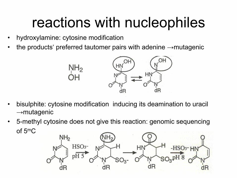
reactions with nucleophiles • hydroxylamine: cytosine modification
• the products‘ preferred tautomer pairs with adenine →mutagenic
• bisulphite: cytosine modification inducing its deamination to uracil
→mutagenic
• 5-methyl cytosine does not give this reaction: genomic sequencing
of 5mC

reactions with electrophiles
• attacking N and/or O atoms
• nitrous acid (HNO2) causes base deamination (C→U,
A→I) – affecting base pairing, mutagenic
• aldehydes: reactions with primary amino groups
• formaldehyde: two step reaction

DNA alkylation
• hard or soft alkylating agents
• hard ones attack both N and O atoms, soft only N
• dimethyl sulfate: typical soft alkylating agent
• N-alkyl-N-nitroso urea: typical hard alkylating agent
• modifies all N + O in bases as well as phosphate groups (forming
phosphotriesters)
• analytical use (sequencing, foorprinting)

Biological consequences of base alkylation
• N-alkylation: the primary site = N7 of guanine (accessible in both ss and dsDNA) – does not change base pairing; easily repairable
• N3 of adenine or guanine: located in minor groove – cytotoxic modification (DNA/RNA polymerization blocked)
• N1 of guanine: interferes with base pairing
• O-alkylation (G-O6, T-O6) the bases „locked“ in enol forms → improper base pairing → mutagenic

• chloro- (bromo-) acetaldehyde: two reactive centres
(aldehyde and alkylhalogenide)
• reaction with C or A
• chemical probes (react only with unpaired bases)
• diethyl pyrocarbonate: acylation of purines (primarily
A) at N7
• modification leads to opening of the imidazole ring
• chemical DNA probing

Metabolically activated carcinogens
• some substances became toxic after their metabolic conversion
• detoxifying machinery of the organism acts here as a bad fellow
• microsomal hydroxylase complex, cytochrome P450
• the role of this system is to introduce suitable reactive groups into xenobiotics enabling their conjugation with other molecules followed by removal from the organism
• but….

Metabolically activated carcinogens
• aromatic nitrogenous compounds (amines, nitro- or azo- compounds):
• aromatic amines are converted into either (safe) phenols, or (dangerous) hydroxylamine derivatives
• azo- compounds: „cleaved“ into amines
• nitro- compounds: reduced into hydroxylamines

Metabolically activated carcinogens
• polycyclic aromatic hydrocarbons like benzo[ ]pyrene:
three-step activation
– P450 introduces epoxy group
– epoxide hydrolase opens the epoxide circle
– P450 introduces second epoxy group
• DNA adduct formation (primarily -NH2 of guanine, then
G-N7, G-O6 and A-N6)
• similar pathway of
aflatoxin activation
bay region

anticancer drugs • some types of antineoplastic agents act via formation of
DNA adducts
• metallodrugs: mainly platinum complexes
(ineffective)
N
NN
O
NH2
Pt
N
NN
O
NH2
NH2NH
2
N N
N
NN
O
NH2
Pt
N
NN
NH2NH
2
N N
NH2

cisplatin: reaction with DNA in certain
sequence motifs
GN
G
Pt
NH
NH
Intrastrand GpG (~65%)
Intrastrand GpNpG (6-10%)
3’5’
5’3’
GG
Pt
5’ 3’
3’5’
GG Pt
NH
NH
Intrastrand ApG (~25%)
Interstrand G-G (1-2%)
5’ 3’
3’
5’
G
Pt NH
NH
A
5’ 3’
3’5’
NH 3
NH3
3
3
3
3
3
3
5´-AATTGGTACCAATT- 3´ 5´-AATTAGTACTAATT- 3´
3´-TTAACCATGGTTAA- 5´ 3´-TTAATCATGATTAA- 5´
(-GG-) (-AG-)
5´-AATTGATATCAATT- 3´ 5´-AATTATGTGTATAT- 3´
3´-TTAACTATAGTTAA- 5´ 3´-TTAATACACATATA- 5´
(-GA-) (-GTG-)
5´-AATTATGCATAATT- 3´ 5´-AATTAACGTTAATT- 3´
3´-TTAATACGTATTAA- 5´ 3´-TTAATTGCAATTAA- 5´
(-GC-) (-CG-)
some adduct types preferred
(and/or more stable than
others)
1,2-GG and 1,2-AG IACs =
the main cytotoxic lesions

other platinum complexes tested as cytostatics

mitomycin C
• reactive aziridine group, quinone group
• reductive activation
• bifunctional adducts
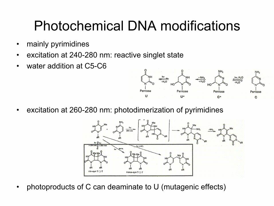
Photochemical DNA modifications • mainly pyrimidines
• excitation at 240-280 nm: reactive singlet state
• water addition at C5-C6
• excitation at 260-280 nm: photodimerization of pyrimidines
• photoproducts of C can deaminate to U (mutagenic effects)

effects of ionizing radiation
• mostly indirect – through water radiolysis
• each 1,000 eV produces ~27 •OH radicals that attack
DNA
• sugar damage:abstraction of hydrogen atoms from C-H
bonds
• a series of steps resulting
in strand breakage

effects of ionizing radiation
• base damage: hydroxylation and/or (under aerobic
conditions) peroxylation
pyrimidine products
purine products (mainly C8
hydroxylation or opening of the
imidazole ring)

chemical nucleases
species containing redox active metal ions mediating production of
hydroxyl radicals (or othe reactive oxygen species) via Fenton and/or
Haber-Weiss processes
bleomycine Fe or Cu
Men + H2O2 → Men+1 + •OH + OH-
iron/EDTA complex
Cu(phen)2 complex

Chemical approaches in Chemical approaches in
DNA studiesDNA studies
(several examples)(several examples)

Maxam and Gilbert method Maxam and Gilbert method
of DNA sequencingof DNA sequencing

HCOOH
(acid depurination)
DMS
(preferential methylation of G
at N7; spontaneous
depurination)
G
A
T
C hydrazine
(modification
&breakdown of the
pyrimidine ring)
hydrazine+NaCl
(selective modification of cytosine)
at sites of base modification (removal) the sugar-phosphate
backobone is labile towards alkali
treatment with hot piperidine → cleavage at such sites
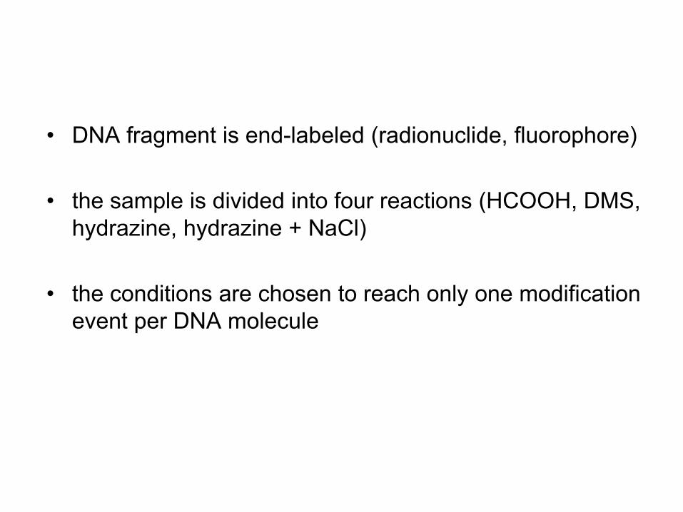
• DNA fragment is end-labeled (radionuclide, fluorophore)
• the sample is divided into four reactions (HCOOH, DMS,
hydrazine, hydrazine + NaCl)
• the conditions are chosen to reach only one modification
event per DNA molecule

HCOOH
G
A
T
C
G
A
T
C
A
T
C
G
A
T
C
G
T
C
G
A
T
C
G
A
T
C
A
T
C
G
A
T
C
G
T
C
or or or

only the „subfragment“
bearing the label is
„visible“ in the following
detection step
G
A
T
C
A
T
C
piperidine
G
A
T
C
A
T
C

G
A
T
C
G
A
T
C
A
T
C
G
A
T
C
T
C
G
A
T
C
A
T
C
T
C
or
or or
HC
OO
H
pip
eri
din
e
PA
GE
au
tora
dio
gra
ph
y

HC
OO
H
DM
S
G
A
T
C
G
A
T
C h
yd
razin
e
Hyd
razin
e
+N
aC
l


Sangerovo („dideoxy“) sekvenování

značení a sekvenování DNA
F. Sanger
2‘,3‘-dideoxynukleotidy:
terminátory syntézy DNA (není
kam napojit další nukleotid)
značené dideoxy:
navázání značky podle
komplementární báze
napojování nukleotidů přes
5‘-OH a 3‘-OH

templát
primer
syntetizovaný úsek
Syntéza DNA in vitro („primer extension“)
Deoxynukleotid trifosfáty (dNTP)
DNA polymeráza

templát
primer
ZNAČENÉ dNTP
DNA polymeráza
vložení značek do DNA pomocí DNA polymeráz
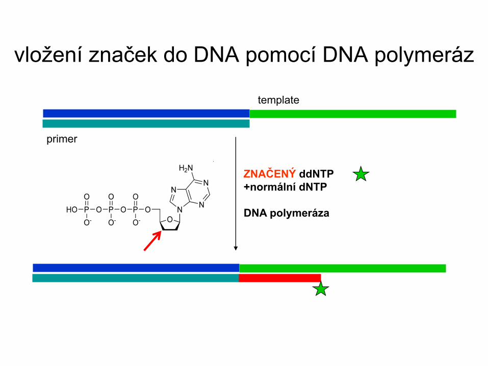
template
primer
ZNAČENÝ ddNTP
+normální dNTP
DNA polymeráza
vložení značek do DNA pomocí DNA polymeráz

značení a sekvenování DNA
F. Sanger
-směs normálních deoxy
a značených dideoxy
-pro každou bázi jiná barva
-různě dlouhé produkty,
dělení elektroforézou
-značka (barva) odpovídá
koncové bázi

templát
primer
Detekce a identifikace mutací a polymorfismů
-důležité pro diagnostiku (určité mutace v určitých pozicích jsou spojeny s
určitým onemocněním)
-zjišťuje se, jaká báze je v konkrétní pozici
G
G C
C T A
T A

DNA „footprinting“: determination of binding sites
of other molecules (e.g. proteins)
at the DNA sequence level

complex free DNA
DMS (guanine) piperidine
DNaseI
UO2 UV
Cu(phen)2
OH radicals

single strandsingle strand--selective selective
chemical probeschemical probes

Open local structures in negatively supercoiled DNA
relaxed
circular DNA
negatively supercoiled
DNA (linking deficit) stress related to the
negative superhelicity
(the linking deficit) can be
absorbed in local open
structures

DNA segments of specific sequence can adopt „alternative“ local
structures
cruciform DNA (inverted repeat)
Open local structures in negatively supercoiled DNA
unpaired bases

Triplex DNA
(homopurine·homopyrimidine stretch with mirror symmetry)
e.g. TTTTTTTTTTTTTTTTTTTTTTTTTTTTTTTTTTTTTTTT
AAAAAAAAAAAAAAAAAAAAAAAAAAAAAAAAAAAAAAAA
TTTTTTTTTTTTTTTTTTTTTTTTTTTTTTTTTTTTTTTT
(intermolecular triplex)
Open local structures in negatively supercoiled DNA

Otevřené lokální struktury v negativně
nadšroubovicové (sc) DNA
Intramolecular triplex
(homoPu•homoPy segment within negatively supercoiled DNA)
T
T T T
T T
A A
A
A A
A A

Chemicals selectively reacting with unpaired bases:
N
NH
O
O
CH3
Os, bipy
N
NH
O
O CH3
O
O
Os
O
O
N
NR R
osmium tetroxide complexes
(Os,L)
(T, more slowly C)
chloroacetaldehyde
(CAA)
(A, C)
diethyl pyrocarbonate
(DEPC)
(A, G)

footprinting of CGC+
triplex by DMS

Using the Maxam-Gilbert technique, it is possible to
determine with a high preciseness which nucleotides are
forming the local structure
modification of supercoiled DNA
restriction cleavage, radiactive labeling
hot piperidine
sequencing PAGE
the structure can be
deduced from the
modification pattern
TTTTTTTTTTTTTTTTTTTTTTTTTTTTTTTTTTTTTTTT
AAAAAAAAAAAAAAAAAAAAAAAAAAAAAAAAAAAAAAAA

DNA damage and repairDNA damage and repair

DNA: the genetic material DNA: the genetic material
ensuringensuring
preservation of the preservation of the
genetic informationgenetic information
its transfer to progeny its transfer to progeny
its transcription and its transcription and
translation into proteinstranslation into proteins
Damage to DNA mayDamage to DNA may
lead to change of the lead to change of the
genetic information genetic information
(mutation)(mutation)
affect gene expressionaffect gene expression
have severe health have severe health
impactsimpacts
Why is it important to study Why is it important to study
„DNA damage“?„DNA damage“?

DNA damage, mutation, lesion, mismatch…?DNA damage, mutation, lesion, mismatch…?
mutation may arise from (among others) DNA damage which is mutation may arise from (among others) DNA damage which is
not repaired prior to DNA replication, e.g..not repaired prior to DNA replication, e.g..
AGCCGATTAACTTAGCTTAGT
TCGGCTAATTGAATCGAATCT
AGCCGATTAGCTTAGCTTAGT
TCGGCTAATCGAATCGAATCT
semi-conservative replication (two cycles without repair)
AGCCGATTAGCTTAGCTTAGT
TCGGCTAATCGAATCGAATCT
„wild type“ (homo)duplex DNA
„wild type“ homoduplex mutated homoduplex
AGCCGATTAGCTTAGCTTAGT
TCGGCTAATUGAATCGAATCT
cytosine deamination
heteroduplex containing a single
base mismatch
DNA damage
lesion/mismatch
mutation (base pair substituted)

mutations arisemutations arise from unrepaired DNA damagefrom unrepaired DNA damage ((or from or from
replication errors)replication errors)
damageddamaged DNA is not DNA is not mutatedmutated yet! yet! (damage is usually (damage is usually
repaired in time i.e. before replication repaired in time i.e. before replication –– lesions and/or lesions and/or
mismatches are recognized by the reparation systems)mismatches are recognized by the reparation systems)
DNA with DNA with mutatedmutated nucleotide sequence does not nucleotide sequence does not
behave as behave as damageddamaged!! All base pairs in such DNA are All base pairs in such DNA are
„OK“ (no business for the DNA repair machinery) but the „OK“ (no business for the DNA repair machinery) but the
genetic information is genetic information is (hereditably)(hereditably) altered.altered.
DNA damage, mutation, lesion, mismatch…?DNA damage, mutation, lesion, mismatch…?

DNA in the cells is permanently exposed to various chemical DNA in the cells is permanently exposed to various chemical
or physical agentsor physical agents
endogenous - products and intermediates of metabolism
exogenous - environmental (radiation, pollutants)
Scharer, O. D. (2003) Chemistry and biology
of DNA repair, Angew. Chem. Int. Ed. 42,
2946-74.

single-strand break double-strand break
interruptions of DNA sugar-phosphate
backbone
abasic sites
interruption
of the N-glykosidic
linkage
reactive oxygen species
action of nucleases
consequence of base damage
spontaneous hydrolysis
(depurination)
consequence of base
damage
Most frequent products of DNA damage („lesions“)Most frequent products of DNA damage („lesions“)

N
N
NH
N
O
NH2
R
NH
N
R
O
O
CH3N
R
NH2
O
N
N
N
NH
N
O
NH
R
OH
OH
OH
N
N
NH
N
O
NH2
R
Pt
N
N
NH
N
O
NH2
R
NH3H
3N
NH
N
R
O
O
NH
N
R
O
O
NH
N
R
O
O
CH3CH
3
guanin
N
N
N
N
NH2
R
N
N
NH
N
O
NH2
R
CH3
N+
N
NH
N
O
NH2
R
CH3
N
N
N
N+
NH2
RCH3
adenin
NH
N
R
O
O
OH
OH
CH3
N
N
NH
N
O
NH2
O
R
N
NN
N
R
N
cytosin thymin
base damage:
chemical modifications
alkylation
oxidative damage
deamination
damage by UV radiation
(sunlight)
metabilically activated
carcinogens
anticancer drugs
Most frequent products of DNA damage („lesions“)Most frequent products of DNA damage („lesions“)

• estimated number of DNA-damage events in a
single human cell: 104-106 per day!!
• only a small number of base pairs alterations in
the genome are in principle sufficient for the
induction of cancer
• DNA-repair systems must effectively counteract
this threat
• in an adult human (1012 cells) about 1016–1018
repair events per day
Importance of DNA repair

p53 and others
DNA damage
cell cycle arrest
DNA replication
postponed until
DNA repair apoptosis
only then DNA
replication
followed by
cell division
damaged cell
eliminated
genomic
instability
mutations
cancer…
if unrepairable? if everything fails

DNA repair pathways
• direct reversal of damage
• base excision repair
• nucleotide excision repair
• mismatch repair
• repair of double strand breaks

Direct reversal of DNA damage
• photolyases: repair of cyclobutane dimers
• O6-alkylguanine transferase: reverses O6-alkylguanine to guanine
by transferring the alkyl group from DNA to a reactive cysteine group
of the protein
NH
N
R
O
O
NH
N
R
O
O
CH3CH
3
NH
N
R
O
O
CH3
NH
N
R
O
O
CH3 UV
photolyase

Base excision repair
• repair of damage by deamination (U, I), oxidation (8-oxoG), and alkylation
• initiated by DNA glycosylases, which recognize damaged bases and excise them from DNA by hydrolyzing the N-glycosidic bond
• substrate specificity of the glycosylases: developed to repair expectable „errors“?
• second enzyme is AP-lyase introducing single strand break next to the abasic site
• replacement of the abasic sugar by proper nucleotide
• sealing the break
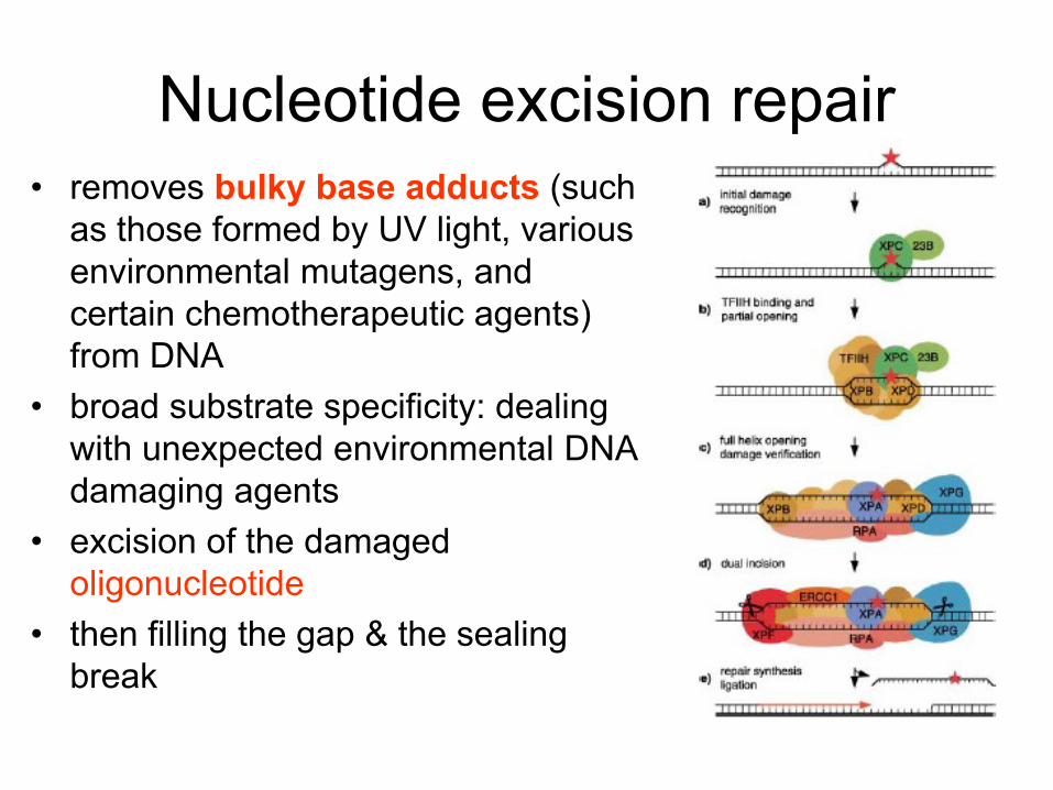
Nucleotide excision repair
• removes bulky base adducts (such
as those formed by UV light, various
environmental mutagens, and
certain chemotherapeutic agents)
from DNA
• broad substrate specificity: dealing
with unexpected environmental DNA
damaging agents
• excision of the damaged
oligonucleotide
• then filling the gap & the sealing
break

Mismatch repair • dealing with replication errors
• polymerases introduce about one erroneous nucleotide per 105 nucleotide; their 3’→5’- exonuclease activity decreases incidence of the errors to 1:107
• the MMR contributes to replication fidelity by a factor of 103 by removal of base-base mismatches, insertions and deletions (hence the resulting incidence of mutations due to erroneous replication is only 1:1010)
• the system must be able discrimitate between parental and daughter DNA strand!
• MutS binds to mismatches and insertion/deletion loops
• „repairosome“ formation, removal of a part of the daughter strand by 5’→3’- exonuclease
• new DNA synthesis and ligation

Repair of double strand breaks
• consequences of DSBs can be very severe (chromosome aberrations)
• two repair pathways:
• homologous recombination: an intrinsically accurate repair pathway that uses regions of DNA homology (such as sister chromatids) as coding information.

Repair of double strand breaks
• consequences of DSBs can be very severe (chromosome aberrations)
• two repair pathways:
• non-homologous end joining: conceptually simple pathway that involves the religation of broken ends (without using a homologous template
• less accurate: may loss of a few nucleotides at the damaged DNA ends

Examples of techniques used
to detect DNA damage

1. Techniques involving complete DNA hydrolysis followed
by determination of damaged entities by chromatography or
mass spectrometry

HPLC: 8-oxo guanine determination

32P-postlabeling: analysis of base adducts

1. Techniques involving complete DNA hydrolysis followed
by determination of damaged entities by chromatography or
mass spectrometry
2. Monitoring of changes in whole (unhydrolyzed) DNA
molecules: electrophoretic and immunochemical techniques

detection of strand breaks:
relaxation (and/or linearization) of plasmid supercoiled DNA
scDNA
(intact)
ocDNA (ssb)
linDNA (dsb)
(damaged)

„comet assay“ (dsb)
„alkaline elution assay“ (ssb + alkali-labile sites)

imunochemical techniques
when antibodies against the adducts
available
ELISA
In situ techniques
8-oxo guanine detection in situ in kidney tissue