Chemical, experimental, and morphological evidence · PDF fileChemical, experimental, and...
6
Chemical, experimental, and morphological evidence for diagenetically altered melanin in exceptionally preserved fossils Caitlin Colleary a,b , Andrei Dolocan c , James Gardner d , Suresh Singh a , Michael Wuttke e , Renate Rabenstein f , Jörg Habersetzer f , Stephan Schaal f , Mulugeta Feseha g , Matthew Clemens h , Bonnie F. Jacobs h , Ellen D. Currano i , Louis L. Jacobs h , Rene Lyng Sylvestersen j , Sarah E. Gabbott k , and Jakob Vinther a,d,l,1 a School of Earth Sciences, University of Bristol, Bristol BS8 1RJ, United Kingdom; b Department of Geosciences, Virginia Polytechnic Institute and State University, Blacksburg, VA 24060; c Texas Materials Institute, University of Texas at Austin, Austin, TX 78712; d Jackson School of Geosciences, University of Texas at Austin, Austin, TX 78712; e Department for the Conservation of the Cultural Heritage of Rhineland-Palatinate, 55116 Mainz, Germany; f Department of Palaeoanthropology and Messel Research, Senckenberg Research Institute, 60325 Frankfurt am Main, Germany; g Paleoanthropology and Paleoenvironment Program, School of Earth Sciences, College of Natural Sciences, Addis Ababa University, Addis Ababa, Ethiopia; h Roy M. Huffington Department of Earth Sciences, Southern Methodist University, Dallas, TX 75275; i Departments of Botany and Geology & Geophysics, University of Wyoming, Laramie, WY 82071; j Division of Natural History, MuseÒum, 7800 Skive, Denmark; k Department of Geology, University of Leicester, Leicester LE1 7RH, United Kingdom; and l School of Biological Sciences, University of Bristol, Bristol BS8 1TQ, United Kingdom Edited by Donald E. Canfield, Institute of Biology and Nordic Center for Earth Evolution, University of Southern Denmark, Odense M., Denmark, and approved August 26, 2015 (received for review May 19, 2015) In living organisms, color patterns, behavior, and ecology are closely linked. Thus, detection of fossil pigments may permit inferences about important aspects of ancient animal ecology and evolution. Melanin-bearing melanosomes were suggested to preserve as organic residues in exceptionally preserved fossils, retaining distinct morphology that is associated with aspects of original color patterns. Nevertheless, these oblong and spherical structures have also been identified as fossilized bacteria. To date, chemical studies have not directly considered the effects of diagenesis on melanin preserva- tion, and how this may influence its identification. Here we use time-of-flight secondary ion mass spectrometry to identify and chemically characterize melanin in a diverse sample of previously unstudied extant and fossil taxa, including fossils with notably different diagenetic histories and geologic ages. We document signatures consistent with melanin preservation in fossils ranging from feathers, to mammals, to amphibians. Using principal compo- nent analyses, we characterize putative mixtures of eumelanin and phaeomelanin in both fossil and extant samples. Surprisingly, both extant and fossil amphibians generally exhibit melanosomes with a mixed eumelanin/phaeomelanin composition rather than pure eumelanin, as assumed previously. We argue that experimental maturation of modern melanin samples replicates diagenetic chemical alteration of melanin observed in fossils. This refutes the hypothesis that such fossil microbodies could be bacteria, and demonstrates that melanin is widely responsible for the organic soft tissue outlines in vertebrates found at exceptional fossil localities, thus allowing for the reconstruction of certain aspects of original pigment patterns. paleocolor | melanosome | mass spectrometry | diagenesis | pigmentation S ince melanosomes were first described in fossil feathers (1), a number of studies have made inferences about original colors of extinct vertebrates, which were based on structural and mo- lecular analyses (2–8). However, these interpretations have been questioned (9–11). Animals synthesize two chemically distinct types of melanin: eumelanin (black to dark brown in color) and phaeomelanin (rufous, brown, and buff in color), occurring in eumelanosomes and phaeomelanosomes, respectively. Melanin is an extremely recalcitrant, complexly cross-linked polymer, which is mainly known to degrade through oxidation (12). The nature and function of eumelanin in organisms has been suggested as a possible explanation for its high preservation potential (1). In extant mam- mals and birds, eumelanosomes are oblong, with a higher aspect ratio than phaeomelanosomes, which are smaller and more spherical (3, 13). It is not clear whether other amniotes or fishes produce morphologically distinct melanosomes, but chemical evidence distinguishes pure eumelanin and phaeomelanin in turtles (14), and phaeomelanin has been tentatively reported in frogs (15), fish (16), and chitons (Mollusca) (17). Melanosome morphology currently serves as the primary basis for interpretations of fossil feather color, as it correlates with several distinct melanin- based colors and iridescence in modern birds (1, 3, 4). However, maturation experiments simulating diagenesis in recent feathers suggest that the dimensions of melanosomes are altered when sub- jected to high pressures and temperatures (5, 18), suggesting that melanosome morphology may not be a reliable indicator of color in fossil feathers (19). Further, outside of mammals and birds (13), the extent to which melanosome morphology is a reliable indicator of melanin chemistry, and therefore color, is unknown. Recent postulations have suggested that oblong and spheroi- dal microstructures preserved in carbonaceous films are of bac- terial origin rather than fossilized melanosomes (10). It was argued that bacteria cultured on modern feathers do not appear to differ in gross morphology from melanosomes (10). However, Moyer et al. (10) failed to factor in the arrangement and size of the observed bacteria, which were several times larger than the Significance Melanin is a widespread pigment that provides black to red- dish brown hues to organisms. Recent evidence has shown that melanin is retained in exceptionally preserved fossils, including feathered dinosaurs, allowing the reconstruction of ancient color patterns. However, little is known about the chemical preser- vation of melanin or its distribution in the fossil record. Here, we show that melanin is preserved in a number of soft-bodied fossils, but its burial under high pressure and temperature for millions of years alters its original chemistry. The widespread occurrence of melanin substantiates the applicability of recon- structing aspects of original color patterns and allows us to dismiss the alternative suggestion that these structures are mi- crobial in origin. Author contributions: J.V. designed research; C.C., A.D., and J.V. performed research; A.D., J.G., M.W., R.R., J.H., S. Schaal, M.F., M.C., B.F.J., E.D.C., L.L.J., R.L.S., and S.E.G. contributed new reagents/analytic tools; C.C., A.D., S. Singh, and J.V. analyzed data; and C.C., A.D., and J.V. wrote the paper. The authors declare no conflict of interest. This article is a PNAS Direct Submission. 1 To whom correspondence should be addressed. Email: [email protected]. This article contains supporting information online at www.pnas.org/lookup/suppl/doi:10. 1073/pnas.1509831112/-/DCSupplemental. 12592–12597 | PNAS | October 13, 2015 | vol. 112 | no. 41 www.pnas.org/cgi/doi/10.1073/pnas.1509831112
Transcript of Chemical, experimental, and morphological evidence · PDF fileChemical, experimental, and...
Chemical, experimental, and morphological evidencefor diagenetically altered melanin in exceptionallypreserved fossilsCaitlin Collearya,b, Andrei Dolocanc, James Gardnerd, Suresh Singha, Michael Wuttkee, Renate Rabensteinf,Jörg Habersetzerf, Stephan Schaalf, Mulugeta Fesehag, Matthew Clemensh, Bonnie F. Jacobsh, Ellen D. Curranoi,Louis L. Jacobsh, Rene Lyng Sylvestersenj, Sarah E. Gabbottk, and Jakob Vinthera,d,l,1
aSchool of Earth Sciences, University of Bristol, Bristol BS8 1RJ, United Kingdom; bDepartment of Geosciences, Virginia Polytechnic Institute and StateUniversity, Blacksburg, VA 24060; cTexas Materials Institute, University of Texas at Austin, Austin, TX 78712; dJackson School of Geosciences, University ofTexas at Austin, Austin, TX 78712; eDepartment for the Conservation of the Cultural Heritage of Rhineland-Palatinate, 55116 Mainz, Germany; fDepartmentof Palaeoanthropology and Messel Research, Senckenberg Research Institute, 60325 Frankfurt am Main, Germany; gPaleoanthropology andPaleoenvironment Program, School of Earth Sciences, College of Natural Sciences, Addis Ababa University, Addis Ababa, Ethiopia; hRoy M. HuffingtonDepartment of Earth Sciences, Southern Methodist University, Dallas, TX 75275; iDepartments of Botany and Geology & Geophysics, University of Wyoming,Laramie, WY 82071; jDivision of Natural History, Muse�um, 7800 Skive, Denmark; kDepartment of Geology, University of Leicester, Leicester LE1 7RH,United Kingdom; and lSchool of Biological Sciences, University of Bristol, Bristol BS8 1TQ, United Kingdom
Edited by Donald E. Canfield, Institute of Biology and Nordic Center for Earth Evolution, University of Southern Denmark, Odense M., Denmark, and approvedAugust 26, 2015 (received for review May 19, 2015)
In living organisms, color patterns, behavior, and ecology are closelylinked. Thus, detection of fossil pigments may permit inferencesabout important aspects of ancient animal ecology and evolution.Melanin-bearing melanosomes were suggested to preserve asorganic residues in exceptionally preserved fossils, retaining distinctmorphology that is associated with aspects of original color patterns.Nevertheless, these oblong and spherical structures have also beenidentified as fossilized bacteria. To date, chemical studies have notdirectly considered the effects of diagenesis on melanin preserva-tion, and how this may influence its identification. Here we usetime-of-flight secondary ion mass spectrometry to identify andchemically characterize melanin in a diverse sample of previouslyunstudied extant and fossil taxa, including fossils with notablydifferent diagenetic histories and geologic ages. We documentsignatures consistent with melanin preservation in fossils rangingfrom feathers, to mammals, to amphibians. Using principal compo-nent analyses, we characterize putative mixtures of eumelanin andphaeomelanin in both fossil and extant samples. Surprisingly, bothextant and fossil amphibians generally exhibit melanosomes witha mixed eumelanin/phaeomelanin composition rather than pureeumelanin, as assumed previously. We argue that experimentalmaturation of modernmelanin samples replicates diagenetic chemicalalteration of melanin observed in fossils. This refutes the hypothesisthat such fossil microbodies could be bacteria, and demonstrates thatmelanin is widely responsible for the organic soft tissue outlines invertebrates found at exceptional fossil localities, thus allowing for thereconstruction of certain aspects of original pigment patterns.
paleocolor | melanosome | mass spectrometry | diagenesis | pigmentation
Since melanosomes were first described in fossil feathers (1), anumber of studies have made inferences about original colors
of extinct vertebrates, which were based on structural and mo-lecular analyses (2–8). However, these interpretations have beenquestioned (9–11). Animals synthesize two chemically distinct typesof melanin: eumelanin (black to dark brown in color) andphaeomelanin (rufous, brown, and buff in color), occurring ineumelanosomes and phaeomelanosomes, respectively. Melanin isan extremely recalcitrant, complexly cross-linked polymer, which ismainly known to degrade through oxidation (12). The nature andfunction of eumelanin in organisms has been suggested as a possibleexplanation for its high preservation potential (1). In extant mam-mals and birds, eumelanosomes are oblong, with a higher aspectratio than phaeomelanosomes, which are smaller and morespherical (3, 13). It is not clear whether other amniotes or fishesproduce morphologically distinct melanosomes, but chemical
evidence distinguishes pure eumelanin and phaeomelanin inturtles (14), and phaeomelanin has been tentatively reported infrogs (15), fish (16), and chitons (Mollusca) (17). Melanosomemorphology currently serves as the primary basis for interpretationsof fossil feather color, as it correlates with several distinct melanin-based colors and iridescence in modern birds (1, 3, 4). However,maturation experiments simulating diagenesis in recent featherssuggest that the dimensions of melanosomes are altered when sub-jected to high pressures and temperatures (5, 18), suggesting thatmelanosome morphology may not be a reliable indicator of color infossil feathers (19). Further, outside of mammals and birds (13), theextent to which melanosome morphology is a reliable indicator ofmelanin chemistry, and therefore color, is unknown.Recent postulations have suggested that oblong and spheroi-
dal microstructures preserved in carbonaceous films are of bac-terial origin rather than fossilized melanosomes (10). It wasargued that bacteria cultured on modern feathers do not appear todiffer in gross morphology from melanosomes (10). However,Moyer et al. (10) failed to factor in the arrangement and size ofthe observed bacteria, which were several times larger than the
Significance
Melanin is a widespread pigment that provides black to red-dish brown hues to organisms. Recent evidence has shown thatmelanin is retained in exceptionally preserved fossils, includingfeathered dinosaurs, allowing the reconstruction of ancient colorpatterns. However, little is known about the chemical preser-vation of melanin or its distribution in the fossil record. Here, weshow that melanin is preserved in a number of soft-bodiedfossils, but its burial under high pressure and temperature formillions of years alters its original chemistry. The widespreadoccurrence of melanin substantiates the applicability of recon-structing aspects of original color patterns and allows us todismiss the alternative suggestion that these structures are mi-crobial in origin.
Author contributions: J.V. designed research; C.C., A.D., and J.V. performed research; A.D.,J.G., M.W., R.R., J.H., S. Schaal, M.F., M.C., B.F.J., E.D.C., L.L.J., R.L.S., and S.E.G. contributednew reagents/analytic tools; C.C., A.D., S. Singh, and J.V. analyzed data; and C.C., A.D.,and J.V. wrote the paper.
The authors declare no conflict of interest.
This article is a PNAS Direct Submission.1To whom correspondence should be addressed. Email: [email protected].
This article contains supporting information online at www.pnas.org/lookup/suppl/doi:10.1073/pnas.1509831112/-/DCSupplemental.
12592–12597 | PNAS | October 13, 2015 | vol. 112 | no. 41 www.pnas.org/cgi/doi/10.1073/pnas.1509831112
melanosomes recorded in any known vertebrate integument anddo not exhibit the unique arrangement or alignment that mela-nosomes display in feathers (20). Bacterial remains—includingmicrobial mats (21)—occur throughout the fossil record from theArchean to the present (22). These may be preserved via anumber of taphonomic pathways, e.g., phosphatization (23) orsilicification (24), but are not confidently known to be preserved assolid carbonaceous bodies similar to melanosomes.Analytical techniques that detect biomolecules indicate that
melanin granules occur in fossil cephalopods (25) and thatmelanin-bearing melanosomes are preserved in fossil fish (7, 26) andmarine reptiles (8). The most conclusive and comprehensive chem-ical study to date shows the presence of chemically intact melanin inquantities of ∼10% in Jurassic cephalopod ink sacs (25, 27). Otherstudies have demonstrated the applicability of time-of-flight sec-ondary ion mass spectrometry (TOF-SIMS), a surface-sensitivetechnique, which collects in situ mass spectrometric data fromfossil samples with only minor alteration of the sample surface (7, 8,28). The secondary ion spectra of putative fossil melanosomes con-tain the representative markers that are found in extant melaninsamples qualitatively (7, 8), and are distinct from the spectra acquiredon bacterial samples and on aromatic organic molecules similar tomelanin, such as porphyrins (7, 8). However, when quantified usingprincipal component analysis (PCA), the fossil and extant melaninsamples are also distinct (8), indicating that they consist of moleculesof different composition, phase, or structure.We hypothesize that fossil melanosomes contain diageneti-
cally altered melanin and explore the chemical composition offossil melanosomes by analyzing pure samples of extant melaninand previously unstudied vertebrate fossils using TOF-SIMS. Totest our hypothesis, we performed maturation experiments of mel-anin. We subjected pure melanin extracts to high pressure andtemperatures (200 °C/250 bar and 250 °C/250 bar, respectively, for
24 h) in sealed gold tubes to mimic burial conditions and theresulting chemical alterations (29, 30). We compare the TOF-SIMSspectra of fresh, artificially matured, and fossil samples by PCA. Wechose a diverse and complementary array of fossils, with soft tissuepreservation and discernible melanosomes, from a wide range oflocalities to understand the significance of diagenesis and taxonomyin fossil melanin composition.
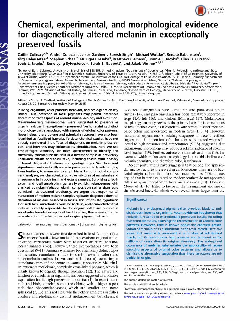
ResultsMelanosome Morphology and Inferred Original Chemistry. The fos-sils in our analyses include amphibians, birds, mammals, fish, andcephalopods that preserve putative melanosomes or melaningranules (Fig. 1 and SI Appendix, Table S1), and which range inage from Carboniferous (Fig. 1 G and H) to Miocene (Fig. 1 Uand V). Material from the Eocene Messel Formation is partic-ularly diverse, including frog eyes (Fig. 1 Q and R) and skin (Fig.1 S and T), mammalian hair (two bat species, Palaeochiropteryxand Hassianycteris) (Fig. 1 I−L), and two feathers (Fig. 1 A, B, E,and F), which serve to test for variation in melanin composition(eumelanin−phaeomelanin) within a single depositional andtaphonomic system. We infer that the Messel feathers (Fig. 1 A,B, E, and F) are iridescent based upon the morphologies of theputative melanosomes, which were analyzed by quadratic dis-criminant analysis, as previously used by Li et al. (3, 4) for birds.Likewise, we infer that the two bats (Fig. 1 I−L) were brown,based upon observations of small (<650 nm), subrounded pu-tative melanosomes, suggesting a predominantly phaeomelanin-based composition.
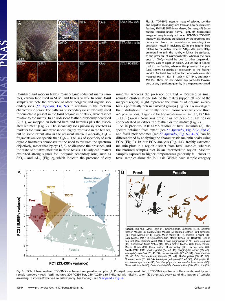
TOF-SIMS and PCA. Overall, the mass spectra of the fossils qualita-tively resemble those observed in previous studies (7, 8) relative toextant melanin reference samples herein (nine bird species, frogs,and a cephalopod ink) and our chosen negative control samples
Fig. 1. Range of fossils analyzed with TOF-SIMS imaged using photography and under SEM. (A and B) Fossil complete bird with associated feathers,Messelornis, Messel, Eocene, Germany. (C and D) Articulated bird associated with feathers, undescribed, Fur Formation, Eocene, Denmark. (E and F )Isolated feather previously described as iridescent (2), Messel, Eocene, Germany. (G and H) Complete cyclostome fish preserving the eye, detailed, MazonCreek, Carboniferous. (I and J) The bat Hassianycteris, Messel, Eocene, Germany. (K and L) The bat Palaeochiropteryx, Messel, Eocene, Germany. (M and N )Fossil ink sac, Lyme Regis, Lower Jurassic, England. (O and P) Fossil octopus (?Keuppia) associated with ink sac, Cretaceous, Hakel, Lebanon; UV imagecourtesy of Jonathan Jackson. (Q and R) Fossil frog preserving eyes, Paleobatrachus, Messel, Eocene, Germany. (S and T ) Same frog as in Q, detailing skinpreserved on the hind leg, Messel, Eocene, Germany. (U and V ) Fossil frog, Pipidae, Mush Valley, Miocene, Ethiopia. (W and X ) Fossil tadpole, Pelobates,Oligocene, Enspel, Germany. Specimens were photographed under normal light, except in O, which is imaged under UV light as well as scanning electronmicroscopy images. See SI Appendix, Table S1, for details. (Scale bars, 1 μm.)
Colleary et al. PNAS | October 13, 2015 | vol. 112 | no. 41 | 12593
EART
H,A
TMOSP
HER
IC,
ANDPL
ANET
ARY
SCIENCE
SEV
OLU
TION
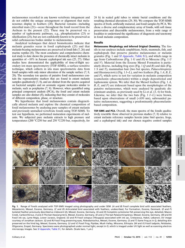
(fossilized and modern leaves, fossil organic sediment matrix sam-ples, carbon tape used in SEM, and bakers yeast). In some fossilsamples, we note the presence of other inorganic and organic sec-ondary ions (SI Appendix, Fig. S2) in addition to the melanincharacteristic peaks. The patterns of secondary ions previously listedfor eumelanin present in the fossil organic imprints (7) were distinctrelative to the matrix. In an iridescent feather, previously described(2, 31), we mapped an isolated barb and barbules plus the associ-ated sediment (Fig. 2). The secondary ions previously selected asmarkers for eumelanin were indeed highly expressed in the feather,but to some extent also in the adjacent matrix. Generally, CnH−fragments are less specific than CnN−. The lack of specificity of suchorganic fragments demonstrates the need to evaluate the spectrumobjectively, rather than by eye (7, 8), to diagnose the presence andthe state of putative melanin in these fossils. The adjacent matrixexhibited strong signals for inorganic secondary ions, such asSiO3− and Al+, (Fig. 2), which indicate the presence of clay
minerals, whereas the presence of CO2H− localized in smallrounded clusters at one side of the matrix (upper left side of themapped region) might represent the remains of organic micro-fossils potentially rich in carboxyl groups (Fig. 2). To investigatethe distribution of bacterially derived biomarkers, we chose threem/z positive ions, diagnostic for hopanoids (m/z = 149.113, 177.164,191.18) (32–34). None was present in noticeable quantities orconcentrated in either the feather or the matrix (Fig. 2).As in previous TOF-SIMS studies of fossil melanin (8), the
spectra obtained from extant (see SI Appendix, Fig. S2 E and F)and fossil melanosomes (see SI Appendix, Fig. S2 A−D) can bedifferentiated by analyzing the characteristic melanin peaks usingPCA (Fig. 3). In our PCA analysis (Fig. 3A), freshly extractedmelanin plots in a region distinct from fossil samples, whereasthe matured samples plot in an intermediate region. Modernsamples exposed to higher temperatures generally fall closer tofossil samples along the PC1 axis. Within each sample category
Fig. 2. TOF-SIMS intensity maps of selected positiveand negative secondary ions from an Eocene iridescentfeather, SMF-ME 3850 fromMessel, Germany. (A) Entirefeather imaged under normal light. (B) Microscopicimage of sample analyzed under TOF-SIMS. TOF-SIMSintensity distributions are labeled by the predicted sec-ondary ion. Note the correlation of secondary ions,previously noted in melanins (7) in the feather barbrelative to the matrix, whereas SiO3−, Al+, and CHO2−are more intense in the matrix, which can be attributedto the presence of aluminosilicates, whereas the pres-ence of CHO2− could be due to other organic-richsources, such as algae or pollen. Sodium (Na+) is local-ized to the feather, whereas the presence of copper(Cu+) shows no particular correlation to the featherimprint. Bacterial biomarkers for hopanoids were alsomapped: m/z = 149.113+, m/z = 177.164+, and m/z =191.18+. These did not exhibit any particular localiza-tion, or any significant quantity in the spectra obtained.
PC
2 (1
5.68
6% v
aria
nce
)
PC1 (23.436% variance)
Fossils: Ink sac, Lyme Regis (1), Cephalopods, Lebanon (2, 3), Isolated feather, Messel (4), Messelornis, Messel (5), Isolated feather, Fur Formation (6), Frogs, Messel (7, 8), Frogs, Mush Valley (9, 10), Tadpole, Enspel (11), Bats, Messel (12, 13), Cyclostome fish, Mazon Creek (14) Control: Recent oak leaf (15), Sequoia (18), Fossil leaf, Mush Valley (19), Rock matrix, Messel (20), Rock matrix, Mazon Creek (21), Rock matrix, Mush Valley (22), Carbon tape (23)Fresh, 200°, 250°: Gallus gallus (24, 40, 48), Troglodytes aedon (25, 49), Anas platyrhynchos (26, 41, 50), Junco hyemalis (27, 42, 51), Columba livia(28, 43, 52), Dumetella carolinensis (29, 44), Gallus gallus (30, 45, 53), Corvus corone (31, 46, 54), Meleagris gallopavo (32, 47, 55), Pelophylax kl.esculentus eye tissue (33, 34), Pelophylax kl. esculentus liver tissue (35), Sepia officianalis (36), Columba livia (37), Columba livia (38), Pica pica (39)
A B
Fig. 3. PCA of fossil melanin TOF-SIMS spectra and comparative samples. (A) Principal component plot of TOF-SIMS spectra with the area defined by eachsample category (fresh, fossil, matured 200 °C/250 bar, 250 °C/250 bar) indicated with distinct color. (B) Schematic overview of distribution of samplesaccording to inferred/observed color/taxonomy. For loadings, see SI Appendix, Fig. S4.
12594 | www.pnas.org/cgi/doi/10.1073/pnas.1509831112 Colleary et al.
(fresh, 200 °C/250 bar, 200 °C/250 bar), eumelanin- and phaeo-melanin-rich samples separate along the PC2 axis (Fig. 3 A and B).The fossil melanin samples show a trend in which the squid ink(Jurassic, Cretaceous) and the two iridescent feathers (iridescenceis generated by eumelanosomes) from Messel plot on one end ofthe PC1 axis, whereas the presumed brown (phaeomelanin-rich)bat samples, also from Messel, plot opposite. Amphibians (skinand eyes, Eocene−Miocene, including Messel), the feather fromEocene of the Fur Fm., and the Carboniferous fish eye plot inintermediate positions (Fig. 3). No clear trend can be attributed togeologic age or burial history of the samples, but there is distinctclustering by phylogeny and presumed original melanin composi-tion as inferred from melanosome morphology (Fig. 3B).The chemical distinction between phaeomelanin-rich and
eumelanin-rich melanosomes of both the fresh and maturedfeathers sorts along the PC2 axis. The fossil samples inferred toconsist of eumelanin and phaeomelanin spread along the PC1 axisrather than the PC2 axis. Nevertheless, they have a similar trend intaxonomic distribution: Squid ink and iridescent feathers plot atone end, and putative phaeomelanin-rich melansomes from thefossil bats plot at the other, with amphibians in between. It ap-pears, from the maturation experiments, that the variations ineumelanin and phaeomelanin experience a clockwise rotationin the spread of the spectra in the PCA plot, an observation de-serving further scrutiny.We further notice a distinct trend in the melanin TOF-SIMS
spectra when comparing the maturation experiments to the freshsamples. The relative intensity of peaks within the spectrum ismore attenuated in the matured samples (see SI Appendix, Fig. S1)compared with the fresh ones (the differences between strong andweak peaks are greater). A similar trend is observed in fossilsamples and can probably be ascribed to the loss of smaller, volatilecompounds, such as NH4, H2O, and CO2, by dehydration of themelanin (35).
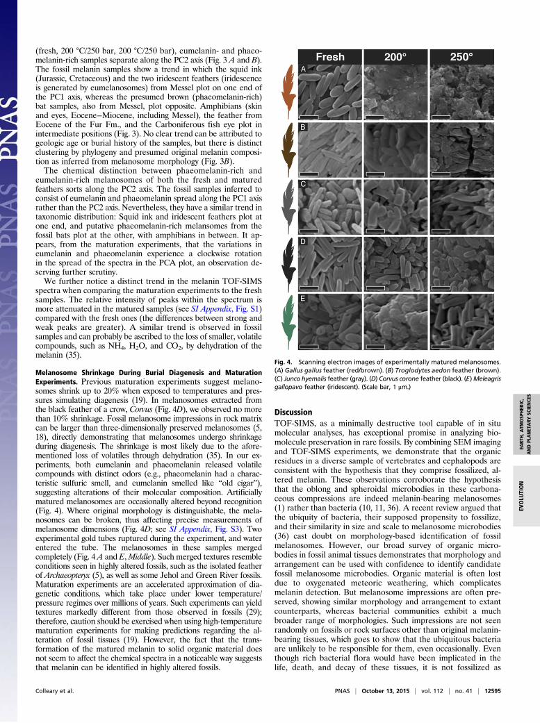
Melanosome Shrinkage During Burial Diagenesis and MaturationExperiments. Previous maturation experiments suggest melano-somes shrink up to 20% when exposed to temperatures and pres-sures simulating diagenesis (19). In melanosomes extracted fromthe black feather of a crow, Corvus (Fig. 4D), we observed no morethan 10% shrinkage. Fossil melanosome impressions in rock matrixcan be larger than three-dimensionally preserved melanosomes (5,18), directly demonstrating that melanosomes undergo shrinkageduring diagenesis. The shrinkage is most likely due to the afore-mentioned loss of volatiles through dehydration (35). In our ex-periments, both eumelanin and phaeomelanin released volatilecompounds with distinct odors (e.g., phaeomelanin had a charac-teristic sulfuric smell, and eumelanin smelled like “old cigar”),suggesting alterations of their molecular composition. Artificiallymatured melanosomes are occasionally altered beyond recognition(Fig. 4). Where original morphology is distinguishable, the mela-nosomes can be broken, thus affecting precise measurements ofmelanosome dimensions (Fig. 4D; see SI Appendix, Fig. S3). Twoexperimental gold tubes ruptured during the experiment, and waterentered the tube. The melanosomes in these samples mergedcompletely (Fig. 4 A and E,Middle). Such merged textures resembleconditions seen in highly altered fossils, such as the isolated featherof Archaeopteryx (5), as well as some Jehol and Green River fossils.Maturation experiments are an accelerated approximation of dia-genetic conditions, which take place under lower temperature/pressure regimes over millions of years. Such experiments can yieldtextures markedly different from those observed in fossils (29);therefore, caution should be exercised when using high-temperaturematuration experiments for making predictions regarding the al-teration of fossil tissues (19). However, the fact that the trans-formation of the matured melanin to solid organic material doesnot seem to affect the chemical spectra in a noticeable way suggeststhat melanin can be identified in highly altered fossils.
DiscussionTOF-SIMS, as a minimally destructive tool capable of in situmolecular analyses, has exceptional promise in analyzing bio-molecule preservation in rare fossils. By combining SEM imagingand TOF-SIMS experiments, we demonstrate that the organicresidues in a diverse sample of vertebrates and cephalopods areconsistent with the hypothesis that they comprise fossilized, al-tered melanin. These observations corroborate the hypothesisthat the oblong and spheroidal microbodies in these carbona-ceous compressions are indeed melanin-bearing melanosomes(1) rather than bacteria (10, 11, 36). A recent review argued thatthe ubiquity of bacteria, their supposed propensity to fossilize,and their similarity in size and scale to melanosome microbodies(36) cast doubt on morphology-based identification of fossilmelanosomes. However, our broad survey of organic micro-bodies in fossil animal tissues demonstrates that morphology andarrangement can be used with confidence to identify candidatefossil melanosome microbodies. Organic material is often lostdue to oxygenated meteoric weathering, which complicatesmelanin detection. But melanosome impressions are often pre-served, showing similar morphology and arrangement to extantcounterparts, whereas bacterial communities exhibit a muchbroader range of morphologies. Such impressions are not seenrandomly on fossils or rock surfaces other than original melanin-bearing tissues, which goes to show that the ubiquitous bacteriaare unlikely to be responsible for them, even occasionally. Eventhough rich bacterial flora would have been implicated in thelife, death, and decay of these tissues, it is not fossilized as
Fig. 4. Scanning electron images of experimentally matured melanosomes.(A) Gallus gallus feather (red/brown). (B) Troglodytes aedon feather (brown).(C) Junco hyemalis feather (gray). (D) Corvus corone feather (black). (E)Meleagrisgallopavo feather (iridescent). (Scale bar, 1 μm.)
Colleary et al. PNAS | October 13, 2015 | vol. 112 | no. 41 | 12595
EART
H,A
TMOSP
HER
IC,
ANDPL
ANET
ARY
SCIENCE
SEV
OLU
TION
organic microbodies except when encapsulated in cherts. Ar-chaea has never been found fossilized, yet its biomarkers areubiquitous (35). Although bacterial activity are relevant inother aspects of soft tissue fossilization, such as in pyrite (23)and calcium phosphate (23, 24) mineralization, the latter ofwhich appears to occasionally preserve microbial fabrics andpotentially also bacterial microbodies, the suggestion (36) thatbacteria only fossilize locally in melanin-bearing tissues, to theexclusion of other, more decay-prone tissues, is untenable. In fact,the argument could be considered reductio ad absurdum, the pre-dictions of which are effectively falsified by the results of ourexperiments and previous work demonstrating the unique ar-rangement of melanosomes in pigmented and iridescent feathers,which bacteria would not replicate (1, 2).Glass et al. (25, 27) documented, using pyrolysis GC-MS, that
Jurassic cephalopod ink melanin produced sulfur-containing com-pounds, such as thiophenes and alkylated thiophenes, which are notexpected phaeomelanin products. This suggests a role for earlydiagenetic alteration and preservation reactions in euxinic (sulfidic)environments, which essentially vulcanize organic substances. Itcould be surmised that these reactions mainly affect marine fossilsdue to the higher sulfate content of marine settings. We note,however, that feathers and hair from the freshwater locality,Messel oil shale, are locally enriched in pyrite framboids, whichsuggests availability of reactive hydrogen sulfide, as confirmed byother studies (38). A more pressing concern is that the data fromthe TOF-SIMS analyses used in melanin characterization (7, 8)are based on assemblages of fragment ions from larger molecules.Individually, these small fragments have little diagnostic value andcould potentially lead to conflation of original sulfur-containingmelanin (phaeomelanin) and the products of secondarily sulfurized(eu)melanin. However, the apparent grouping of fossil melano-somes by taxonomy and chemistry, rather than by environmentalsetting, suggests that diagenetic sulfur incorporation, if it occurs,is negligible.In addition, we document the presence of melanosomes in
fossil mammals for the first time, to our knowledge, both chemi-cally and morphologically, showing that two species of bats fromthe Eocene of Messel (48.2 Ma) were originally brown. Amphibianmelanosomes are intermediate in morphology between eumela-nosomes and phaeomelanosomes (Fig. 1 Q−Y). These resultscontradict recent studies arguing that melanosomes outside ofparavian dinosaurs (including birds) and mammals lack morpho-logical distinction (13), and that they can reflect differences inphysiology. Rather, amphibian melanosomes are generally ofmixed composition, and thus intermediate in morphology. Thefossil Carboniferous fish shows a similar trend in our PCA plot,also suggesting a mixed melanin composition. Another studyused pyrolysis GC-MS to study a fossil tadpole from Enspel (38)and could not conclusively present evidence for melanin due tolack of comparisons to extant, fossil, or artificially maturedmelanin samples and to previous studies on fossil eumelaninusing the same experimentation (25, 27). However, we show herethat, in our comparative framework, the Enspel melanosomes doretain melanin residues, contrary to earlier studies suggesting amicrobial origin (39).The emerging picture, suggesting a more widespread occur-
rence of phaeomelanin in groups other than mammals and birds,is of interest. Phaeomelanin was thought to be restricted towarm-blooded animals, presumably evolving convergently formetabolic reasons (40). However, it appears that phaeomelaninis actually more widespread in ectothermic organisms than pre-viously thought (14–17). As proposed recently (20), the morepure melanins in mammals and birds could reflect the markedshift in external integument with the evolution of hair andfeathers for insulation in these warm-blooded animals ratherthan physiology (13). With the evolution of insulating featurescovering the body, more-colorful chromatophores were lost,
and selection pressure to evolve more-radical variants of melaninsto provide diverse colors, iridescence, and patterns came into play.Despite the advances in the identification of melanin by mass
spectrometric methods, confident reconstruction of ancient colorrelies on a combination of both chemical analyses and interp-retations of fossil melanosome morphology. Maturation experi-ments confirm the presence of diagenetically altered melanin byreconstructing the diagenetic pathways that lead to fossil melanin.Furthermore, we show here that TOF-SIMS is able to distinguishbetween different mixtures of eumelanin and phaeomelanin,which are congruent with the observed melanosome morphol-ogy. Electron microscopy is still necessary for inferring ancientmelanin-based color patterns (1), as melanosome chemistry doesnot distinguish gray from brown colors (Fig. 3B), the relativebrightness of a color pattern, or the propensity for melanosomesto form iridescent nanostructures. It is necessary to do a detailedtaphonomic study of each case to understand the degree of al-teration and shrinkage of the melanosomes within the fossil (20).Statistical quadratic discriminant analyses of melanosome mor-phology allow for inferring melanin-based colors (black, brown,gray) and iridescence in birds with high accuracy (3, 4). Relativebrightness can be assessed through the characterization of mela-nosome density (6).
Materials and MethodsSpecimens. For details on specimens, see SI Appendix, Table S1.
Melanin Extractions. A modified protocol (41) for enzyme melanin extractionwas used to obtain purified melanin samples from a variety of tissues. Birdsamples used in maturation experiments were similar taxonomically to thosepublished recently (42). Additional samples (100−500 mg) were washed threetimes with 1 mL acetone and once with biomolecular grade water. They werethen added to 1.5 mL of phosphate buffer and DTT. Samples were then in-cubated for 24 h at 37.5 °C and stirred at 200 rpm. Then, 1.5 mL phosphatebuffer (15 mL), DTT (5 μL), and Proteinase-K (5 mg) were added, and thesamples were again incubated for 24 h at the same temperature and speed. Thesamples were removed and centrifuged for 3 min at 16.1 relative centrifugalforce before the pellets were washed in biomolecular-grade water six times.Then, 1.5 mL of phosphate buffer (15 mL), DTT (15 μL), and Papain (5 mg) wereadded to each sample, and samples were incubated for 24 h. Samples werethen washed six times with biomolecular-grade water; 1.5 mL of phosphatebuffer, DTT, and Proteinase-K were added in the same quantities as before, andsamples were incubated for 24 h. Triton X-100 (0.05 mL/1 mL) was added, andsamples were stirred for 4 h. Samples were then washed nine times with bio-molecular-grade water, and 1.5 mL of phosphate buffer, DTT, and Proteinase-Kwere added before they were incubated for 24 h. Samples were washed withacetone and washed six times with biomolecular-grade water. Then, 1.5 mLof phosphate buffer, DTT, and Proteinase-K were added, and the sampleswere incubated for 24 h. This step was repeated for three additional days fortissue samples. On the final day, specimens were washed three times withbiomolecular-grade water and left to dry for 1 h under a laminar flow hood.
Maturation Experiments.Weperformed 20maturation experiments on feathersof nine species of birds, based on a protocol recently used in the analysis ofmorphological changes in melanosomes (19). A portion of the melanosomeextract was left unaltered for analysis of fresh melanin. Each experiment con-sisted of 1.0–6.6 mg of feather, weighed into 3-mm-o.d. gold capsules that hadbeen welded closed at one end using an oxyacetylene torch, and then eachcapsule was welded shut at the other end. Each capsule was then placed insidea Ni alloy pressure vessel, along with a Ni filler rod, and pressurized with waterto 25 MPa. Each pressure vessel was then placed inside a furnace and heated toeither 200 °C or 250 °C, and held at 25 MPa and temperature for 24 h. Pressurewas measured to ±0.1 MPa; the K-type thermocouples used have been found tobe precise to ±5 °C. The use of the Ni rod fixed the oxygen fugacity at aboutNi−NiO. The experiment was quenched after 24 h by removing the pressurevessel from the furnace and, first, blowing on the vessel with compressed airfor about 30 s, and then immersing it into a water bath. All capsules wereremoved from their pressure vessels and weighed to check that no leaks oc-curred during the experiment. The capsules were then cracked open, and thesamples were extracted and mounted on SEM stubs for TOF-SIMS analysis andSEM analysis.
12596 | www.pnas.org/cgi/doi/10.1073/pnas.1509831112 Colleary et al.
TOF-SIMS and PCA. An ION-TOF TOF-SIMS.5 was used with a pulsed (18 ns,10 kHz) analysis ion beam consisting of Bi3
+ clusters at 30-kV ion energy,which was raster-scanned over areas that typically varied between 100 ×100 μm2 and 500 × 500 μm2, depending on the quality (i.e., corrugation andconductivity) of the sample surface. The polyatomic sputtering was selectedto further enhance the signal and reduce fragmentation of large organicmolecules (43–45). To reduce the sputtering-induced sample charging, aconstant energy (21 eV) electron beam was shot on the sample during thedata acquisition. All detected secondary ions had negative polarity and anaverage mass resolution of ∼1–3,000 (m/δm). The base pressure during ac-quisition was <1 × 10⁻⁸ mbar. In addition, we used the Burst Alignmentmode on one sample (SMF ME 3850) to create high lateral resolution mapsof the species of interest. (Fig. 2). Some samples were coated with gold (Au)before analysis, which does not seem to affect our results. Mass calibrationwas performed by identifying the following carbon cluster peaks: C−, C2−, C3−,C4−, C5−, C6−, C7−, C8−, and C9−. In some spectra, some of these peaks weredifficult to identify and thus not chosen. Appropriate regions of interestwere selected from the initially acquired data to ensure that the massspectra were representative for the samples under investigation and tominimize effects of topography. Following previous work (8), 55 melanincharacteristic peaks were analyzed for each sample, and PCA (Fig. 3 and
SI Appendix, Fig. S4) was performed on the ensemble to determine thecorrelations between samples. To give each peak the same weight in thePCA, the peak intensity of a certain mass was normalized to the standarddeviation of all peak intensities of the same mass from all samples enteringthe analysis.
ACKNOWLEDGMENTS. Matthew Shawkey and Liliana D’Alba (University ofAkron) provided duplicate melanosome samples used previously (42). Wethank Derek E. G. Briggs (Yale), Roger Summons, Luke Parry, and the re-viewers for discussion and suggestions. Jo Kaye (University of Bristol) pro-vided tissues of frog (Pelopylax kl. esculentus), Matt Brown [University ofTexas at Austin (UT Austin)] assisted with sampling, Charlie Navarro (Univer-sity of Bristol) assisted with data processing, and Drew Muscente (VirginiaPolytechnic Institute and State University) commented on manuscript.Martin Munt and Zoe Hughes are thanked for access to the collections atthe Natural History Museum, London. The research was initiated during, andlargely funded by, a distinguished postdoctoral fellowship to J.V. at theJackson School of Geosciences, UT Austin, as well as a Waitts Grant fromNational Geographic (to J.V.). The National Science Foundation (NSF) GrantDMR-0923096 is acknowledged in connection to the TOF-SIMS instrument atTexas Materials Institute, UT Austin. NSF EAR-1053549 is acknowledged forthe Mush Valley exploration.
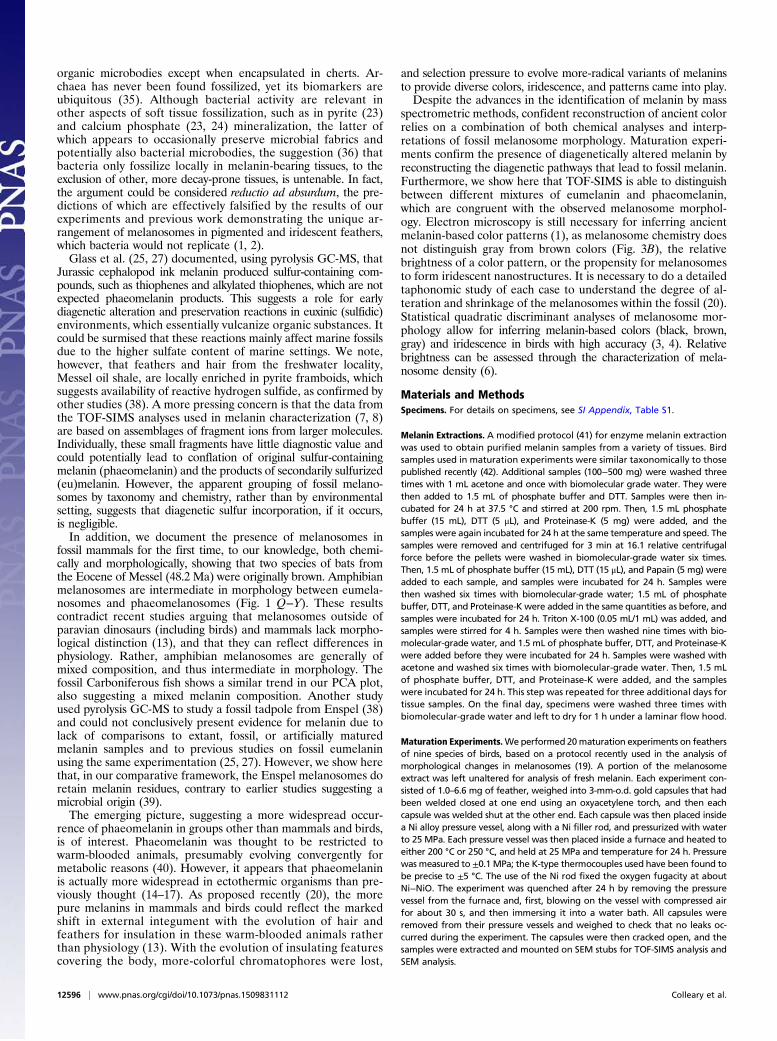
1. Vinther J, Briggs DEG, Prum RO, Saranathan V (2008) The colour of fossil feathers. BiolLett 4(5):522–525.
2. Vinther J, Briggs DEG, Clarke J, Mayr G, Prum RO (2010) Structural coloration in afossil feather. Biol Lett 6(1):128–131.
3. Li Q, et al. (2010) Plumage color patterns of an extinct dinosaur. Science 327(5971):1369–1372.
4. Li Q, et al. (2012) Reconstruction of Microraptor and the evolution of iridescentplumage. Science 335(6073):1215–1219.
5. Carney RM, Vinther J, Shawkey MD, D’Alba L, Ackermann J (2012) New evidence onthe colour and nature of the isolated Archaeopteryx feather. Nat Commun 3:637.
6. Field DJ, et al. (2013) Melanin concentration gradients in modern and fossil feathers.PLoS One 8(3):e59451.
7. Lindgren J, et al. (2012) Molecular preservation of the pigment melanin in fossilmelanosomes. Nat Commun 3:824.
8. Lindgren J, et al. (2014) Skin pigmentation provides evidence of convergent melanismin extinct marine reptiles. Nature 506(7489):484–488.
9. Mcnamara ME, Orr PJ (2008) Experimental degradation of vertebrates: Taphonomyof keratinous tissues and implications for the fossil record. Palaeontol Assoc Newsl69:29.
10. Moyer AE, et al. (2014) Melanosomes or microbes: testing an alternative hypothesisfor the origin of microbodies in fossil feathers. Sci Rep 4:4233.
11. Mcnamara ME, et al. (2010) Exceptionally preserved tadpoles from the Miocene ofLibros, Spain: Ecomorphological reconstruction and the impact of ontogeny upontaphonomy. Lethaia 43(3):290–306.
12. Wakamatsu K, Ito S (2002) Advanced chemical methods in melanin determination.Pigment Cell Res 15(3):174–183.
13. Li Q, et al. (2014) Melanosome evolution indicates a key physiological shift withinfeathered dinosaurs. Nature 507(7492):350–353.
14. Roulin A, Mafli A, Wakamatsu K (2013) Reptiles produce pheomelanin: Evidence inthe eastern Hermann’s tortoise (Eurotestudo boettgeri). J Herpetol 47(2):258–261.
15. Wolnicka-Glubisz A, Pecio A, Podkowa D, Kolodziejczyk LM, Plonka PM (2012)Pheomelanin in the skin of Hymenochirus boettgeri (Amphibia: Anura: Pipidae).Exp Dermatol 21(7):537–540.
16. Kottler VA, Künstner A, Schartl M (2015) Pheomelanin in fish? Pigment Cell MelanomaRes 28(3):355–356.
17. Speiser DI, DeMartini DG, Oakley TH (2014) The shell-eyes of the chiton Acantho-pleura granulata (Mollusca, Polyplacophora) use pheomelanin as a screening pig-ment. J Nat Hist 48(45-48):2899−2911.
18. Clarke JA, et al. (2010) Fossil evidence for evolution of the shape and color of penguinfeathers. Science 330(6006):954–957.
19. McNamara ME, Briggs DE, Orr PJ, Field DJ, Wang Z (2013) Experimental maturation offeathers: Implications for reconstructions of fossil feather colour. Biol Lett 9(3):20130184.
20. Vinther J (2015) A guide to the field of palaeo colour: Melanin and other pigmentscan fossilise: Reconstructing colour patterns from ancient organisms can give newinsights to ecology and behaviour. BioEssays 37(6):643–656.
21. Gehling JG (1999) Microbial mats in terminal Proterozoic siliciclastics: Ediacaran deathmasks. Palaios 14(1):40–57.
22. Westall F (1999) The nature of fossil bacteria: A guide to the search for extraterrestriallife. J Geophys Res 104(E7):16437−16451.
23. Briggs DEG (2003) The role of decay and mineralization in the preservation of soft-bodied fossils. Annu Rev Earth Planet Sci 31:275–301.
24. Muscente A, Hawkins AD, Xiao S (2015) Fossil preservation through phosphatizationand silicification in the Ediacaran Doushantuo Formation (South China): A compara-tive synthesis. Palaeogeogr Palaeoclimatol Palaeoecol 434:46–62.
25. Glass K, et al. (2012) Direct chemical evidence for eumelanin pigment from the Ju-rassic period. Proc Natl Acad Sci USA 109(26):10218–10223.
26. Tanaka G, et al. (2014) Mineralized rods and cones suggest colour vision in a 300 Myr-old fossil fish. Nat Commun 5:5920.
27. Glass K, et al. (2013) Impact of diagenesis and maturation on the survival of eume-lanin in the fossil record. Org Geochem 64:29–37.
28. Greenwalt DE, Goreva YS, Siljeström SM, Rose T, Harbach RE (2013) Hemoglobin-derived porphyrins preserved in a Middle Eocene blood-engorged mosquito. ProcNatl Acad Sci USA 110(46):18496–18500.
29. Stankiewicz B, et al. (2000) Alternative origin of aliphatic polymer in kerogen. Geology28(6):559–562.
30. Gupta NS, Michels R, Briggs DEG, Evershed RP, Pancost RD (2006) The organic preservationof fossil arthropods: An experimental study. Proc Biol Sci 273(1602):2777–2783.
31. Vitek NS, Vinther J, Schiffbauer JD, Briggs DE, Prum RO (2013) Exceptional three-dimensional preservation and coloration of an originally iridescent fossil feather fromthe Middle Eocene Messel Oil Shale. Palaeontol Z 87(4):493−503.
32. Steele A, Toporski J, Avci R, Guidry S, McKay D (2001) Time of flight secondary ionmass spectrometry (ToFSIMS) of a number of hopanoids. Org Geochem 32(7):905–911.
33. Siljeström S, et al. (2009) Detection of organic biomarkers in crude oils using ToF-SIMS. Org Geochem 40(1):135–143.
34. Leefmann T, et al. (2013) Spectral characterization of ten cyclic lipids using time-of-flightsecondary ion mass spectrometry. Rapid Commun Mass Spectrom 27(5):565–581.
35. Briggs DE, Summons RE (2014) Ancient biomolecules: Their origins, fossilization, androle in revealing the history of life. BioEssays 36(5):482–490.
36. Lindgren J, et al. (2015) Interpreting melanin-based coloration through deep time: acritical review. Proc R Soc B 282(1813):20150614.
37. Bauersachs T, Schouten S, Schwark L (2014) Characterization of the sedimentary or-ganic matter preserved in Messel oil shale by bulk geochemistry and stable isotopes.Palaeogeogr, Palaeoclimatol, Palaeoecol 410:390–400.
38. Barden HE, et al. (2015) Bacteria or melanosomes? A geochemical analysis of micro-bodies on a tadpole from the Oligocene Enspel Formation of Germany. PalaeobioPalaeoenv 95(1):33–45.
39. Toporski J, et al. (2002) Morphologic and spectral investigation of exceptionally well-preserved bacterial biofilms from the Oligocene Enspel formation, Germany. GeochimCosmochim Acta 66(10):1773–1791.
40. Galván I, Ghanem G, Møller AP (2012) Has removal of excess cysteine led to theevolution of pheomelanin? Pheomelanogenesis as an excretory mechanism for cys-teine. BioEssays 34(7):565–568.
41. Liu Y, et al. (2003) Comparison of the structural and physical properties of human haireumelanin following enzymatic or acid/base extraction. Pigment Cell Res 16(4):355–365.
42. Liu SY, Shawkey MD, Parkinson D, Troy TP, Ahmed M (2014) Elucidation of the chemicalcomposition of avian melanin. RSC Adv 4(76):40396–40399.
43. Zimmerman JD, et al. (2013) Control of interface order by inverse quasi-epitaxialgrowth of squaraine/fullerene thin film photovoltaics. ACS Nano 7(10):9268–9275.
44. Sai N, et al. (2012) Understanding the interface dipole of copper phthalocyanine(CuPc)/C60: Theory and experiment. J Phys Chem Lett 3(16):2173–2177.
45. Chou H, Ismach A, Ghosh R, Ruoff RS, Dolocan A (2015) Revealing the planar chem-istry of two-dimensional heterostructures at the atomic level. Nat Commun 6:7482.
Colleary et al. PNAS | October 13, 2015 | vol. 112 | no. 41 | 12597
EART
H,A
TMOSP
HER
IC,
ANDPL
ANET
ARY
SCIENCE
SEV
OLU
TION