CHEMICAL CONSTITUENTS AND BIOACTIVITIES OF Garcinia...
39
CHEMICAL CONSTITUENTS AND BIOACTIVITIES OF Garcinia griffithii T. ANDERSON NIK SHAZWANI AFIFAH BINTI NIK SAZALI UNIVERSITI TEKNOLOGI MALAYSIA
Transcript of CHEMICAL CONSTITUENTS AND BIOACTIVITIES OF Garcinia...
CHEMICAL CONSTITUENTS AND BIOACTIVITIES OF
Garcinia griffithii T. ANDERSON
NIK SHAZWANI AFIFAH BINTI NIK SAZALI
UNIVERSITI TEKNOLOGI MALAYSIA

CHEMICAL CONSTITUENTS AND BIOACTIVITIES OF
Garcinia griffithii T. ANDERSON
NIK SHAZWANI AFIFAH BINTI NIK SAZALI
A thesis submitted in fulfillment of the
requirements for the award of the degree of
Master of Science (Chemistry)
Faculty of Science
Universiti Teknologi Malaysia
APRIL 2014
iii
Dedicated to
My most beloved mother and father
My dearest sisters and brothers
My friends
Who always supportand inspire me all the time.
iv
ACKNOWLEDGEMENTS
First and foremost, I thank Allah S.W.T. for His love and care which kept me
going forward and made me strong to complete this research. I would like to convey
my gratefulness and appreciationto my supervisor, Assoc. Prof. Dr. Farediah Ahmad
for her constant encouragement, generosity, guidance and patient throughout the
completion of this research.
I want to express my gratitude to all Organic Chemistry lectures, thanks for
the opinion and knowledge in helping out in this project. I would like to
acknowledge all the organic master students for all their guidance and assistance in
completion some part of this research. I also insist to thanks all the laboratory
assistances for their helps and cooperation.
I also like to extend my sincere appreciation and heartfelt gratitude to all my
laboratory mates. I am greatly thankful for their willingness to share useful
knowledge, help, ideas, support and guidance given. The moments we spent together
in overcoming the challenges throughout our project have truly making us to know
more each other.
My deepest appreciation goes to my beloved family members especially to
my parents, Nik Sazali Nik Ibrahim and Norizah Busu, who have been my constant
inspirational source to further my studies. Abundle of thousand thanks for their
continuous advices, support and motivationfor me for finish this research well. Not
forgetting to my siblings Nadhirah, Naim, Izzat, Izyan and Fadhli for motivated me.
Last but not least, thank you to those being with me during my master’s study
and also to the entire individual who directly and indirectly helped me in
accomplishing this research.
v
PREFACE
This thesis is the result of my work carried out in the Department of chemistry,
Universiti Teknologi Malaysia under the supervision of Assoc. Prof. Dr. Farediah
Ahmad. Parts of my work described in this thesis have been reported in the
following publications:
1. Nik Shazwani Afifah Nik Sazali and Farediah Ahmad (2011). Chemical
Constituents and Biological Activities of The Leaves of Garcinia Griffithii
(Guttiferae). Poster presented at International Conference on Natural
Products (ICNP) 2011, Palm Garden Hotel, IOI Resort, Putrajaya. 13 – 16
November 2011.
vi
ABSTRACT
Chemical and bioactivity investigations were carried out on the leaves and stem barks of Garcinia griffithii T. Anderson from Guttiferae family. Sample for each part of the plant was extracted consecutively with increasing polarity of solvents by Soxhlet method. Vacuum liquid chromatography and column chromatography were used to purify the crude extracts. The pure compounds were elucidated by using combined spectroscopic techniques which include UV, IR, NMR (1D and 2D) and MS. Chromatographic purification of the leaves extracts have afforded nine pure compounds identified as squalene, 28-hydroxyfriedelan-3-one, friedooleanan-3-one, olean-12-en-3-ol, amento-4'-methyl ether, 3,8''-binaringenin, 3,8''-binaringenin-7''-O-glucoside, morelloflavone and morelloflavone-7''-O-glucoside. Chromatographic purification of the ethyl acetate and methanol extracts of the stem barks yielded two compounds identified as amento-4'-methyl ether and morelloflavone. The crude extracts and pure compounds isolated from methanol crude extract of the leaves were screened for various types of antioxidant assay and tyrosinase inhibition activities. The antioxidant assay on 2,2-diphenyl-1-picrylhydrazyl (DPPH) radical showed that the crude n-hexane extract of the stem barks had the highest radical scavenging activity with IC50 = 96.43 ± 2.69 µg/mL, while morelloflavone was found to be the strongest antioxidant compound with IC50 = 57.57 ± 0.53 µg/mL compared to other compounds. The crude methanol extract of the stem barks showed the highest total antioxidant with 260.81 ± 2.21 mg/g of ascorbic acid equivalent (AAE/L) and 871.43 ± 6.62 mg/g of butylated hydroxyl toluene equivalent (BHTE/L) while the crude methanol extract of the leaves showed the highest total phenolic content with 444.10 ± 6.67 mg/g of gallic acid equivalent (GAE/L) and 423.10 ± 6.67 mg/g of (± )-cathechin equivalent (CE/L). Morelloflavone showed the highest value for both assays with values 58.50 ± 3.15 mg/g of AAE/L and 264.50 ± 9.45 mg/g of BHTE/L; and 841.33 ± 38.28 mg/g of GAE/L and 822.97 ± 33.93 mg/g of CE/L, respectively. The crude extracts and all compounds were found to have weak anti-tyrosinase activity. The antimicrobial assay of all the crude extracts were carried out by using disc diffusion method, followed by minimum inhibition concentration (MIC) and minimum bactericidal concentration (MBC). The methanol crude extract of the leaves showed the most significant antimicrobial activity towards E. faecalis and K. pneumoniae with MIC and MBC value ranged between 225 – 450 µg/mL compared to the other crude extracts.
vii
ABSTRAK
Kajian kimia dan bioaktiviti telah dijalankan ke atas daun dan batang pokok Garcinia griffithii T. Anderson daripada keluarga Guttiferae. Sampel untuk setiap bahagian pokok telah diekstrak secara berturutan dengan menggunakan pelarut mengikut kekutuban menaik melalui kaedah Soxhlet. Kromatografi turus vakum dan
kromatografi turus graviti telah digunakan untuk penulenan ke atas ekstrak mentah. Struktur sebatian tulen dikenalpasti dengan menggunakan kaedah spektroskopi UV, IR, NMR (1D dan 2D) dan MS. Penulenan dan pencirian ke atas ekstrak mentah n-heksana daripada daun telah Berjaya menemukan Sembilan sebatian tulen yang dikenalpasti sebagai skualena, 28-hidroksifriedelan-3-on, friedooleanan-3-on, olean-12-en-3-ol, amento-4'-metil eter, 3,8''-binaringenin, 3,8''-binaringenin-7''-O-glukosida, morelloflavon dan morelloflavon-7''-O-glukosida. Penulenan secara kromatografi ke atas ekstrak etil asetat dan methanol batang pokok menghasilkan dua sebatian yang dikenalpasti sebagai amento-4'-metil eter dan morelloflavon. Ekstrak mentah dan sebatian tulen yang telah diasingkan daripada ekstrak mentah methanol daun disaring untuk beberapa ujian antioksidan dan aktiviti rencatan tirosinase. Ujian antioksidan ke atas radikal bebas 2,2-difenil-1-pikrilhidrazil (DPPH) menunjukkan bahawa ekstrak mentah n-heksana daripada batang pokok mempunyai aktiviti radikal tertinggi dengan nilai IC50 = 96.43 ± 2.69 µg/mL, manakala sebatian morelloflavon ditemui sebagai sebatian antioksidan terkuat dengan nilai IC50 = 57.57 ± 0.53 µg/mL berbanding dengan sebatian lain. Ekstrak mentah methanol daripada batang pokok menunjukkan nilai jumlah antioksidan yang paling tinggi dengan nilai 260.81 ± 2.21 mg/g setara dengan asid askorbik (AAE/L) dan 871.43 ± 6.62 mg/g setara dengan butil hidroksitoluena (BHTE/L) manakala ekstrak mentah methanol daripada daun mempunyai jumlah kandungan fenol yang paling tinggi, iaitu 444.10 ± 6.67 mg/g setara dengan asid galik (GAE/L) and 423.10 ± 6.67 mg/g setara dengan (± )-katekin (CE/L). Sebatian morelloflavon menunjukkan nilai tertinggi bagi kedua-dua saringan masing-masing dengan nilai 58.50 ± 3.15 mg/g AAE/L dan 264.50 ± 9.45 mg/g BHTE/L; dan 841.33 ± 38.28 mg/g GAE/L dan 822.97 ± 33.93 mg/g CE/L. Ekstrak mentah dan kesemua sebatian tulen didapati mempunyai aktiviti anti-tirosinase yang lemah. Saringan antimikrob telah dijalankan ke atas ekstrak mentah dengan menggunakan kaedah pembauran cakera, diikuti dengan penentuan nilai rencatan minimum (MIC) dan kepekatan bakterisida minimum (MBC). Ekstrak mentah methanol daripada daun menunjukkan aktiviti antimikrob yang paling signifikan terhadap E. faecalis and K. pneumonia dengan nilai MIC dan MBC antara 225 – 450 µg/mL berbanding dengan ekstrak mentah yang lain.
viii
TABLE OF CONTENTS
CHAPTER TITLE PAGE
DECLARATION
ii
DEDICATION iii
ACKNOWLEDGEMENTS iv
PREFACE v
ABSTRACT vi
ABSTRAK vii
TABLE OF CONTENTS viii
LIST OF TABLES xiii
LIST OF FIGURES xv
LIST OF SCHEMES xvi
LIST OF ABBREVIATIONS xvii
LIST OF APPENDICES xix
1 INTRODUCTION
1.1 General Introduction 1
1.2 Guttiferae Family 2
1.3 Garcinia griffithii 4
1.4 Problem Statement 5
1.5 Objectives of Research 5
1.6 Scope of Research 5
2 LITERATURE REVIEW
2.1 Phytochemical Studies on Genus Garcinia 7
2.1.1 Xanthones 7
ix
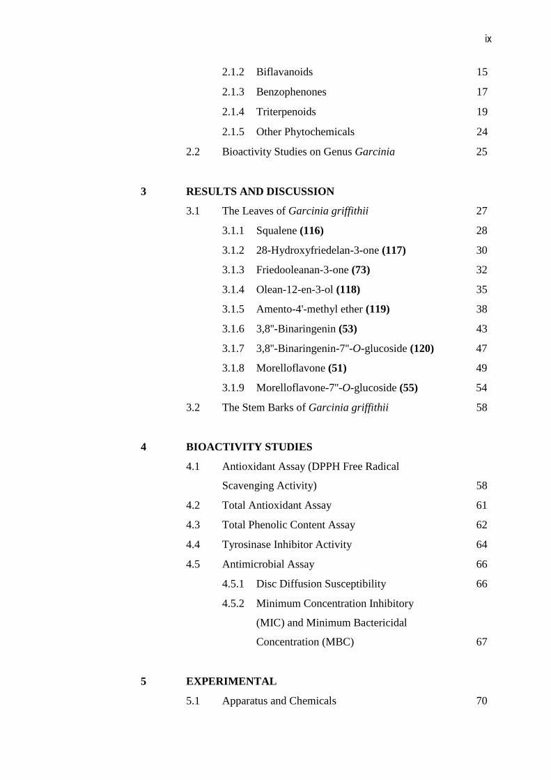
2.1.2 Biflavanoids 15
2.1.3 Benzophenones 17
2.1.4 Triterpenoids 19
2.1.5 Other Phytochemicals 24
2.2 Bioactivity Studies on Genus Garcinia 25
3 RESULTS AND DISCUSSION
3.1 The Leaves of Garcinia griffithii 27
3.1.1 Squalene (116) 28
3.1.2 28-Hydroxyfriedelan-3-one (117) 30
3.1.3 Friedooleanan-3-one (73) 32
3.1.4 Olean-12-en-3-ol (118) 35
3.1.5 Amento-4'-methyl ether (119) 38
3.1.6 3,8''-Binaringenin (53) 43
3.1.7 3,8''-Binaringenin-7''-O-glucoside (120) 47
3.1.8 Morelloflavone (51) 49
3.1.9 Morelloflavone-7''-O-glucoside (55) 54
3.2 The Stem Barks of Garcinia griffithii 58
4 BIOACTIVITY STUDIES
4.1 Antioxidant Assay (DPPH Free Radical
Scavenging Activity)
58
4.2 Total Antioxidant Assay 61
4.3 Total Phenolic Content Assay 62
4.4 Tyrosinase Inhibitor Activity 64
4.5 Antimicrobial Assay 66
4.5.1 Disc Diffusion Susceptibility 66
4.5.2 Minimum Concentration Inhibitory
(MIC) and Minimum Bactericidal
Concentration (MBC)
67
5 EXPERIMENTAL
5.1 Apparatus and Chemicals 70
x
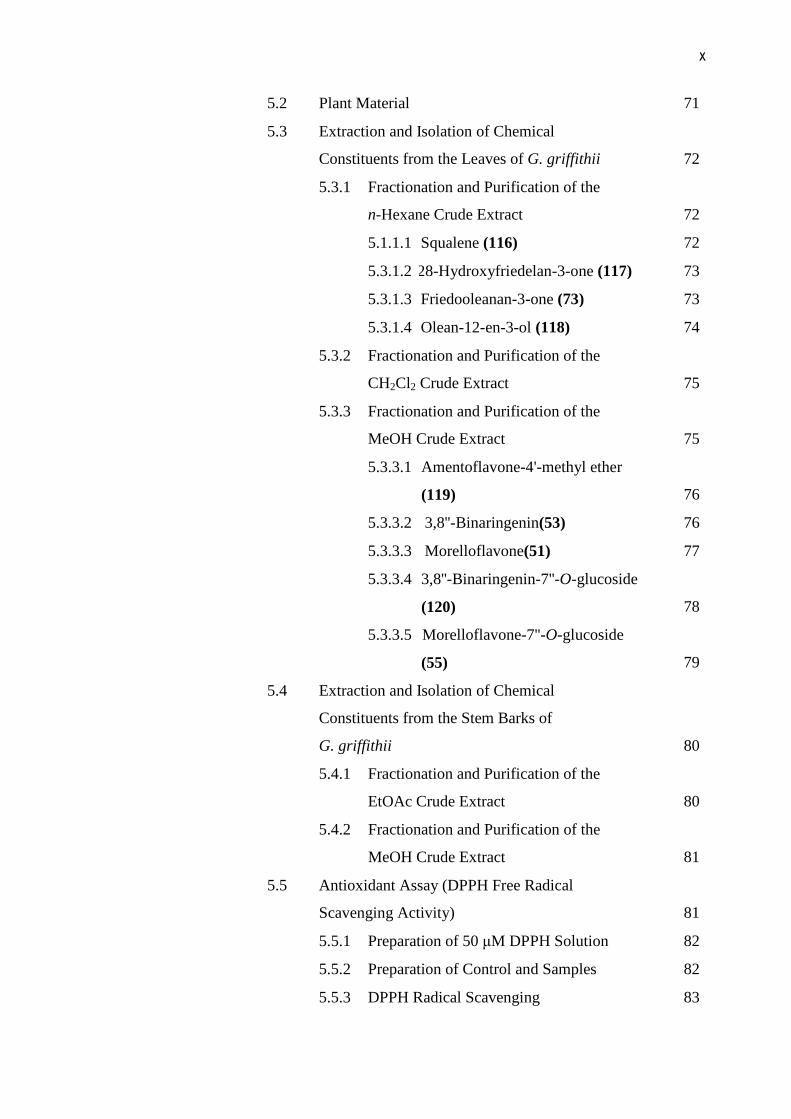
5.2 Plant Material 71
5.3 Extraction and Isolation of Chemical
Constituents from the Leaves of G. griffithii
72
5.3.1 Fractionation and Purification of the
n-Hexane Crude Extract
72
5.1.1.1 Squalene (116) 72
5.3.1.2 28-Hydroxyfriedelan-3-one (117) 73
5.3.1.3 Friedooleanan-3-one (73) 73
5.3.1.4 Olean-12-en-3-ol (118) 74
5.3.2 Fractionation and Purification of the
CH2Cl2 Crude Extract
75
5.3.3 Fractionation and Purification of the
MeOH Crude Extract
75
5.3.3.1 Amentoflavone-4'-methyl ether
(119)
76
5.3.3.2 3,8''-Binaringenin(53) 76
5.3.3.3 Morelloflavone(51) 77
5.3.3.4 3,8''-Binaringenin-7''-O-glucoside
(120)
78
5.3.3.5 Morelloflavone-7''-O-glucoside
(55)
79
5.4 Extraction and Isolation of Chemical
Constituents from the Stem Barks of
G. griffithii
80
5.4.1 Fractionation and Purification of the
EtOAc Crude Extract
80
5.4.2 Fractionation and Purification of the
MeOH Crude Extract
81
5.5 Antioxidant Assay (DPPH Free Radical
Scavenging Activity)
81
5.5.1 Preparation of 50 µM DPPH Solution 82
5.5.2 Preparation of Control and Samples 82
5.5.3 DPPH Radical Scavenging 83
xi
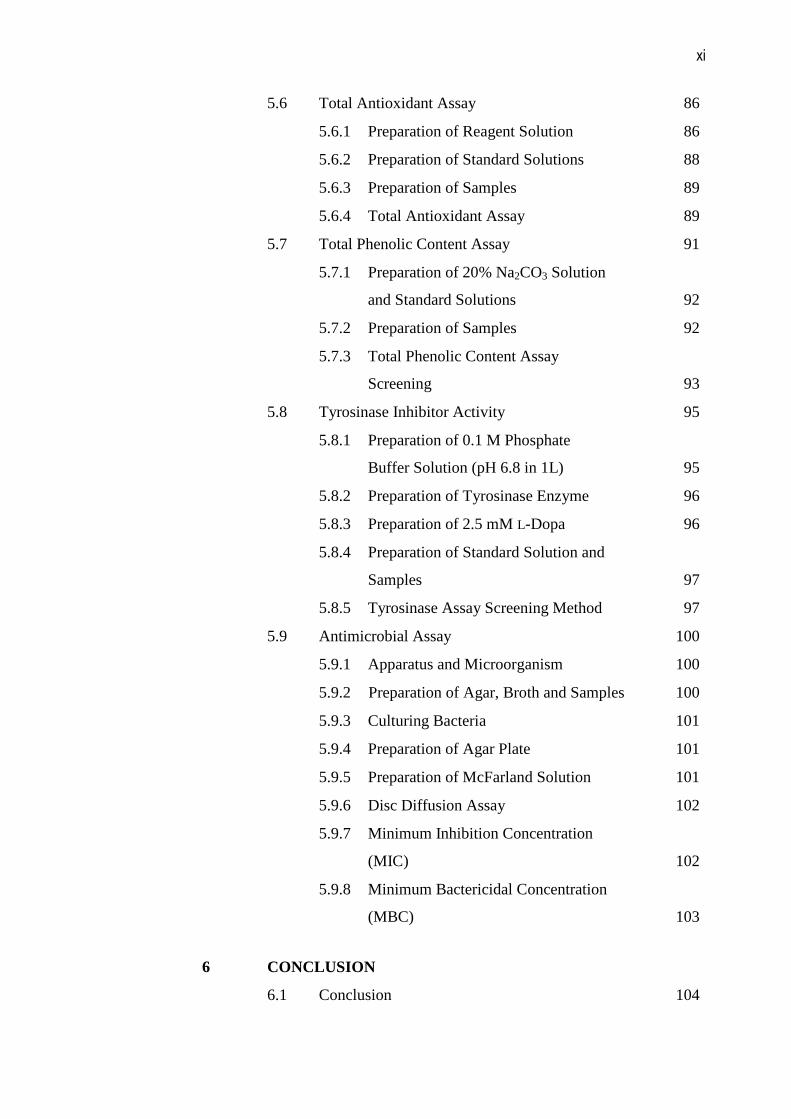
5.6 Total Antioxidant Assay 86
5.6.1 Preparation of Reagent Solution 86
5.6.2 Preparation of Standard Solutions 88
5.6.3 Preparation of Samples 89
5.6.4 Total Antioxidant Assay 89
5.7 Total Phenolic Content Assay 91
5.7.1 Preparation of 20% Na2CO3 Solution
and Standard Solutions
92
5.7.2 Preparation of Samples 92
5.7.3 Total Phenolic Content Assay
Screening
93
5.8 Tyrosinase Inhibitor Activity 95
5.8.1 Preparation of 0.1 M Phosphate
Buffer Solution (pH 6.8 in 1L)
95
5.8.2 Preparation of Tyrosinase Enzyme 96
5.8.3 Preparation of 2.5 mM L-Dopa 96
5.8.4 Preparation of Standard Solution and
Samples
97
5.8.5 Tyrosinase Assay Screening Method 97
5.9 Antimicrobial Assay 100
5.9.1 Apparatus and Microorganism 100
5.9.2 Preparation of Agar, Broth and Samples 100
5.9.3 Culturing Bacteria 101
5.9.4 Preparation of Agar Plate 101
5.9.5 Preparation of McFarland Solution 101
5.9.6 Disc Diffusion Assay 102
5.9.7 Minimum Inhibition Concentration
(MIC)
102
5.9.8 Minimum Bactericidal Concentration
(MBC)
103
6 CONCLUSION
6.1 Conclusion 104

xii
6.2 Future Works 106
REFERENCES 107
Appendices 1-79 118
xiii
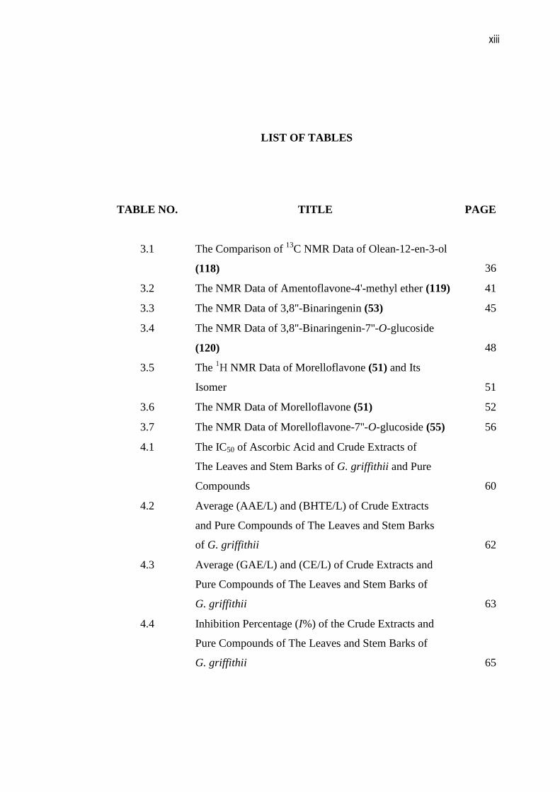
LIST OF TABLES
TABLE NO. TITLE PAGE
3.1
The Comparison of 13C NMR Data of Olean-12-en-3-ol
(118)
36
3.2 The NMR Data of Amentoflavone-4'-methyl ether (119) 41
3.3 The NMR Data of 3,8''-Binaringenin (53) 45
3.4 The NMR Data of 3,8''-Binaringenin-7''-O-glucoside
(120)
48
3.5 The 1H NMR Data of Morelloflavone (51) and Its
Isomer
51
3.6 The NMR Data of Morelloflavone (51) 52
3.7 The NMR Data of Morelloflavone-7''-O-glucoside (55) 56
4.1 The IC50 of Ascorbic Acid and Crude Extracts of
The Leaves and Stem Barks of G. griffithii and Pure
Compounds
60
4.2 Average (AAE/L) and (BHTE/L) of Crude Extracts
and Pure Compounds of The Leaves and Stem Barks
of G. griffithii
62
4.3 Average (GAE/L) and (CE/L) of Crude Extracts and
Pure Compounds of The Leaves and Stem Barks of
G. griffithii
63
4.4 Inhibition Percentage (I%) of the Crude Extracts and
Pure Compounds of The Leaves and Stem Barks of
G. griffithii
65
xiv
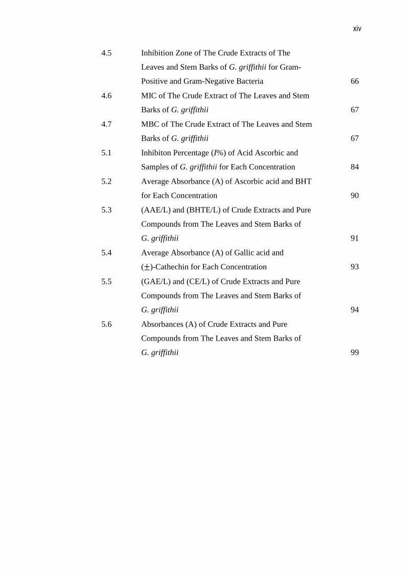
4.5 Inhibition Zone of The Crude Extracts of The
Leaves and Stem Barks of G. griffithii for Gram-
Positive and Gram-Negative Bacteria
66
4.6 MIC of The Crude Extract of The Leaves and Stem
Barks of G. griffithii
67
4.7 MBC of The Crude Extract of The Leaves and Stem
Barks of G. griffithii
67
5.1 Inhibiton Percentage (I%) of Acid Ascorbic and
Samples of G. griffithii for Each Concentration
84
5.2 Average Absorbance (A) of Ascorbic acid and BHT
for Each Concentration
90
5.3 (AAE/L) and (BHTE/L) of Crude Extracts and Pure
Compounds from The Leaves and Stem Barks of
G. griffithii
91
5.4 Average Absorbance (A) of Gallic acid and
(±)-Cathechin for Each Concentration
93
5.5 (GAE/L) and (CE/L) of Crude Extracts and Pure
Compounds from The Leaves and Stem Barks of
G. griffithii
94
5.6 Absorbances (A) of Crude Extracts and Pure
Compounds from The Leaves and Stem Barks of
G. griffithii
99
xv
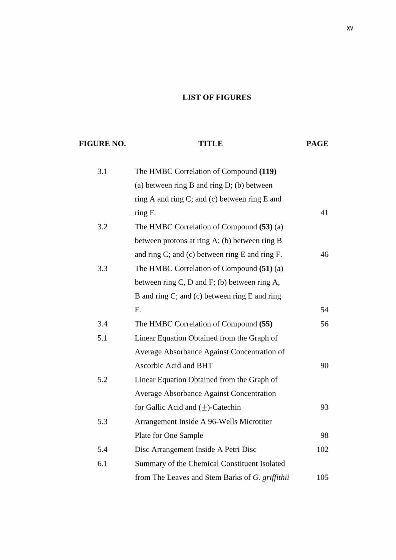
LIST OF FIGURES
FIGURE NO. TITLE PAGE
3.1
The HMBC Correlation of Compound (119)
(a) between ring B and ring D; (b) between
ring A and ring C; and (c) between ring E and
ring F.
41
3.2 The HMBC Correlation of Compound (53) (a)
between protons at ring A; (b) between ring B
and ring C; and (c) between ring E and ring F.
46
3.3 The HMBC Correlation of Compound (51) (a)
between ring C, D and F; (b) between ring A,
B and ring C; and (c) between ring E and ring
F.
54
3.4 The HMBC Correlation of Compound (55) 56
5.1 Linear Equation Obtained from the Graph of
Average Absorbance Against Concentration of
Ascorbic Acid and BHT
90
5.2 Linear Equation Obtained from the Graph of
Average Absorbance Against Concentration
for Gallic Acid and (±)-Catechin
93
5.3 Arrangement Inside A 96-Wells Microtiter
Plate for One Sample
98
5.4 Disc Arrangement Inside A Petri Disc 102
6.1 Summary of the Chemical Constituent Isolated
from The Leaves and Stem Barks of G. griffithii
105
xvi
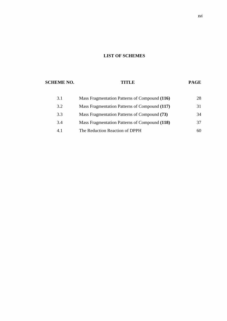
LIST OF SCHEMES
SCHEME NO. TITLE PAGE
3.1
Mass Fragmentation Patterns of Compound (116)
28
3.2 Mass Fragmentation Patterns of Compound (117) 31
3.3 Mass Fragmentation Patterns of Compound (73) 34
3.4 Mass Fragmentation Patterns of Compound (118) 37
4.1 The Reduction Reaction of DPPH 60
xvii
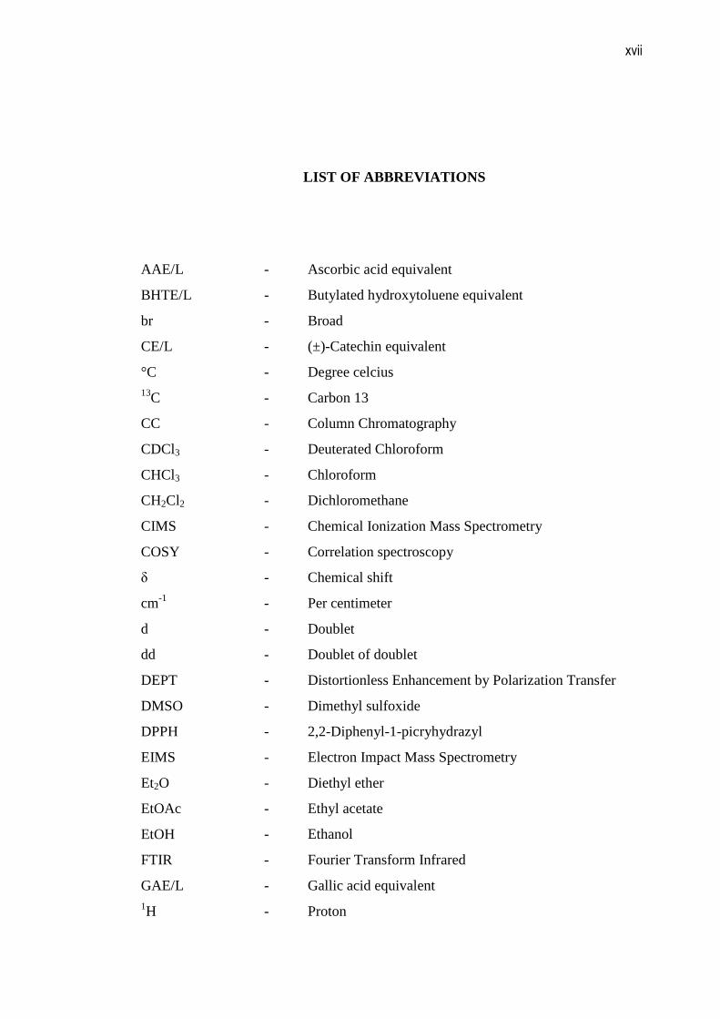
LIST OF ABBREVIATIONS
AAE/L - Ascorbic acid equivalent
BHTE/L - Butylated hydroxytoluene equivalent
br - Broad
CE/L - (±)-Catechin equivalent
°C - Degree celcius 13C - Carbon 13
CC - Column Chromatography
CDCl3 - Deuterated Chloroform
CHCl3 - Chloroform
CH2Cl2 - Dichloromethane
CIMS - Chemical Ionization Mass Spectrometry
COSY - Correlation spectroscopy
δ - Chemical shift
cm-1 - Per centimeter
d - Doublet
dd - Doublet of doublet
DEPT - Distortionless Enhancement by Polarization Transfer
DMSO - Dimethyl sulfoxide
DPPH - 2,2-Diphenyl-1-picryhydrazyl
EIMS - Electron Impact Mass Spectrometry
Et2O - Diethyl ether
EtOAc - Ethyl acetate
EtOH - Ethanol
FTIR - Fourier Transform Infrared
GAE/L - Gallic acid equivalent 1H - Proton
xviii
H2O - Water
H3BO3 - Boric acid
HMBC - Heteronuclear Multiple Bond Correlation
HMQC - Heteronuclear Multiple Quantum Coherence
Hz - Hertz
IC50 - Inhibition concentration for 50%
IR - Infrared
J - Coupling constant
KBr - Potassium bromide
L - Liter
λ - Lambda
m/z - Mass to charge ratio
m - Multiplet
m.p. - Melting point
µg - Microgram
mg - Miligram
mL - Mililiter
MBC - Minimum bactericidal concentration
MeOH - Methanol
MIC - Minimum inhibitory concentration
nm - Nanometer
NA - Nutrient agar
NB - Nutrient broth
NMR - Nuclear Magnetic Resonance
ppm - Parts per million
quint - Quintet
Rf - Retention factor
s - Singlet
SiO2 - Silica gel
t - Triplet
TLC - Thin Layer Chromatography
UV - Ultraviolet
VLC - Vacuum Liquid Chromatography
xix
LIST OF APPENDICES
APPENDIX NO. TITLE PAGE
1
GCMS Spectrum of Squalene (116)
119
2 IR Spectrum of Squalene (116) 120
3 1H NMR Spectrum of Squalene (116) 121
4 COSY Spectrum of Squalene (116) 122
5 13C NMR Spectrum of Squalene (116) 123
6 DEPT Spectra of Squalene (116) 124
7 IR Spectrum of 28-Hydroxyfriedelan-3-one (117) 125
8 1H NMR Spectrum of 28-Hydroxyfriedelan-3-one (117) 126
9 COSY Spectrum of 28-Hydroxyfriedelan-3-one (117) 127
10 HMQC Spectrum of 28-Hydroxyfriedelan-3-one (117) 128
11 13C NMR Spectrum of 28-Hydroxyfriedelan-3-one (117) 129
12 DEPT Spectra of 28-Hydroxyfriedelan-3-one (117) 130
13 EIMS Spectrum of 28-Hydroxyfriedelan-3-one (117) 131
14 IR Spectrum of Friedooleanan-3-one (73) 132
15 1H NMR Spectrum of Friedooleanan-3-one (73) 133
16 COSY Spectrum of Friedooleanan-3-one (73) 134
17 13C NMR Spectrum of Friedooleanan-3-one (73) 135
18 DEPT Spectra of Friedooleanan-3-one (73) 136
19 HMQC Spectrum of Friedooleanan-3-one (73) 137
20 GCMS Spectrum of Friedooleanan-3-one (73) 138
21 13C NMR Spectrum of Olean-12-en-3-ol (118) 139
22 DEPT Spectra of Olean-12-en-3-ol (118) 140
23 1H NMR Spectrum of Olean-12-en-3-ol (118) 141
24 COSY Spectrum of Olean-12-en-3-ol (118) 142
xx
25 HMQC Spectrum of Olean-12-en-3-ol (118) 143
26 GCMS Spectrum of Olean-12-en-3-ol (118) 144
27 IR Spectrum of Olean-12-en-3-ol (118) 145
28 CIMS Spectrum of Amentoflavone-4'-methyl ether (119) 146
29 UV Spectrum of Amentoflavone-4'-methyl ether (119) 147
30 IR Spectrum of Amentoflavone-4'-methyl ether (119) 148
31 1H NMR Spectrum of Amentoflavone-4'-methyl ether
(119)
149
32 COSY Spectrum of Amentoflavone-4'-methyl ether (119) 150
33 13C NMR Spectrum of Amentoflavone-4'-methyl ether
(119)
151
34 DEPT Spectra of Amentoflavone-4'-methyl ether (119) 152
35 HMQC Spectrum of Amentoflavone-4'-methyl ether
(119)
153
36 Expansion of HMQC Spectrum of Amentoflavone-
4'-methyl ether (119)
154
37 HMBC Spectrum of Amentoflavone-4'-methyl ether
(119)
155
38 Expansion of HMBC Spectrum (I) of Amentoflavone-
4'-methyl ether (119)
156
39 HMBC Spectrum (II) of Amentoflavone-4'-methyl
ether (119)
157
40 Expansion of HMBC Spectrum (II) of Amentoflavone-
4'-methyl ether (119)
158
41 UV Spectrum of 3,8''-Binaringenin (53) 159
42 CIMS Spectrum of 3,8''-Binaringenin (53) 160
43 IR Spectrum of 3,8''-Binaringenin (53) 161
44 13C NMR Spectrum of 3,8''-Binaringenin (53) 162
45 DEPT Spectra of 3,8''-Binaringenin (53) 163
46 1H NMR Spectrum of 3,8''-Binaringenin (53) 164
47 COSY Spectrum of 3,8''-Binaringenin (53) 165
48 HMQC Spectrum of 3,8''-Binaringenin (53) 166
49 HMBC Spectrum of 3,8''-Binaringenin (53) 167
xxi
50 CIMS Spectrum of 3,8''-Binaringenin-7''-O-glucoside
(120)
168
51 UV Spectrum of 3,8''-Binaringenin-7''-O-glucoside (120) 169
52 IR Spectrum of 3,8''-Binaringenin-7''-O-glucoside (120) 170
53 13C NMR Spectrum of3,8''-Binaringenin-7''-O-glucoside
(120)
171
54 DEPT Spectra of 3,8''-Binaringenin-7''-O-glucoside
(120)
172
55 1H NMR Spectrum of 3,8''-Binaringenin -7''-O-glucoside
(120)
173
56 COSY Spectrum of 3,8''-Binaringenin-7''-O-glucoside
(120)
174
57 HMQC Spectrum of 3,8''-Binaringenin-7''-O-glucoside
(120)
175
58 UV Spectrum of Morelloflavone (51) 176
59 CIMS Spectrum of Morelloflavone (51) 177
60 IR Spectrum of Morelloflavone (51) 178
61 1H NMR Spectrum of Morelloflavone (51) 179
62 Expansion of 1H NMR Spectrum of Morelloflavone
(51)
180
63 COSY Spectrum of Morelloflavone (51) 181
64 13C NMR Spectrum of Morelloflavone (51) 182
65 DEPT Spectra of Morelloflavone (51) 183
66 HMQC Spectrum of Morelloflavone (51) 184
67 HMBC Spectrum of Morelloflavone (51) 185
68 HMBC Spectrum (I) of Morelloflavone (51) 186
69 Expansion of HMBCSpectrum (I) of Morelloflavone
(51)
187
70 HMBC Spectrum (II) of Morelloflavone (51) 188
71 DEPT Spectra of Morelloflavone-7''-O-glucoside (55) 189
72 CIMS Spectrum of Morelloflavone-7''-O-glucoside (55) 190
73 IR Spectrum of Morelloflavone-7''-O-glucoside (55) 191
74 1H NMR Spectrum ofMorelloflavone-7''-O-glucoside (55) 192
xxii
75 COSY Spectrum of Morelloflavone-7''-O-glucoside (55) 193
76 13C NMR Spectrum ofMorelloflavone-7''-O-glucoside
(55)
194
77 DEPT Spectra of Morelloflavone-7''-O-glucoside (55) 195
78 HMQC Spectrum ofMorelloflavone-7''-O-glucoside (55) 196
79 HMBC Spectrum ofMorelloflavone-7''-O-glucoside (55) 197

CHAPTER 1
INTRODUCTION
1.1 General Introduction
A natural product is a chemical compound or a substance produced by living
organisms, plants, animals, insects and microbes. These chemical constituents
usually have pharmacological or bioactivities that can be applied in pharmaceutical
and drug design [1]. They have, until recently, been the primary source for the
commercial medicines and drug research development [2]. Natural products
chemistry has always been concerned with the discovery of bioactive constituents
and it remains one of the main keys that play an important role in the continuous
research for new drugs in the industrial drug discovery process [3]. Besides, many
natural products have reached the market without chemical modification, thus the
potential to commercialize these small, drug-like molecules are very economical [4].
The abundance of natural renewable supply of plants and herbs indeed is a great
source of affordable drugs, which is complimentary to the modern medicine [5].
The practices of modern medicine have yielded numerous purified
compounds with medicinal properties as the result of the chemical investigations and
purification of extracts of plants. These compounds have been developed into
pharmaceutical agents [6]. To date about 25% of all available modern drugs such as
morphine and salicylates are derived directly or indirectly from higher plants. These
interesting compounds developed major classes of the analgesic drugs, namely,
opioids which are classified as central nervous system depressants and non-steroidal
anti-inflammatory drugs. Over the years, natural products and their derivatives are
2
not only used clinically, but also play an important role in discovery of new targets
such as receptors, enzymes, transporters or ion channels involved in relevant
physiological and pathological processes [7].
The analysis of plant components should thus begin with bioactivity-directed
screening and bioactivity-directed fractionation leading to the isolation and
characterization of pure biologically active compounds [8]. With the advancement of
spectroscopic methods, numerous types of active compounds can be isolated from
plants and are structurally characterized. In due course, many of these compounds
are synthesized in the laboratory. Sometimes, better-tolerated drugs are produced by
chemical modifications or by total synthesis of analogues of the active principles [9].
As a result, the findings from this area of research will assist in contributing to the
evolution in practice of traditional and modern medicineby utilizing them on a larger
commercial basis to eliminate health problems. Therefore, today, an increase of
global interest for industrial production concurrently meet the demand for
conserving biodiversity to enhance the development of renewable natural products
for medicine [10].
1.2 Guttiferae Family
Guttiferae family is one of the families in order Guttiferals. The family
contains about 48 genera and over 1000 species of perennial herbs, shrubs and trees,
widely distributed by the tropical and temperate regions of the world [11]. Most
members of the family are in view of their economical and medicinal purposes in
many parts of the world for treatment of different illnesses [12]. In Malaysia,
Guttiferae is an important component of the Malaysian Rain Forest as the second-
storey forest trees which include some well-known and important trees such as
ironwood tree, mangosteen and penaga laut. Whitemore [11] identified and
classified four genera and 121 species that can be found in Peninsular Malaysia
which are Calophyllum (45 species), Garcinia (49 species), Mammea (23 species)
and Mesua (4 species).
3
The trees or shrubs of this family have inner barks with yellow or white latex
in droplets. The bark is smooth, fissured or scaly patter and the bole rarely with stilt
roots or buttresses [13]. The leaves are mostly opposite without true stipule and the
secondary nerves often numerous, close and parallel. Flowers are bisexual or
sometimes unisexual, have scented and can be on the twigs behind the leaves. The
sepals and petals are four to five each and overlapping whereas, the stamens usually
connate in bundles. The fruits of this family can be a drupe for Calophyllum and
Mammea, a nut for Mesua or a berry with the seeds embedded in pulp, not splitting
with wall leathery of fleshy for Garcinia [11, 14].
Primarily, the genus Garcinia is the biggest genus with the common village
fruit-trees such as G. atroviridis (asam gelugor), G. cowa (village kandis) and
G. prainiana (kechupu). The trees are small to medium, rarely taller than 30 m;
therefore the trees are almost completely confined to the interior of the forest, in
shade. Many species of Garcinia have very similar leaves but differ in flower and
fruit characters [11]. The fruit hull of G. mangostana has found many uses in
traditional medicine such as for healing skin infections and wounds in Thai folk
medicine [15].
The second largest genus is Calophyllum called ‘bintangor’ by Malays. It
provides timber such as C. coriaceum (bintangor gunung daun besar), C. cuneatum
(bintangor gunung daun kecil) and C. inophylloide (bintangor batu). Trees of this
genus have the largest size among the other three genera with heights of up to
30 – 36m and are common of the lowland and mountain forest. C. inophyllum has
wide variety of cures found from its oil to its roots. Its gum, bark, leave, and all
other parts of it are found to be curative in medicine hence known as the 'All Heal'
plant [16]. The seed oil of C. inophyllumis reported to cure rheumatism and skin
affections in Indian traditional medicine [17].
The Malay name for genus Mesua is ‘penaga’ and also consists of valuable
timber with very hard and heavy wood such as M. grandis (penaga sabut),
M. lepidota (penaga tikus) and M. nuda (penaga lilin). The trees comprise small to
medium trees with height up to about 23 m [11]. M. ferreais a very well-known
4
species; the word ‘ferrea’ is from the Latin word, ‘ferrum’ which means ‘iron’
referring to its extremely hard wood [18]. The heartwood is very hard, dense, strong,
heavy and durable like ebony and used extensively for heavy construction, in
machinery work, vehicles and agricultural implements [19]. In Malaysia, the kernels
of M. ferrea are pounded with the seed oil and applied to wounds as poultice [18].
The last genus identified in Malaysia is Mammea consists of M. brevipes,
M. malayana, M. siamensis and M. odorata. The Malaysian wild Mammea species
are rare trees and restricted to the lowland. The genus has small trees with about
15 m height and has little important uses to human. M. siamensis has a big flower
which smell of violets and planted in temple. Its pollen is reputed to be used as a
cosmetic. The well-known species in this genus is M. americana, the sole American
species which produces large edible apricot-like fruits called as the Mammea apple
[11].
1.3 Garcinia griffithii
Species of Garcinia griffithii (apple-kandis or kandis gajah) is a small to
medium tree that may reach 23 m tall and conspicuous from the large leaves and
fruits. Its latex is yellowish white and the inner bark with opaque yellow exudate.
The sapwood and heartwood cannot be distinct and in dark red-brown colour. The
leaves have very large blade with size from 15×7 to 28×16 cm, broady elliptic,
strongly ribbed and pointed. The edges incurved and has base rounded. The young
leaves are pink and the drying leaves are blackish green with thin-texture. The
flowers mainly in short woody on the twigs behind the leaves and have four sepals
and petals with yellow flushed red colours at the base. The fruits are
characteristically globose, faintly ribbed, subsessile, fattened at the top, clustered on
the branched, edible and turning brownish yellow with watery acid flesh like green
apple. The stigma is usually sunken entirely, flat or slightly convex. The species is
common in Peninsular Malaysia at the low land forest [11, 16, 20].
5
1.4 Problem Statement
To date, there are two reports on the phytochemicals and antiplasmodial
activity of Garcinia griffithii collected from Indonesia [21] and Singapore [22].
However, the bioactivities of the plants were not studied thoroughly. Although the
climate and ecology of Singapore and Indonesia are similar with Malaysia, this
study is still being continued hoping to get variations of chemical constituents with
interesting bioactivities. The data obtained will be utilized for future research on the
chemical markers of Garcinia species from Malaysia.
1.5 Objectives of Research
The objectives of this research are:
• To isolate the chemical compounds from the leaves and stem barks of
G. griffithii.
• To elucidate the structures of the chemical compounds by combined
spectroscopic methods.
• To carry out the antioxidant, antimicrobial and tyrosinase inhibition
activities on the crude extracts and pure compounds of G. griffithii.
1.6 Scope of Research
This research will focus on the leaves and stem barks of G. griffithii. The
sample will be extracted by Soxhlet apparatus using n-hexane, dichloromethane,
ethyl acetate and methanol as the solvent to afford the crude extracts. Separation and
purification of the natural compounds from the crude extracts will be carried out
with chromatographic techniques such as vacuum liquid chromatography and

6
column chromatography. The structure of the pure compounds will be elucidated
with various spectroscopic techniques such as UV, IR, NMR (1D and 2D) and MS.
The crude extracts will be screened for bioactivity focusing on antioxidant,
tyrosinase inhibition and antimicrobial activities. The antioxidant assay by DPPH
method, the total antioxidant assay by formation of the green phosphomolybdenum
complex and the total phenolic content assay using Folin-Ciocalteaumicro method
will be used to screen the antioxidant activities of the samples. The antimicrobial
assay will be carried out by disc diffusion method against two Gram-positive
bacteria: Enterococcus faecalis and Bacillus subtilis and two Gram-negative
bacteria: Escherichia coli and Klebsiella pneumoniae. Further evaluation of the
antimicrobial activity will be carried out by determination of minimum inhibition
concentration (MIC) and minimum bactericidal concentration (MBC). The isolated
compounds will be screened for antioxidant as above and tyrosinase inhibition
activities only.
REFERENCES
1. Newman, D.J. and Cragg, G.M. (2007). Natural Products as Sources of New
Drugs over the Last 25 Years. Journal of Natural Products.70, 461-477.
2. Cseke, L.J., Kirakosyan, A., Kaufman, P.B., Warber, S.L., Duke, J.A.,
Brielman, H.L. (2006). Natural Products from Plants. Boca Raton, FL. CRC
Press Taylor & Francis Group. 2-3.
3. Steven, M.C. and Russell, J.M. (1993). Bioactive Natural Products: Detection,
Isolation and Structural Determination. Boca Raton, FL. CRC Press Taylor &
Francis Group. 2-8, 10-49.
4. Lixin, Z. and Arnold, L.D. (2005). Natural Products: Drug Discovery and
Therapeutic Medicine. Totowa, NJ. Humana Press. 4.
5. Sujata, V.B., Nagasasampagi, B.A., Meeenakshi, S. (2009). Natural Products:
Chemistry and Applications. Oxford, UK. Alpha Science International Ltd. 3.
6. Balandarin, M.F., Kinghorn, A.D., Fransworth, N.R. (1993). Chapter 1: Plant-
Derived Natural Products in Drugs Discovery and Development, An
Overview. Human Medical Agents from Plants. Washington, DC. American
Chemical Society. 2-12.
7. Calixto, J.B., Kassuya, C.A., Andre, E., Ferreira, J. (2005). Contribution of
Natural Products to the Discovery of the Transient Receptor Potential (TRP)
Channels Family and Their Functions. Pharmacology & Therapeutics.
106(2), 179-208.
108
8. Anderson, J.E., Goetz, C.M., McLaughlin, J.L., Suffness, M. (1991). A Blind
Comparison of Simple Bench-Top Bioassays and Human Tumor Cell
Cytotoxicities as Tumor Antitumor Prescreens. Phytochemical Analysis. 2,
107-111.
9. Dewick, P.M. (2009). Medical Natural Products: A Biosynthetic Approach.
3rd Edition. University of Nottingham, UK. John Wiley & Sons, Ltd. 7-8.
10. Tringali, C. (2012). Bioactive Compounds from Natural Sources: Natural
Products as Lead Compounds in Drug Discovery. 2nd Edition. Boca Raton,
FL. CRC Press Taylor & Francis Group. 2.
11. Whitemore, T.C. (1972). Malayan Forest Records No. 26: Tree of Malaya: A
Manual for Foresters. Kuala Lumpur. Longman.2, 162-236.
12. Sultanbawa, M.U.S. (1980). Xanthonoids of Tropical Plants. Tetrahedron. 36,
1465-1506.
13. Kochummen, K.M. (1997). Malay Forest Records No. 44: Tree Flora of
Pasoh Forest. Kuala Lumpur. Forest Research Institute Malaysia. 255.
14. Hsuan, K. (1978). Orders and Family of Malayan Seed Plants. Singapore.
Singapore University Press. 88.
15. Parveen, M., Khan, N.U., Acchari, B., Dutta, P.K. (1991). A Triterpene from
Garcinia mangostana. Phytochemistry. 30, 361-362.
16. Corner, E.J.H. (1940). Wayside Trees of Malaya in Two Volumes. Singapore.
Government Printing Office. 310-321.
17. Govindachari, T.R., Viswanathan, N., Pai, B.R., Rao, U.R., Srinivasan, M.
(1967). Triterpenes of Callophyllum inophyllum Lin. Tetrahedron. 23, 1901-
1910.
109
18. Wee, Y.C. (2003). Tropical Trees and Shrubs: A Selection for Urban
Planting. Singapore. Suntree Marketing Pte Ltd. 93.
19. Foxworthy, F.W. (1927). Malayan Forest Records No. 3: Commercial Timber
Trees of The Malay Peninsula. Singapore. Fraser & Neave, Ltd. 131-137.
20. Desch, H.E. (1941). Malayan Forest Records No. 15: Manual of Malayan
Timbers. Kuala Lumpur. Caxton Press Ltd. 218-220.
21. Nilar, L.-H., Nguyen, D., Ganpathi, V., Sim, K.-Y., Harrison, L.J. (2005).
Xanthones and Benzophenones from Garcinia griffithii and Garcinia
mangostana. Phytochemistry. 66, 1718-1723.
22. Elfita, E., Muharni, M., Madyawati, L., Darwati, D., Ari, W., Supriyatna, S.,
Husein, H.B., Dachriyanus, D., Paul, C., Louis, M., Kenne, F., Sandra, A.,
Luc, P. (2009). Antiplasmodial and Other Constituents from Four Indonesian
Garcinia spp. Phytochemistry. 70, 907-912.
23. Gautam, B. (2009). Natural Product: Chemistry, Biochemistry and
Pharmocology. Oxford, UK. Alpha Science International Ltd. 326-327.
24. Bennett, G.J. and Lee, H.J. (1989). Review Article Number 43: Xanthones
from Guttiferae. Phytochemistry. 28(4), 967-998.
25. Waterman, P.G. and Hussain, R.A. (1983). Systematic Significance of
Xanthones, Benzophenones and Biflavonoids in Garcinia. Biochemical
Systematics and Ecology. 11, 21-28.
26. Hill, J.R. (1915). Mangostin: A Crystalline Substance Allied to The Resins.
Journal of the Chemical Society, Transactions. 107, 595-601.
27. Arunrattiyakorn, P., Suksamrarn, S., Suwannasai, N., Kanzaki, H. (2011).
Microbial Metabolism of α-Mangostin Isolated from Garcinia mangostana L.
Phytochemistry. doi: 10.1016/j.phytochem.2011.02.007.
110
28. Nilar, L.-H. and Harrison, L.J. (2002). Xanthones from The Heartwood of
Garcinia mangostana. Phytochemistry. 60, 541-548.
29. Kinghorn, A.D., Chai, H.-b., Sung, C.K., Keller, W.J. (2011). The Classical
Drug Discovery Approach to Defining Bioactive Constituents of Botanicals.
Fitoterapia. 82, 71-79.
30. Pedraza-Chaverri, J.,Cardenas-Rodriguez, N., Orozco-Ibarra, M.,Perez-Rojas,
J.M. (2008). Review: Medicinal Properties of Mangosteen (Garcinia
mangostana). Food and Chemical Toxicology. 46, 3227-3239.
31. Xu, Y.-J., Cao, S.-G., Wu, X.-H., Lai, Y.-H., Tan, B.H.K., Pereira, J.T., Goh,
S.H., Venkatraman, G., Harrison, L.J., Sim, K.-Y. (1998). Griffipavixanthone,
A Novel Cytotoxic Bixanthone from Garcinia griffithii and G. parvifolia.
Tetrahedron Letters. 39, 9103-9106.
32. Rukachaisirikul, V., Trisuwan, K., Sukpondma, Y., Phongpaichit, S. (2008).
A New Benzoquinone Derivative from The Leaves of Garcinia parvifolia.
Archives of Pharmacal Research. 31(1), 17-20.
33. Rukachaisirikul, V., Naklue, W., Phongpaichit, S., Towatana, N.H.,
Maneenoon, K. (2006). Phloroglucinols, Depsidones and Xanthones from The
Twigs of Garcinia parvifolia. Tetrahedron. 62, 8578- 8585.
34. Klaiklay, S., Sukpondma, Y., Rukachaisirikul, V., Phongpaichit, S. (2013).
Friedolanostanes and Xanthones from The Twigs of Garcinia hombroniana.
Phytochemistry. 85, 161-166.
35. Hui, Y., Mario, F., Satoshi, T., Scott, B., Bei, J., Margaret, J.B., Bernard,
I.W., Edward, J.K. (2010). Benzhophenone and Biflavonoids from Garcinia
livingstonei Fruits. Journal of Agriccultural and Food Chemistry. 58, 4749-
4755.
111
36. Sordat-Diserens, I., Rogers, C., Sordat, B., Hostettmann, K. (1992).
Prenylated Xanthones from Garcinia livingstonei. Phytochemistry. 30(1),
313-316.
37. Heim, F., Maheu, J., Matrod, L. (1919). The Value of The Pericarps of
Garcinia mangostana L. in The Tanning Industry. Monthly Bulletin of
Agriculture Intellgence and Plant Disease. Monthly Bulletin of Agriculture
Intelligence and Plant Diesease. 10, 1256-1257.
38. Murakami, M. (1932). Constitution of Fukugetin and Garcinin. Proceedings
of The Imperial Academy (Tokyo). 8, 500-502. ISSN: 0369-9846.
39. Gunatilaka, A.A.L., Silva, A.M.Y.J.D., Sotheeswaran, S., Balasubramaniam,
S., Wazeer, M.I.M. (1984). Terpenoid and Biflavonoid Constituents of
Calophyllum calaba and Garcinia spicata From Sri Lanka. Phytochemistry.
23(2), 323-328.
40. Konoshima, M. and Ikeshiro, Y. (1970). Fukugiside, The First Biflavonoid
Glycoside from Garcinia spicata. Tetrahedron Letters. 20, 1717-1720.
41. Konoshima, M., Ikeshiro, Y., Nishinaga, A., Matsuura, T., Kubota, T.,
Sakamoto, H. (1969).The Constitution of Flavonoids from Garcinia spicata
Hook. Tetrahedron Letters. 2, 121-124.
42. Nguyen, H.D., Trinh, T.D., Tran, Q.N., Nguyen, H.D., Pham, H.D., Hansen,
P.E., Duus, F., Connolly, J.D., Nguyen, L.-H.D. (2011). Friedolanostane,
Friedocycloartane and Benzophenone Constituents of The Bark and Leaves of
Garcinia benthami. Phytochemistry. 72, 290-295.
43. Elya, B., He, H.P., Kosela, S., Hanafi, M., Hao, X.J. (2006). A New
Benzophenone from The Stem Bark of Garcinia benthami. Natural Product
Research. 20(12), 1059-1062.

112
44. Rukachaisirikul, V., Adair, A., Dampawan, P., Taylor, W.C., Turner, P.C.
(2000). Lanostane and Friedolanostane from The Pericarp of Garcinia
hombroniana. Phytochemistry. 55, 183-188.
45. Rukachaisirikul, V., Saelim, S., Karnsomchoke, P., Phongpaichit, S. (2005).
Friedolanostanes and Lanostanes from The Leaves of Garcinia hombroniana.
Journal of Natural Products. 68, 1222-1225.
46. Vieira, L.M.M., Kijjoa, A., Silva, A.M.S., Mondranondra, I.-O., Kengthong,
S., Gales, L., Damas, A.M., Herz, W. (2004). Lanostanes and Friedolastones
from The Barks of Garcinia speciosa. Phytochemistry. 65, 393-398.
47. Vieira, L.M.M., Kijjoa, A., Wilairat, R., Nascimento, M.S.J., Gales, L.,
Damas, A.M., Silva, A.M.S., Mondranondra, I.-O., Herz, W. (2004).
Bioactive Friedolanostanes and 11(10→8)-Abeolanostanes from The Bark of
Garcinia speciosa. Journal of Natural Products. 67, 2043-2047.
48. Lee,L.-T., Tsai, Y.-F., Hu, N.-Y., Wang, C.-W., Huang, K.-K., Hsiao, J.-K.,
Shih, Y.-C., Munekazu, L. (2013). Anti-Arthritis Effect of Mangostins from
Garcinia mangostana. Biomedicine & Preventive Nutrition.3(3), 227-232.
49. Keiser, J., Vargas, M., Winter, R. (2012). Anthelminthic Properties of
Mangostin and Mangostin Diacetate. Parasitology International. 61, 369-
371.
50. Lee, L.-T., Tsai, H.-P., Wang, C.-C., Chang, C.-N., Liu, W.-C., Hsu, H.-C.,
Hsieh, C.-T., Chen, Y.-C., Tseng, H.-W., Gau, R.-J., Liu, S.-H., Chen, I-S.,
Iiluma, M. (2013). Guttiferone F from The Fruit of Garciniamultiflora and Its
Anti-Hepatocellular Carcinoma Activity. Biomedicine & Preventive Nutrition.
3, 247-222.
51. Wu, Y.-P., Zhao, W., Xia, Z.-Y., Kong, G.-H., Lu, X.-P., Hu, Q.-F., Gao, X.-
M. (2013). Three New Xanthones from The Stems of Garcinia oligantha and
Their Anti-TMV Activity. Phytochemistry Letters. 6, 629-632.
113
52. Pavia, D.L., Lampman, G.M., Kriz, G.S., Vyvyan, J.R. (2009). International
Student Edition: Introduction to Spectroscopy. 4th Edition. United State.
Brooks/Cole, Cengage Learning. 33, 55.
53. Suga, T. and Shishibori, T. (1975). The Stereospecificity of Bisynthesis of
Squalene and β-amyrin in Pisumsativum. Phytochemistry.14, 2411-2417.
54. Ragasa, C.Y., Levida, R.M., Don, M.-J., Shen, C.-C. (2012). Cytotoxic
Isothiocyanates from Moringa oleifera Lam Seeds. Philippine Science
Letters. 5(1), 46-52.
55. Nozaki, H., Suzuki, H., Hirayama, T., Kasai, R., Wu, R.-Y., Lee, K.-H.
(1986). Antitumor Triterpenes of Maytenus diversifolia. Phytochemistry.
25(2), 479-485.
56. Shaiq Ali, M., Mahmud, S., Perveen, S., Uddin Ahmad, V., Hafeez Rizwani,
G. (1999). Epimers from The Leaves of Calophyllum inophyllum.
Phytochemistry. 50, 1385-1389.
57. Hui, W.-H., Ko, P.D.S., Lee, Y.-K., Li, M.-M., Arthur, H.R. (1975).
Triterpenoids from The Ten Lithocarpus Species of Hong Kong.
Phytochemistry. 14, 1063-1066.
58. Betancor, C., Freire, R., Gonzalez, A.G., Salazar, J.A., Pascard, C., Prange, T.
(1980). Three Triterpenes and Other Terpenoids from Catha cassinoides.
Phytochemistry. 19, 1989-1993.
59. Quieroga, C.L., Silva, G.F., Dias, P.C., Possenti, A., Carvalho, J.E. (2000).
Short Communication: Evaluation of the Antiulcerogenic Activity of
Friedelan-3β-ol and Friedelin Isolated from Maytenus ilicifolia (Celastraceae).
Journal of Ethnopharmacology. 72, 465-468.
114
60. Tahany, M.A.A.-R., Hegazy, A.K., Sayed, A.M., Kabiel, H.F., El-Alfy, T.,
El-Komy, S.M. (2010). Full Length Research Paper: Study on Combined
Antimicrobial Activity of Some Biologically Active Constituents from Wild
Moringa peregrine Forssk. Journal of Yeast and Fungal Research. 1(1), 15-
24.
61. Vazquez, L.H., Palazon, J. and Navarro-Ocana, A. (2012). The Pentacyclic
Triterpenesα, β-amyrins: A Review of Resources and Biological Activities.
Phytochemicals – A Global Perspective of Their Role in Nutrition and Health
by Dr. Venketeshwer Rao (Ed). ISBN: 978-953-51-0296-0. InTech.
62. Ryu, Y.B., Jeong, H.J., Kim, J.H., Kim, Y.M., Park, J.-H., Kim, D., Naguyen,
T.T.H., Park, S.-J., Chang, J.S., Park, K.H., Rho, M.-C., Lee, W.S. (2010).
Biflavonoids from Torreya nucifera displaying SARS-CoV 3CLpro Inhibition.
Bioorganic & Medicinal Chemistry.18, 7940-7947.
63. Markham, K.R. (1982). Techniques of Flavonoid Identification. UK, London.
Academic Press. 39, 42-48.
64. Markham, K.R., Sheppard, C., Geiger, H. (1987). 13C NMR Studies of Some
Naturally Occuring Amentoflavone and Hinokiflavone Biflavonoids.
Phytochemistry. 72 (12), 3335-3337.
65. Feng, W.-S., Zhu, B., Zheng, X.-K., Zhang, Y.-L., Yang, L.-G., Li, Y.-J.
(2011). Chemical Constituents of Selaginella stautoniana. Chinese Journal of
Natural Medicines. 9 (2), 108-111.
66. Chen, F.-C., Lin, Y.-M., Hung, J.-C. (1975). Phenolic Compounds from The
Heartwood of Garcinia multiflora. Phytochemistry.14, 300-303.
67. Crichton, E.G. and Waterman, P.G. (1979). Manniflavanone, A New 3,8-
Linked Flavanone Dimer from The Stem Bark of Garcinia manni.
Phytochemistry. 18, 1553-1557.

115
68. Duddeck, H., Snatzke, G., Yemul, S.S. (1978). 13C NMR and CD of Some
3,8''-Biflavanoids From Garcinia Species And of Related Flavanones.
Phytochemistry. 17, 1369-1373.
69. Chen, F.-C., Lin, Y.-M., Hung, J.-C. (1975). A New Biflavanone Glucoside
from Garcinia multiflora. Phytochemistry. 14, 818-820.
70. Chiruvella, K.K., Mohammed, A., Dampuri, G., Ghanta, R.G., Raghavan,
S.C. (2007). Phytochemical and Antimicrobial Studies of Methyl Angolensate
and Luteolin-7-O-glucoside Isolatef from Calluc Cultures of Soymida
febrifuga. International Journal of Biomedicinal Science. 3(4), 269-278.
71. Li, X.-C., Joshi, A.S., Tan, B., ElSohly, H.N., Walker, L.A., Zjawiony, J.K.,
Ferreira, D. (2002). Absolute Configuration, Conformation and Chiral
Properties of Flavanone-(3→8'')-Flavone Biflavonoids from Rheedia
acumianata. Tetrahedron. 58, 8709-8717.
72. Konoshima, M. and Tkeshiro, Y. (1970). Fukugiside, The First Biflavonoid
Glycoside from Garcinia spicata Hook. Tetrahedron Letters. 20, 1717-1720.
73. Padmaa, M.P. and Ravichandra, V.D. (2012). Antioxidant Activity of Some
Plants Native to Karnataka: A Review. Medicinal Plants as Antioxidant
Agents: Understanding Their Mechanism of Action and Therapeutic Efficacy.
Research Signpost. 59-81. ISBN: 978-81-308-0509-2.
74. Prakash, A. (2001). Antioxidant Activity. Analytical Progress. 19, 1-6.
75. Rice-Evans, C.A., Miller, N.J., Paganga, G. (1997). Antioxidant Properties of
Phenolic Compounds. Trends in Plant Science Reviews. 2(4), 152-159.
PII S1360-1385(97)01018-2.
76. Gandhiappan, J. and Rengasamy, R. (2012). Comparative Study On
Antioxidant Activity of Different Species of Solanaceae Family. Pelagia
116
Research Library. Advances in Applied Science Research. 3(3), 1538-1544.
ISSN: 0976-8610.
77. Kyoung, O.C. and Kim, D.-O. (2004). Consideration On Equivalent
Chemicals In Total Phenolic Assay of Chlorogenic Acid-Rich Plums. Food
Research International. 37, 337-342.
78. Tsong-Min, C. (2012). Tyrosinase and Tyrosinase Inhibitors. Journal of
Biocatalysis & Biotransformation. 1:2. doi: 10.4172/2324-9099.1000e106
79. Ohad, N., Ramadan, M., Soliman, K., Snait, T., Jacob, V. (2004). Chalcones
as Potent Tyrosinase Inhibitors: The Effect of Hydroxyl Positions and
Numbers. Pytochemistry. 65, 1389-1395.
80. Te-Sheng, C. (2009). Review: An Updated Review of Tyrosinase Inhibitors.
International Journal of Molecular Sciences. 10, 2440-2475.
81. Salie, F., Eagles, P.F.K., Leng, H.M.J. (1996). Preliminary Antimicrobial
Screening of Four South African Asteraceae Species. Journal of
Ethnopharmacology. 52, 27-33.
82. Magina, M.D.A., Dalmarco, E.M., Wisniewski Jr., A., Siminatto, E.L.,
Dalmacro, J.B., Pizzolatti, M., Brighente, I.M.C. (2009). Chemical
Composition and Antibacterial Activity of Essential Oils of Eugenia Species.
Journal of Natural Medicines. 63, 345-350.
83. Tagashira, M. and Ohtake, Y. (1998). A New Antioxidative 1,2-Benzodioxole
from Melissa officinalis. Planta Medica. 64, 555-558.
84. Prieto, P., Pineda, M., Aguilar, M. (1999). Spectrophotometric Quantitation
of Antioxidant Capacity Through The Formation of A Phosphomolybdenum
Complex: Specific Application to The Determination of Vitamin E1.
Analytical Biochemistry. 269, 337–341.
117
85. Slinkard, K. and Singleton, V.L. (1977).Total Phenol Analysis: Automation
and Comparison with Manual Methods. American Journal of Enology and
Viticulture. 28,49-55.
86. Likhitwitayawuid, K. and Sritularak, B. (2001). A New Dimeric Stilbene with
Tyrosinase Inhibitory Activity from Artocarpus gomezianus. American
Chemical Society and American Society of Pharmacognosy. doi:
10.1021/np0101806.
87. Hsing-Tan, L., Syun-Wun, R., Jin-Cherng, H., Hsin-Liang, C., Chung-Yi, C.
(2012). Antioxidant and Tyrosinase Inhibitor from Leucaena leucocephala.
African Journal of Biotechnology. 11(77), 14182-14185. ISSN: 1684-5315.
88. Zavala, M.A., Perez, G.S., Perez, G.R.M. (1997). Antimicrobial Screening of
Some Medicinal Plants. Phytotherapy Research. 11, 368-371.
89. Petrus, E.M., Tinakumari, S., Chai, L.C., Ubong, A., Tunung, R., Elexson, N.,
Chai, L.F., Son, R. (2011). A Study on The Minimum Inhibitory
Concentration and Minimum Bactericidal Concentration of Nano Colloidal
Silver on Food-Borne Pathogens. International Food Research Journal. 18,
55-66.