Chemical Composition and Structure of Natural Lignocellulose
48
Chapter 2 Chemical Composition and Structure of Natural Lignocellulose Abstract The wide variety of natural cellulosic materials has complex and uneven components. Cellulose, hemicellulose, and lignin comprise the main composition of cell walls of plants and are important components of natural lignocellulosic materials. Cellulose molecules determine the cell wall framework, and pectin is located between the cellulose microfilaments of the cell wall. In addition, cellulosic materials contain rich cell wall protein, pigment, and ash. Understanding of the chemical composition and structure of natural lignocellulosic materials, characteris- tics of each component, and interrelationships between various components would contribute to the research and development regarding natural cellulose materials. This chapter mainly describes the chemical composition and structure of natural cellulosic materials. Keywords Cellulose • Hemicellulose • Lignin • Cell wall protein • Biological properties 2.1 Main Components of Natural Lignocellulosic Materials Cell walls of plants consist mainly of three organic compounds: cellulose, hemicel- lulose, and lignin. These compounds are also major components of natural ligno- cellulosic materials. Cellulose molecules arrange regularly, gather into bundles, and determine the framework of the cell wall. Fibers are filled with hemicellulose and lignin. The structure of the plant cell wall is compact. There is different bonding among cellulose, hemicellulose, and lignin. Cellulose and hemicellulose or lignin molecules are mainly coupled by a hydrogen bond. In addition to the hydrogen bond, there is the chemical bonding between hemicellulose and lignin, which results in the lignin, isolated from natural lignocelluloses, always contains a small amount of carbohydrates. The chemical bonds between the hemicellulose and lignin mainly refer to the chemical bonds between galactose residues, arabinose residues on the side chains of hemicellulose molecules and lignin, and carbohydrates, with this H. Chen, Biotechnology of Lignocellulose: Theory and Practice, DOI 10.1007/978-94-007-6898-7__2, © Chemical Industry Press, Beijing and Springer ScienceCBusiness Media Dordrecht 2014 25
Transcript of Chemical Composition and Structure of Natural Lignocellulose

Chapter 2Chemical Composition and Structureof Natural Lignocellulose
Abstract The wide variety of natural cellulosic materials has complex and unevencomponents. Cellulose, hemicellulose, and lignin comprise the main compositionof cell walls of plants and are important components of natural lignocellulosicmaterials. Cellulose molecules determine the cell wall framework, and pectin islocated between the cellulose microfilaments of the cell wall. In addition, cellulosicmaterials contain rich cell wall protein, pigment, and ash. Understanding of thechemical composition and structure of natural lignocellulosic materials, characteris-tics of each component, and interrelationships between various components wouldcontribute to the research and development regarding natural cellulose materials.This chapter mainly describes the chemical composition and structure of naturalcellulosic materials.
Keywords Cellulose • Hemicellulose • Lignin • Cell wall protein • Biologicalproperties
2.1 Main Components of Natural Lignocellulosic Materials
Cell walls of plants consist mainly of three organic compounds: cellulose, hemicel-lulose, and lignin. These compounds are also major components of natural ligno-cellulosic materials. Cellulose molecules arrange regularly, gather into bundles, anddetermine the framework of the cell wall. Fibers are filled with hemicellulose andlignin. The structure of the plant cell wall is compact. There is different bondingamong cellulose, hemicellulose, and lignin. Cellulose and hemicellulose or ligninmolecules are mainly coupled by a hydrogen bond. In addition to the hydrogenbond, there is the chemical bonding between hemicellulose and lignin, which resultsin the lignin, isolated from natural lignocelluloses, always contains a small amountof carbohydrates. The chemical bonds between the hemicellulose and lignin mainlyrefer to the chemical bonds between galactose residues, arabinose residues on theside chains of hemicellulose molecules and lignin, and carbohydrates, with this
H. Chen, Biotechnology of Lignocellulose: Theory and Practice,DOI 10.1007/978-94-007-6898-7__2, © Chemical Industry Press, Beijingand Springer ScienceCBusiness Media Dordrecht 2014
25

26 2 Chemical Composition and Structure of Natural Lignocellulose
Table 2.1 Structure and chemical composition of cellulose, hemicellulose, and lignin in cell wallsof plants [2]
Lignin Hemicellulose Cellulose
Subunits Guaiacylpropane (G),syringylpropane (S), p-hydroxyphenylpropane(H)
D-Xylose, mannose,L-arabinose, galactose,glucuronic acid
D-Pyran glucose units
Bonds betweenthe subunits
Various ether bonds andcarbon-carbon bond,mainly “-O-4 etherbond
“-1,4-Glycosidic bonds inmain chains; “-1.2-,“-1.3-, “-1.6-glycosidicbonds in side chains
“-1,4-Glycosidicbonds
Polymerization 4,000 Less than 200 Several hundred totens of thousands
Polymer G lignin, GS lignin, GSHlignin
Polyxylose,galactoglucomannan(Gal-Glu-Man),glucomannan (Glu-Man)
“-Glucan
Composition Amorphous,inhomogeneous,nonlinearthree-dimensionalpolymer
Three-dimensionalinhomogeneousmolecular with a smallcrystalline region
Three-dimensionallinear molecularcomposed of thecrystalline regionand the amorphousregion
Bonds betweenthreecomponents
Contain chemical bondwith hemicellulose
Contains chemical bondwith lignin
Without chemicalbond
knowledge gained through research on the separated lignin-carbohydrate complexes(LCCs) [1, 2]. Table 2.1 shows the chemical composition and structure of cellulose,hemicellulose, and lignin.
Cell walls mainly consist of cellulose, hemicellulose, and lignin in a 4:3:3 ratio.This ratio differs from sources such as hardwood, softwood, and herbs. Besidesthese three components, natural lignocellulosic materials contain a small amountof pectin, nitrogenous compounds, and the secret ash. For instance, the elementcontent of wood is about 50 % carbon, 6 % hydrogen, 44 % oxygen, and 0.05–0.4 %nitrogen.
2.2 Biological Structure of Plant Cell Walls
One of the most important components in the plant cell wall is cellulose, whichdetermines the wall structure. Cellulose is a natural high molecular polymercomposed of glucose residues, with cellobiose as the basic coupling unit. It isthe most abundant renewable resource in nature, and cellulose metabolism is an

2.2 Biological Structure of Plant Cell Walls 27
important part of the biosphere’s carbon cycle [3]. Gao et al. used cotton fiber asa raw material for research on the structure of cellulose in plant cell walls. Cottonfiber is the only natural pure cellulose; its cellulose content can reach 95–97 %,and its crystallinity is about 70 %. Scanning electron microscopy showed that thediameter of fibrils is about 500 nm, so it is the largest structural unit of cellulose.A fibril is composed of entwined microfibrils, which makes cellulose stronger thansteel wire of the same thickness. Microfibrils would entwine into a network as thebasic framework of the cell wall; their diameter is about 10–25 nm. The microfiber isformed with elementary fibrils arranged in parallel. The diameter of the elementaryfibril is approximately between 2 and 4 nm, the structural unit of which is cellulosemolecules linked by “-1,4-glycosidic bonds [3]. In some regions of the microfibrils,cellulose molecules are arranged in an orderly fashion, so the cellulose has crystalproperties. This regular arrangement of cellulose molecules in microfibrils is calledthe micelle. Some noncellulose molecules also exist in the network structures ofcellulose, including hemicelluloses, pectin, and so on.
Another important component in the cell wall is lignin. Except for cellulose, itis the most abundant large-molecule polymer in the cell wall. Botanically, ligninencloses the bundle cells, such as wood fibers and sclerenchyma cells. From achemistry point of view, phenylpropanoid derivatives are the basic units of thelignin; they combine into high molecular substances by ether bonds or carbon-carbon bonds. According to the physical characteristics, lignin is hard, whichincreases the hardness of the cell wall. Commonly, the cell wall of plants with asupporting function and mechanical action always contains a high lignin content.The lignin content is about 27–32 % in woody plants and about 14–25 % inherbaceous plants [4].
The cell wall of protective tissue usually also contains cutin, suberin, wax, andother fatty substances. For example, the cell wall surfaces of the epidermic cell arecovered with cutin; the cell walls of cork cells in secondary protective tissue containsuberin, cutin, and suberin, often combined with wax. These components greatlyreduce water loss from the plants.
Depending on the time of formation and chemical composition, the cell wallcan be divided into the primary wall and the secondary wall. Plant cell wallformation follows after cell division; the primary cell wall is formed in the newcell plate, and intercellular layers are formed between primary cell walls. As cellsdifferentiate, secondary cell walls are formed inside primary cell walls and outsidethe protoplast; with the further differentiation of the cells, the structure of the cellwall gradually adapts to the function of the cells. The intercellular layer is formedoutside the primary wall, but it is difficult to identify the boundaries between them,especially after the secondary wall has been formed. The intercellular layer is mainlycomposed of pectic substances, which are amorphous colloids and have stronghydrophobicity and plasticity. Multicell plants rely on the pectin substances to bondneighboring cells together. Pectins are easily broken down by acids or enzymes,resulting in the isolation of cells. When the cells are lignified, the sequence oflignification degree is middle lamella (ML), primary wall, then secondary wall [5].

28 2 Chemical Composition and Structure of Natural Lignocellulose
In the process of cell growth, the primary wall is formed from some protoplastsecretions. The main components of the primary cell wall are polysaccharides,proteins (such as the expansins), and many other enzymes, glycoproteins, andsome ions (such as calcium). Main polysaccharides of primary walls are cellulose,hemicellulose, and pectin. Cellulose accounts for 15–30 % dry weight of theprimary cell wall. The hemicellulose interacts with celluloses, forming a networkwith microfibrils. Pectin accounts for about 30 % of polysaccharide of the primarycell wall [6]. Those cells with an active division property usually do not have theprimary wall, which is similar to those mature cells relating to photosynthesis,respiration, and secretory action. These cells without secondary cell walls canchange specialized forms and restore the ability to divide and differentiate intodifferent cell types. Therefore, these cells that only have primary walls are relevantto callus reaction and regeneration. Usually, when the primary wall grows, itthickens unevenly. There would be a thin field in the primary wall called theprimary pit field. Plasmodesmata, which connect protoplasts of adjacent cells,tend to be concentrated in this field [5]. The main function of the primary cellwall is to provide structural and mechanical support, maintain and determinecell morphology, withstand cell swelling pressure, control the rate and directionof cell growth, promote plant morphogenesis, regulate material diffusion in ML,reserve carbohydrates, maintain resistance to pathogens, resist dehydration, andactivate the interaction between source signal molecules and cells [6]. In plants,many cells only have primary walls, but many others have both primary andsecondary walls. When cells stop growing and the superficial area of the primarywall no longer increases, the secondary wall begins to form. Some substancesproduced in the metabolic process of protoplasts deposit on the inside of cellwalls, then form secondary cell walls next to the plasma membrane. The secondarywall cells are formed inside the primary cell wall and have some differences incomposition compared to the primary cells. In addition to containing cellulose andhemicellulose, the secondary wall contains lignin. Lignin could highly cross-linkwith each other to enhance mechanical support for the plants to grow upward [6].The secondary wall is particularly important for those specialized cells that arerelated to mechanical reinforcement and water transportation. The secondary wallhas more celluloses than the primary wall but lacks pectin. Therefore, the secondarycell wall is harder, is less extended than the primary wall, and has no enzymesand glycoprotein. The basic component of the secondary cell wall is hemicellulose.It usually can be divided into three layers: inner layer (S3), middle layer (S2),and outer layer (S1). Different layers have differences in composition, structure,microfibrillar angle, and so on. A large amount of solar energy and carbon fixed byplants is stored in secondary walls. The accumulated biomass in secondary wallsaccounts for the vast majority of the total plant biomass, which is the main formof biomass on Earth, and are also fiber materials and bioenergy raw materials forhuman life.
The primary pit field is not covered by the secondary cell wall component,resulting in the formation of many sunken areas called pits. Sometimes, the pitscan also occur in the absence of a primary pit field. Pits on the cell wall are often

2.3 Cellulose 29
opposite the pits on the adjacent cell walls; the intercellular layer between the twopits and two layers of primary walls make up the pit membrane, and two oppositepits and pit membrane make up the pit pair. Pits on the secondary wall have twotypes: the simple pit and the bordered pit. The basic difference between them isthat the secondary thickened wall uplifts toward the central part, hangs over thepit cavity, and forms a dome-shaped edge so that the pit aperture is significantlysmaller, but this kind of dome-shaped edge does not exist in the simple pit.
The growth of the cell wall includes an increase in surface area and thickness; thegrowth process is strictly controlled by biochemical reactions in the protoplast. Thegrowth of the cell wall should be in a relaxed state and have a high respirationrate, protein synthesis rate, and water absorption rate. Most newly synthesizedmicrofibrils are superimposed on the original cell wall, but a few insert into theoriginal cell wall. In those cells that grow evenly, such as marrow cells, storage cells,and culture cells, cell wall microfibrils randomly arrange in various directions andform an irregular network. In contrast, in the extended-growth cells, the depositiondirection of microfibrils on the side walls makes an acute angle with the extendeddirection of the cells. When the surface area of the cell is increased, the externalmicrofibrils arranged direction gradually is parallel with the long axis of thecells. Substrates (such as pectin and hemicellulose) and glycoprotein are mainlytransported to the cell wall by the Golgi vesicles. The type of substrate is dependenton the development stage of the cell. For example, at the expanding stage of cells,the pectin is predominant in the matrix; otherwise, hemicelluloses predominate atthe shrinking stage [5].
2.3 Cellulose
Cellulose is the most abundant renewable organic resource on Earth and iswidespread in higher plants, bacteria, marine algae, and other biomass. The totalannual amount of cellulose is several billion tons, revealing the huge economicvalue of it. Cellulose is the main component of the plant cell. Although someanimals (such as tunicates) and some bacteria contain cellulose, the content ofcellulose in these species is negligible when compared with plants. Cellulose wasfirst separated by Anselme Payen (1839) from timber that was alternately treatedwith nitric acid and sodium hydroxide solution. It is a “-1,4-linked linear polymerof glucose units and is insoluble in water, dilute acidic solutions, and dilute alkalinesolutions at normal temperatures. Although the structure and composition of thecell walls of plants vary widely, the cellulose content usually accounts for 35–50 %of dry weight and, peculiarly, almost 100 % for cotton. Study of the supramolecularstructure of natural cellulose showed that the crystalline and noncrystalline phasesintertwine to form the cellulose. The noncrystalline phase assumes an amorphousstate when tested by X-ray diffraction because most hydroxyl groups on glucoseare amorphous. However, large amounts of hydroxyl groups in the crystalline phase

30 2 Chemical Composition and Structure of Natural Lignocellulose
HO
HO
HO HO
HOOH
CellobioseGlucose
OH
OH
OH OH
OHOH OH
O
O
O
OO
O
O
O
Fig. 2.1 Molecular chain structure of cellulose [8]
form many hydrogen bonds, and these hydrogen bonds construct a huge networkthat directly contributes the compact crystal structure [7]. In most conditions, thecellulose is wrapped by hemicellulose (dry matter accounting for 20–35 %) andlignin (dry matter accounting for 5–30 %). Cellulose has become an important rawmaterial for the pulp and paper, textile, and fibrous chemical industries. Predictably,bioenergy generated from lignocellulosic materials will become clean energy in thefuture.
2.3.1 Chemical Structure of Cellulose
Cellulose is a linear homopolymer composed of D-glucopyranose units linked by“-1,4-glycosidic bonds. It mainly contains carbon (44.44 %), hydrogen (6.17 %),and oxygen (49.39 %). The chemical formula of cellulose is (C6H10O5)n; n, calledthe degree of polymerization (DP), represents the number of glucose groups,ranging from hundreds to thousands or even tens of thousands. In the twentiethcentury, it was proved that cellulose consists of pure dehydrated repeating units ofD-glucoses (as shown in Fig. 2.1), and the repeating unit of the cellulose is calledcellobiose.
Sodium hydroxide solution at different concentrations and different temperaturescould dissolve cellulose with different DP. According to different solubilities underspecific conditions, cellulose can be divided into three types: ① ’-cellulose, whichis dissolved in 16.5 % NaOH at 20 ıC; ② “-cellulose, which is deposition extractedafter neutralizing the acid solution and the remaining alkaline solution; and ③ ”-cellulose, which is the soluble remainder in the neutralized solution. Staudingerused a viscosity method to measure the DP of these three celluloses. The resultsindicated that the DPs of ’-cellulose, “-cellulose, and ”-cellulose were more than200, between 10 and 200, and less than 10, respectively. In industry, ’-celluloseusually is used to express the purity of cellulose. Traditionally, “-cellulose and ”-cellulose are together called industrial hemicellulose. Holocellulose refers to all thecarbohydrates in natural cellulose material, which also is the sum of cellulose andhemicellulose [2].
2.3 Cellulose 31
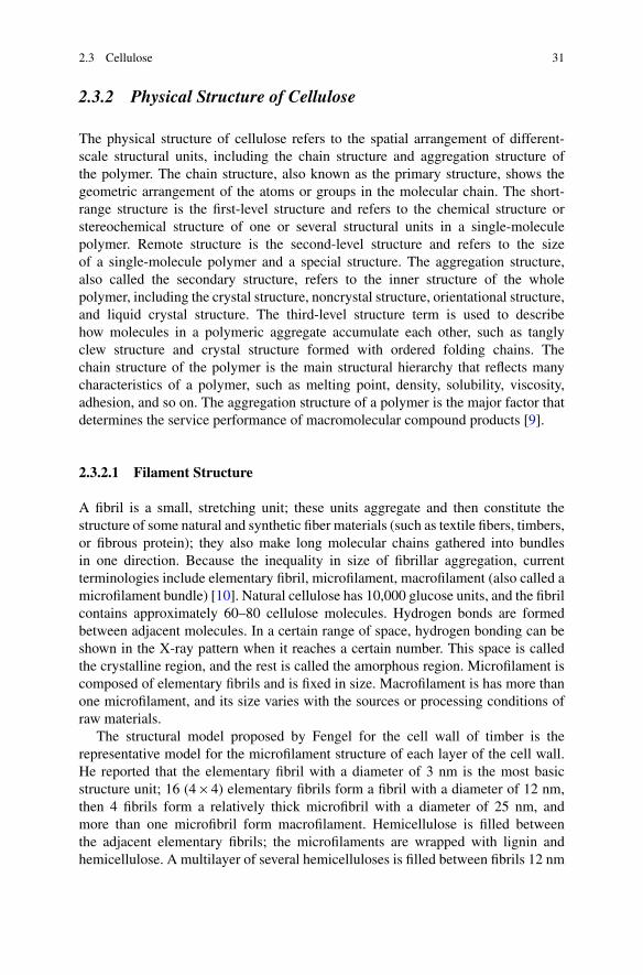
2.3.2 Physical Structure of Cellulose
The physical structure of cellulose refers to the spatial arrangement of different-scale structural units, including the chain structure and aggregation structure ofthe polymer. The chain structure, also known as the primary structure, shows thegeometric arrangement of the atoms or groups in the molecular chain. The short-range structure is the first-level structure and refers to the chemical structure orstereochemical structure of one or several structural units in a single-moleculepolymer. Remote structure is the second-level structure and refers to the sizeof a single-molecule polymer and a special structure. The aggregation structure,also called the secondary structure, refers to the inner structure of the wholepolymer, including the crystal structure, noncrystal structure, orientational structure,and liquid crystal structure. The third-level structure term is used to describehow molecules in a polymeric aggregate accumulate each other, such as tanglyclew structure and crystal structure formed with ordered folding chains. Thechain structure of the polymer is the main structural hierarchy that reflects manycharacteristics of a polymer, such as melting point, density, solubility, viscosity,adhesion, and so on. The aggregation structure of a polymer is the major factor thatdetermines the service performance of macromolecular compound products [9].
2.3.2.1 Filament Structure
A fibril is a small, stretching unit; these units aggregate and then constitute thestructure of some natural and synthetic fiber materials (such as textile fibers, timbers,or fibrous protein); they also make long molecular chains gathered into bundlesin one direction. Because the inequality in size of fibrillar aggregation, currentterminologies include elementary fibril, microfilament, macrofilament (also called amicrofilament bundle) [10]. Natural cellulose has 10,000 glucose units, and the fibrilcontains approximately 60–80 cellulose molecules. Hydrogen bonds are formedbetween adjacent molecules. In a certain range of space, hydrogen bonding can beshown in the X-ray pattern when it reaches a certain number. This space is calledthe crystalline region, and the rest is called the amorphous region. Microfilament iscomposed of elementary fibrils and is fixed in size. Macrofilament is has more thanone microfilament, and its size varies with the sources or processing conditions ofraw materials.
The structural model proposed by Fengel for the cell wall of timber is therepresentative model for the microfilament structure of each layer of the cell wall.He reported that the elementary fibril with a diameter of 3 nm is the most basicstructure unit; 16 (4 � 4) elementary fibrils form a fibril with a diameter of 12 nm,then 4 fibrils form a relatively thick microfibril with a diameter of 25 nm, andmore than one microfibril form macrofilament. Hemicellulose is filled betweenthe adjacent elementary fibrils; the microfilaments are wrapped with lignin andhemicellulose. A multilayer of several hemicelluloses is filled between fibrils 12 nm

32 2 Chemical Composition and Structure of Natural Lignocellulose
in diameter; the monolayer of hemicelluloses is filled between 3-nm elementaryfibrils. Because the microfilament is formed before the lignification of the cell wall,lignin is surrounded by only microfilament with a diameter of 25 nm [10]. It isgenerally thought that the movement of the liquid in the cell wall occurs mainlyat the elementary fibril level of hemicellulose; usually, contraction and swellingprocesses also mainly occur at this level.
In recent years, elementary fibrils with a diameter of 1.7 nm have been foundwith high-resolution electron microscopy. Because the fibrils are surrounded byhemicellulose and the microfilaments are surrounded by a large amount of lignin,the microfilament can be observed after delignification, and the elementary fibril canbe found only after the hydrolysis of hemicellulose. The measurement results forelementary fibrils according to most investigators indicated that the diameter of ele-mentary fibrils is between 30 and 35 Å, and an elementary fibril is composed of 40cellulose macromolecular chains. The ordered region of cellulose macromoleculesis the crystalline region; irregular regions form an incomplete crystalline structure.When the crystal diameter is about 3 nm, a monolayer of hemicellulose wouldexist around the crystal, and several cellulose crystals combine to form the cellulosecrystal beam called the nanofiber. The diameter of a nanofiber is about 2–3 nm, andhemicellulose and lignin are around it. In summary, the fiber cell walls are composedof many fibers, and larger microfibrils always consist of smaller elementary fibrils.
2.3.2.2 Aggregation Structure
The aggregation state of cellulose, also called the supramolecular structure ofcellulose, mainly refers to how cellulose molecules arrange to form crystal andamorphous structure, then elementary fibril, fibril, and microfibril structures. X-ray diffraction studies showed that, in the aggregates of cellulose macromolecules,molecules in crystal structure arrange regularly and display a clear X-ray pattern, sothe density of cellulose in the crystalline region is high (1.588 g�cm�3). Molecularchains in the amorphous region arrange irregularly and loosely, so the distancebetween molecules is large. The density of cellulose in the amorphous region islow, 1.500 g�cm�3. However, the molecule chain is almost parallel with the mainspindle of cellulose. The cellulose crystallinity, generally between 30 and 80 %,refers to the percentage of all the cellulose occupied in the crystalline region [2].
The crystallization of cellulose shows pleomorphism. There are five kinds ofcrystal modification in solid cellulose, whose characteristics can be reflected bycharacteristics of their unit cells. Under certain conditions, cellulose crystals canbe converted into many crystal variants. Type I is the crystal form of the naturalcellulose. Types II, III, IV, and X are those crystal forms of “artificial” celluloseunder artificial processing. Now, the commonly accepted cell structure of type Iis the monoclinic unit cell model introduced by Meyer and Misch in 1937 [9].Extensive chemical treatment and heat treatment will change the crystal form; forexample, ball milling can destroy crystal lattice completely. There is no distinctboundary from the crystalline region to the amorphous region. Each crystalline

2.3 Cellulose 33
region is called a microcrystal (also called a micel or micella). Since free hydroxylsat position 2, 3, and 6 of glucosyl in cellulose microcrystal regions have formedhydrogen bonds, only amorphous regions contain some free hydroxyls.
2.3.3 Physicochemical Properties of Cellulose
2.3.3.1 Chemical Properties of Cellulose
Every glucosyl ring of cellulose has three active hydroxyls: one primary hydroxylgroup and two secondary hydroxyl groups. Thus, cellulose may have a seriesof chemical reactions related to hydroxyl. However, these hydroxyl groups alsocan form hydrogen bonds between molecules, which has a profound influence onthe morphology and reactivity of cellulose chains, especially the intermolecularhydrogen bond formed by oxhydryl at C3 and oxygen at an adjacent molecule ring.These hydrogen bonds not only can enforce the linear integrity and rigidity of thecellulose molecule but also can make molecule chains range closely to form a highlyordered crystalline region [10]. The accessibility of cellulose refers to the difficultyreagents have in arriving at the cellulose hydroxyl. In heterogeneous reactions,the accessibility is mainly affected by the ratio of the cellulose crystalline regionsto the amorphous regions. The reactivity of cellulose is the reactive capability ofthe primary hydroxyl and the secondary hydroxyl at the cellulose ring. Generally,because of the smallest steric hindrance, the reactivity of the primary hydroxylgroups is higher than for the secondary hydroxyl groups, so the reactivity ofhydroxyl at C6 with a bulky substituting group is higher. For example, esterificationof toluenesulfonyl chloride chiefly occurs in the primary hydroxyl. The reversiblereaction occurs mainly in the hydroxyl group at C6, and an irreversible reactionalways occurs in the hydroxyl group at C2. Thus, for the esterification of thecellulose, the reactivity of the hydroxyl group at C6 is the highest, but for theetherification, C2 is the highest [10].
The degradation of cellulose is an important reaction that can be used toproduce cellulose products. Acid degradation, microbial degradation, and alkalinedegradation are mainly to break the glycosidic bonds between two adjacent glucosemolecules; an alkali peeling reaction and oxidation-reduction reaction of celluloseusually act on reducing ends of celluloses, and the oxidative degradation of thecellulose occurs mainly in dissociating hydroxyls at C2, C3, and C6 of the glucosylring. Cellulose molecule chains will form carbonyls at C2 when oxidized to somedegree and then be degraded in the following alkali treatment process by theelimination reaction of “-alkoxy. After disconnecting the glycosidic bond, thereaction product is formed and then degraded to a series of organic acids [9].
Esterification and etherification reactions of cellulose act on three alcoholichydroxyls of cellulose molecule monomer. They can greatly change the propertiesof cellulose, thereby producing many valuable derivatives of cellulose, such assulfonic ester, cellulose acetate, cellulose nitrate, and cellulose ether (carboxyl

34 2 Chemical Composition and Structure of Natural Lignocellulose
methyl cellulose, methyl cellulose, ethyl cellulose). To enhance the reactivity ofester and the ether bond of cellulose in multiphase medium and improve the qualityof cellulose ester and ether, some pretreatments need to be performed. The mainmethods include the following: ① Preswelling treatment of celluloses can weakenthe hydroxyl-binding forces between cellulose molecules to increase the reagents’diffusion velocity in cellulose, such as being immersed in concentrated caustic solu-tion, activated by glacial acetic acid, and so on. ② The elimination of crystallinityby the ethamine can only change the DP by 20 % (usually, the concentration ishigher than 1 %) when the concentration of ethamine is more than 71 %. Therefore,it was analyzed that ethylamine only enters into the microfilaments, only makesthe amorphous region swell, and does not greatly change crystallization regions.③ Cellulose derivatives with a high degree of substitution and many hydroxylgroups are substituted substantially, so the total free hydroxy declines and waterabsorbability decreases. So, actually some cellulose derivatives with a low degreeof substitution have higher water absorbability, such as methyl, ethyl, hydroxyethyl,hydroxymethyl cellulose ether, and so on. These groups lead to the swelling ofthe cellulose structure and binding force decrease in macromolecules. They furtherresult in the increase of water absorbability, degree of hydrolysis, and wrinkleresistance. The improvement of wrinkle-resistant property can be used to enhancethe stiffness and moisture resistance of cardboard; also, it can improve the burststrength and the dimensional stability of paper [9].
2.3.3.2 Physical Properties of Cellulose
Free hydroxyls of cellulose have a strong attraction to many solvents and solutions,but adsorbed water only exists in the amorphous region, not the crystalline region.In the moisture sorption process, the hydrogen bonds of the amorphous region inthe dry cellulose constantly could be broken; the hydrogen bonds in the cellulosemolecules are replaced by the hydrogen bonds between cellulose molecules andwater molecules, even forming new hydrogen bonds, and some hydrogen bonds incellulose molecules remain. In the desorption process, because of the obstructionfrom inside, hydrogen bonds between cellulose molecules and water moleculescannot be broken completely and reversibly, resulting in hysteresis. Some waterabsorbed by cellulose enters into the amorphous region of cellulose and forms thewater bound by hydrogen bonds, called bound water. Molecules of bound waterattracted by hydroxyl of cellulose are arranged in a certain direction and have ahigh density, making swelling the cellulose and generating a heat effect. Whenthe celluloses absorb water that reaches the fiber saturation point, water moleculescontinue to enter into the cell lumina and pores of cellulose to form a main layeradsorbed water or capillary water, which is called free water. No heat effect andswellability of cellulose exist when absorbing free water [9].
When solids absorb liquids, the configuration homogeneity does not change, butsolids become soft with the decrease of the inner cohesive force and increasedvolume. This phenomenon is known as the swellability. Swellability of cellulose

2.3 Cellulose 35
is divided into swellability in the crystalline regions and swellability betweencrystalline regions. The former refers to the fact that the swelling agent can onlyreach the surface of crystalline and amorphous regions, and the X-ray pattern ofcellulose does not change. The latter refers to the fact that the crystallization regionsof microfilaments are permeated with the swelling agent and then swell to generatenew crystalline lattice and display a new X-ray pattern. Unlimited swelling of thecellulose is dissolution. The hydroxyl groups in the cellulose have polarities. As aswelling agent, the greater polarity the liquid has, the greater degree of swelling thecellulose has. The metal ion in the alkali solution is usually in the form of aquo ions,which is more favorable for entering the crystallization region. Usually, 15–20 %NaOH will cause swelling within crystalline regions. If the alkali concentrationis increased, the radius of aquo ions is reduced because the ion density is toohigh, resulting in the drop of swellability. Except for alkali, the swellability ofother swelling agents, sorted from strong to weak, is as follows: phosphoric acid,water, polar organic solvents, and so on. Cellulose is saturated in a concentratedsolution of NaOH to generate alkali cellulose. Although alkali cellulose is washedwith water and dried, such changes cannot restore it to its original condition. Alkalicellulose may have a crystalline form of hydrate cellulose that is more stable thanthat of natural cellulose, which would increase its absorbability and make it easy toreact with a variety of reagents. Using alkali to impregnate celluloses is also calledmercerization. In addition, alkali cellulose is the important intermediate product forthe production of viscose fibers and derivatives of cellulose ether [9].
Characteristics of polymer compounds are high molecular weight and a strongcohesive force. They have movement difficulties in the system and a poor diffusioncapacity, so they cannot be dispersed in a timely manner in the solvent. Thesolvent dissolved with celluloses is not the real cellulose solution, but the mixedproduct is obtained by mixing celluloses and components in liquids. The solventsof the cellulose can be divided into two categories: aqueous and nonaqueous.Aqueous solvents include the following: ① Inorganic acids, such as H2SO4 (65–80 %), HCl (40–42 %), H3PO4 (73–83 %), and HNO3 (84 %) can lead to thehomogeneous hydrolysis of cellulose. Concentrated HNO3 (66 %) does not dissolvethe cellulose but forms an addition compound with cellulose. ② Lewis acids, suchas LiCl, ZnCl2, Be(ClO4)2, thiocyanate, iodide, bromide, and others, could dissolvecelluloses with a low DP. ③ Inorganic bases, such as NaOH, hydrazine and sodiumzincate, NaOH, and others can only dissolve cellulose with a low DP. ④ Organicbases, such as quaternary ammonium bases (CH3)4NOH, amine oxides, and others,are also aqueous solvents. The application of amine oxide solvent to dissolvecellulose can be used to manufacture the man-made fibers. ⑤ Complexes, such ascopper oxide ammonia (cuoxam), copper ethylenediamine (cuen), cobalt hydroxideethylenediamine (cooxen), zinc ethylenediamine (zincoxen), cadmium ethylenedi-amine (cadoxen), and the iron–tartaric acid–sodium complex (EWNN, an aqueousalkaline solution of iron sodium tartrate) are included as aqueous solvents [9].
A nonaqueous solvent of cellulose refers to a nonaqueous or less-aqueous solventthat is based on the organic solvents. It consists of activators and organic liquids.The organic solvents can be used as a component of the active agent and as a
36 2 Chemical Composition and Structure of Natural Lignocellulose
solvent of the activator, which can make the solvent have a larger polarity todissolve cellulose. Therefore, the mechanism of cellulose dissolved in a nonaqueoussolvent system cannot be easily explained by swelling theory, as in aqueoussolvents. The detailed mechanism of this process can be expressed as follows: ①
An oxygen atom and a hydrogen atom of cellulose hydroxyl participate in theinteraction of the EDA; the oxygen atom and the hydrogen atom act as a -electron donor and a •-electron acceptor, respectively. ② The active agent in thesolvent system has an electron donor center and an electron receiving center; thespatial location of these two centers is suitable for interaction with the oxygenatom and hydrogen atom of cellulose hydroxyl. ③ There is necessarily a suitablescope for the interaction strength of the EDA, causing the centers of the donorand acceptor to interact in polar organic solvents. When the hydroxyl chargeseparates to some extent, the complex of cellulose molecular chains is separatedand dissolved.
Several different systems of nonaqueous solvents of cellulose exist: ①
Paraformaldehyde/dimethyl sulfoxide (PF/DMSO) is an excellent new solventsystem that is not biodegradable. PF resolves into formaldehyde by heating,and then formaldehyde reacts with the hydroxyl group of cellulose to generatehydroxymethylcellulose, which is dissolved in DMSO. ② Dinitrogen tetroxide/dimethylformamide (N2O4/DMF or DMSO) is an intermediary derivative of thereaction of N2O4 with cellulose to generate nitrite esters; it can be dissolved in DMFor DMSO. ③ Amine oxides directly dissolve cellulose without the intermediatederivatives. ④ Liquid ammonia/ammonium thiocyanate restricts the dissolution ofthe cellulose; the solvent consisting of 72.1 % (w/w) NH4SCN, 26.5 % (w/w)NH3, and 1.4 % (w/w) H2O has the maximum dissolving ability. ⑤ Lithiumchloride/dimethylacetamide (LiCl/DMAC) also directly dissolves cellulose withoutthe intermediate derivatives. At room temperature, the LiCl/DMAC solution isstable and can be used for reeling off raw silk and film forming. Recently, researchon nonaqueous solvents of cellulose has been active; they not only can be used toproduce artificial fiber and films but also can be available for processing cellulosicmaterials and for the use of cellulose in homogeneous conditions to producecellulose derivatives. The problems of cellulose solvents are the low solubilityof cellulose, high price and low recovery of solvents, and environmental pollution.
Thermal decomposition of cellulose is in the narrow temperature range of 300–375 ıC. Different products depend on different temperatures. Heated at a lowtemperature (200–280 ıC), cellulose mainly dehydrates into dewatering celluloseand then forms charcoal and gas products. Heated at higher temperatures, celluloseseparates into flammable volatile products (tar). The most important intermediateproduct of cellulose high-temperature thermal degradation is laevoglucose, whichcan be further degraded into low molecular products and tar-like products. Tar-like products can be polymerized into an aromatic ring structure similar to graphitestructure at high temperature (400 ıC or higher). Mechanical degradation of thecellulose occurs because cellulose in the mechanical process can effectively absorb

2.3 Cellulose 37
mechanical energy, causing changes of morphology and microstructure; thesechanges are shown as decreased DP and crystallinity and significantly increasedaccessibility [10].
2.3.4 Biosynthesis of Cellulose
2.3.4.1 Cellulose Synthesizing Site
Some research has already forecast that assembling of cellulose microfilaments isfinished in the enzyme complex located in the extending top of the cellulose. Then,scientists hypothesized that a cellulose synthase complex was made up of manysubunits, and each subunit synthesized single-chain glucose, then polymerized itto the ordered particles of cellulose. But, until 1976, through the freeze-etchingtechnique, the complex located in the end of the cellulose microfilament was firstobserved in green algae. This verified the hypothesis that assembling of cellulosemicrofilaments is finished in the enzyme complex located in the extending top ofthe cellulose. The subunits of the complex are arranged linearly in three lines andform the linear enzyme complex where cellulose is synthesized [11]. The alternatingself-aggregation and dispersion of the complex determine that the microfilamentarrangement direction changes periodically, resulting in different levels of micro-filaments arranged perpendicular to each other. Later, similar terminal complexeswere observed in bacteria, mosses, ferns, green algae, and microtubule plants, butin corns, spherical complexes were found [12].
A terminal complex like a rosette has been observed in higher plants andconcentrates in the cellulose gathering place. Each six cellulose synthase sub-units of rosette synthesizes 6 glucose chains and then forms microfilamentswith 36 chains. The microfilament directions are mutually different in the dif-ferent levels of the cell wall, which makes the cell wall in any direction havehigh mechanical strength [13]. The rosette complex not only has the functionof synthase but also can bring glucose chains to the surface of cytoplasm. Acomplete rosette complex is essential for the synthesis of crystalline cellulose.The terminal complex would disappear or be changed when EDTA (ethylenedi-aminetetraacetic acid) is used to handle oysters or Congo red is used to han-dle banana cells, further causing the interruption or disturbance of cellulosebiosynthesis. Once the cellulose synthesis recovers, the terminal complex reap-pears. The mutation of the CesAI gene in Arabidopsis heat-sensitive mutants(RSWL) will lead to the reduction of the cellulose content, the content of antacid“-glucan, and the number of rosette complexes in the cell membrane, perhapsbecause the mutation of this enzyme disrupts the structure of the rosette complex.This indicates that the RSW1 (a radial swelling phenotype) maybe one componentconstituting the rosette complex [14]. Kimura et al. [15] used the polyclonalantibody technique on the central area in cotton CesA protein to verify that theplasma membrane has a rose-like structure, which was the complex of cellulosesynthase, and was CesA protein located in rosettes. This finding demonstrated that

38 2 Chemical Composition and Structure of Natural Lignocellulose
the CesA gene has an important role in cellulose synthesis and provided directevidence for the hypothesis that cellulose biosynthesis takes place in the terminalcomplex of the rosette [11].
It is uncertain whether the rosette complex is composed of identical subunits ordifferent subunits. Key information on assembly also still cannot be clarified. Thebacteria linear terminal complex goes through the cell membrane and lipopolysac-charide layer and mainly synthesizes the 1’ types of cellulose I, which is themetastable monoclinic system. But, the rosette terminal complex in plants is partof the cell membrane, which mainly synthesizes the 1“ type of cellulose I, which isa stable monoclinic form. Therefore, it is generally believed that cellulose synthesisin cotton also occurs in the cellulose synthase complex connected with the plasmamembrane.
In the process of cellulose biosynthesis, in addition to the terminal complex,another polypeptide with a molecular mass of 18 kDa also plays an important role.This polypeptide does not exist in the plasma membrane but is loosely connectedwith the plasma membrane. So, it is unlikely to be the component with catalyticactivity in the cellulose synthase complexes. However, it may have regulating effectbecause it can combine with 2,6-dichloro-phenyl nitrile, which is the inhibitor ofcellulose synthesis.
2.3.4.2 Substrate for Cellulose Synthesis
Identifying the substrate of cellulose synthesis has been difficult. Previous studiesreported that callosum generated in the translating period was the substrate of cel-lulose synthesis. This result was derived from the fact that, with in vivo conditions,the speed of synthesizing callosum from the substrate that can supply a radioactivelabel is higher than the predictable accumulation level, and the conversion of calloseradioactivity is consistent with the change of cellulose. The conversion may occurbecause of the transglycosylation of “-l,3-glucose polymerase; the discovery that thecell wall had the activity of “-l,3-glucose polymerase also supports the hypothesismentioned. Callose is a homopolymer of “-1,3-linked glucoses. It plays an importantrole in the regulation of vital processes, such as metabolism of the sieve tube, thedevelopment of the gametophyte, and so on. The composition and resolution ofcallose are directly related to the normal growth of plant metabolic processes [16].
The precursor for the biosynthesis of cellulose is uridinediphosphate-D-glucose(UDPG). However, in the past, UDPG was thought to be obtained by the catalysisof UDPG pyrophosphorylase. Now, with research on cotton fiber development, ithas been found that the catalytic reaction by the sucrose synthase could also provideUDPG. In the formation stage of the secondary wall of the cotton fibers, sucrosesynthase is connected with cellulose synthase, which may be used as carbon path.Two sets of evidence support this view. It has been proved that in vitro biosynthesisof cellulose takes cellulose synthase from the cell membrane of cotton fiber as theenzyme source and UDPG as a substrate. The products are always “-1,3-glucan and

2.3 Cellulose 39
Fig. 2.2 Biosynthetic pathway of plant cellulose [18, 19]
“-1,4-glucan, a few calloses, and a small amount of cellulose. When taking sucroseas a substrate, the synthetic rate of cellulose is equal to the synthetic rate of callose,and sometimes is more than the synthetic rate of callose; the absence of Ca2C ismore conducive to cellulose synthesis. This shows that cellulose synthase can onlyuse the UDGP directly from the catalytic reaction by sucrose synthase, but callosesynthase can directly use the free UDPG. The other evidence is that, in the mutantof cotton without fibers, there is no gene expression of sucrose synthase in the ovuleepidermal cell, but there is large gene expression of sucrose synthase in wild-typefiber cells; this shows that sucrose synthase has a close relation to the developmentof cotton fiber [17].
2.3.4.3 Cellulose Synthesis Process
Currently, there are different hypotheses about the mechanism of cellulose synthe-sis. One hypothesis suggests that the extension of the glucan chain is caused bythe moving glycosyltransferase catalyzing several glucosyl residues to connect tothe end of the growing cellulose chain. The synthesis of acetobacter cellulose maybelong to such a mechanism. The other hypothesis claims that some short glucancould polymerize with lipid or protein and form the mature cellulose polymer. Penget al. (2001) found that CesA protein adhered to the end of noncrystalline fibersin fiber cell wall fragments treated with herbicide. Meanwhile, a small amount ofthe attached glucose chain was detected in these fragments, indicating that CesAglycosyltransferase probably took sitosterol-“-glucoside (SG) as primers to startglucan polymerization. First, the SG and UDP-glucose is used as the substrate togenerate sitosterol cellodextrin (SCD) and continue polymerization by the cellulosesynthase, then enters into the crystallization process of cellulose (Fig. 2.2) [18, 19].Schrick et al. [20] studied the relationship of biosynthesis of Arabidopsis sitosteroland biosynthesis of cellulose; they found that sitosterol is important in cellulose
40 2 Chemical Composition and Structure of Natural Lignocellulose
Glucose
sucrose +UDP
SUSY
CESA CESA
Pm
vascular
micofilament
KOR
UDPG
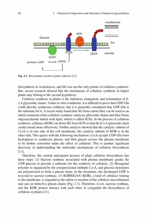
Fig. 2.3 Biosynthetic model of plant cellulose [21]
biosynthesis in Arabidopsis, and SG was not the only primer of cellulose synthesis.But, recent research showed that the mechanism of cellulose synthesis in higherplants may belong to the second hypothesis.
Cellulose synthesis in plants is the initiation, elongation, and termination of “-1,4-glycosidic chains. Under in vitro conditions, it is difficult to prove that UDP-Glucould directly synthesize cellulose, but it is generally considered that UDP-Glu isthe substrate for it. A recent study found that SG from cotton fiber can be used as aninitial extension when cellulose synthase catalyzes glucoside chains and then formsoligosaccharide linked with lipid, which is called SCDs. In the process of cellulosesynthesis, cellulase (KOR) cut down SG from SCD so that the “-1,4-glucoside chaincould extend more effectively. Further analysis showed that the catalytic subunit ofCesA is in one side of the cell membrane; the catalytic subunit of KOR is in theother side. This agrees with the following mechanism: CesA accepts UDP-Glu fromhyaloplasm to synthesize glucan, and then glucan crosses the plasma membraneto be further converted under the effect of cellulose. This is another significantdiscovery in understanding the molecular mechanisms of cellulose biosynthesis[11].
Therefore, the current anticipated process of plant cellulose biosynthesis hasthree steps: (1) Sucrose synthase associated with plasma membrane guides theUDP-glucose to provide a substrate for the synthesis of cellulose. (2) Hexagonalpolymer is organized by the coexpressional multiple CesA, and glucose monomersare polymerized to form a glucan chain; in the meantime, the discharged UDP isrecycled to sucrose synthase. (3) KORRIGAN (KOR), a kind of cellulase relatingto the membrane, is regarded as the editor or monitor of the cellulose microfilamentand can cut defective glucan chains (Fig. 2.3). Therefore, CesA, sucrose synthase,and the KOR protein interact with each other to coregulate the biosynthesis ofcellulose in plants [21].
2.3 Cellulose 41
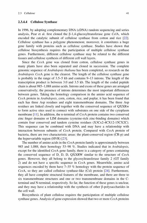
2.3.4.4 Cellulose Synthase
In 1996, by adopting complementary DNA (cDNA) random sequencing and seriesanalysis, Pear et al. first cloned the “-1,4-glucosyltransferase gene CelA, whichencoded the catalytic subunit of cellulose synthase from cotton and rice [22].Cellulose synthase has a polygene phenomenon; moreover, it constitutes a hugegene family with proteins such as cellulose synthase. Studies have shown thatcellulose biosynthesis requires the participation of multiple cellulose synthasegenes. Furthermore, different cellulose synthase may be related to the differenttissues and cellulose synthesis of different cell wall layers.
Since the CesA gene was cloned from cotton, cellulose synthase genes inmany plants have also been separated and cloned in succession. The completegenomic sequence of Arabidopsis thaliana has been completed, so research on theArabidopsis CesA gene is the clearest. The length of the cellulose synthase geneis probably in the range of 3.5–5 kb and contains 9–13 introns. The length of thetranscription product is between 3.0 and 3.5 kb. The length of the coded peptidechain is about 985–1,088 amino acids. Introns and exons of these genes are arrangedconservatively; the presence of introns determines the most important differencesbetween genes. Taking the homology comparison in the amino acid sequence ofCesA protein in Arabidopsis, corn, cotton, rice, and poplar, it has been found thateach has three Asp residues and eight transmembrane domains. The three Aspresidues are linked closely and together with the conserved sequence of QXXRwto form active sites used to connect with substrates on one side of the cytoplasmmembrane [11]. In addition, the n-terminal of CesA protein contains two conservedzinc finger domains or LIM domains (cysteine-rich zinc-binding domains) whichcontain four conserved and tandem cysteine residues (X3Cx2-4CX12-15CX2C).This sequence can be combined with DNA and may have a relationship withinteraction between subunits of CesA protein. Compared with CesA protein ofbacteria, there are two characteristic areas: the plant-conserved region (CR-p) andthe hypervariable region (HVR) [23].
The number of amino acids in the CesA protein family is approximately between985 and 1,088; their homology 53–98 %. Studies indicated that in Arabidopsis,except for the identified CesA gene family, there is a category of genes containingthe conserved sequence of D, D, D, QXXRW similar to the structure of CesAgenes. However, they all belong to the glycosyltransferase family 2 (GT family2) and do not have a specific sequence in CesA genes. Meanwhile, amino acidsequences encoded by them have 7–35 % homology with the protein sequence ofCesA, so they are called cellulose synthase-like (Csl) protein [24]. Furthermore,they all have complete structural features of the membrane, and there are three tosix transmembrane structures and one or two transmembrane domains in the C-terminal and N-terminal, respectively. So far, the function of these genes is unclear,and they may have a relationship with the synthesis of other “-polysaccharides inthe cell wall.
Biosynthesis of plant cellulose requires the participation of multiple cellulosesynthase genes. Analysis of gene expression showed that two or more CesA proteins

42 2 Chemical Composition and Structure of Natural Lignocellulose
are involved in the biosynthesis of cellulose in the same developmental stage ofthe same cell. Phenotypes of the IRX1 mutant are similar to IRX3; both appearedto have irregular xylem and a declining cellulose content; it also has been foundthat expression sites and periods of these two genes were identical. PtrCesA2and PtrCesA1 of poplar are homologous with IRX1 and IRX3. Their expressionsites and period are the same. They both are expressed at the secondary cell wallsynthesis of xylem. So, it was suggested that these two genes may be expressedin the same cell and related to the formation of the second wall [21]. Currently,as most studies involved with cellulose biosynthetic genes are focus on the CesAgene, the CesA gene has been cloned from microbes and many plants. Furtherstudy showed that the mechanism of cellulose biosynthesis was complicated. Exceptfor cellulose synthase, sitosterol glycosyltransferase, cellulase, sucrose synthase(SUSY), cytoskeletal protein, and Rac13 proteins were likely related to cellulosesynthesis. Sucrose synthase is related to the supply of substrate from cellulosebiosynthesis. The experiments proved that, in three different heterotrophic systems,sucrose synthase could improve the efficiency of cellulose biosynthesis. It cancatalyze the reaction of sucrose and UDP to produce UDP-glucose and fructose,which can directly offer substrate to improve the biosynthesis of the cellulose. In2004, Konishi et al. [25] further confirmed that sucrose synthase could use sucroseto synthesize UDP-glucose, which could be used directly for cellulose synthesis.
In short, cellulose biosynthesis is a highly complex biological process; thoroughclarification of its mechanism will require extensive research.
2.4 Hemicellulose
Hemicellulose is another main component in plant fiber materials. In 1891, Schulz[26] thought that polysaccharides that easily separated from plant tissue weresemifinished products of cellulose or precursor molecules of cellulose, so they werenamed hemicellulose. He also found that this component was easy to be hydrolyzedto monosaccharides in hot, dilute mineral acid or cold 5 % NaOH solution. Tohemicellulose, this concept is vague in terms of both chemical structure andbiological function. In recent years, people learned more about hemicellulose withimprovements of polysaccharide purification as well as application of various typesof chromatography, spectroscopy, nuclear magnetic resonance, mass spectrometry(MS), and electron microscopy. Aspinall in 1962 defined that hemicellulose wasderived from polysaccharides of plants and included the basic chain containingresidues of D-xylose, D-mannose, D-glucose, or D-galactose and other glycosyls asbranched chains linked to this basic chain. The purification of hemicelluloses wasconducted according to the different alkaline solubilities with cellulose. So, in 1978,Whistler thought that hemicellulose was the polysaccharide extracted by an alkalisolution, except cellulose and pectin. Unlike cellulose, hemicellulose is a copolymercomposed of different amounts of several saccharide molecules [2].

2.4 Hemicellulose 43
2.4.1 Chemical Structure of Hemicellulose
The content and structure of hemicellulose in various plants are different. Theresearch on the chemical structure is mainly about the composition of the main chainand branched chains of glucans in hemicellulose. The main chain may consist of oneor more types of glycosyls, and the connections between glycosyl are also different.Raw materials from different producing areas and different parts have differentglycan compositions. Therefore, to illustrate the chemical structure, glycans mustbe classified first. It is generally believed that hemicellulose is the glucan in thematrix of the cell, and the main components are xylan, xyloglucan, glucomannan,manna, galactomannan, callose, and so on [27].
2.4.1.1 Chemical Structure of Xylan Hemicelluloses
Almost all plants contain xylan. D-Xylosyls are linked with each other to formhomopolymer linear molecules as the main chain. Xylan hemicellulose is the glucanwith a backbone of 1,4-“-D-xylopyranose and with branch chains of 4-oxymethyl-glucuronic acid.
The hemicellulose of hardwoods and gramineous forbs is mainly composedof this kind of polysaccharide. The hemicellulose of Gramineae also contains L-arabinofuranose linking to the main chain as branch chains. The number of branchchains depends on different kinds of plants. The typical molecular structure ofhemicellulose of Gramineae is chiefly composed of “-D-xylopyranosyl, which islinked by “-1,4-glucosidic bonds. Branch chains consist of L-arabinofuranosyland D-glucuronopyranosyl, respectively, on C3 and C2 of the main chain; thereare also branch chains composed of xylosyl and acetyl (xylosyl acetate). TheDP of hemicellulose in Gramineae is less than 100. Xylan hemicellluloses intimber are composed of linear xylans linked by “-1,4-glucosidic bonds, with somedifferent short-branch chains linked to the main chain, similar to the Gramineae.However, average polymerization is higher than 100. In addition, hemicellulosefrom softwoods and hardwoods also has the distinction. Hemicellulose of hardwoodis chiefly acidic xylans that have been partly acetylized; for example, the content ofthis hemicellulose in birch is about 35 %, while this content in Euonymus bungenusis only 13 % [28]. Xylan hemicellulose in softwoods is 4-O-methyl-glucuronic acidarabinose-xylan with almost no acetyl, while O-acety-L-4-O-methyl-glucuronicacid xylan is the most important hemicellulose in hardwoods [2].
2.4.1.2 Chemical Structure of Mannan Hemicellulose
Softwoods contain the highest content of mannan hemicellulose; some hardwoodsalso have mannan hemicellulose, but grass has little. Mannose and glucose arelinked by “(1!4) bonds to form inhomogeneous polymer as the main chain.
44 2 Chemical Composition and Structure of Natural Lignocellulose
The main chain of mannan hemicellulose in hardwoods is composed of glucoseand mannose; the proportion of these two glycosyls is 1.5–2:1, and the average DPis 60–70; whether it is acetylated remains unclear. For the mannan hemicellulose ofsoftwoods, glucose and mannose in a ratio of 3:1 arrange randomly to form the mainchain, galactosyl is linked to the glucose or mannose of the main chain by ’(1!6)bonds, and acetyl seems to be evenly distributed on the C2 and C3 of mannose. Theaverage DP is more than 60; some can reach 100 [2].
2.4.1.3 Chemical Structure of Xyloglucan
The main chains of xyloglucan and cellulose are composed of D-glucopyranoselinked with “-(1 ! 4) bonds. The difference is that 75 % glucosyl residues arereplaced by ’-D-xylopyranose at O-6 on the main chain of the former. Xyloglucanmainly contains glucose, xylose, and galactose, and its residual ratio is approxi-mately 4:3:1. According to differences in family, xyloglucan in plants may alsocontain fucose and arabinose. The main chain of xyloglucan in the dicotyledon is “-(1 ! 4) glucans. ’-Xylose residues are linked to the O-6 of the “-glucose residues.The terminal galactose is linked to the O-2 site of xylose residues with “ bonds.If fucose is contained, it is linked to the O-2 site of galactose residues with ’
bonds. Sometimes, arabinose exists in xyloglucan, but the amount is small. Thecontent of xyloglucan in the monocotyledon is much different; generally, terminalgalactose does not exist, and the contents of xylose and galactose are lower than inthe dicotyledon.
2.4.1.4 Chemical Structure of Mannan
Mannan compounds include mannan, galactomannan, glucomannan, glucuronicacid mannan, and so on. Mannose residues are connected by a “-(1 ! 4) bond toform mannan, but they form galactomannan if linked to galactose residues by an ’-(1 ! 6) bond. The backbone of glucomannan is composed of glucose and mannose,which are linked by “-(1 ! 4) bonds with the residual ratio of 1:3. Glucomannanalso contains one galactose residue as a branch chain. Therefore, it is sometimescalled galactoglucomannan. Furthermore, the hydroxyl group of mannose residuesmay also be acetylated. Glucuronic acid is prevalent in the cell wall, but its contentis low. Mannose residues linked by ’-(1 ! 4) bonds and glucuronic acid residueslinked by “-(1 ! 2) bonds exist alternately in the main chain of glucuronic acid,whose side chains not only have “-(1 ! 6)–linked xylose or galactose but also have1 ! 3–linked arabinose [2].
2.4.1.5 Chemical Structures of Galactan and Arabinogalactan
Galactose residues are connected by “-(1 ! 4) bonds to form the backbone ofgalactan; galactose residues, as the side chain, are attached to the O-6. There are

2.4 Hemicellulose 45
two types of arabinogalactan: The common type has terminal galactose residueslinked at O-3 or O-6, galactose residues linked at O-3 or O-6, and arabinofuranoseresidues linked at O-3 or O-5. Another type has galactose residues linked by O-4or O-3 and O-4 bonds and arabinofuranose residues linked terminally or by an O-5bond. Arabinogalactan may also be oligosaccharide constituted by several arabinoseresidues. Further, ferulic acid may be linked to some arabinose and galactoseresidues. Arabinogalactan in the cell walls may be an independent molecule or asthe side chains on the polysaccharide molecules of pectin [2].
2.4.1.6 Chemical Structure of Arabinan
Arabinan entirely consists of arabinose, and ’-L-arabinofuranosyl residues arelinked with each other at C-5, forming the main chain. Arabinan contains manybranched chains; some side chains of arabinofuranosyl are linked to O-2 or O-3 orsimultaneously connected to O-2 and O-3, and some side chains are composed ofarabinose.
2.4.2 Chemical Properties of Hemicellulose
Because of the low DP and few crystalline structures, hemicellulose is moreeasily degraded in acidic medium than cellulose. But, the category of glycosyl inhemicellulose varies, including the pyran type, furan type, ’-glycoside bond-linkedtype, “-glycoside bond-linked type, L- configuration type, D- configuration type,and so on. The ways of linkage between glycosyls are various, such as 1-2, 1-3, 1-4 and 1-6 links [2]. Most studies showed that hydrolysis of methyl-rabopyranoseis the fastest; the others are arranged in decreasing speed as follows: methyl-D-galactopyranoside, methyl-D-xylopyranoside, methyl-D-mannopyranoside, andmethyl-D-galactopyranoside, which is the most stable. The “-D type of glycoside iseasier to hydrolyze than the ’-D type. Generally, the hydrolysis rate of the furan typeis faster than that of the pyran type. The hydrolysis rate of glucuronide is 40,000;perhaps the carboxyl has positive control in the glucoside bond.
Hemicellulose is an inhomogeneous glycan composed of a variety of glycosyls,so the reducing ends have many kinds of glycosyls and some branch chains.Similar to cellulose, hemicellulose can have a peeling reaction under mild alkalineconditions. At high temperature, it would have alkaline hydrolysis. Researchshowed that the speed of alkaline hydrolysis of furan glycosides was many timesfaster than that of pyran glycosides. Hemicellulose can dissolve in both alkalisolution (5 % Na2CO3 solution) and acid solution (2 % HCl solution). It has arelative affinity to water, which can make it form a viscous state or become a gellingagent. In rheologic studies of the viscosity of hemicellulose, this phenomenon canbe well observed. For example, when the concentration of hemicelluloses in waterreaches 0.5 %, the aqueous solution of hemicellulose has a certain consistency

46 2 Chemical Composition and Structure of Natural Lignocellulose
that is the same as in human saliva; when the concentration is 2 %, the solutioncannot flow because of the viscosity generated. When the concentration reaches4 %, the solution is to be regarded as a gel. The affinity of hemicellulose is closelyrelated to its pentose; for example, arabinose and xylose are responsible for fixingwater masses on to different structures of the hemicellulose. The greatest benefitbrought by this characteristic is to apply pentose in food technology. This featurealso illustrates that if the percentage of the pentose in the hemicellulose is too low,the spatial organization keeps pentose away from water, resulting in low affinity ofhemicellulose to water [2].
2.4.3 Biosynthesis of Hemicelluloses
Research showed that, in the plant cells obtained, the Golgi apparatus controlsthe biosynthesis of hemicellulose. Proteins synthesized on the ribosome of theendoplasmic reticulum of the plant cell can be transferred to the Golgi apparatusand form glycosides; the hemicellulose produced is contained in the Golgi vesiclesand moved to the cell surface (moved to the cell membrane). In the cell membrane,the Golgi vesicles inosculate to the continuous plasma membrane, further causingthe hemicellulose to be stuck to the cell wall. The Golgi apparatus can producehemicellulose because it can produce the enzymes needed for its synthesis [2].
At the initial stage of polysaccharide biosynthesis in the cell wall, a certainprimer is required to accept sugar residues under the effect of polysaccharidesynthase. It can be speculated that the primer of polysaccharide biosynthesis incell wall is a protein because that protein is the biosynthesis primer of manypolysaccharides, such as starch, glycogen, and so on. First, the reducing end of thefirst sugar residue is linked to protein. Then, the protein accepts monosaccharideresidues from sugar nucleotides under the effect of polysaccharide synthase,extending the polysaccharide chain. Actually, at the initial stage of the biosynthesisof glucorono-xylan in the pea and xyloglucan biosynthesis in the suspension cultureof bean, it was found that some protein primers indeed participated. Inositol can beused as a primer at the initial stage of callose biosynthesis. However, regardlessof the circumstance mentioned, it is not clear whether these primers have been“cut” before polysaccharide enters the cell walls or enters the cell wall togetherwith polysaccharide. Therefore, this interesting issue should be studied further.
2.4.4 Physiological Function of Hemicelluloses
Although some data indicate that some hemicellulose, like starch, exerts a functionas a storage polysaccharide, combination with lignin and cellulose moleculesincreases the resistance to enzymatic degradation of the cell wall and the insolubility

2.5 Lignin 47
of components in the cell wall. Some reports also indicated that xyloglucan is relatedto plant morphogenesis. However, the generally accepted view is that the mainfunction of hemicellulose is to take part in building the cell wall structure and theregulation of the cell growth process.
2.5 Lignin
Lignin is one of the most abundant organic polymers in plants, just behind cellulose.It is the exclusive chemical composition of gymnosperm and angiosperm. Thecontent of lignin in wood and Gramineae is 20–40% and 15–20 %, respectively.Lignin is the name of a group of substances; their inhomogeneity is manifestedin different species of plants, length of growing season, and different parts of theplants. Even in the different morphologies of cells of the same xylem or differentcell wall layers, the structures of lignin are not the same [29].
Lignin is a complex composed of complicated phenylpropane units nonlinearlyand randomly linked; three main monomers are coumaryl alcohol, coniferyl alcohol,and sinapyl alcohol. Because of the different monomers, lignin can be divided intothree types (Fig. 2.4): syringyl lignin polymerized by syringyl propane, guaiacyllignin polymerized by guaiacyl propane, and hydroxy-phenyl lignin polymerizedby hydroxy-phenyl propane. Usually, gymnosperm mainly contains guaiacyl (G)lignin; the dicotyledon mainly contains guaiacyl-syringyl (GS) lignin; the mono-cotyledon mainly contains guaiacyl-syringyl-hydroxy-phenyl (GSH) lignin [30].
At one time, lignin in plant was divided into softwood, hardwood, and grasslignins. Based on the structure of lignin, Gibbs divided lignin into G ligninand GS lignin. G lignin is chiefly formed through dehydrated oligomerization ofconiferyl alcohol, and its structure is homogeneous. This kind of lignin has negativeMaule reaction because less than 1.5 % of syringaldehyde and about 5 % ofp-hydroxybenzaldehyde were generated when oxidized by nitrobenzene. Mostlignin in softwood belongs to G lignin, which is copolymerized by guaiacyl and hasa positive Maule reaction. GSH lignin is the result of the dehydrated oligomerizationof coniferyl alcohol and sinapyl alcohol; the content of lignin is 17–23 %. The ratioof syringyl propane to guaiacyl propane is 0.5�0.1; it also contains 7–12 % estergroups. p-Coumaryl alcohol in it is linked to lignin in the form of ester [10].
Fig. 2.4 Basic structural unit of lignin
48 2 Chemical Composition and Structure of Natural Lignocellulose
2.5.1 Distribution of Lignin
Early studies indicated that the lignin concentration in the complex middle lamella(CML) layer is above 50 % (mass ratio), while it is about 20 % in the secondwall (S layer). However, because the volume of the S layer is far greater than thevolume of the ML layer, most of lignin still presents in the second wall. The ligninconcentration of the cell corner of the middle lamella (CCML) layer is generallyhigher than that of the CML layer, even more than 70 % [31]. The lignin density ofthe CCML layer is about four times that of the S layer; that is, the CCML layer isfull of lignins. The ultrastructure of Salix psammophila and the lignin distribution invarious layers was researched by Xu et al. using optical (or light) microscopy (LM),transmission electron microscopy (TEM), scanning electron microscopy (SEM),electron microscopy/ energy dispersive X-ray analysis (EM-EDXA), and confocallaser scanning microscopy (CLSM). The results showed that the cell wall of Salixpsammophila was divided into the primary (P) wall, ML, and secondary (S) walllayer; the lignin concentration ratio in the CCML, CML, and S2 was 1.96:1.33:1.CLSM pictures (530 nm) demonstrated that the lignin concentration in the vesseland ML was higher than that in fiber cells [32]. Wu et al. proposed that thecomposition of the lignin molecule was affected by cell type, the site of the samegrowth ring, vessel arrangement, and the producing area in the 25 kinds of hardwoodstudied [33].
Because lignin structural units differ according to timber assortments and mea-surement regions, the measurement of lignin distribution is uncertain and difficult.Lignin measurement methods include ultraviolet (UV) absorption spectroscopy,EM-EDXA, the interference microscopy (IM) method, CLSM, and so on. The UVmethod determines the distribution of lignin in the cell wall because that lignin hasa typical absorption at 270–280 nm in the UV spectra. The EM-EDXA methodcan provide distribution information for lignin in different regions. Simultaneouslyusing UV and EM-EDXA to analyze samples not only could provide information onlignin microdistribution but also offer information on the ratio of G-type and S-typestructural units in the different microareas. The IM method measures the opticalpath difference first by the inference microscope and then calculates the refractiveindex; finally, it obtains the volume percentage and mass percentage of lignin. TheCLSM method can be used quickly to measure the biological structure of samplesand obtain good measurement results in the Z direction [31].
2.5.2 Structure of Lignin
Lignin is a polyphenolic polymer with a three-dimensional network. Because almostall of the delignification process includes covalent bond rupture of natural lignin,with different separation methods and separation conditions, the lignin structure

2.5 Lignin 49
would have great differences. Therefore, a structural model was usually used topresent the structure of lignin. This kind of structural model only describes ahypothetical structure inferred from the average results. Further, different plantsources, or even lignin isolated from the same plant but in different ways, wouldhave different categories of linkages and composition of functional groups, resultingin the complicated lignin structure. Through nearly two decades of research onlignin structure, a dozen structural models have been proposed. Figure 2.4 is astructural model of lignin from softwoods. It can be seen that the lignin has acomplicated structure [34].
Through the study of various types of lignin structural models, lignin is a com-plicated amorphous polymer with three-dimensional network, which is basicallycomposed of phenylpropane units linked to each other by the irregular coupling ofC–C and C–O. Lignin includes three basic structural monomers: p-phenyl monomer(H type) derived from coumaryl alcohol, guaiacyl monomer (G type) derived fromconiferyl alcohol, and syringyl monomer (S type) derived from sinapyl alcohol(Fig. 2.4). The structural formula is as follows:
Although lignin only has three basic structures, the quantity proportions of thesebasic structures vary greatly in different families of plants. Lignin of hardwoodincludes large amounts of syringyl units. In the UV-photodegradated productionfrom Eucalyptus urophylla lignin, ¨ (syringyl-type compounds) is 58.10 %, and¨ (guaiacyl-type compounds) is 18.75 %. Compared with eucalyptus lignin, thepyrolysis products of sulfate pulp lignin are rich in guaiacol. In the sulfate pulplignin, M (syringyl)/m (guaiacyl) is 4.3:1; in the eucalyptus lignin, this ratio is6.4:1. Structural units of softwood lignin are mainly guaiacyl-type units; a smallamount of p-hydroxyphenyl-type units remains. Wheat straw lignin mainly consistsof noncondensed guaiacyl units, noncondensed syringyl units, and other condensedunits; the ratio of n (noncondensed guaiacyl units) to n (noncondensed syringylunits) to n (condensed units) is 1.44:1:3.24. The content of the –OCH3 group inbamboo lignin is similar to the content in hardwood lignin, for example, in Bai jiabamboo (ph, nidularia Mu) lignin, the ratio of n (guaiacyl units) to n (syringyl units)to n (hydroxy-phenyl units) is 1:1.15:0.54 [35].
The coupling modes between each basic unit include “-O-4, “-5, “-1, and so on.Figure 2.5 is a partial section of a softwood lignin structure.
Ether bonds in lignin include phenol-ether bonds, alkyl-ether bonds, dialkylbonds, diaryl ether bonds, and so on. About two thirds to three quarter phenyl-propane units of lignin are linked to the adjacent structural units by ether bonds;only a small part is present in the form of free phenolic hydroxyl. Phenol-ether bonds account for 70–80 % in these groups, guaiacyl glycerol-“-aryl ethers(“-O-4) account for about half of phenol-ether bonds, followed by the guaiacylglycerol-’-aryl ethers (’-O-4), also containing other types of ether bonds. Lignin insoftwood and hardwood mainly contains aryl glycerol-“-aryl-(“-O-4) ether bonds,approximately reaching half of the lignin in softwood and more than 60 % inhardwood. In the C–C bonds of lignin, the dominant coupling type is “-5, “-“linkage, followed by “-1, “-2, 5–5, and so on [36].

50 2 Chemical Composition and Structure of Natural Lignocellulose
OH
Lignin-O OMe
O
OMeO
OMeO
OH
OH
HOO
HO
O
O O
OMe
OMe
OHLignin
OMe
O O
HO
OOH
HOO
OH
OH
OMe
OMe
OMeO
OH
CHO
OMe
OMe
O
O
O
OOH
OMe
OMe
OMe
OH
O
O OH
OH
O
OMe
OMe
OMeOMe
HO
HO
O
OO
OMe
OH
OH
OMeOMe
OMe
OH
HO
O
HO HOOMe
OH(CHO)
HO
HO
OH
OH
HO
HO
HO
HOOH
OH
HO
HOHO
HO
HO
HO
HO
O
O
O
HOOMe
HO
OMe
HO
Fig. 2.5 Structural model of a section of cork lignin [34]
The main bond types of grass lignin are the same as those of lignin in wood. Themain bond type of the structural unit is aromatic glycerol-“-aryl-ether bonds; theseare fewer than hardwood and are similar to softwood. The proportion of carbon–carbon bonds, such as “-5 and “-“ in structural units, is higher than in hardwood. Inthe structure of grass lignin, a considerable part of p-hydroxyphenylpropane unitsconnects with phenylpropane units in its ester form. Taking straw as an example,60 % p-hydroxyphenylpropane units are connected in the form of its esters. Inaddition, grass lignin still contains a small amount of ferulic acid esters.
Because the types and positions of functional groups in different types of ligninare different, lignin of gymnosperms has different chemical characteristics. Ligninin gymnosperm mainly contains guaiacyl lignin (G), the G-structure unit of whichhas a methoxy group, and is hard to remove in the papermaking process because astable C–C linkage was formed by linkage with other monomers. Monocotyledonmainly contains GSH lignin [37].

2.5 Lignin 51
2.5.3 Physicochemical Properties of Lignin
2.5.3.1 Chemical Properties of Lignin
The chemical properties of lignin include halogenation, nitration, and oxidationreactions on the phenyl ring; reactions on the benzyl alcohol, the aryl ether bond,and an alkyl ether bond in the side chain; lignin-modified chromogenic reaction;and so on. The chemical reactions of the lignin structural unit are divided into twomajor categories: nucleophilic reactions and electrophilic reactions.
(1) Chemical reactions of lignin structural unit on the side chainReactions on the lignin side chains are associated with pulping and lignin modifi-cation; the reaction is a nucleophilic reaction. The following reagents can conductnucleophilic reactions with lignin:
① In alkaline medium, the effect of HO-, HS-, and S2-nucleophilic reagents leadsto the cleavage of the main ether bond (e.g., ’-aryl ether bond, a phenol-type ’-alkoxy ether bond, and phenol-type “-aryl ether bond) and fragmentation andpartial dissolution of macromolecule lignin. In alkaline medium, the phenol-type structural unit is separated into phenolate anions, and an oxygen atom thataffects the benzene ring by induction and a conjugative effect, which activatestheir ortho- and para- positions and thereby affects the stability of the CObond and breaks the ’-aryl ether bond, then generating a methylene quinoneintermediate and resulting in the aromatization of methylene quinone to generatea 1,2-diphenylethene structure.
② In neutral medium, reaction with nucleophile HSO3� or SO3
2� leads to breakingof the ether bond and brings SO3
2� groups in the degradation of lignin fragments.③ Acidic media mainly relate to the lignin fragmentation reaction of the acidic
sulfite pulping process. SO2 aqueous solution is taken as an affinity reagent,leading to the breakage of phenol-type and nonphenolic ’-aryl ether bonds,sulfonation of ’-carbon, and increased lignin hydrophilicity. Phenol-type andnonphenolic ’-alkoxy ether bonds may also have a similar reaction. In addition,C1, C5, and C6 on the high electron density centers of the aromatic ringcould also have a condensation reaction with methylene quinone intermediates[10, 36].
(2) Chemical reaction of the aromatic ring in the lignin structureChemical reactions of the aromatic ring in the structural unit of lignin are closelyrelated to the lignin-bleaching process and its modification and have been dividedinto electrophilic and nucleophilic reactions.
① Electrophilic substitution reaction: This mainly refers to substitution and oxida-tion reactions. Electrophilic reagents include chlorine, chlorine dioxide, oxygenmolecule, ozone, nitro cation, nitroso cation, and so on. The electrophilic reagentreplacement breaks the side chains of lignin and leads to the oxidative cleavage of“-aryl ether linkages. The aliphatic side chain is oxidized into a carboxylic acid,

52 2 Chemical Composition and Structure of Natural Lignocellulose
and the aromatic ring is oxidized into the compound of the o-quinone structure,which will finally be oxidized into dicarboxylic acid derivatives.
② Nucleophilic reaction: Nucleophilic reagents that can react with the aromaticring of lignin include hydroxide ions, hypochlorite ions, and hydrogen peroxideions. These nucleophilic reagents can react with the chromophoric groups in thedegraded lignin fragments, breaking the chromophoric structure to some extent[10].
(3) Lignin chromogenic reaction
① The condensation reaction of lignin with concentrated inorganic acids is mainlyrelated to the coniferyl aldehyde structure in the lignin structure.
② Mäule chromogenic reaction: Hardwood lignin becomes reddish violet whenfirstly treat with KMnO4 and HCl and then ammonia. The syringyl ringgenerates methoxy o-dihydroxybenzene under the effect of KMnO4 and HCl,then generates a methoxy-o-quinone structure, which is purple after ammoniatreatment.
③ Cross-Bevan reaction: Timber without extractives is treated with chlorine in thewet state; lignin will be converted into chlorinated lignin after sulfinic acid andsodium sulfite treatment, turning hardwood lignin red-purple [29].
2.5.3.2 Physical Properties of Lignin
(1) Molecular weight and polydispersityAny type of separation method may cause some partial degradation and changesin lignin. Accordingly, the molecular weight of the original lignin is unsure. Themolecular weight of the separated lignin varies with the separation method andconditions. Its molecular weight distribution can range from several hundred toseveral million. Under the effect of mechanical action, enzymes, or chemicalreagents, the three-dimensional net structure is degraded into different size ligninfragments, which leads to the molecular weight polydispersity of lignin [29]. Themolecular weight of separated lignin varies greatly; taking milled wood lignin(MWL) from spruce as an example, depending on the grinding time and extractionmethods, its weight average molecular weights include 2,100, 7,100, and 11,000.The weight average molecular weight of the high molecular weight fraction ofMWL from spruce and hardwood is 40,000 and 18,000, respectively, and the lowmolecular weight fraction is between 3,700 and 5,000. The molecular weight oflignosulfonate ranges from 103 to 105; the maximum is beyond 106. Kraft ligninhas a much lower molecular weight [10]. Determination of lignin molecular weightincludes the osmometric method, light-scattering method, supercentrifugation, gelpermeation chromatography, high-pressure liquid chromatography, and so on. Asfor insoluble lignin, such as lignin obtained by acid hydrolysis, their molecularweights are measured depending on the linear relationship between lgMw (the base-10 logarithm of molecular weight) and the heat-softening temperature Ts [10].

2.5 Lignin 53
(2) SolubilityHydroxyls and many polar groups exist in the lignin structure, resulting in strongintramolecular and intermolecular hydrogen bonds, and making the intrinsic lignininsoluble in any solvent. Condensation or degradation make the separated lignin ableto be divided into soluble lignin and insoluble lignin; the former has an amorphousstructure, and the latter is the morphological structure of the raw material fibers. Thepresence of phenolic hydroxyl and carboxyl makes the lignin able to be dissolvedin alkaline solution. Separated Brauns lignin and organosolv lignin can be dissolvedin dioxane, DMSO, methanol, ethanol, acetone, methyl cellosolve, and pyridine.Alkali lignin and lignosulfonate usually can be dissolved in a dilute alkali, water,and salt solution. Brauns lignin, phenol lignin, and many organosolv lignins can becompletely dissolved in dioxane. Acid lignin is not soluble in any solvents. The bestsolvents for most separated lignin are acetyl bromide and hexafluoroisopropanol inacetic acid [29].
(3) Thermal propertiesLignin is an amorphous thermoplastic polymer. It has slight friability under hightemperature and cannot form film in solution. It also has glassy transfer properties.Under the glassy transfer temperature, lignin is in the solid glass phase; it beginsto move when it is above the glassy transfer temperature. The lignin is softened tobecome sticky and has adhesive force. The glassy transfer temperature of separatedlignin varies with the raw materials, separation method, molecular weight, and watercontent. The softening temperature of absolutely dried lignin ranges from 127 to129 ıC, which remarkably decreased with increased water content, indicating thatwater acts as a plasticizer in lignin. The higher the lignin molecular weight is, thehigher the softening point is. For example, for lignins with a MW of 85,000 and4,300, the softening points, respectively, are 176 and 127 ıC [29].
(4) Relative densityThe relative density of lignin is roughly between 1.35 and 1.50. Values vary with theliquid for measurement if measured by water. The relative density of sulfuric acidlignin isolated from pine is 1.451 and is 1.436 if measured by benzene. The relativedensity of dioxane lignin is 1.33 when measured by water at 20 ıC and is 1.391by dioxane. Lignin prepared by different methods has different relative densities,such as the relative density of pine glycol lignin is 1.362, but it is 1.348 for pinehydrochloride lignin [29].
(5) ColorIntrinsic lignin is a white or nearly colorless substance; the color of lignin we cansee is the result of the separation and preparation process. For example, the colorof lignin isolated by Brauns and named after him is light cream, and the colors ofacid lignin, copper ammonia lignin, and periodate lignin vary from fawn to deep tan[29].

54 2 Chemical Composition and Structure of Natural Lignocellulose
Cinnamic acid
Coumaric acid
4CL
coscoA
CCoAOMT
coscoA
CCH
CCR CCR
CAD CAD CAD
Caffeoyl CoA Feruloyl CoACoumaroyl CoA
Coumaraldehyde
Coumaryl alcohol Coniferyl alcohol
Peroxidase/Laccase
G-lignin S-ligninH-lignin
Sinapyl alcohol
ConiferaldehydeCAId5H AldOMT5- hydroxy
coniferaldehydeMustard aldehyde
coscoA
4CL 4CL
Caffeic acid Ferulic acid
PALPhenylalanine
tyrosine
COMT
TAL
C3H
C4H
OH
CO2H CO2H CO2H
OCH3
OCH3
OH
OH
OH
OH
OH OH OH
Fig. 2.6 Biosynthetic pathway of lignin monomer [38]. PAL phenylalanine ammonia lyase, TALtyrosine deaminase, Cald5H coniferaldehyde-5-hydroxylase, C4H cinnamic acid-4-hydroxylase,CCH coumaroyl CoA-3-hydroxylase, 4CL 4-coumarate CoA ligase, CCoAOMT caffeoyl CoA 3-O-methyltransferase, C3H coumarate-3-hydroxylase, CCR cinnamoyl CoA reductase, AldOMT5-hydroxy coniferaldehyde-O-methyltransferase, CAD cinnamoyl alcohol dehydrogenase
2.5.4 Lignin Synthesis
2.5.4.1 Biosynthesis of Monolignols
Biosynthesis of monolignols is conducted through the phenylpropanoid pathway.In the shikimate pathway, glucose generated from the carbon dioxide by photosyn-thesis first is converted to the final intermediate: shikimic acid. Then, through theprephenic acid, the shikimic acid is converted into the final products of the shikimatepathway: phenylalanine and tyrosine. These two amino acids, which are widelypresent in plants, are starting materials for the cinnamic acid pathway. Under theeffects of various enzymes, three monomers of lignin are finally synthesized after aset of reactions, such as deamination, hydroxylation, methylation, reduction, and soon (Fig. 2.6) [37, 38].
Cinnamoyl CoA reductase (CCR) catalyzes the first step of the redox reaction oflignin biosynthesis, which may be the rate-limiting step, controlling lignin synthesisand the pathway from which carbon can go into lignin biosynthesis. Cinnamyl alco-hol dehydrogenase (CAD) catalyzes the redox reaction of another step in the ligninsynthesis process, which may control the reduction of coniferaldehyde. Sinapyl

2.5 Lignin 55
alcohol dehydrogenase (SAD), ferulic acid 5-hydroxylase (F5H), and bispecific caf-feic acid/5-hydroxyferulic acid O-methyltransferase (COMT) are immunolocalizedin cells and tissues that have S lignin. CAD is located in tissues with precipitationof G lignin. It is speculated that the last step of redox reactions of different typesof lignin may be passed through different synthetic pathways and catalyzed bydifferent enzymes [39].
Two hydroxylases are found in the phenylpropanoid pathway. C3H can catalyzecoumaric acid to caffeic acid, and the other hydroxylase is F5H. Arabidopsis fah1mutant without F5H activity is almost full of S lignin. Now, it has been recognizedthat F5H can catalyze the hydroxylation of coniferaldehyde and coniferyl alcohol,converting them to monomers of S lignin.
Lignin monomer synthesis needs to go through two-step methylation reactionsat the 30and 50 site, respectively. COMT and CCoAOMT (caffeoyl CoA 3-O-methyltransferase) are methylases on two different substrate levels. 5-Hydroxy-ferulic acid can be converted into sinapic acid under the methylation effect ofCOMT to involve in the synthesis of S lignin. Much research is more inclined tosupport that, in lignin biosynthesis, COMT can catalyze the methylation of caffeicacid, 5-hydroxyl coniferyl aldehyde, and 5-hydroxyl coniferyl alcohol into ferulicacid, sinapic acid, and sinapic alcohol, respectively, and CCoAOMT can catalyzethe methylation of caffeoyl-CoA to generate feruloyl-CoA.
2.5.4.2 Macromolecular Synthesis of Lignin
It has been proven that the polymerization of lignin macromolecules by phenyl-propane units is dehydro-oligomerization. The German scientist Freudenberg firstproduced synthetic lignin by coniferyl alcohol and laccase collected from mush-rooms under aerobic conditions. Later, peroxidase (POD) was found to catalyzethis polymerization reaction effectively. But, there is no direct evidence to provethat POD and laccase could catalyze the polymerization of various lignols [38].Hans et al. discovered that POD and manganese ion could be used to synthesizelignin in vitro, with their structures similar to natural lignin. In this process, redoxshuttles of Mn3C-Mn2C played an important role in lignin biosynthesis. POD did notdirectly react with lignol to participate in the synthesis of lignin. It catalyzed Mn2Coxidized to Mn3C. The latter (Mn3C) may contact many kinds of lignin precursorsby diffusion to catalyze the formation of free radicals in terminal groups of lignin.The latter also would participate in the formation of various covalent bonds in ligninmolecules [39].
There have been several different hypotheses to explain lignification; one isa random coupling model, which proposed that lignol molecules are graduallyconnected to the lignin polymer by oxidative coupling, and the formation of lignin isadjusted according to the categories, amount, and chemical coupling characteristicsof lignol at the terminal of the lignin molecule. The random coupling hypothesis isreasonable because it explains the plasticity of lignin biosynthesis in mutants andtransgenesis research. Another hypothesis involves the dirigent-like protein model,

56 2 Chemical Composition and Structure of Natural Lignocellulose
which considered that the lignified process is strictly under the manipulation ofdirigent-like protein by controlling the formation of particular chemical bonds ofthe lignin molecules. The hypothesis showed that the metabolism of lignin shouldbe an orderly life process, which explains the large numbers of O-4 linkages inlignin molecules. Dixon et al. believed that the formation of sinapyl alcohol andconiferyl alcohol was a relatively independent process by which enzymes involvedin sinapyl alcohol synthesis were associated with each other to form a multienzymecomplex. This multienzyme complex was combined in the endomembrane systemthrough C4H, C3H, C5H, and other P450 proteins, which made a series of reactionsassociated with mustard alcohol synthesis pathway run in an orderly fashion. Thesynthesis of coniferyl alcohol is conducted in the cytoplasm another way. Thephenylalanine ammonia lyase (PAL), caffeoyl CoA methyltransferase (CCOMT),CCR, and CAD distribute in the cytoplasm, catalyzing the synthesis of coniferylalcohol [39].
Peroxidase is widespread in plants with multiformity. So, how it participates inlignin monomer polymerization needs further studies. So far, it remains uncertainwhether the POD or laccase catalyzes the polymerization of lignin monomers in theplant or if they work synergistically [38].
2.5.4.3 Deposition of Lignin in Plant Tissues
Lignin biosynthesis in vascular plants is controlled by morphogenesis and the envi-ronment. The lignification process is the deposition process of lignin in plant tissues.Fluorescence detection indicated that, in the normal development of Arabidopsisstems, lignin mainly was deposited in the fiber cell wall between the vessel cell walland vascular bundles. Dystopia anchorage genes ELP1 were isolated from lignindeposited mutants of Arabidopsis pith. In mutant stems, lignin could be depositedin the usual parts and ectopically deposited in the pith of parenchyma cells. Suchdystopia anchorage would exist in both young stems and mature stems. Associatedwith this dystopia anchorage, enzyme activity related to lignin synthesis wouldincrease, and COMT could be ectopically expressed in the central cells of the pith.These results suggest that ELP1 genes inhibit lignin synthesis in the pith. Lignindeposition is related to coniferyl alcohol because that methylated ester generated byconiferyl alcohol can be covalently coupled with hydroxyproline-rich glycoproteins(HRGPs), resulting in the deposition of lignin in the cell wall [40].
2.5.4.4 Factors Influencing Lignin Synthesis
Different parts of plants would have a different lignin content and composition. Forexample, the lignin content and structure are significantly different in the node andinternode of reed (Arundo donax); the node has a higher density than the internodebecause of the high content of phenolic acids (p-coumaric acid and ferulic acid).The lignin content in different parts of lettuce is different depending on the harvest-
2.5 Lignin 57
ing time. Compared with the normal harvest, lignin in the middle of overmatureharvested lettuce buds is increased most obviously, secondarily increased in thebasal area, and not significantly increased at the top [40]. The reasons for thisdifference are varied. For instance, methyl jasmonate can significantly improve thePOD activity of seedlings and the lignin content. Both jasmonic acid and gaseousmethyl jasmonic acid could induce the expression of chalcone synthase (CHS),thereby increasing the lignin content. In general, overuse of nitrogen fertilizer willreduce the lignin content and thus postpone the lignification of the plant. However,nitrogen nutrition can increase the lignin content in the mulch covering of Pinussylvestris L. and Picea abies (L.) Karst. Phosphate fertilizer will greatly limit theperiod of rapid growth of plants but increase the lignin content [40].
2.5.4.5 Regulation of Lignin Biosynthesis
Lignin biosynthesis may be regulated by changing the activities of differentenzymes two main ways. One is to regulate the synthesis process of ligninmonomers by reducing enzyme activities that participate in the synthesis processof lignin, such as those of CCR, and CAD, and by reducing the lignin content.The second way is to regulate certain particular enzyme activity to influence thecomposition and chemical structure of lignin. It is generally believed that thedegradation of GS lignin, which is composed of guaiacyl monomers and syringylmonomers, is easier than that for G lignin, which is simply composed of guaiacylmonomers. Therefore, activate the enzyme activities of CAld5H and AldOMTfor the coniferaldehyde substrate to compete with CAD, make additional syringyllignin monomers synthesized via sinapyl aldehyde, and finally relatively reduce thesynthesis of guaiacyl G monomer.
In the gene regulation of lignin biosynthesis, the search for and separation ofbiological enzymes related to lignin synthesis is the first task, and then the aminoacid sequence of zymoprotein is analyzed to obtain the sequence of its messengerRNA (mRNA), which is the coding sequence of the functional gene exon. Genetransfer methods include agrobacterium-mediated indirect conversion and the genegun technique. The antisense technology is also known as antisense DNA andantisense RNA. It can transfer the constructed or amplified antisense DNA orantisense RNA to the living body, then combine the exogenous antisense mRNAor antisense mRNA transcribed from antisense DNA with positive sense mRNAto form the duplexes, which could prevent the transportation of mRNA from thenucleus and combination with ribosome, consequently inhibiting the natural geneexpression of endogenous mRNA and achieving the regulation of target genes.Antisense technology can be utilized for regulation of forest lignin. First, anoligonucleotide sequence is constructed that is antisense to the lignin synthase; then,the sequence is transferred into plants in direct or indirect ways, and it interacts withgenes in plants to influence the translation and reduce the activity of zymoprotein.This is currently the most common and effective transgenic breeding technology ingenetic engineering of lignin regulation. Finding and discovering a natural mutantin plants is also an equally effective and direct means [41].

58 2 Chemical Composition and Structure of Natural Lignocellulose
2.5.5 Biological Function of Lignin
2.5.5.1 The Relationship Between Metabolism of Lignin and CellDifferentiation
Lignin metabolism in plants has physiological significance, which was mainlypresent as the close relationship between changes of its enzyme activity, theincrease of intermediate and lignin contents and cell differentiation, the resistanceto pathogen infection, and other physiological activities in plant development [37].Plant cell differentiation is a core problem in developmental biology. The processof cell differentiation is essentially the changing process of cell physiology andmorphology; cell differentiation and organ growth have some relation with phenyl-propanoid metabolism and its products. Since the 1970s, there have been manyreports about the relationship between lignin metabolism and cell differentiation;Oven found that when the plant was injured, the callus first formed lignifiedparenchyma cells, followed by formation of short vessels and traumatic resin canals.This process required the participation of the phenylpropanoid metabolites. Malaet al. thought that phenylalanine metabolism had a closer relationship with furtherdevelopment of an embryo.
The structure of cell walls from outside to inside is as follows: ML, primarywall, the first secondary wall (S1), the second secondary wall (S2), and the thirdsecondary wall (S3). The structure of S1 and S2 of the wild-type tobacco cell wallis dense. However, the structure of the anti-CCR transgenic tobacco cell wall varieswith types of cells. For example, in fiber cells, the fiber framework of S2 becomessignificantly loose, but in vessel cells, the structures of S2 and S3 are changed.However, in the anti-COMT gene tobacco plants, structural changes of the cell wallare not detected. In the hybrid generation of these two transgenic plants, only theS3 structure of the vessel cell wall has slight changes. The immunocytochemistryanalysis of the lignin subunit showed that the G and S units of wild-type plantsconcentrated on S2 in the fiber cells, G and S units of wild-type plants decreasedin the hybrid generation, and cell walls became thin. In the vessel cells, G and Sunits in wild-type plants were unevenly distributed in the S1, S2, and S3, mostlyin S2. However, G and S units of hybrid generation were distributed in S1 andS3. Corresponding with this microscopic change, plants with antisense CCR genegrow shorter and have smaller leaves. But, plants with anti-COMT gene have slightchanges in height and leaf size, the height and leaf features of hybrid generation arebetween those of CCR gene plants and anti-COMT gene plants.
Many studies have confirmed that the formation of tracheary elements is accom-panied by the synthesis of lignin. Therefore, research on the metabolism physiologyof lignin is an important part in research on the differentiation physiology oftracheary elements. The results for the differentiation of wheat root tip (meristematiczone, elongation zone, maturation zone) showed that the tracheary element is mainlyformed in the maturation zone. There was only preliminary differentiation and asmall amount of lignin deposition in the elongation zone. Lignin is the basic factor

2.5 Lignin 59
that causes the differentiation of totracheary elements from the apical cell of wood.In the construction of cambium and catheter, the middle structure of a nonlignifiedcell wall contains an irregular fine network structure, which may contain pectinand hemicellulose. After the lignification, the middle structure quickly becomesdense, fully lignified surface structure becomes more compact, and parts of surfacestructure are partially covered with a spherical structure [40].
2.5.5.2 Relationship Between Metabolism of Lignin and FruitDevelopment
Phenolics in the fruit can be mainly divided into water-soluble phenolic substancesand water-insoluble phenolic substances. The former includes a variety of phenolicacids, water-soluble tannin, anthocyanin, and so on. They are soluble in both waterand ethanol, but after oxidation, they cannot be redissolved in water, only in ethanol.The latter consists of condensed tannin and lignin. Lignin is the main ingredient forthe formation of the secondary cell wall. The peel and pith of apple are vigorousmetabolic areas of phenolics; anthocyanin is mainly synthesized in the flesh cellsof peel and adjacent to peel. The large amount of lignin is in the fruit core, and thephenolics content is the lowest in the flesh.
According to reports, the content of lignin would change with the developmentalstage of the apple [42]. Simple phenols and flavonoids (anthocyanin) are synthesizedin the young fruit period of apple, and lignin is gradually synthesized with thebeginning of the cell enlargement period. Lignin content in the peel is positivelycorrelated with the development time of the fruit and the activity of POD [43]. Incontrast, the lignin content in the yellow apple peel is slightly more than that of redapple. In addition, a low temperature has a significant impact on the lignin contentin peel [40].
The harvest time also affects the lignin content. Ju et al. [44] found that the lignincontent in the late-harvested Laiyang pears was the highest because many phenolicswould be converted to lignin during the period of fruit development. Also, theconversion rate in flesh is higher than that in the pith tissue. Bagging also affects thelignin content. PAL is a photo-induced enzyme; its activity is positively correlatedwith light. However, after bagging the fruit, the activity of PAL decreases, and thelignin content in peel and pulp can both be reduced by 32 %.
2.5.5.3 Relationship Between Metabolism of Lignin and Plant DiseaseResistance
Many studies have shown that, when the plant is infected or resistance is induced,the activity of enzymes related to lignin synthesis and the content of lignin wouldboth increase, thereby enhancing the resistance of plants. Research on Erysiphegraminis f.sp. tritici showed that there was a positive correlation between theresistance of different types of wheat and the increase of lignin content that

60 2 Chemical Composition and Structure of Natural Lignocellulose
depended on the types of wheat. Wheats different in disease resistance all havethe PALactivity peak after the inoculation of powdery mildew 12 hours later. Theactivity peaks of enzymes related to disease resistance are high and have a longduration. The activity peak of susceptible cultivars would decline quickly andbecome close to the normal level after 24 h. PAL and POD activity increasedsharply after bacterial infection in the leaves of mung bean. After inoculatingVerticillium dahliae toresistant varieties of cotton in SBSI (Seabrook Sea Island)and susceptible varieties in Rowden, the expression of mRNA, encoding PAL andCOMT, was higher than that of the reference substance. After the cotyledon, leaf,and stem of Arabidopsis thaliana, Piplotaxis muralis, Piplotaxis tenuifolia, andRaphanus raphanistrum and the cotyledon of Sisymbrium loeselii were inoculatedwith Leptosphaeria maculans, they all had strong resistance characteristics to tissuebrowning and lignin deposition.
In recent years, research on the metabolism of lignin and plant resistancehas been plentiful. Pot-planting experiments of three poplar clones from Aigeirossp. under waterlogging conditions showed that, in comparison to their respectivecontrols, the activities of PAL and the contents of organic components in the basalstem phloem of all clones tested had obvious changes. When the waterlogging timewas prolonged to 40 days, the metabolism within the plant was confused resultingin the abnormal contents of organic components, the PAL activity increased, and theplant growth decreased significantly. Through research on the overwintering shoots,as the temperature decreased, the PAL activities of the terminal bud, cortex, andxylem steadily increased until December, when it reached peak activity. The PALactivity of different strains in the coldest month was different [45].
In the interaction with phytopathogenic organisms, the lignification of thesecondary cell wall is one of the characteristics of the disease resistance response.The lignin, as an important physical antibacterial substance in plants, can bedeposited in the HRGP of cell wall as structural barriers, reinforcing the cell walland protecting cells from infection by phytopathogenic organisms. The infection ofulcer germs can induce accumulation of HRGP and lignin deposition in the cell wallof poplar. The accumulation of different varieties of HRGP and lignin in the poplarcell wall is different and has a positive correlation with disease resistance. Treatingtobacco seedlings with jasmonate acid not only can improve the ability of seedlingsto be resistant to anthracnose but also can significantly increase seedling PODactivity and lignin content. POD does not play a critical role in the disease resistanceinduced by methyl jasmonate, but lignin is closely related to the disease resistance.In addition, the lignin content in leaves of grape and cucumber has a relationshipto disease resistance. Xu et al. reported that, when watermelon was infected byFusarium oxysporum, lignin in seedling leaves and stems/roots, HRGP content,and POP and PAL activity would increase in different degrees with the infectionin resistant varieties, and the increase of resistant varieties was significantly higherthan for the susceptible varieties.
The biosynthetic pathway of lignin is a branch of the phenylpropanoid materialsynthesis pathway of a plant. There are reciprocal chiasmata on enzymes involved inthese synthesis pathways. Therefore, in those susceptible and under waterlogging,

2.5 Lignin 61
low-temperature, and other adverse conditions, lignin synthesis is correlated withsynthesis of other phenylpropanoid substances. Chen et al. [46] reported that thetotal phenolic content and the pisatin and phaseoline content in the flavonoids weresignificantly higher than those in susceptible varieties. Zhou et al. [47] reportedthat, under noninfective Verticillium wilt, PAL activity and the content of phenoland lignin of the rootstock and grafted strains were obviously higher than those ofthe control strains. After infection with Verticillium wilt, the increasing amplitudeof PAL activity, phenol content, and lignin content was higher than that of thecontrol group. PAL activity and phenol content have some relevance to diseaseresistance to Verticillium spp. In buckwheat seedlings and different tissues, theflavonoid content always varies with the PAL activity, and this relationship is alsoreflected in the time sequence. As can be seen, in disease resistance and adversityresistance, nonlignin metabolism has some relevance for lignin metabolism in thephenylalanine metabolism. But, relatively little research currently is available on thedegree of relevance in the different plants and the degree of relevance under normalcircumstances.
2.5.5.4 Relationship Between Metabolism of Lignin and Plant Growthand Development
The growth and differentiation of plants are closely related to the activities of the cellwall, and lignin is one of the main components of plant cell walls. Since the 1970s,there have been many reports on the relationship between lignin metabolism andplant growth and development. Suzuki and Itoh [48] analyzed the lignifications ofbamboo terminal buds and 2- to 14-year-old stem nodes, results of which indicatedthat the lignification of terminal buds was prior to the extension growth of stems;the stem node growth was closely related to the degree of lignifications. Through thestudy in the clones of the annual cutting alnus, they found that the sequence of thePAL activity of different clones corresponded to the sequence of the high growth ofclones. Throughout the entire growth period, the PAL activity of alnus clones had asignificant positive correlation with the high growth.
Lignin, as a secondary metabolite in plant growth and development, has impor-tant biological functions in the growth and development and disease resistanceof plants. In the cell wall lignification process, lignin penetrates into the cellwalls and fills in their framework, which increases the hardness of the cell wall,enhancing the mechanical and the compressive strengths, promoting the formationof mechanical tissues, and consolidating the plant body and water conduction.Because of the chemical characteristics of the lignin, such as the insolubility andcomplex phenolic polymer, lignin has hydrophobicity of the cell wall and higherresistance to pests. Clarifying the physiological effects of lignin would contributeto better understanding of many biological phenomena of the plant and providea scientific basis for industrial and agricultural production, but further studies areneeded in this field.

62 2 Chemical Composition and Structure of Natural Lignocellulose
2.6 Ash
In plants, except for carbon, hydrogen, oxygen, and other basic elements, thereare many kinds of other elements that are indispensable materials in living plantcells, such as nitrogen, sulfur, phosphorus, calcium, magnesium, iron, potassium,sodium, copper, zinc, manganese, chlorine, and more. When the sample is dried at105 ıC and further processed at 750 ıC in a high temperature furnace, elementssuch as carbon, hydrogen, oxygen, nitrogen, sulfur, etc. disappear in the form ofgaseous compounds, and the residue is the ash which contains many types of mineralelements in the form of oxide. Different types of plants and growth environmentwould lead to different types and contents of elements. These elements are absorbedfrom soil and generally exist as ions. It is well known that different parts (organs) ofthe plants have different functions in the process of plant growth, causing differentdistributions of metal ions. Residues from reed and bamboo are mainly parenchymacells. Table 2.2 shows that, except Mn and Fe, the content of other metal elementsin the residues of reed and bamboo are all significantly higher than those of wheatand bagasse. In the process of standard sieve-screening classification of bagasse,with the increase of the mesh number, the particle size becomes finer, the fibercontent is reduced, the content of parenchyma cells increases (cells through the 80mesh are mainly parenchyma cells), and the metal contents rapidly increase. Themetal elements and silicon content in the joints, leaves, and ears of wheat straw aresignificantly higher than in the internode stem [2].
In the combustion process, a small part of the mineral elements may also belost through gasification. The content and elemental proportion of ash vary with thevarieties of raw materials, the kind of plant organs, the age of the plants, and soon. The elements in the growth environment of the plant would also have influenceon the content and elemental composition of ash. Generally, wood has a low ashcontent, usually less than 1 % (the majority have 0.3–0.5 %, dry raw material). The
Table 2.2 Comparison of metal ion contents from several grass families [2]
Sample Mn Cu Fe Ca Mg Al Na Si Co Ni
Reed Reed chip 460:2 9:9 481:4 1,569 625 763 134 9,413 <0.1 <0.2Reed residue 305:2 102:2 243:9 4,285 1,569 1;628 805 24,285 <0.1 12.1
Bamboo Bamboo chip 251:3 2:5 36:4 593 440 39:1 21 8,674 <0.1 0.5Bamboo residue 379:8 11:1 2;181:9 2,901 1,110 353 98 17,810 0.5 4.7
Wheat Internode stem 9:1 5:4 64 2,325 1,535 63:3 122 14,005 <0.1 1.78Joint 16 4:27 106 3,995 3,937 170:9 294 17,827 <0.1 1.61Leaf 40 4:61 329 5,061 4,376 54:1 357 4,032 <0.1 2.35Panicle 30 3:0 279 1,421 1,421 662:5 132 19,422 <0.1 1.21
Bagasse >20 mesh 65 4:4 90 582 1,060 71:9 44 2,644 <0.1 0.5620/40 mesh 98 6:3 267 929 1,490 204:4 57 7,366 <0.1 0.6540/80 mesh 161 19:1 547 1,124 1,895 360:4 68 11,551 <0.1 0.78Over 80 mesh 312 118:1 3;004 3,084 3,075 138 209 4,408 <0.1 9.08

2.6 Ash 63
ash content in Gramineae and bark is high, with most having 2–5 % (straw ashcontent is 10–15 %) [2]. It is worthwhile to note that, compared with other plantmaterials, SiO2 accounts for more than 60 % of straw ash, even more than 80 % inrice straw.
Silicon is a necessary nutrient in corn, rice, and other grass families [49, 50]There are several forms of silicon in the soil, but the silicon that can be directly usedby higher plants mainly is the monomeric silicic acid [Si(OH)4] in the molecularstate. After Si(OH)4 enters plants, the hydroxyl can form the SiO(OH) unitsthrough weak intermolecular forces with a variety of the hydrophilic components;then, the units are hydrolyzed to silica sol-gel. Finally, the SiOn(OH)4-2n sphericalnanoparticles are assembled into the silicon in any shape [51, 52]. It is consideredthat, according to the Si content, the cultivated plants can be divided into threecategories. The first is plants that contain a high amount of Si, with the Si contentranging from 5 to 20 %, such as in rice. The second category involves plants thatcontain a moderate amount of Si, with the Si content ranging from 2 to 4 %, suchas in wheat, barley, oats, and the like. The third category has plants with a lowamount of Si; the Si content is below 1 %, such as in legumes and dicotyledonousplants [53]. The Si content in plants often is calculated using the percentage (%)of SiO2 to the plant’s dry weight; different plant species have different Si contents.For example, Takahashi et al. analyzed components of 175 plants cultivated on thesame soil; the results showed that Si has the largest difference between plant species(the difference of Si content was as high as 200 times between the top 10 high-Si-containing plants and the top 10 low-Si-containing plants) [2].
The presence of silicon in plant materials may influence their subsequent use.For example, a large amount of Na2SiO3, generated from silicon of raw materialin the alkaline pulping process, dissolved in alkali waste liquid would increasethe viscosity of the waste liquid, lower the extraction rate of black liquor in pulpwashing, and bring adverse effects in the evaporation, combustion, cauterization,and lime mud recovery processes and so on [2]. Chen et al. reported that the presenceof ash in the raw materials would affect enzymatic hydrolysis and fermentation ofstraw. Therefore, according to the distribution characteristics of SiO2, Chen andcolleagues introduced a fractionated processing technology that included steamexplosion, carding, and screening (referred to as steam-exploded fractionation) torealize the high-value utilization of lignocellulose. First, the abundant nanosilicawas isolated without destroying the chemical composition of lignocellulose. Then,the lignocellulose that remained was used to prepare ethanol, butanol, and otherhigh-value-added products through biological fermentation [54, 55]. In addition,studies showed that pulps from any source would contain a complex systemcomposed of a variety of metal ions, with part of the metal ions coming from plantmaterials. In these ions, transition metal ions such as Cu, Fe, or Mn will exert anadverse influence on pulp color; decompose H2O2, O2, O3, and other bleachingagents; produce some •OH radicals; degrade carbohydrates; and finally reduce theeffect of bleaching. Alkaline earth metal ions represented by Ca and Mg couldhave a stabilization effect on bleaching agents and protect carbohydrates. However,excessive alkaline earth metal ions would stabilize the lignin, thereby reducing

64 2 Chemical Composition and Structure of Natural Lignocellulose
brightness [2]. Therefore, some appropriate pretreatments need to be performed toimprove ion distribution in the pretreated materials, further improving the utilizationefficiency of the raw material.
2.7 Pectin
Pectin is a major component of the plant cell wall; it is located between the cellulosemicrofilaments of the cell wall, mainly condensed by galacturonic acid and itsmethyl ester and composed of rhamnose and arabinose. The ML of plant cells ismainly composed of pectin, which can combine adjacent cells. According to theconditions of combination and physicochemical characteristics, the pectin can bedivided into pectic acid, pectin, and protopectin. Pectic acid is a straight chain linkedby an ’-1,4 – bond by 100 galacturonic acids. Pectic acid is soluble in water andeasily acts with calcium to generate the gel of pectate calcium. Pectin is the maincomponent of intercellular substances in the higher plants; it is a long-chain polymercompound linked by ’-1,4-glycosidic bonds by galacturonic acid ester and a smallamount of galacturonic acid. The molecular weight of pectin ranges from 25,000to 50,000, with each chain containing more than 200 galacturonic acid residues.Pectin can be dissolved in water and mainly exists in the middle and primary wall;some even is present in the cytoplasm or vacuole [56]. The molecular weight ofprotopectin is higher than that of pectin acid and pectin. The protopectin mainlyexists in the primary wall and is insoluble in water, but it can be converted tosoluble pectin under the effects of dilute acid and protopectinase. Because of theformation of calcium bridges between pectin substances, pectin molecules cross-link with each other and form a network structure that plays a bonding role as themiddle layer between the cells and allows water molecules to pass through freely.The gel formed by pectin substances has viscosity and elasticity. The increase of thecalcium bridge will cause decreased fluidity of the cell wall matrix. Along with theincrease of the esterification degree, the formation of the calcium bridge would bereduced, and the elasticity of the cell wall would increase [57].
Pectinase is a complex enzyme system consisting of pectate lyase, pectinesterase, polygalacturonase, and so on. Polygalacturonase is also known as thepectinase and includes exonuclease and endonuclease. These can catalyze thehydrolysis of ’-(1,4)-polygalacturonic acid by breaking pectin molecules downinto small molecules [58]. A study found that polygalacturonase was related to thematurity of fruits; its main function is to decompose the ML of the cell wall. Manymature fruits accumulated a large number of polygalacturonases. Pectin esterasecould deesterify the esterified part of polygalacturonic acids and change them toacidic pectin molecules so that the calcium bridge is formed in the polygalacturonicacid chains, making the pectin firm and strengthening the cell wall structure.Therefore, the esterification and deesterification of the polygalacturonic acid mayaffect the expansion of the wall. Pectinase not only exists in fruit tissue but also

2.8 Pigment 65
exists in leaves. It is related to physiological processes such as fruit ripening andsoftening and leaf abscission. Pectinase is also the earliest enzyme secreted when afungus invades the plant cell wall, which plays a key role in the utilization of naturalfibrous materials by microorganisms [59].
2.8 Pigment
Pigment is also an important substance of lignocellulosic materials. Wood rays existin the cambium, which is the meristematic tissue of wood existing between timberand bark. The function of cambium is to transport horizontally and store tissues,which makes the wood have various color changes because of the susceptibilityto colored bacteria. Up to 1 year of age, tissues near the center of the xylemwould be gradually replaced by branches, tannins, pigments, and other substancesand then degrade to dead cells with the lost of physiological activity. Many typesof heartwood have special pigments; for example, ebony heartwood is black,mahogany is red, and pine is brown [2].
Plant pigment is generally generated from lignin and some lignin degradationproducts with benzene ring. Lignin contains the chromophore and auxochrome, butthe current understanding of the lignin chromophore is not uniform. It is generallybelieved that the lignin chromophore mainly contains quinone structures andconjugated systems of carbonyls and double bonds. The results of studies showedthat the content of the lignin quinoid structure is less than the content of catechol.However, as the optical absorption coefficient of the quinone is higher than that of’-carbonyl (680,000 and 800 m2�kg�1, respectively), the quinoid structure has greatoptical absorption; the optical absorption in the visible region accounts for 35–60 %of the total absorption. Therefore, Kringstad et al. reported that the quinoid structurewas the most important chromophore in lignin. The formation and degradation oflignin are involved with formations of paraquinones and O-quinone. The O-quinoneis red, and the paraquinones are yellow; they are the major chromogenic structuresof lignin. Paraquinone structures are mostly dissolved in the unit form instead ofcondensed units form except for a few condensed units, but O-quinone structuresmainly remain in the lignin macromolecules without dissolution [60, 61].
Plants also contain small amounts of flavonoids and anthocyanin. In the transfor-mation of plant materials, these pigment-like substances will be extracted, affectingthe further conversion of lignocellulosic materials. In the pulp and papermakingprocess, the blue stain of pine would exacerbate the wood color and having adverseimpacts on wood products and the papermaking process. The presence of pigmentswould cause a certain degree of inhibition of the properties of enzymolysis andfermentation. Therefore, pretreatments of lignocellulosic feedstock usually need toremove pigments in the hydrolysate; these treatments include macroporous resinadsorption, ion exchange, electrodialysis, and precipitation.

66 2 Chemical Composition and Structure of Natural Lignocellulose
2.9 Cell Wall Protein
In addition to the main components mentioned, the cell wall is enriched withproteins. According to physiological function, the cell wall proteins can be roughlydivided into two categories: structural protein, which is the main portion of thecell wall proteins; zymoprotein, including dozens of proteins with various enzymeactivities [62].
2.9.1 Structural Proteins
(1) ExtensinExtensin is the most important structural protein in the cell wall and a subtribe ofHRGP. It approximately is made up of 300 amino acid residues, with the content ofhydroxyproline (Hyp) is especially high, typically 30–40 %. In addition to the Hyp,the extensin contains serine, valine, threonine, histidine, tyrosine, and so on [63].The main sugar components in extensin are arabinose and galactose, accountingfor 26–65 % of the glycoprotein, and sugars connected to the amino acid play animportant role in maintaining the conformation of extensin. The extensin also cancross-link with hemicelluloses and pectin, conducive to the formation of the networkstructure in the cell wall and consequently increasing the rigidity and strength of thecell wall [64].
(2) Proline-rich proteinProline-rich proteins in plant cell walls contain a repeated proline-proline sequence,which can make proline-rich protein molecules cross-link themselves or withextension and interact with acidic pectin through ionic bonds.
(3) Glycine-rich proteinThe characteristics of glycine-rich protein cell wall protein are a high content ofglycine (up to 70 %), highly repetitive primary structure units, and a similar proteinstructure as the structural protein-pectin in animals and cocoon silk protein in thesilkworm. Arabinogalactan protein is also found in the plant cell wall, where themain sugars are arabinose and galactose. In the corn and other grass families,threonine- and Hyp-rich glycoproteins and histidine- and Hyp-rich glycoproteins arealso found. These two glycoproteins contain threonine and histidine, respectively;the composition and content of the remaining amino acids and sugars are similar tothe stretching protein [65].
2.9.2 Zymoprotein
Active proteins in the cell wall mainly refer to various enzymes and lectins. Theprimary wall of higher plants is enriched with a variety of enzymes that coalesce tothe cell wall through ionic, covalent, and hydrogen bonds and so on. To date, dozens

2.9 Cell Wall Protein 67
of enzymes have been found in the cell walls, such as cellulase. In the plant growthand development process, the degradation of cell wall polysaccharides is mainlysynergistic, completed by exoglucanase, endonuclease, and “-glucosidase [66].POD is involved in the polymerization and cross-linking of various componentsin the cell wall and regulates the expansion of the cell wall. Therefore, POD playsan important role in the composition and plasticity of the cell wall [67]. Extensin is aclass of cell-wall-specific apoenzyme in the cucumber hypocotyl; it was discoveredby Cosgrove in 1989. It can break the hydrogen bonds among the cell wall polymers,inducing the relaxation and acidic growth of the cell wall. At present, more than 100types of extensins have been separated and identified, and they may be prevalent inthe dicotyledon and monocotyledon [68]. Studies have shown that the expressionof extensin genes is subject to strict regulation in time and space. Extensin isinvolved in almost the entire development process of the plant [69]. Enzymaticanalysis indicated that extensin has no cellulase activity but has a synergistic effectwith cellulase in the enzymatic hydrolysis of microcrystalline cellulose, increasinghydrolysis efficiency of microcrystalline cellulose by 50 %. Extensin also can loosenstructures of the filter paper, crystalline cellulose, hemicelluloses, and so on [70].
From the analysis of the plant cell structure mentioned, a large number ofstructural proteins and zymoproteins were found in the plant cell wall and cellmembrane, where enzymes play an important role in cell wall growth, structuralchanges, and other aspects. Although in the aging stage of plants the protein contentwould decrease, as the corn stems stay green while seeds mature, the fresh cornstalkafter harvest is still rich in protein. Currently, the utilization of straw mainly focuseson the utilization of cellulose, lignin, hemicellulose, and other major componentsbut neglects the application of cell wall proteins. After harvest, straw often is driedand stored under natural conditions, which would significantly reduce its proteincontent. Some pretreatments can improve the accessibility of the substrate. But, theoperating conditions of most pretreatment methods are intense, such as for steamexplosion, acid or alkali treatment, or organic solvent treatment, denaturing the cellwall proteins and causing loss of active ingredients.
Han and Chen [71] separated the “-glucosidase from corn straw cell wall protein;the specific activity reached 13.5 IU�mg�1, and the molecular weight was 62.4 kDa,which was similar to the “-glucosidase found in corn by other organizations. ESI-MS (Electron Spray Ionization) analysis showed that the “-glucosidase from cornstraw had the highest similarity to the “-glucosidase from soybean (AAD09291).However, their species source and the molecular weights were different, indicatingthat they were different proteins. The best reaction temperature of “-glucosidase is37 ıC, and the optimum pH is 4.8, which also indicates the important applications insimultaneous saccharification and fermentation of cellulose because simultaneoussaccharification and fermentation can react at the optimum temperature of the“-glucosidase and yeast. Studies of cornstalk simultaneous saccharification andfermentation found that cornstalk “-glucosidase was more suitable for ethanolfermentation than Aspergillus “-glucosidase. Through ammonium sulfate precipi-tation, hydrophobic chromatography, and CM-Sephadex G75, “-exoglucanase witha molecular weight of 63.1 kDa can be isolated from fresh corn straw cell wall

68 2 Chemical Composition and Structure of Natural Lignocellulose
proteins. ESI-MS analysis found that this enzyme had the highest similarity with“-(1–3,1–4)-exoglucanase (AAF79936) in maize coleoptiles. The comprehensivecomparisons of species, molecular weight, enzyme activity, and other characteristicsillustrated that they may be the same protein. The optimum reaction conditions forthis enzyme are 40 ıC and pH 6.0, and this characteristic enables potential appli-cations in simultaneous saccharification and fermentation of cellulose because itrelieves the inconsistent temperature of “-exoglucanase and yeast. “-Exoglucanasecan promote the simultaneous saccharification and fermentation and enzymatichydrolysis, especially simultaneous saccharification and fermentation.
2.9.3 Hydrophobic Protein
Fresh cornstalks contain various proteins; in addition to the structural proteins andzymoproteins, they have hydrophobic proteins. Han and Chen obtained severalkinds of hydrophobic proteins from the total proteins of fresh cornstalks; these pro-teins had strong adsorption of lignin. The molecular weights of these proteins rangedfrom 15 to 35 kDa. ESI-MS analysis of protein with a molecular weight of 18.5 kDashowed that the protein and corn lipid body-associated protein L2 (P21641) had ahigh degree of similarity. It can be concluded that they were the same protein bycomprehensive analysis of the protein characteristics, species source, and so on.Structural analysis showed that the protein had an amphiphilic structure, 46–122amino acids consisted of the significant hydrophobic region, while 1–46 amino acidsat N-terminal and 122–187 amino acids at C-terminal were hydrophilic. Therefore,it can be inferred that, in the hydrophilic-hydrophobic interface, the protein will self-assemble into amphiphilic film. Hydrophobic protein is without cellulase activity. Itcan be adsorbed onto lignin and steam-exploded straw but cannot be adsorbed ontomicrocrystalline cellulose. It has a synergistic effect with cellulase in the enzymatichydrolysis of steam-exploded straw but has no effect on the enzymatic hydrolysisof microcrystalline cellulose. Principle analysis showed that hydrophobic proteinmay play a role in the enzymatic hydrolysis of steam-exploded straw through thecombination with lignin, reducing the nonspecific adsorption of cellulase on thesubstrate, particularly exonuclease and beta-glucosidase.
In view of the plentiful proteins in plants, as well as the possible synergy betweenthe plant zymoprotein and exogenous cellulase in the enzymatic hydrolysis oflignocellulose, Chen et al. studied the application of vegetable protein in cellulosedegradation for several years and had some success. Di Lu performed some researchon the change of content of the apoplast protein of corn straw during storage and theimpact on Penicillium expansum cellulase activity; it was found that the apoplastprotein and Penicillium expansum cellulase had obvious synergies, with the newharvest straw apoplast protein having the most obvious synergies [72]. Han andChen [71, 73] found that in vitro autolysis producing glucose as the main productalso happened in the fresh corn straw; autolysis substrates accounted for 2.56 %of the straw dry weight; these autolysis substrates were mainly cellulose and the

References 69
“-(1–3, 1–4) glucan of hemicellulose. According to this characteristic, he developedthe new technology of straw enzymolysis and fermentation based on the use of cellwall proteins in fresh corn straw; glucose and ethanol production increased by 61.2and 112.7 %, respectively, compared with the control. Fresh cornstalks were used asthe substrate for enzymatic hydrolysis and fermentation; the cell wall proteins alsoplayed an additive effect on Trichoderma viride cellulase.
With further research and development of the cell wall proteins in the future,the application of the plant cell wall proteins in straw biotransformation willincreasingly be broadened, achieving prominent and significant results.
References
1. Chen HZ. Ecological high value-added theory and application of crop straws. Beijing:Chemical Industry Press; 2006.
2. Yang SH. Plant fiber chemistry. Beijing: China Light Industry Press; 2008.3. Zhang YZ, Liu J, Gao P. Scanning tunneling microscopy of the ultrastructure of native
cellulose. Acta Biophys Sin. 1997;13(3):375–9.4. Chen HQ, Gong Y, Fang Z. The situation and aspect of application for macromolecule (I)-
cellulose, lignin and starch. Yunnan Chem Technol. 1996;11(1):41–6.5. Li XB, Wu Q. Plant cell wall. Beijing: Peking University Press; 1993.6. Song DL, Shen JH, Li LG. Cellulose synthesis in the cell walls of higher plants. Plant Physiol
J. 2008;44(4):791–7.7. Zhang JQ, Lin L, Sun Y, Mitchell G, Liu SJ. Advance of studies on structure and decrystalliza-
tion of cellulose. Chem Ind For Prod. 2008;28(6):109–14.8. Zugenmaier P. Conformation and packing of various crystalline cellulose fibers. Prog Polym
Sci. 2001;26(9):1341–417.9. Zhan HY. Fiber chemistry and physics. Beijing: Science Press; 2005.
10. Gao J, Tang LG. Cellulose science. Beijing: Science Press; 1996.11. Tai FJ, Li XB. Cellulose biosynthesis in plant and the enzymes involved in it. Chin J Cell Biol.
2004;26(5):490–4.12. Mueller SC, Brown Jr RM, Scott TK. Cellulosic microfibrils: nascent stages of synthesis in a
higher plant cell. Science. 1976;194(4268):949–51.13. Reiter WD. Biosynthesis and properties of the plant cell wall. Curr Opin Plant Biol.
2002;5(6):536–42.14. Arioli T, Peng L, Betzner AS, Burn J, Wittke W, Herth W, Camilleri C, Höfte H,
Plazinski J, Birch R. Molecular analysis of cellulose biosynthesis in Arabidopsis. Science.1998;279(5351):717–20.
15. Kimura S, Laosinchai W, Itoh T, Cui X, Linder CR, Brown RM. Immunogold labeling ofrosette terminal cellulose-synthesizing complexes in the vascular plant Vigna angularis. PlantCell Online. 1999;11(11):2075–85.
16. Wang YJ. Callose in plants. Bull Biol. 2005;40(1):18–9.17. Bian HY, Zhou ZG, Chen BL, Jiang GH. Biological synthesis of cellulose during cotton fiber
thickening process. Cotton Sci. 2004;16(6):374–8.18. Zhong R, Burk DH, Ye ZH. Fibers. A model for studying cell differentiation, cell elongation,
and cell wall biosynthesis. Plant Physiol. 2001;126(2):477–9.19. Kimura S, Kondo T. Recent progress in cellulose biosynthesis. J Plant Res. 2002;115:297–302.20. Schrick K, Fujioka S, Takatsuto S, Stierhof YD, Stransky H, Yoshida S, Jürgens G.
A link between sterol biosynthesis, the cell wall, and cellulose in Arabidopsis. Plant J.2004;38(2):227–43.

70 2 Chemical Composition and Structure of Natural Lignocellulose
21. Yan SP, Wang QY, Yang CP. Research advances in the plant cellulose biosynthesis. J AnhuiAgric Sci. 2008;36(21):9049–51.
22. Pear JR, Kawagoe Y, Schreckengost WE, Delmer DP, Stalker DM. Higher plants containhomologs of the bacterial celA genes encoding the catalytic subunit of cellulose synthase.Proc Natl Acad Sci U S A. 1996;93(22):12637–42.
23. Doblin MS, Kurek I, Jacob-Wilk D, Delmer DP. Cellulose biosynthesis in plants: from genesto rosettes. Plant Cell Physiol. 2002;43(12):1407–20.
24. Richmond TA, Somerville CR. The cellulose synthase superfamily. Plant Physiol.2000;124(2):495–8.
25. Konishi T, Ohmiya Y, Hayashi T. Evidence that sucrose loaded into the phloem of a poplar leafis used directly by sucrose synthase associated with various “-glucan synthases in the stem.Plant Physiol. 2004;134(3):1146–52.
26. Zhang P, Hu HR, Shi SL. Application of hemicellulose. Tianjin Pap Mak. 2006;2:16–8.27. Yin ZF, Fan RW. The research progress of plant cell wall. Bull Bot Res. 1999;19(4):407–14.28. Xu F, Sun RC, Zhan HY. Progress in non-wood hemicellulose research. Trans Chin Pul Pap.
2003;18(1):145–52.29. Jiang TD. Lignin. Beijing: Chemical Industry Press; 2001.30. Wei JH, Song YR. Recent advances in study of lignin biosynthesis and manipulation. J Integr
Plant Biol. 2001;43(8):771–9.31. Lv WJ, Xue CY, Cao CY, Zhang Y. Lignin distribution in wood cell wall and its testing
methods. J Beijing Univ. 2010;32(1):136–41.32. Xu F, Zhong XC, Sun RC, Jones GLL. Lignin distribution and ultrastructure of Salix
psammophila. Trans Chin Pul Pap. 2005;20(1):6–9.33. Wu J, Fukazawa K, Ohtani J. Distribution of syringyl and guaiacyl lignins in hardwoods in
relation to habitat and porosity form in wood. Holzforschung-Int J Biol Chem Phys TechnolWood. 1992;46(3):181–6.
34. Qiu WH, Chen HZ. Structure, function and higher value application of lignin. J Cellul SciTechnol. 2006;14(1):52–9.
35. Zheng DF, Qiu XQ, Lou HM. The structure of lignin and its chemical modification. Fine Chem.2005;22(4):249–52.
36. Tao YZ, Guan YT. Study of chemical composition of lignin and its application. J Cellul SciTechnol. 2003;11(1):42–55.
37. Li W, Xiong J, Chen XY. Advances in the research of physiological significances and geneticregulation of lignin metabolism. Acta Bot Boreali-Occidentalia Sin. 2003;23(4):675–81.
38. Geng S, Xu CS, Li YC. Advance in biosynthesis of lignin and its regulation. Acta Bot Boreali-Occidentalia Sin. 2003;23(1):171–81.
39. Lin ZB, Ma QH, Xu YY. Lignin biosynthesis and its molecular regulation. Prog Nat Sci.2003;13(5):455–61.
40. Yu MG, Yang HQ, Zhai H. Lignin and physiological function in plant. J Shandong Agric Univ.2003;34(1):124–8.
41. Chen Y, Tan X, Clapham D. Lignin biosynthesis and genetic regulation. Acta Agric UnivJiangxiensis. 2003;25(4):613–17.
42. Yu M. Lignin metabolism and its regulation in apple rootstock M. Hupehensis Rehd [disserta-tion]. Tai’an: Shandong Agricultural University; 2002.
43. Guo XF, Zhang YL, Liu H. Yearly changes of phenol content in Danxia apple tree. J Fruit Sci.2004;21(6):606–8.
44. Ju ZG, Yan SP. Laiyang pear phenolic substances synthetic regulation and its effect on thequality of fruit. Sci Agric Sin. 1993;26(4):44–8.
45. Cheng SW, Tang LZ, Xiao Y, Xu X. Pal activity and organic components in basal stemphloem of poplar clones under waterlogging and flooding condition. J Nanjing For Univ.1997;21(1):51–5.
46. Chen HM, Liu JY, Ran B, Zhou J, Li T. Dynamics of some related enzymes of tobacco infectedwith brown spot. J Yunnan Agric Univ. 1995;10(1):1–6.
47. Zhou BL, Lin GR, Gao YX. The resistance of grafted eggplant to vertillium wilt and itsfunction. J Shenyang Agric Univ. 2000;31(1):57–60.
References 71
48. Suzuki K, Itoh T. The changes in cell wall architecture during lignification of bamboo,Phyllostachys aurea Carr. Trees-Struct Func. 2001;15(3):137–47.
49. Liu WG, Wang LQ, Bai YH. Research progress in the beneficial elements-silicon for plants.Acta Bot Boreali-Occidentalia Sin. 2003;23(12):2248–53.
50. Xu CX, Liu ZP, Liu YL. The physiological function of silicon in plants. Plant Physiol Commun.2004;40(6):753–7.
51. Ma JF, Yamaji N. Silicon uptake and accumulation in higher plants. Trends Plant Sci.2006;11(8):392–7.
52. Wang LJ, Wang YH. Nanostructures SiO2 in plant body. Chin Sci Bull. 2001;46(8):625–32.53. Ji XE, Zhang MS, Yu HQ, Bai BZ. Silicon nutrient of plants. Agric Technol. 1998;18(2):11–3.54. Yu B, Chen HZ. Effects of steam-exploded fractionation on the structure and distribution of
silicon dioxide in corn straw. Trans Chin Soc Agric Eng. 2008;24(10):190–4.55. Chen HZ, Yu B. A preparation method of nano-silica by straw. China patent 200710062669.
2007.56. Du JY, Bai L, Bai B. Chemical composition and basic characteristics of pectin. Agric Technol.
2002;22(5):72–6.57. Hardell H, Leary G, Stoll M, Westermark U. Variations in lignin structure in defined
morphological parts of birch [Betula verrucosa, middle lamella, primary wall, secondary wall,ray cells, vessels]. Svensk papperstidning. 1980;83(2):44–9.
58. Lu SM, Xi Y, Jin YF, Zhang Y. Structure and function of plant polygalacturonases. Acta HorticSin. 1999;26(6):369–75.
59. Xue CH, Zhang YQ, Li ZJ, Li ZJ. Recent development of pectin and pectolytic enzyme. J FoodSci Biotechnol. 2005;24(6):94–9.
60. Xi W, Li XP. An investigation to reaction properties of hydrogen peroxide with “-O-4 ligninquinoid chromophoric group. China Pulp Pap Ind. 2008;29(16):32–5.
61. Li XP, Wu S. Research development of the reaction characteristic between lignin quinonoidchromophoric group and hydrogen peroxide. J Cellul Sci Technol. 2006;14(4):52–6.
62. Han YJ, Chen HZ. Plant cell wall proteins & enzymatic hydrolysis of lignocellulose. ProgChem. 2007;19(7/8):1153–8.
63. Li LC, Wang X, Jing JH. The existence of expansion and its properties in the hypocotyls ofsoybean seedlings. Acta Bot Sin. 1998;40(7):627–34.
64. Lee SJ, Saravanan RS, Damasceno C, Yamane H, Kim BD, Rose JKC. Digging deeper into theplant cell wall proteome. Plant Physiol Biochem. 2004;42(12):979–88.
65. Li XB, Yang ZH. Structure, function, crossing linking and biosythesis of extensions. PlantPhysiol Commun. 1990;3:7–13.
66. Sandgren M, Ståhlberg J, Mitchinson C. Structural and biochemical studies of GH family12 cellulases: improved thermal stability, and ligand complexes. Prog Biophys Mol Biol.2005;89(3):246–91.
67. Jamet E, Canut H, Boudart G, Pont-Lezica RF. Cell wall proteins: a new insight throughproteomics. Trends Plant Sci. 2006;11(1):33–9.
68. Tong B, Rao JP, Ren XL, Li JR. Studying progress of plant cell wall proteins expansions. ChinAgric Sci Bull. 2005;21(9):112–15.
69. Shcherban TY, Shi J, Durachko DM, Guiltinan MJ, McQueen-Mason SJ, Shieh M, CosgroveDJ. Molecular cloning and sequence analysis of expansins—a highly conserved, multigenefamily of proteins that mediate cell wall extension in plants. Proc Natl Acad Sci U S A.1995;92(20):9245–9.
70. Whitney SEC, Gidley MJ, McQueen-Mason SJ. Probing expansin action using cellu-lose/hemicellulose composites. Plant J. 2001;22(4):327–34.
71. Han YJ, Chen HZ. Characterization of “-glucosidase from corn stover and its application insimultaneous saccharification and fermentation. Bioresour Technol. 2008;99(14):6081–7.
72. Lu D, Chen HZ, Ma RY. Effect of straw apoplast protein on cellulase activity. Chin JBiotechnol. 2006;22(2):257–62.
73. Han YJ, Chen HZ. Synergism between corn stover protein and cellulase. Enzyme MicrobTechnol. 2007;41(5):638–45.

http://www.springer.com/978-94-007-6897-0