Chemical composition and anti-proliferative properties of ...ifrj.upm.edu.my/20 (03) 2013/28 IFRJ 20...
6
© All Rights Reserved *Corresponding author. Email: [email protected] International Food Research Journal 20(3): 1229-1234 (2013) Journal homepage: http://www.ifrj.upm.edu.my 1 Neda, G. D., 1* Rabeta, M. S. and 2 Ong, M. T. 1 Food Technology Division, School of Industrial Technology, 2 Institute for Research in Molecular Medicine, Universiti Sains Malaysia Chemical composition and anti-proliferative properties of flowers of Clitoria Ternatea Abstract Aqueous and methanol extracts of the flowers of Clitoria ternatea (CT), a popularly plant consumed for blue colour in Nasi Kerabu was selected to explore its cytotoxic effect on six types of normal and cancer-origin cell lines. These included the hormone- dependent breast cancer cell line (MCF-7), non-hormone-dependent breast cancer cell line (MDA-MB-231), human ovary cancer cell line (Caov-3), human cervical cancer cell line (Hela), human liver cancer cell line (HepG2) and human foreskin fibroblast cell line (Hs27). The anti-proliferation activities of the extracts were examined by employing colorimetric MTT (3-(4,5-dimethylthiazol-2-yl) 2,5 diphenyltetrazolium bromide) assay through time periods of 24, 48 and 72 hours. Preliminary results showed that the water extracted of CT had significant effects (p < 0.05) against MCF-7 with an IC 50 value of 175.35 μg/ml. Furthermore, the aqueous and methanolic extracts were investigated by Gas Chromatogram-Mass spectrometry (GC-MS). The GC-MS chromatogram analysis of the water extracted had shown five peaks that represented components in the water extract namely mome inositol (38.7%) and pentanal (14.3%). Fifteen chemical constituents were identified in the methanol extract and the major chemical constituents were mome inositol (33.6%), cyclohexen, 1-methyl-4-(1- methylethylideme)- (7.1%), acetic acid, cyano- (6.5%) and hirsutene (5.7%). Heavy metals tested were at very low levels. The analysis conducted on the flowers provides a strong basis for emphasizing the medicinal and nutritional value of CT. Introduction The World Health Organization (WHO) has declared that total mortality due to cancer will increase to 12 million in 2030 whereas this number was only 7.6 million in 2005 (Farooqui et al., 2011). In year 2006, National Cancer Registry had reported about 21,773 cancer cases were diagnosed in Peninsular Malaysia (Zainal et al., 2006). Recently, cancer chemoprevention has developed as a major attention for researchers (Kim et al., 2010). Treatment used against cancer such as chemotherapy, radiation, hormone therapy and immunotherapy can kill both cancer and normal cells (Cooper, 1993). Hence the potential usage of natural products as anticancer treatment has been explored intensively by the scientists (Wan-Nor Izzah et al., 2009). CT is originally related to the Leguminosae (Fabaceae) family (Morris, 2009). The vines of this plant are climbing, herbaceous, tall and slender with five leaflets, while the flower colour ranges from white to blue with a white or yellowish center (Jain et al., 2003). It is known as butterfly pea and commonly known as Bunga telang in Malaysia. Flowers of CT are consumed to make Nasi Kerabu blue in colour, which is a famous local dish in Malaysia. All parts of CT are useful for medical treatments and have been used in folk medicines and for curing different diseases (Mukherjee et al., 2008). The primary objectives of this study were to determine the chemical composition of the CT’s flowers and examine the effect of its aqueous and methanolic extracts on normal and cancer cell lines. Materials and Method Flowers of CT, harvested in October 2010 Keywords Proximate analysis mineral and heavy metal content MTT assay cancer cell line Gas Chromatogram-Mass Spectrometry Article history Received: 4 October 2012 Received in revised form: 7 January 2013 Accepted: 11 January 2013
Transcript of Chemical composition and anti-proliferative properties of ...ifrj.upm.edu.my/20 (03) 2013/28 IFRJ 20...
© All Rights Reserved
*Corresponding author. Email: [email protected]
International Food Research Journal 20(3): 1229-1234 (2013)Journal homepage: http://www.ifrj.upm.edu.my
1Neda, G. D., 1*Rabeta, M. S. and 2Ong, M. T.
1Food Technology Division, School of Industrial Technology, 2Institute for Research in Molecular Medicine, Universiti Sains Malaysia
Chemical composition and anti-proliferative properties of flowers of Clitoria Ternatea
Abstract
Aqueous and methanol extracts of the flowers of Clitoria ternatea (CT), a popularly plant consumed for blue colour in Nasi Kerabu was selected to explore its cytotoxic effect on six types of normal and cancer-origin cell lines. These included the hormone-dependent breast cancer cell line (MCF-7), non-hormone-dependent breast cancer cell line (MDA-MB-231), human ovary cancer cell line (Caov-3), human cervical cancer cell line (Hela), human liver cancer cell line (HepG2) and human foreskin fibroblast cell line (Hs27). The anti-proliferation activities of the extracts were examined by employing colorimetric MTT (3-(4,5-dimethylthiazol-2-yl) 2,5 diphenyltetrazolium bromide) assay through time periods of 24, 48 and 72 hours. Preliminary results showed that the water extracted of CT had significant effects (p < 0.05) against MCF-7 with an IC50 value of 175.35 μg/ml. Furthermore, the aqueous and methanolic extracts were investigated by Gas Chromatogram-Mass spectrometry (GC-MS). The GC-MS chromatogram analysis of the water extracted had shown five peaks that represented components in the water extract namely mome inositol (38.7%) and pentanal (14.3%). Fifteen chemical constituents were identified in the methanol extract and the major chemical constituents were mome inositol (33.6%), cyclohexen, 1-methyl-4-(1-methylethylideme)- (7.1%), acetic acid, cyano- (6.5%) and hirsutene (5.7%). Heavy metals tested were at very low levels. The analysis conducted on the flowers provides a strong basis for emphasizing the medicinal and nutritional value of CT.
Introduction
The World Health Organization (WHO) has declared that total mortality due to cancer will increase to 12 million in 2030 whereas this number was only 7.6 million in 2005 (Farooqui et al., 2011). In year 2006, National Cancer Registry had reported about 21,773 cancer cases were diagnosed in Peninsular Malaysia (Zainal et al., 2006). Recently, cancer chemoprevention has developed as a major attention for researchers (Kim et al., 2010).
Treatment used against cancer such as chemotherapy, radiation, hormone therapy and immunotherapy can kill both cancer and normal cells (Cooper, 1993). Hence the potential usage of natural products as anticancer treatment has been explored intensively by the scientists (Wan-Nor Izzah et al., 2009).
CT is originally related to the Leguminosae
(Fabaceae) family (Morris, 2009). The vines of this plant are climbing, herbaceous, tall and slender with five leaflets, while the flower colour ranges from white to blue with a white or yellowish center (Jain et al., 2003). It is known as butterfly pea and commonly known as Bunga telang in Malaysia. Flowers of CT are consumed to make Nasi Kerabu blue in colour, which is a famous local dish in Malaysia. All parts of CT are useful for medical treatments and have been used in folk medicines and for curing different diseases (Mukherjee et al., 2008). The primary objectives of this study were to determine the chemical composition of the CT’s flowers and examine the effect of its aqueous and methanolic extracts on normal and cancer cell lines.
Materials and Method
Flowers of CT, harvested in October 2010
Keywords
Proximate analysismineral and heavy metal contentMTT assaycancer cell lineGas Chromatogram-Mass Spectrometry
Article history
Received: 4 October 2012 Received in revised form: 7 January 2013Accepted: 11 January 2013
1230 Neda et al./IFRJ 20(3):1229-1234
were obtained from Kampung Seronok, Bayan Lepas, Pulau Pinang, Malaysia. Hs27 (ATCC® CRL-1634™, human foreskin fibroblast cell line), MCF-7 (ATCC® HTB-22™, hormone-dependent breast cancer cell line), MDA-MB-231 (ATCC® HTB-26™, non-hormone-dependent breast cancer cell line) Caov-3(ATCC® HTB-75™, human ovary cancer cell line), Hela (ATCC® CCL-2TM, human cervical cancer cell line) and HepG2(ATCC® HB-8065™, human liver cancer cell line) were purchased from the American Type Culture Collection (ATTC), USA. Phosphate Buffer Solution (PBS) tablets were obtained from AMRESCO INC, Cleveland, Ohio, USA. The media used was Dulbecco’s Modified Eagle Medium (DMEM with low glucose and high glucose) and Foetal Bovine Serum (FBS), penicillin–streptomycin and trypsin were obtained from Gibco®, InvitrogenTM, USA. MTT (3-(4,5-dimethylthiazol-2-yl) 2,5 diphenyltetrazolium bromide) labelling reagent was obtained from Molecular Probes®, InvitrogenTM, Oregon, USA.
Sample preparation Flowers of CT were separated from the stem and
sun dried at room temperature (20oC ± 5oC) for 4 days. Plant powders were kept in an airtight polyester container at -20°C before use.
Proximate analysisProximate Analysis was determined by using the
method stipulated by Official Agricultural Chemists (AOAC) International (1990). The six analyses are including moisture, ash, fat, protein content, fiber and carbohydrate as well.
Determination of mineral and heavy metals content by Inductively Coupled Plasma Optical Emission Spectrophotometer (ICP-OES)
The mineral and heavy metal content determined in the flowers of CT included calcium (Ca), zinc (Zn), iron (Fe), sodium (Na), manganese (Mn), copper (Cu), nickel (Ni), chromium (Cr), lead (Pb), boron (B), calcium (Ca), cobalt (Co), potassium (K), magnesium (Mg), selenium (Se), arsenic (As) and cadmium (Cd) as well. This analysis was performed by using Inductively Coupled Plasma Optical Emission Spectrophotometry (ICP-OES) (OPTIMA 7000DV, Perkin Elmer, USA).
Digestion and sample preparation Powder dry samples were weighted 1 gram (n =
3) in microwave digester tubes and digested in (10:1) mixture of nitric acid 65% (HNO3) and perchloric acid (HCLO4). Samples were made to undergo digestion in mineral digester for 60 minutes. After digestion,
a few drops of concentrated hydrochloric acid (HCl) were added. The solution was heated gently and then filtrated. Subsequently, the entire filtrate were transferred into a 100 mL volumetric flask and marked up with de-ionized water. The dilute filtrate solutions was transferred into medicine bottles and then injected into the ICP-OES.
Extraction of sample for anti-proliferative propertiesHot water was used for the aqueous extracts
and methanol was used for the organic extracts. Extraction was done by soaking the CT flowers in boiling distilled water in the proportion of 1:20 (w/v) for 4 hours. The resulting crude extracts were filtered and lyophilized (Huang et al., 2003).
The methanol extract was obtained by maceration of the powdered flowers in 95% methanol for 24 hours. The methanol fraction was collected and the residual solvent eliminated by reduced pressure at 40oC by using a rotary evaporator. The residue obtained was dried in a desiccator until it reached a constant weight (Wicaksono et al., 2009). The extract produced was used to screen the antiproliferative properties and stored at -20oC until use. The extract was diluted in PBS and then sterilized before assays. Final serial dilution was contained in DMEM with 20% FBS.
Cell culture MCF-7, MDA-MB-231, Caov3, Hela and
HepG2 were grown in DMEM with low glucose, and the Hs27 was grown in DMEM supplemented with additional 4.5 g/L of glucose that was used as a comparison. The cells were cultured in the growth medium (supplemented with 10% FBS and 1% penicillin-streptomycin) and incubated overnight at 37oC with 5% CO2 using 25 cm2 tissue culture flasks (McAteer and Douglas, 1979).
Cell subcultureThe cells were grown to 70-80% confluency by the
method of Freshney (1994). Firstly, the old medium was removed, and subsequently the cells were rinsed with PBS twice to wash the cells. The subconfluent monolayer was trypsinized and incubated at 37oC and 5% CO2 for 5 minutes. About 1-2 ml of medium was added into the flask and the cells were collected in growth medium containing serum. The cells were then re-suspended in growth medium, and counted. The total number of viable cells was counted by a haemocytometer to prepare the cell suspension. Sixty microlitres of suspension containing 3 x 10³ cells/ ml was added to each well of a 96-well microtiter plate. The plate was then incubated overnight at 37oC with 5% CO2.
Neda et al./IFRJ 20(3):1229-1234 1231
Measurement of the growth inhibitory effectEach of the cancer cell lines was grown in a 96-
well microtiter plate (Nunc, Denmark) in a volume of 60 μL culture medium per well. The normal and cancer cells were then treated with 60 μL extracted of CT flowers, which contained a serial dilution at doses of 0.78, 1.56, 3.125, 6.25, 12.5, 25, 50 and 100 μg/mL and the temperature was maintained at 37oC with CO2 for 24–72 hours. The cells in the first row of the 96-well microtiter plate were feed with fresh growth medium for control. After the incubation period, 24 μL of MTT - formazan labelling reagent was added to each well. The microtiter plate was then incubated again for 4 hours at 37oC with 5% CO2. At the end of the drug period, the medium and MTT were removed from all of the wells. Subsequently, the remaining MTT- formazan crystals were solubilised with 100 μL of acidified-isopropanol. One hundred microlitre distilled water was added into each well for further colour development. The absorbance of viable cells was measured using a spectrophotometric plate reader (Multiskan spectrum, Thermo Electron Co., Waltham, Massachusetts, USA) at 570 nm immediately, due to unstable product. To calculate the IC50, the processes giving below were followed.
Cell viability (%) = OD of drug- tested sample - OD of Blank x100 OD of Control – OD of Blank
Dose response curve were constructed using probit analysis (Finney, 1962) on a finney computer program Bio StatTM 2009 (AnalystSoft Inc., Vancouver, Canada) to obtain IC50 value.
Gas Chromatogram- Mass Spectrometry analysis GC-MS analyses were conducted to analyze
volatile compounds by using a GC system coupled to a mass selective detector. The column was VB-1 (30 m x 320 µm). The temperature programming for the operating condition was: initial oven temperature, 50oC for 0 min increased up to 260oC at a rate of 10 oC/min and held for 9 min; Injector temperature, 260oC; split ratio, 100:1; carrier gas, helium, solvent delay for 1.70 min; transfer temperature, 260oC; ion source temperature, 260oC and mass range 28 to 400 Da.
Statistical analysis Results for percentage cell viability were reported
as means ± standard error of triplicate measurements. Significant differences for multiple comparisons were determined by one-way analysis of variance (ANOVA) followed by Duncan test with α = 0.05 using the SPSS statistical package (ver.19.0).
Results and Discussion
Results of the proximate analysis, mineral and heavy metal content of the CT flowers are presented in Tables 1. In proximate analysis, the parameters determined were moisture content, ash, crude fat, crude protein and crude fiber as well. The flowers of CT contained appreciable amount of crude fiber (2.1 ± 0.2) and fat (2.5 ± 0.1). A dietary pattern containing low-fat and high-fiber products has been associated with reduced risks of breast cancer (Kushi et al., 2012; Rabeta et al., 2009). Results also indicate that the flowers of CT are rich in calcium (3.09 mg/g) and magnesium (2.23 mg/g). The potassium, zinc, sodium and iron concentrations of the flowers on this plant were clearly high (1.25, 0.59, 0.14 and 0.14 mg/g), respectively (p < 0.05) than most of the other parameters analyzed (< 0.01 mg/g). In addition, minerals such as calcium and magnessium are necessary for growth, skeletal development and other vital processes within the body. Iron is useful for the prevention of anemia and other related diseases (Oluyemi et al., 2006) while zinc plays arole in protein synthesis, normal body development
and recovery from illness (Muhammad et al., 2011). Deficiency of these nutrients and minerals can be detrimental to human health. The heavy metals tested were at very low levels thus making the plants relatively safe for consumption.
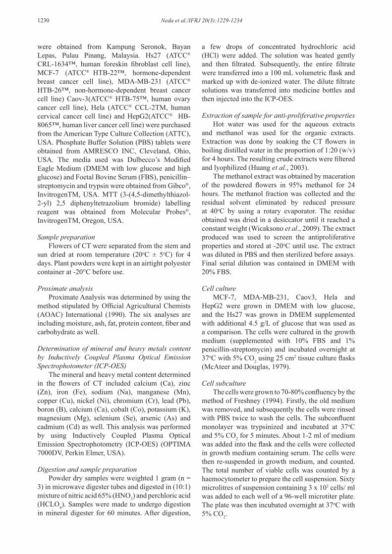
Figure 1 shows the percentage inhibition exerted by the water extracted on normal cell line (Hs27) and various human cancer-origin cell lines such as, MCF-7, MDA-MB-231, Caov-3, Hela, and HepG2. Based on The inhibition of cancer-origin cell lines increased steadily with increasing concentrations of the water extract and time duration. Growth
Proximate Mineral Mineral Heavy metal
Moisture 92.4±0.1 Boron 0.0150±0.002 Magnesium 2.2306±0.134 Cadmium < 0.0001Ash 0.45±0.15 Calcium 3.0953±0.09 Manganese 0.0249±0.003 Arsenic < 0.0001Fat 2.5±0.1 Cobalt < 0.0001 Molybdenum 0.0001±10-4×5.7 Lead 0.002333±0.0002Protein 0.32±0.03 Chromium 0.0007±0.0 Sodium 0.1413±0.003 Nickel 0.001267±0.0001Crude Fibre 2.1±0.2 Cupper 0.0103±0.0004 Selenium < 0.0001Carbohydrate 2.23±0.3 Iron 0.1441±0.007 Zinc 0.5980±0.006
Potassium 1.2506±0.235
Table 1. Proximate analysis, mineral and heavy metal content of flowers of CT
Data are mean values ± standard deviation (SD) of triplicate results; for proximate analysis, dry basis and are expressed in percentage (%) and results for mineral and heavy metal (mg/g).
1232 Neda et al./IFRJ 20(3):1229-1234
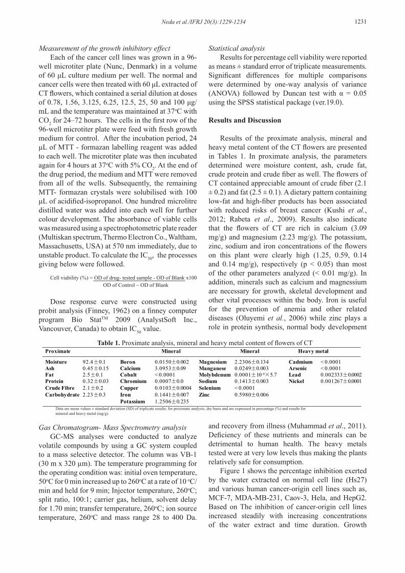
inhibition of the cancer origin cell line was most significant (p < 0.05) at 72 hours. The water extract had no effect on normal (non cancer-origin) cell growth. After treatment with the water extracted, the inhibition showed more anti proliferations for MCF7, Caov3, HepG2 and MDA-MB-231 with IC50 values of 175.3 µg/mL, 224.5 µg/mL, 236.3 µg/mL, and 304.7 µg/mL at 72 hours, respectively but the growth of Hela wasn’t effected with the water extract (Table 2). Figure 2 emphasizes the growth inhibitory effect of the methanol extract on MCF-7 cell lines (IC50= 536.01 µg /mL), and MDA-MB-231 cell lines (IC50= 561.3 µg/mL) at the 72 hours mark (p < 0.05). In contrast, the methanol extracted had no effect on the growth of Caov-3, Hela and HepG2 (Table 2). The water extracted of CT was found to have a stronger antiproliferative effect in comparison with the methanolic extract (p < 0.05). This may be due to the presence of more active compounds into the water extract.
Several studies have shown that the cytotoxicity and anticancer properties of natural plant are mainly due to the presence of flavonoids. Phenolic compounds, including flavonoids are especially promising candidates for cancer prevention. This could have
contributed to the susceptibility of the cells to be aqueous extract of CT flowers. Furthermore, Rajan et al., (2011) reported similar result that showed extracts of petroleum ether from CT had cytotoxic activity against HepG2 cell line. Shyam Kumar and Bhat (2011) also added that petroleum ether extracted and ethanolic extract of CT flowers had cytotoxic activity against the Dalton’s Lymphoma Ascites (DLA) cell
Figure 1. Inhibition of water extract of flowers from CT on MCF-7, MDA-MB-231, Caov3, Hela, HepG2 and Hs27 respectively. A) Treated in 24 hours. B) Treated in 48 hours. C) Treated in 72 hours. Values are expressed as mean ± standard error (SE) of triplicate measurements. (a, b, c) Same letters in each concentration are not statistically
significant from each other at p < 0.05
Table 2. The IC50 of water and methanol extract from flowers of CT on cancer cell lines
Cell lines 24 hr 48 hr 72 hrWater Methanol Water Methanol Water Methanol
Caov3 8386.5 41333.1 857.1 2109.9 224.5 947.2
Hela 42274.3 20381.7 18835.4 6281.1 51513.7 2095.7
HepG2 1438512 40674.6 481.5 23880 236.3 5214.1
MDA-MB-231 55355.3 - 481.5 4343.6 304.7 536.01
MCF-7 42567.6 - 1159.2 1983.4 175.35 561.3Using Probit analysis on a finney computer program Bio StatTM 2009.
Probit analysis is a type of regression used to analyze binomial response variables.
Figure 2. Inhibition of methanol extract of flowers from CT on MCF-7, MDA-MB-231, Caov3, Hela, HepG2 and Hs27 respectively. A) Treated in 24 hours. B) Treated in 48 hours. C) Treated in 72 hours. Values are expressed as mean ± standard error (SE) of triplicate measurements. (a, b, c) Same letters in each concentration are not statistically
significant from each other at p < 0.05
Neda et al./IFRJ 20(3):1229-1234 1233
line. Based on the growth inhibitory properties of the extracts observed, GC-MS analysis were obtained.
The components presented in the water extract from flowers of CT were mome inositol (38.7%) and pentanal (14.3%) (Table 3). The GC-MS chromatogram of the methanol extracte (Table 4) showed 15 peaks indicating the presence of three phytochemical constituents. The components presented in the methanol extract from flowers of CT were mome inositol (33.6%), cyclohexen, 1-methyl-4-(1-methylethylideme)- (7.1%), acetic acid, cyano- (6.5%) and hirsutene (5.7%). Furthermore, Studies from in vitro experiments, animal studies, and limited clinical experiences, claim that inositol may be used
effectively against some types of cancer, particularly when used in combination with phytic acid (Vucenik and Shamsuddin, 2003). These compounds may be responsible for the anti-cancer activity observed during in this study. However, further in vivo study is needed to confirm our findings and evaluating actual anti-proliferative properties in the CT flowers.
Conclusion
Conclusively, better inhibitions of cancer cell lines were observed in the water extract (IC50 of 175.3 μg/ml for MCF7). Knowing the exact compounds responsible for the plant’s anticancer properties will
Table 3. Potocomponents identified in the water extract CT flowers by GC-MS
Table 4 . Potocomponents identified in the methanol extract from flowers of CT by GC-MS
1234 Neda et al./IFRJ 20(3):1229-1234
help in formulating anticancer agents. In addition, it results from the proximate and mineral constituent analysis at the plant has provided pertinent information for food formulations. Acknowledgements
We would like to acknowledge the excellent technical guidance and support Ms. Lam Kit Lay from Institute for Research in Molecular Medicine (INFORMM). The authors are thankful to Universiti Sains Malaysia Short Term Grant 304/PTEKIND/6310065 and School of Industrial Technology, USM.
References
AOAC. 1990. Official Methods of Analysis of AOAC International (15th ed.), Washington, DC: AOAC.
Cooper, G. M. 1993. The cancer book: a guide to understanding the causes, prevention and treatment of cancer, p. 99-144. Jones and Bartlett Learning.
Farooqui, M., Hassali, M., Shatar, A., Shafie, A. and Seang, T. 2011. A Qualitative Exploration of Malaysian Cancer Patients’ Perspectives on Cancer and its treatment. BMC Public Health 11(1): 525.
Finney, D.J. 1962. Probit analysis. University Press, Cambridge.
Freshney, R. I. 1994. Culture of animal cells: a manual of basic technique: John Wiley and Sons, Inc.
Huang, S. T., Yang, R. C., Yang, L. J., Lee, P. N. and S Pang, J. H. 2003. Phyllanthus urinaria triggers the apoptosis and Bcl-2 down-regulation in Lewis lung carcinoma cells. Life sciences 72(15): 1705-1716.
Jain, N. N., Ohal, C., Shroff, S., Bhutada, R., Somani, R., Kasture, V. and Kasture, S. 2003. Clitoria ternatea and the CNS. Pharmacology Biochemistry and Behavior 75(3): 529-536.
Kim, Y. A., Kong, C. S., Yea, S. S. and Seo, Y. 2010. Consitituents of Corydalis heterocarpa and their antiproliferative effects on human cancer cells. Food and Chemical Toxicology 48:722-728
Kushi, L. H., Doyle, C., McCullough, M., Rock, C. L., Demark-Wahnefried, W., Bandera, E. V., Gapstur, S., Patel, A. V., Andrews, K. and Gansler, T. 2012. American Cancer Society guidelines on nutrition and physical activity for cancer prevention. CA: A Cancer Journal for Clinicians 62(1): 30-67.
McAteer, J. A. and Douglas, W. H. J. 1979. Monolayer culture techniques. Methods in enzymology 58: 132-140.
Muhammad, A., Dangoggo, S. M., Tsafe, A. I., Itodo, A. U. and Atiku, F. A. 2011. Proximate, minerals and anti-nutritional factors of Gardenia aqualla (Gauden dutse) fruit pulp. In Pakistan Journal of Nutrition 10: 577-581.
Morris, J. 2009. Characterization of butterfly pea (Clitoria ternatea L.) accessions for morphology,
phenology, reproduction and potential nutraceutical, pharmaceutical trait utilization. Genetic Resources and Crop Evolution 56(3): 421-427.
Mukherjee, P. K., Kumar, V., Kumar, N. S. and Heinrich, M. 2008. The Ayurvedic medicine Clitoria ternatea-From traditional use to scientific assessment. Journal of Ethnopharmacology 120(3): 291-301.
Oluyemi, E. A., Akilua, A. A., Adenuya, A. A. and Adebayo, M. B. 2006. Mineral contents of some commonly consumed Nigerian foods. Science Focus 11: 153-157.
Rabeta, M. S., Suzana, S. and Ahmad Rohi, G. 2009. Low fiber intake increased risk of breast cancer in pre menopausal women. 14th National confrance on medical and health sciences.
Rajan, M. S. D., Vijaya, T. and Thenmozhi, D. V. 2011. In vitro cytotoxic activity of Clitoria Ternatea. International Journal of Universal Pharmacy and Life Sciences 1(1): 19-28.
Shyam K. B. and Bhat, D. K. I. 2011. In-vitro cytotoxic activity studies of Clitoria ternatea Linn flower extracts. International Journal of Pharmaceutical Sciences Review and Research 6(2): 120.
Vucenik, I. and Shamsuddin, A. K. M. 2003. Cancer inhibition by inositol hexaphosphate (IP6) and inositol: from laboratory to clinic. The Journal of Nutrition 133(11): 3778S-3784S.
Wan-Nor Izzah, W. M. Z., Asmah, R., Fauziah, O. and Taufiq-Yap, Y. 2009. Antiproliferative properties of Clausine-B against different cancer cell lines. Malaysian Journal of Medical Sciences 16(3): 29-34.
Wicaksono, B. D., Handoko, Y. A., Arung, E. T., Kusuma, I. W., Yulia, D., Pancaputra, A. N. and Sandra, F. 2009. Antiproliferative effect of the methanol extract of Piper crocatum ruiz and pav leaves on human breast (T47D) cells in-vitro. Tropical Journal of Pharmaceutical Research 8(4) : 345-352.
Zainal, A., Zainuddin, M. and Nor Saleha, I. 2006. Malaysian Cancer Statistics-Data and Figure Peninsular Malaysia 2006. National Cancer Registry, Ministry of Health Malaysia.
![(14) IFRJ-2010-059 Azrina UPM[1]](https://static.fdocuments.in/doc/165x107/577cde151a28ab9e78ae5968/14-ifrj-2010-059-azrina-upm1.jpg)


![(5) IFRJ-2010-076 Thawien Thailand[1]](https://static.fdocuments.in/doc/165x107/577cc3c31a28aba711971255/5-ifrj-2010-076-thawien-thailand1.jpg)